
Back to Journals » International Journal of Nanomedicine » Volume 20
Metal Nanoparticles: From Endophytic Fungi-Mediated Biosynthesis to Their Therapeutic Applications in Oncology
Authors Xie K  , Du Y, Zhang J, Liu X, Gan X, Xie N, Hu T, Yang X
Received 21 August 2025
Accepted for publication 11 December 2025
Published 24 December 2025 Volume 2025:20 Pages 15599—15625
DOI https://doi.org/10.2147/IJN.S562203
Checked for plagiarism Yes
Review by Single anonymous peer review
Peer reviewer comments 3
Editor who approved publication: Professor Eng San Thian
Kangwei Xie,1 Yusha Du,1 Jiatong Zhang,1 Xinling Liu,1 Xiujuan Gan,1 Niqi Xie,2 Tieyi Hu,3 Xingyong Yang1
1College of Pharmacy, Chengdu University, Chengdu, 610106, People’s Republic of China; 2Department of Laboratory Medicine, the Affiliated Dazu’s Hospital of Chongqing Medical University, Chongqing, 402360, People’s Republic of China; 3Department of Neurology, the Affiliated Dazu’s Hospital of Chongqing Medical University, Chongqing, 402360, People’s Republic of China
Correspondence: Xingyong Yang, College of Pharmacy, Chengdu University, Chengdu, 610106, People’s Republic of China, Email [email protected] Tieyi Hu, Department of Neurology, the Affiliated Dazu’s Hospital of Chongqing Medical University, Chongqing, 402360, People’s Republic of China, Email [email protected]
Abstract: Metal nanoparticles possess unique properties and usage patterns compared to traditional materials owing to their distinctive structures. In recent years, their application scenarios and dosages have considerably expanded. Biosynthetic nanoparticles, particularly those derived from endophytic fungi that help host organisms in adapting to heavy metal environments, hold substantial value and potential for application. This is largely attributed to their simplicity, cost-effectiveness, and energy efficiency. The present review provides an overview of the entire process of metal nanoparticle biosynthesis by plant endophytic fungi and illustrates various scenarios of their applications in oncology treatment. In addition to focusing on the preparation of metal nanoparticles using plant endophytic fungi, this review also explores the characterization of these nanoparticles and clarifies the synthesis mechanisms, including the synthetic pathways and the roles of fungal enzymes. It also comprehensively summarizes the application of biosynthetic metal nanoparticles in cancer, covering their role in diagnosis, enhancement of drug biocompatibility, and improvement of therapeutic efficacy. These nanoparticles exhibit toxicity toward cancer cells by generating reactive oxygen species and inducing oxidative stress, ultimately leading to the death of malignant cells. The biosynthesis of metal nanoparticles by plant endophytic fungi represents a promising, green, and environmentally friendly approach with potential applications in various fields, including cancer treatment, in the future.
Keywords: endophytic fungi, metal nanoparticles, biosynthesis, cancer therapy
Introduction
Nanotechnology represents an interdisciplinary field operating at the nanoscale, integrating principles from chemistry, biology, physics, and materials science. Nanoparticles are minute particles with sizes ranging from 1 to 100 nm. They can be categorized into various types based on their chemical and physical characteristics, including metal, ceramic, and polymer nanoparticles.1 Nanomaterials address certain limitations associated with traditional materials and enhance the functionality of various products, owing to their unique properties and diverse applications.2 Specifically, their high surface-to-volume ratio enables broader applications in structural and functional materials, such as residential buildings, automotive components, solar cells, and coatings.3 Additionally, nanomaterials possess a high refractive index, resulting in visible light scattering, making them advantageous for use in dyes and ultraviolet protection at approximately 50 nm.4 Their superior mobility enables effective nanoparticle-based drug delivery and therapeutic interventions.5 Moreover, nanocomposites can be tailored through surface modifications to achieve tissue-specific and controlled delivery.6 Notably, the minute size of nanomaterials enables the establishment of innovative and functional structures, devices, and systems, overcoming key issues in contemporary biology.7 Currently, a primary focus in modern nanotechnology is the development of nanoparticles that are stable, effective, and uniform in size.8
Nanoparticles can be prepared through various physicochemical methods.9 However, the synthesis process is increasingly shifting toward non-toxic and environmentally friendly biological methods, particularly for invasive biomedical applications. Successful biosynthetic pathways have been established for nanoparticle synthesis,10 involving reactions between metal salt solutions and biomass. Various sources, such as plants, plant tissues, plant extracts, and seaweed, have been utilized for this purpose.11 Among these natural biological resources, fungi demonstrate remarkable efficiency, making them particularly well-suited for synthesizing metallic nanoparticles.12 Specifically, fungi exhibit relatively rapid growth rates and can produce substantial quantities of proteins, enhancing the productivity and stability of the resulting particles.13 Furthermore, fungal hyphae are resilient in harsh bioreactor environments, facilitating easier processing during downstream manufacturing than plant materials and other microorganisms.14 Consequently, significant advancements have been made in fungal biosynthesis for nanomaterial production, presenting a viable alternative to physicochemical synthesis methods. As natural microorganisms, plant endophytes perform functions analogous to their symbiotic hosts.15 Using endophytes for nanoparticle synthesis can effectively reduce the reliance on chemicals, enhance environmental performance, lower production costs, and support large-scale production, thus aligning with sustainable development principles.16
The present review offers a comprehensive overview of the entire process of metal nanoparticle biosynthesis by plant endophytic fungi and highlights their potential applications in oncology treatment. Specifically, we outlined the methods, conditions, and mechanisms involved in nanoparticle synthesis by plants and their endophytic fungi. We summarized the applications of metal nanoparticles synthesized by endophytic fungi (Figure 1), focusing on promising prospects in cancer research. This review provides a new biosynthetic approach for sourcing anticancer nanoparticles.
 |
Figure 1 Schematic diagram of nanoparticles synthesized by plant fungal endophytes. |
Synthesis Methods
Nanoparticle preparation methods can be categorized into two primary types: top-down and bottom-up (Figure 2).17 Top-down methods involve disintegrating larger materials into nanoscale particles through techniques such as mechanical grinding, soft lithography, and quenching. However, these methods lack precision in controlling the particle shape and size.18 Conversely, bottom-up techniques involve the self-assembly of atoms and their growth into nanoparticles, representing a gradual construction process starting at the atomic level. This group primarily encompasses chemical synthesis and biosynthesis methods. Nanoparticles synthesized through physical, chemical, and biological means each possess specific advantages and disadvantages (Figure 3).19 Among these, the environmentally friendly synthesis of nanoparticles using plant endophytic fungi represents a novel and promising synthetic pathway.
 |
Figure 2 The two strategies for nanoparticle synthesis. (A) Top-down: breaking down large bulk materials into nanoscale particles through energy-driven decomposition. (B) Bottom-up: assembling nanoscale structures by reacting and polymerizing precursor molecules. |
 |
Figure 3 Schematic of the nanoparticle synthesis pathways. Top left: green chemical synthesis. Top right: gas-phase chemistry reaction, precipitation, hydrothermal synthesis, spray pyrolysis, and sol-gel method. Bottom left: actinomycetes, fungus, algae, bacteria, plants. Bottom right: laser ablation, lithography, thermal decomposition, milling, and etching. |
Biosynthesis represents a predominantly bottom-up approach to nanoparticle synthesis, relying on reactions facilitated by organisms, including plants, fungi, bacteria, actinomycetes, and algae. This method allows for precise control over nanoparticle morphology and dimensions by adjusting cultivation parameters,20 making it a preferred approach for sustainable nanoparticle fabrication. Biosynthesis is relatively environmentally friendly, energy-efficient, cost-effective, scalable, biocompatible, easy to manipulate, and avoids the production of harmful by-products.11 Although fungi exhibit a slower growth rate than bacteria, they offer advantages in nanoparticle synthesis owing to the simplicity of their cultivation, substantial biomass production, enzyme excretion, and mycelial network resilience to shear stress.21 Filamentous fungi can produce highly stable nanoparticles that prevent molecular aggregation, even after prolonged storage.22 In addition to biosynthesis, chemical synthesis is an alternative bottom-up technique for nanoparticle production. However, chemical methods often involve or produce toxic and hazardous substances, limiting their utility in biomedical applications.
Plants and Their Endophytic Fungi-Mediated Nanoparticles
Plant Green Biosynthetic Nanoparticles
The ability of plants to reduce metal ions was demonstrated in the early 20th century.23 However, to date, research on the identity of these reducing agents remains limited.24 Different synthetic schemes apply different principles. Synthesizing metal nanoparticles using entire plants involves absorption of metals from the environment, followed by their reduction and cellular accumulation in the form of nanoparticles.11 Extracts from different plant parts, including the roots, stems, bark, leaves, flowers, fruits, and seeds, can be used to synthesize metal nanoparticles.25 During the synthesis process, metal ions are first reduced to metal atoms, which then undergo nucleation to form the smallest nanoparticles before growing into larger nanoparticles. The compounds involved in the reduction of metal ions include hydroxyl, carbonyl, amino, and methoxy functional groups. These compounds interact electrostatically with metal ions and then undergo reduction reactions.26 Plant secondary metabolites, alkaloids, cofactors, enzymes, flavonoids, and polyphenols also play a crucial role in the reduction process.27 Over the last three decades, using plant extracts or tissue organs to reduce metal salts into nanoparticles has gained considerable attention owing to its efficiency and convenience.28
The Potential of Plant Endophytic Fungi in Green Nanoparticle Synthesis
Fungi are invaluable sources of natural products, exemplified by the discovery of penicillin, which sparked increased exploration and utilization of fungal resources. The use of microorganisms for nanoparticle synthesis is rooted in their metal resistance mechanisms.29 Among the fungi that can produce nanoparticles, genera such as Fusarium, Aspergillus, Trichoderma, Verticillium, Rhizopus, and Penicillium are primarily used in research on nanoparticle biosynthesis. Silver and gold nanoparticles are the most commonly biosynthesized nanoparticles produced by fungi. These nanoparticles have been widely applied in various fields, including industry, medicine, and agriculture (Figure 4).30
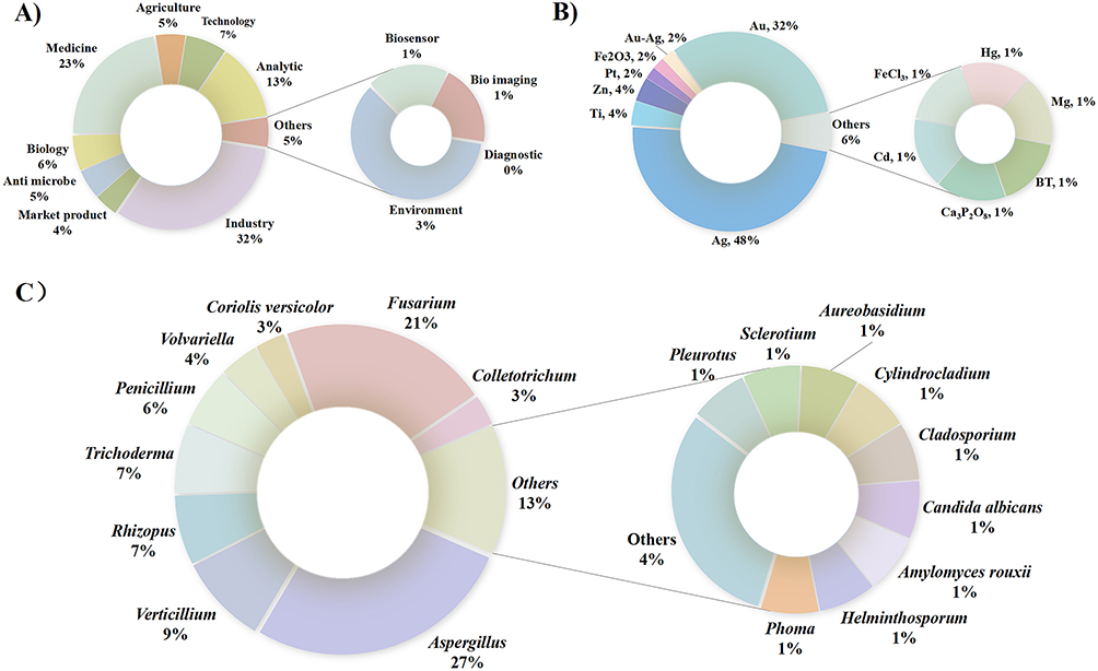 |
Figure 4 Overview of nanoparticle synthesis by fungi. (A) Different applications of fungal-based nanoparticles. (B) Application of different metals in the biosynthesis of nanoparticles by fungi. (C) Frequency of use of different fungal species for nanoparticles biosynthesis. |
The term “endophyte” was initially coined by Bary in 1866.31 Plant endophytes are microorganisms that inhabit within the intercellular and/or intracellular spaces of their host plant throughout the entirety or a portion of the life cycle of the plant, generally without eliciting disease symptoms.32 Using microorganisms for the biosynthesis of high-value secondary metabolites is a simple and economical approach, enhancing product availability and contributing to the reduction of market prices.33 Since the isolation of the paclitaxel-producing endophytic fungus Taxomyces andreanae from the yew tree in 1993,34 plant endophytic fungi have been recognized as a key source of natural bioactive compounds. With nearly 300,000 plant species globally, each harboring a diverse array of endophytes,35 it is estimated that over one million species of endophytic fungi exist in nature.36 Despite numerous reports on the bioactive compounds of endophytic fungi, this field remains in its nascent stages.
Nanoparticle production by plants is typically facilitated by their enzymes, proteins, and amino acids.37 For example, the reduction of silver ions to nanoparticles from Dendranthema morifolium extract is attributed to the presence of H+, NAD+, and ascorbic acid.38 The alkaloids, proteins, enzymes, amino acids, alcohol compounds, and polysaccharides present in Datura stramonium leaf extract reduce silver ions and synthesize highly stable silver nanoparticles (AgNPs).39 Similar compounds produced by plant endophytic fungi can also facilitate the reduction of metal ions and the synthesis of metal nanoparticles, both intracellularly and extracellularly. Their ability to produce nanoparticles is closely related to their metal ion tolerance and bioaccumulation capacities.40 A strain of endophytic fungus Aspergillus sp. SA17 isolated from seagrass can synthesize spherical zinc oxide nanoparticles with an average size of 7.2 nm.41 Nanosilver particles synthesized by Penicillium polonicum PG21, isolated from Rehmannia glutinosa, exhibit antibacterial activity against plant pathogens and significantly promote the growth of safflower (Carthamus tinctorius).42 The endophytic fungus Chaetomium globosum, isolated from Panax notoginseng, biosynthesizes silver nanoparticles that can protect tomatoes from gray mold.43
Endophytic Fungi and Metal Nanoparticle Synthesis
Isolating endophytic fungi is a complex procedure that requires stringent aseptic conditions and depends on various factors, including the host plant species, tissue or organ of the plant, sampling season, endophytic fungi type targeted for isolation, and culture conditions.44 The initial step involves selecting suitable plants and their corresponding tissue parts, with preference given to those known to successfully synthesize nanoparticles. It is advisable to collect plants that thrive in heavy metal-contaminated environments, as they are likely to harbor endophytic fungi capable of synthesizing nanoparticles.45 After collection, the surfaces of the plant tissue samples must be sterilized to eliminate surface microorganisms. The sterilized tissue samples are then cut into uniform pieces of a given size, and their outer layers are disrupted to facilitate the growth of endophytic fungi. These tissue pieces are then placed onto culture medium plates for incubation at a constant temperature (Figure 5).46 Alternatively, plant tissues can be ground under sterile conditions, serially diluted with sterile water, and plated onto isolation media for incubation at a constant temperature to isolate endophytic fungi.47
 |
Figure 5 Schematic diagram illustrating the extraction of endophytic fungi from plants and metal nanoparticle synthesis. (A) Plant nanoparticle synthesis pathways: a, Extraction of metal nanoparticles absorbed and converted by plants from the environment; b, Nanoparticle synthesis directly within plant organs; c, Nanoparticle synthesis using active products derived from plants. (B) Pathways by which plant fungus endophytes synthesize nanoparticles. a, Nanoparticle synthesis from extracellular active products; b, Nanoparticle synthesis from intracellular actives; c, Metabolic synthesis of nanoparticles using fungal biomass. *: Explanation: The biosynthetic capacity of plant organisms is not necessarily related to whether they originate from a heavy metal-rich environment. |
Although isolating endophytic fungi and metal nanoparticle synthesis are two distinct processes, they may be interrelated. Certain endophytic fungi can synthesize specific metal nanoparticles, which may exert beneficial influences on plant growth and development or exhibit other biological activities. A concise overview of the techniques for synthesizing metal nanoparticles using plant endophytic fungi is provided in Figure 5. The procedure is as follows: (1) Endophytic fungi are cultivated in an appropriate medium under controlled temperature conditions with agitation. Centrifugation or filtration is then applied to isolate endophytic fungal cells and extracellular fluid for nanoparticle synthesis. In intracellular synthesis, nanoparticles are formed within the hyphae. In extracellular synthesis, nanoparticles are produced by the cell-free filtrate derived from the fungi.48 (2) Metal salt solutions of varying volumes are combined with endophytic fungal cells or extracellular fluid of different volumes to facilitate metal nanoparticle synthesis. The formation of certain metal nanoparticles can be preliminarily assessed through observable color changes and subsequently verified using nanoparticle characterization methods, such as spectral scanning.49
Optimization of Synthesis Conditions
The physicochemical properties of nanoparticles depend on their size and morphological structure, with both their dimensions and shape significantly influencing their characteristics and functionalities.50 For instance, the surface area of nanosilver is closely correlated with its inhibitory activity against Staphylococcus aureus and Escherichia coli. Plate-like nanoparticles possess a larger surface area than rod-shaped configurations, resulting in superior antibacterial efficacy.51 Moreover, at a size of 12 nm, zinc oxide nanoparticles can effectively inhibit the growth of pathogenic bacteria.52 Therefore, during the fungal synthesis of nanoparticles, it is crucial to optimize various physical parameters, such as temperature, reaction duration, pH level, and metal ion concentration, to achieve effective nanoparticle morphology, size distribution, and yield efficiency (Table 1 and Figure 6).
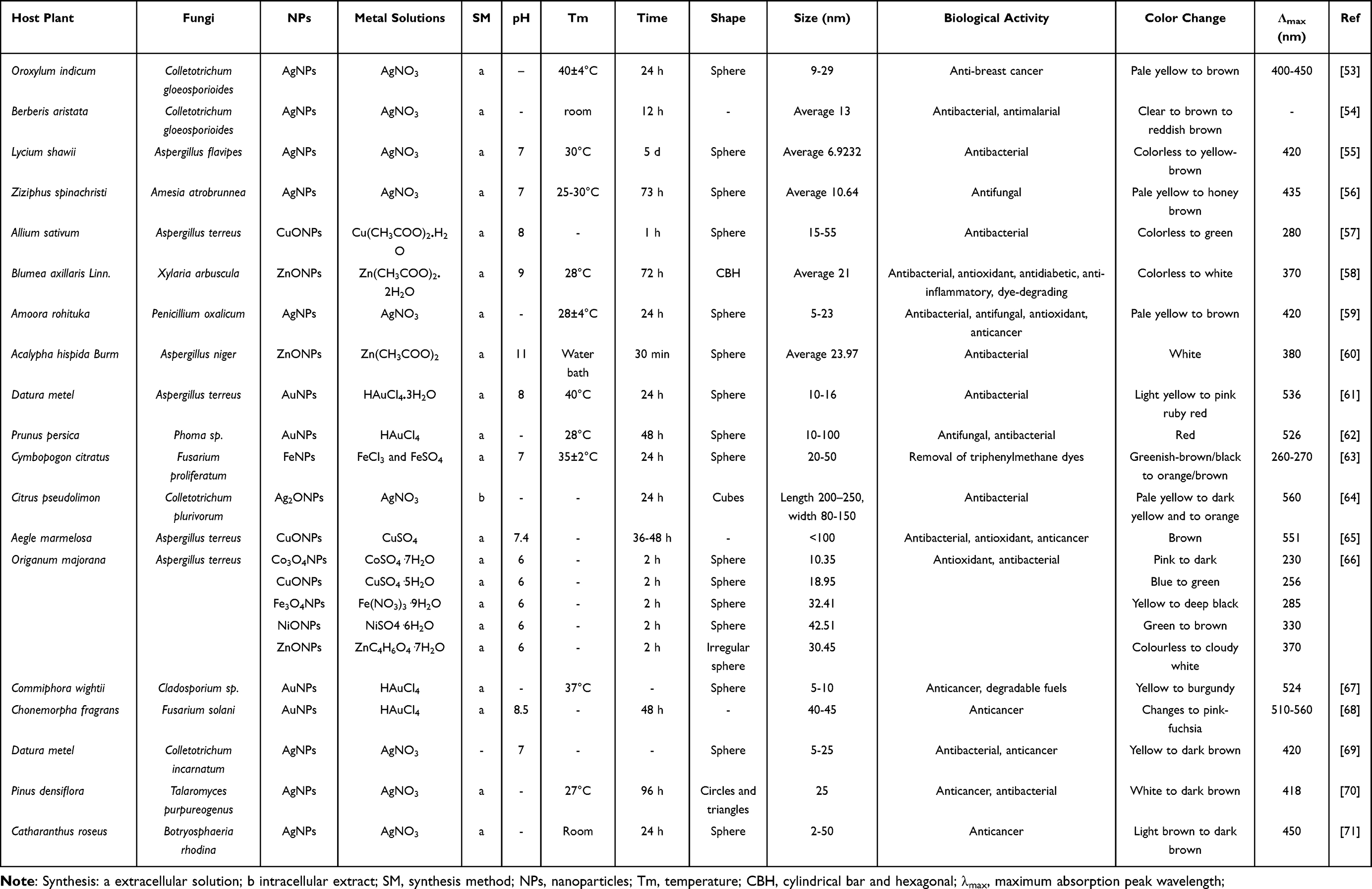 |
Table 1 Characteristics of Nanoparticles Synthesized by Plant-Derived Endophytic Fungi |
 |
Figure 6 Characterization of nanoparticles synthesized by endogenous fungi under optimized conditions. The image of Colletotrichum gloeosporioides is reproduced with permission from Ref.72 and the copyright 2022 Public Library of Science. The characterization images of AgNPs are reproduced with permission from Ref.,53 © 2024 Elsevier B.V. The image of Xylaria arbuscula is reproduced with permission from Ref.,73 © 2024 MDPI (Basel, Switzerland). The characterization images of ZnONPs are reproduced with permission from Ref.,58 © 2023 Informa Healthcare. The image of Cladosporium. sp and the characterization images of AuNPs are reproduced with permission from Ref.,67 © 2020 Elsevier B.V. The image of Fusarium proliferatum is reproduced with permission from Ref.,74 © 2023 Springer Nature. The characterization images of FeNPs are reproduced with permission from Ref.,63 © 2022 Taylor & Francis. The image of Aspergillus terreus is reproduced with permission from Ref.,75 ©2019 MDPI (Basel, Switzerland). The characterization images of CuONPs are reproduced with permission from Ref.,57 © 2023 BMC. The image of A. terreus and the characterization images of Co3O4NPs, Fe3O4NPs, and NiONPs are reproduced with permission from Ref.66 © 2021 Springer Berlin Heidelberg. |
Mechanisms by Which Endophytic Fungi Synthesize Nanoparticles
The capacity of plants to synthesize nanoparticles arises from their diverse mechanisms designed to mitigate the detrimental effects of excessive metal accumulation.11 Compounds with antioxidant properties play a crucial role in these protective mechanisms, as many metals produce reactive oxygen species (ROS) that can lead to significant cellular damage.76 For example, phenolic compounds can enhance the antioxidant capacity of plants. The accumulation, reduction, and stabilization of metals within plant tissues have been confirmed in bioremediation processes, where metals are deposited as nanoparticles within the plant.77
Endophytic fungi play a crucial role in maintaining and supporting the normal functions of their host plants.78 Notably, these fungi can produce metabolites that are either identical or functionally analogous to those produced by their host plants.79 Given the synthetic capabilities of both plants and endophytic fungi, the demonstrated advantages of fungi in nanoparticle synthesis, their symbiotic relationship with host plants, and established successful synthesis protocols (Table 1), we propose the common mechanisms involved in the synthesis of fungal nanoparticles.80 First, the polymers present in the cell wall function as electron carriers in reduction reactions catalyzed by nitrate-dependent reductases and pH-sensitive oxidoreductases. Second, the transfer of electrons from the external environment into the intracellular space can occur through metal reduction, facilitated by low-molecular-weight redox molecules such as nicotinamide adenine dinucleotide (NADH) or flavin dehydrogenase, as well as through direct interaction with cytochrome C oxidoreductase proteins. This process promotes the complex reduction of target metal ions into their corresponding nanoparticles.81 Finally, metal precursors are subjected to oxidation and reduction reactions catalyzed by quinone derivatives derived from naphthoquinone and anthraquinone. These metal precursors are subsequently stabilized through the activity of fungal proteins and chelatins.
Intracellular and Extracellular Synthesis Pathways
Microbiologically mediated nanoparticle synthesis encompasses both extracellular and intracellular methods, using cell-free supernatants and cell lysates or extracts, respectively.82 There are notable differences between these two methods (Figure 7). For example, Fusarium oxysporum produces two distinct forms of nanoparticles: extracellularly secreted and intracellularly synthesized nanoparticles.83 In the intracellular synthesis pathway, F. oxysporum is cultivated under optimal growth conditions to yield sufficient biomass. The harvested mycelium undergoes washing with sterile water, followed by incubation with an appropriate concentration of metal salts. The electromotive force facilitates the attraction and rapid absorption of cations, resulting in the adhesion of metal ions (M+) to lysine residues on the fungal cell wall. This process is further supported by electrostatic interactions with the negatively charged surface of F. oxysporum. Subsequently, M+ is reduced and precipitated by enzymes located within the cytoplasmic membrane, such as ATPase and hydrogenase, as well as those in the cell wall. This process results in nanoparticle production, followed by vesicle aggregation and extracellular secretion.80 Conversely, during extracellular synthesis, fungal enzymes present in the supernatant following liquid culture, including cytochrome b5 reductase, oxidoreductase, FAD-dependent glutathione reductase, and NADH-dependent nitrate reductase, are incubated with an aqueous solution of metal salts to facilitate nanoparticle generation. These nanoparticles are stabilized by nitrogen-containing biomacromolecules or proteins present in the supernatant.59
 |
Figure 7 Schematic illustration of the mechanisms underlying fungi-mediated nanoparticle synthesis, specifically that of silver nanoparticles. (A) Extracellular fungal products reduce silver ions to form nanoparticles. (B) Intracellular substances within the fungus convert absorbed silver ions into nanoparticles. (C) Fungal cell wall proteins, enzymes, and polymers, along with electron shuttles such as naphthoquinones and anthraquinones, catalyze the reduction process through NADPH-dependent nitrate reductase. Subsequently, fungal proteins stabilize the nanoparticles. |
Roles of Fungal Enzymes in Nanoparticle Synthesis
Regardless of whether the synthesis of nanoparticles occurs intracellularly or extracellularly, the cellular constituents of fungi serve as crucial electron donors, playing a crucial role in nanoparticle synthesis through their catalytic activities.84 Fungal constituents include cell walls, membrane-associated molecules, enzymes, cationic proteins, alkaloids, flavonoids, polysaccharides, phenolics, and other organic biomolecules. Fungi can degrade or hydrolyze intricate polymeric substrates through the action of extracellular enzymes, enabling them to assimilate and metabolize these substrates. Compared to other microorganisms, fungi consistently produce enzymes in substantial quantities. Owing to their cost-effective cultivation methods, reliable genetic manipulation techniques, and efficient downstream processing procedures, fungi have received considerable attention across various enzyme-based industries.85
In the green biosynthesis of nanoparticles, enzymes, cofactors, and biomolecules involved in the reduction of metal ions to metal nanoparticles, along with their respective mechanisms of action, are integral components of the process.86 For example, nitrate reductase (NR), a ubiquitous enzyme, functions as a soluble NADPH-dependent assimilatory enzyme in bacteria, fungi, and plants. It facilitates the conversion of nitrate into nitrite, which is then reduced to ammonia by nitrite reductase.87 Notably, NR has been identified as a crucial participant in nanoparticle biosynthesis, making it a focal point in studies investigating the synthesis mechanism. In 2006, Kumar et al used an α-NADPH-dependent NR derived from the fungus F. oxysporum, along with phytochelatin, to successfully synthesize AgNPs in vitro. Under the catalytic action of NR, Ag+ was reduced to form stable silver sols with diameters of 10–25 nm, which were stabilized through the capping peptides.88 Additionally, sulfite reductase (SR) is a key enzyme in the assimilation of sulfur and the dissimilation of oxygen anions such as sulfate and sulfite.89 Sastry et al reported that F. oxysporum secretes SR, converting sulfate ions into sulfide ions. These sulfide ions subsequently react with aqueous Cd2+ to form highly stable cadmium sulfide nanoparticles with sizes of 5–20 nm.90
Proteases constitute a substantial class of enzymes, categorized into six primary categories based on substrate specificity, catalytic mechanism, active site attributes, optimal operational parameters, and thermal stability. These categories include serine proteases, serine carboxypeptidases, cysteine proteases, aspartic proteases, metalloproteases, and metal carboxypeptidases.91 Despite the scarcity of current reports on fungal proteases, bacterial proteases have been successfully used to synthesize nanoparticles, providing insights for investigating nanoparticle synthesis associated with endophytic fungi. For example, Muzikar used purified and overexpressed Escherichia coli harboring the glutathione reductase (GR) gene to synthesize gold nanoparticles (AuNPs), demonstrating that GR can catalyze NADPH-dependent gold reduction. This process resulted in the formation of AuNPs that can bind to the active site, with their size precisely controllable over a broad range.92
Characterization Methods
Structurally, nanoparticles can be classified into decahedra, icosahedra, face-centered cubic, and hexagonal close-packed structures.93 One of the most notable characteristics of nanomaterials is their minuscule size.94 Nanomaterials are composed of minute molecular or atomic units, conferring them with physical or chemical properties that distinguish them from bulk materials consisting of the same chemical elements. These distinctive mechanical, electrical, magnetic, and thermal properties have been widely leveraged in rapidly evolving technological fields.95 Determining the success of nanoparticle synthesis and assessing the quality of the synthesized nanoparticles are crucial steps in advancing nanotechnology. Nanoparticles are characterized by attributes such as size, shape, surface area, and dispersibility,96, 97 and the uniformity of these characteristics is crucial across many applications. Common methods used for characterizing nanoparticles include ultraviolet-visible spectroscopy, dynamic light scattering, scanning electron microscopy, transmission electron microscopy, Fourier transform infrared spectroscopy, powder X-ray diffraction, and energy-dispersive spectroscopy. Each of these techniques has certain advantages in the characterization of nanoparticles, as outlined in Table 2.
 |
Table 2 Various Techniques to Characterize Nanoparticles Along with Their Respective Merits and Demerits |
Application of Biosynthetic Metal Nanoparticles in Cancer Treatment
Cancer poses a significant threat to human life and health, presenting a considerable challenge for the medical community in developing definitive cures. Current clinical treatments for cancer include surgery, chemotherapy, immunotherapy, and radiation therapy.104 Notably, advancements in nanotechnology and nanoparticle biosynthesis using endophytic fungi have driven significant progress in the application of biosynthesized nanomaterials to cancer research and treatment.105 Nanotechnology represents an innovative and promising field that offers exceptional tools for cancer detection and management.106 The applications of nanoparticles in anticancer research (Figure 8) are extensive:107 (1) They provide novel imaging agents that enhance imaging efficacy and seamlessly integrate with existing detection instruments, thereby advancing cancer diagnosis and treatment while minimizing toxic side effects.108 (2) Nanoparticles help overcome biological barriers to directly deliver therapeutic drugs to cancer tissues, making personalized medicine increasingly feasible in routine clinical practice.109 Metal nanoparticles represent a diverse range of nanoscale materials containing various metal ions, such as iron, copper, zinc, gold, silver, and titanium, which can be combined with other organic or inorganic components.110 This combination enhances their pharmacological functions. Moreover, these metal-derived nanocomposites, including antibodies and peptides, can target cancer cells or prolong their circulation time in conjunction with biocompatible polymers for therapeutic purposes.111 (3) Nanoparticles enable tumor visualization and targeted drug delivery, indirectly contributing to cancer diagnosis and treatment. Furthermore, biomedical metal nanoparticles may exhibit direct anticancer properties and serve as pivotal agents in cancer therapy. In this process, metal ions within the nanoparticles can generate ROS and induce oxidative stress, leading to the death of malignant cells.112
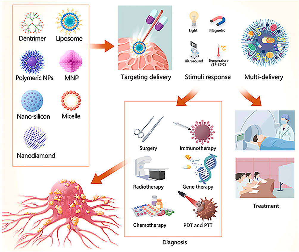 |
Figure 8 An example of the application of nanobiomaterials to the diagnosis and treatment of head and neck cancers (HNCs). Nanomaterials can serve as carriers to encapsulate various agents, including imaging agents, drugs, genes, vaccines, radiosensitizers, and photosensitizers. These nanocarriers enable targeted delivery, stimulus response, and multi-delivery, potentially enhancing the efficiency of HNC diagnosis and treatment. Reproduced with permission from Ref.113 © 2023 Elsevier B.V. |
Cancer Detection and Diagnosis
Traditionally, tumor diagnosis often involves surgical excision for tissue sampling. However, this approach has several limitations, including biodegradation, limited specificity, and potential inaccuracies in results. Subsequent therapeutic interventions are often hindered by short drug retention periods and inadequate targeting capabilities.114 The application of nanotechnology in molecular diagnostics, known as nanodiagnostics,115 involves integrating nanodevices with existing diagnostic methods to enable more precise in vivo and in vitro detection of cancerous lesions (Figure 9A).116 This integration significantly enhances detection sensitivity, facilitating early diagnosis and treatment.117 In terms of imaging modalities, computed tomography (CT) is effective at detecting differences in electronic density between tumors and surrounding tissues, making it a commonly used tool for identifying cancer cells. CT provides comprehensive information about tumors, including their size and precise location.118 However, its sensitivity is often compromised in soft tissues,119 and its application alone is insufficient for effectively detecting potential tumors. Notably, this limitation can be addressed by incorporating contrast agents, such as nanoparticles (Figure 9B).120 Currently, CT utilizes folic acid-cysteine-AuNPs to improve the detection of small and indistinguishable head and neck tumors.113 Magnetic resonance imaging (MRI) is also used for tumor detection, where magnetic nanoparticles or iron oxide nanoparticles serve as contrast agents targeting cancer cells.121 These agents enable the quantification of the magnetization of hydrogen molecules (Figure 9C),122 enhancing the visibility and quantification of images.123 Although magnetic nanoparticles cannot penetrate cancer cells, they can be taken up by healthy Kupffer cells in the liver, resulting in distinct MRI images that differentiate healthy from cancerous cells. Cancer cells appear in high-signal images, whereas healthy cells generate low-signal images.124 This capability has greatly advanced liver imaging, meeting the medical needs of patients with cancerous lesions and those with significant liver and kidney impairments. In addition to standard paramagnetic nanoparticles, the versatility of nanoparticles can be leveraged by embedding tumor-specific markers within them, leading to differentiated images for various cancer types. Such combinations may play a crucial role in the diagnosis and treatment of tumors.125 However, the current availability of biosynthesized nanoparticles for research and application in this field remains limited, highlighting a potential direction for future investigation.
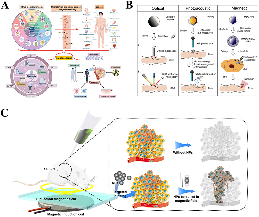 |
Figure 9 Application of nanoparticles to cancer diagnostics. (A) Summary of the diagnostic techniques used in nanomedicine during the therapeutic process. Reproduced with permission from Ref.,106 © 2024 Springer Nature. (B) Metal and metal oxide nanoparticles used in tumor imaging. Reproduced with permission from Ref.,110 © 2020 Elsevier B.V. (C) A schematic illustrating mice undergoing nanoparticle-enhanced diagnostic imaging. Reproduced with permission from Ref.,122 © 2021 Springer US. |
Synergistic Effects of Nanoparticles in Combination with Anticancer Drugs
When combined with existing therapeutic compounds or drugs, nanomaterials can enhance efficacy, particularly by reducing drug side effects and improving treatment safety and stability. Utilizing the synergistic interaction between nanoparticles and drugs represents an effective strategy for increasing the activity of anticancer drugs and enhancing their bioavailability (Figure 10). Specifically, nanoparticles can target cancer cells either passively or actively, exhibiting promise as drug delivery systems.126 Tumors often have large interstitial gaps of approximately 100 to 800 nm, which create defective, barrier-like blood vessels. These vessels allow only small nanoparticles to traverse and accumulate near the tumor. This selective accumulation minimizes interactions between nanoparticles and normal cells, minimizing adverse effects on healthy tissue and enhancing drug efficacy through targeted delivery.126 In addition to passive targeting, which relies on the physical attributes of nanoparticles, the particle surface can be further modified by attaching specific targeting molecules or applying biocompatible molecules and biodegradable polymers.127 However, not all combinations yield synergistic outcomes, as effective nanocarriers for the targeted delivery of anticancer drugs must meet the following prerequisites: (1) possess a strong affinity for anticancer drugs and enable conjugation; (2) ensure complete drug release the intended target site; (3) maintain stability of the drug–nanoparticle complex in serum; and (4) their degradation must pose no risk to the organism.128
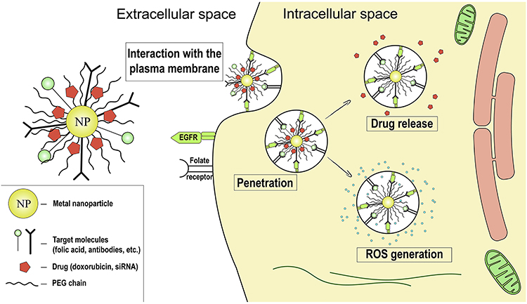 |
Figure 10 Application of metal nanoparticles in chemotherapy. Nanoparticles (NPs) can carry antibodies targeting tumor-specific receptors, directing them to the tumor site. They penetrate the plasma membrane to release chemotherapeutic agents. They exhibit antitumor potential by generating reactive oxygen species (ROS) and inducing tumor cell death. Reproduced with permission from Ref.,110 © 2020 Elsevier B.V. |
The synergistic properties of nanoparticles have been effectively utilized to enhance the efficacy of conventional pharmaceuticals. For example, NGO-AgNPs-PEG, a PEGylated silver-decorated graphene nanocomposite material synthesized using the aqueous extract of Azadirachta indica leaves as a reducing agent and loaded with doxorubicin (DOX), demonstrates significantly improved loading efficiency. Compared to free DOX, NGO-AgNPs-PEG loaded with DOX causes reduced damage to normal cells while exhibiting increased toxicity to HeLa cells.129 Moreover, imatinib-loaded silver nanoparticles (IMAB-AgNPs) exhibited a dose-dependent cytotoxic effect on MCF-7 cells, with half maximal inhibitory concentration (IC50) values of 1.69, 3.02, and 9.63 µM for IMAB-AgNPs, IMAB, and AgNPs, respectively. IMAB-AgNPs also induce the expression of apoptotic proteins.130 In another study, AgNPs, biosynthesized using the water extract of Setaria verticillata seeds, were loaded with the hydrophilic anticancer drugs daunorubicin (DNR) and DOX. Notably, the cytotoxicity of these AgNPs increased with the concentration of seed extract, and the loading efficiency of DNR-AgNPs was 40.25%, whereas that of DOX-AgNPs was 80.50%.131
Direct Intervention Effects of Nanoparticles
To date, relatively few studies have explored the use of biosynthesized nanoparticles in oncological diagnostics. In contrast, bio-sourced nanoparticles have been widely investigated as adjuncts to clinically validated drugs for therapeutic applications. Most research on cancer nanotherapeutics has primarily focused on the effects of nanoparticles on cancer cells. The most direct effects of nanoparticles on these cells include the induction of cell death mechanisms such as apoptosis, cell cycle arrest, and ferroptosis. Moreover, the progression of tumors to advanced stages is largely attributed to the proliferation and spread of cancer cells. Certain nanoparticles exhibit anti-angiogenic and anti-metastatic properties, significantly slowing disease progression. Additionally, Virchow’s hypothesis,132 which suggests a link between lymphoreticular infiltration and cancer development at sites of chronic inflammation, underscores the crucial role of inflammatory processes in regulating cancer progression through inflammatory factors, cytokines, and chemokines within the tumor microenvironment.133 Notably, metal nanoparticles often exert robust anti-inflammatory effects and can be used to inhibit tumor growth and metastatic dissemination associated with chronic inflammation.134
Direct Impacts on Cancer Cells
Inducing apoptosis in tumor cells is a crucial strategy in the development of anticancer drugs.135 When cells are exposed to external, non-receptor-mediated stimuli, the mitochondrial membrane potential becomes disrupted, triggering the apoptotic process.136 In the extrinsic signaling pathway, apoptosis is triggered by the activation of a transmembrane receptor, facilitating the transduction of extracellular signals into the cytoplasm. Importantly, metal nanoparticles can stimulate apoptosis in various cancer cells, including breast, lung, liver, colorectal, bladder, ovarian, and cervical cancer cells (Figure 11A).137
 |
Figure 11 Direct intervention role of nanoparticles. (A) A representative scheme of intrinsic and extrinsic signaling pathways governed by metal nanoparticles (NPs), leading to apoptotic death in cancer cells. Ag, Zn, and Au NPs are the most widely investigated nanoparticles, owing to their ability to induce apoptosis in cancer cells by generating cytotoxic reactive oxygen species (ROS). (B) Metal NPs (Fe and Mn) mediate the induction of ferroptosis in cancer cells via the Fenton reaction. (C) Molecular mechanisms involved in the anti-angiogenic and anti-metastatic actions of metal nanoparticles. (D) Anti-inflammatory activity of metal nanoparticles. Reproduced with permission from Ref.,107 © 2022 Springer Berlin Heidelberg. |
In gastrointestinal digestion, AgNPs can induce apoptosis by upregulating nicotinamide adenine dinucleotide oxidase (4-NOX4), thereby stimulating mitochondrial oxidative stress. When colon cancer cells (HCT116) are treated with AgNPs, pre-treatment with diphenyleneiodonium chloride (DPI) or 4-phenylbutyric acid inhibits several key responses, including the generation of mitochondrial ROS, apoptosis, endoplasmic reticulum stress response, NOX4 expression, and mitochondrial dysfunction.138
Nanoparticle synthesis using plant extracts has witnessed advancements. For example, the reduction of AgNO3 using an extract from Chinese cabbage leaves (Brassica rapa var. japonica) acts as both a reductant and stabilizer, resulting in the production of spherical AgNPs with a size range of 15–30 nm. These nanoparticles exhibit potent growth inhibitory effects against certain bacteria and cause the downregulation of the NF-κB/Akt/mTOR signaling pathways in colorectal cancer (Caco-2) cells, leading to apoptosis and DNA damage.139 Moreover, the endophytic fungus F. chlamydosporum MW341592.1, isolated from healthy leaves of Eucalyptus sideroxylon, can biosynthesize both zinc oxide nanoparticles (ZnONPs) and AuNPs. These nanoparticles can reduce the number of HCT116 human colon cancer cells and Caco-2 human intestinal cancer cells.140 In human breast cancer cells (MCF-7), bismuth(III) oxide nanoparticles demonstrate dose- and time-dependent apoptotic effects by inhibiting the mitochondrial membrane potential and activating caspase-3.141 Moreover, AgNPs biosynthesized by Chaetomorpha linum can downregulate the expression of anti-apoptotic proteins, such as Bcl-2 and Bcl-xl, in MCF-7 cells.142 AgNPs synthesized using a water extract of Bergenia ligulata as a reducing agent can induce apoptosis by upregulating P53-dependent pro-apoptotic markers, including Bax and caspase-3.143 In MCF-7 and mouse breast cancer cells, a significant negative correlation has been observed between the concentration of ZnONPs produced using Cucumis melo extract and cell viability, with ZnONPs including apoptosis by upregulating the expression of caspases 3 and 8.144 Moreover, selenium nanoparticles (SeNPs) have been successfully synthesized using four endophytic fungi present in garlic roots (Allium sativum). These SeNPs exert anticancer activity against PC3 and MCF7 cells, with IC50 values of 225.7 ± 3.6 and 283.8 ± 7.5 µg/mL, respectively, while maintaining biocompatibility with normal WI38 and Vero cells.145 In human ovarian cells, treatment with ZnONPs resulted in the upregulation of the expression of caspase-9, Rad51, γ-H2AX, p53, and LC3, and the downregulation of Bcl-2 expression, leading to significant cytotoxicity, apoptosis, and autophagy.146 In HeLa cells, AuNPs synthesized by Catharanthus roseus induce apoptosis by increasing Bax expression and decreasing that of anti-apoptotic proteins such as Bcl-2 and Bid.147
In addition to their importance in the digestive tract and gynecological health, metal nanoparticles also have applications in other fields. In studies on the treatment of human epidermoid carcinoma cells, zinc oxide nanoparticles, synthesized through the sol-gel method using zinc acetate dihydrate, ethylene glycol, and 2-propanol, induced apoptotic cell death by generating ROS and causing DNA damage.148 Wang et al biosynthesized ZnONPs using Marsdenia tenacissima and demonstrated their role in promoting cell apoptosis in laryngeal cancer cells (Hep-2) by upregulating the expression of Bax and Caspases 3 and 9.149 In human lung epithelial carcinoma (BEAS-2B) cells, titanium dioxide nanoparticles (TiNPs) induce apoptotic death through the mitochondrial apoptosis pathway by increasing the expression of cytochrome C, p53, and caspase 9.150 In human liver cancer, cerium dioxide nanoparticles reduce cell viability and induce apoptotic death by generating ROS and phosphorylating ERK1/2, JNK, and p38 MAPK.151
Additional effects and actions of metal nanoparticles synthesized using endophytic fungi on cancer cells are presented in Table 3. Metal nanoparticles can induce apoptotic cell death and cell cycle arrest. For example, AgNPs biosynthesized using papaya leaf extract downregulate cyclin D1 and upregulate tumor suppressor protein expression, including cip1/p21 and kip1/p27. This results in cell cycle arrest at the G1-S phase, exhibiting cytotoxic activity against human prostate cancer cells (DU145).152 Similarly, spherical AgNPs measuring 12.96 nm in diameter, biosynthesized using Juniperus chinensis extract, decrease the expression level of cyclin D1 protein, leading to cell cycle arrest at the G0/G1 phase in A549 lung cancer cells in vitro.153 AuNPs synthesized with monodisperse properties and high stability, using the water extract of Sesuvium portulacastrum, can induce cell cycle arrest at the G0/G2 phase in A549 cells, thereby inhibiting cell cycle progression.154 Moreover, TiO2NPs induce genotoxicity and cause G2/M cell cycle arrest in A549 cells by upregulating ATM, P53, and cd-2, while downregulating cyclin B1 expression.155 In HCT116 cells, these nanoparticles exhibit cytotoxicity by inducing apoptosis and G1 cell cycle arrest.156 In MCF-7 cells, 30 nm AuNPs significantly decrease overall cell viability and induce G2/M arrest. Compared to the control group, the treated cells exhibit an increased number of apoptotic cells, along with significant upregulation of CDH1 and downregulation of PALB2 expression.157
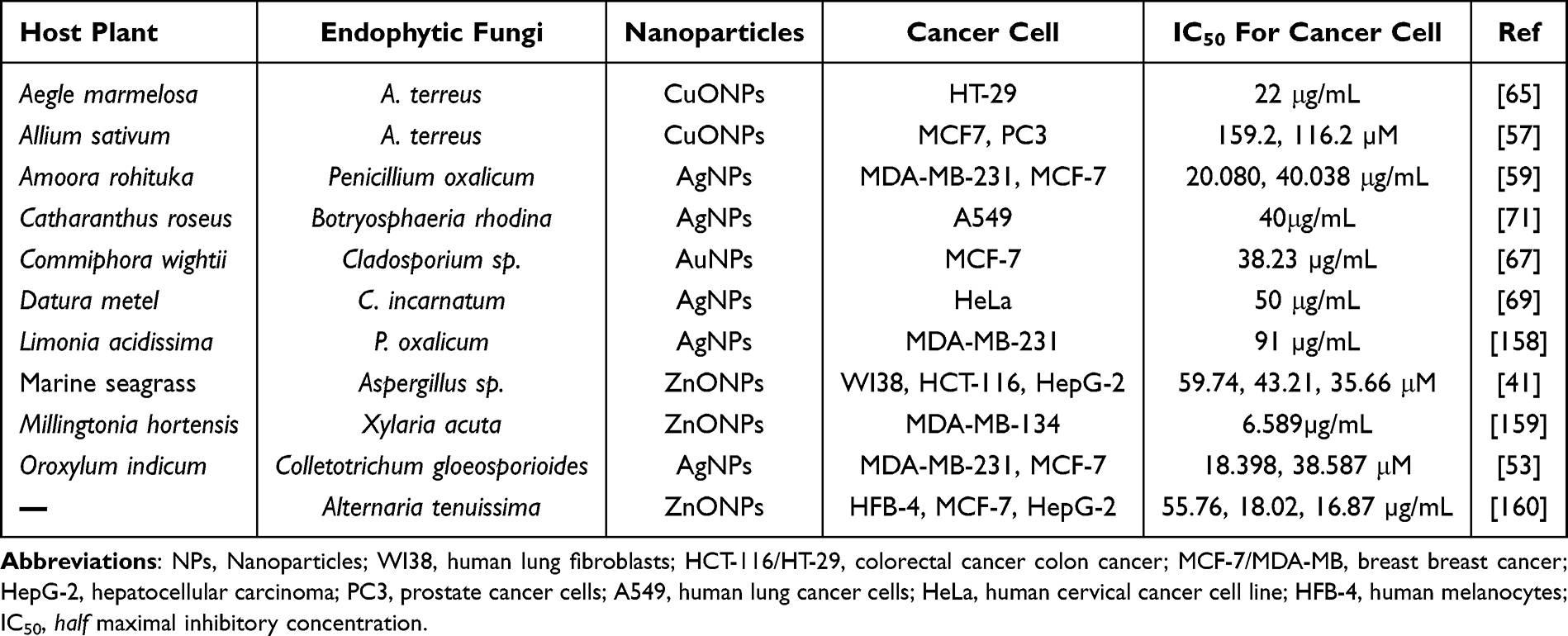 |
Table 3 Anticancer Effects of Metal Nanoparticles Biosynthesised by Plant Endophytic Fungi |
Ferroptosis is a novel, regulated form of cell death caused by an imbalance between oxidative damage and protective mechanisms, holding significant potential as a strategy for cancer treatment.161 In cancer cells, hydrogen peroxide (H2O2) secreted by mitochondria interacts with iron ions, resulting in the generation of harmful free radicals that can induce cell death through iron-mediated toxicity. The primary mechanism underlying ferroptosis involves iron-dependent Fenton reactions that produce ROS.162 Fe3+ ions enter the cell via glycoprotein transferrin receptors and are subsequently reduced and released from transferrin glycoproteins to form Fe2+. The increased availability of Fe ions enhances the Fenton reaction between iron and H2O2, leading to ROS production. These ROS oxidize polyunsaturated fatty acids, resulting in the formation of toxic lipid radicals and ultimately cell death.163 Based on the mechanism of the Fenton reaction, various nanocancer drugs that induce ferroptosis have been developed (Figure 11B). Among these, glutathione peroxidase 4 (GPX4) utilizes glutathione to neutralize phospholipid peroxides, protecting cells from iron overload and serving as a crucial regulator in metal-induced ferroptosis mechanisms.164 Cheng et al designed a manganese-deposited iron oxide nanoparticle platform (Pt-FMO) loaded with the prodrug cisplatin to initiate a series of intracellular reactions, generating ROS to enhance the effects of iron oxidation, and explored the mechanism underlying its induction of ferroptosis.165 The ethanol extract of Cirsium japonicum biosynthesized novel AuNPs that elicit oxidative stress and iron-dependent ferroptosis in human gastric adenocarcinoma cells. These AuNPs achieve their effect by disrupting GPX4-dependent antioxidant defense, leading to the accumulation of mitochondrial ROS, Fe2+, and lipid peroxides, as well as mitochondrial damage.166 These nanotherapeutic agents initiate apoptotic cell death by upregulating the expression of the apoptosis-inducing factor.
Preventing Cancer Cell Metastasis Through Anti-Angiogenesis and Anti-Metastasis Effects
Angiogenesis is the formation of new blood vessels from existing ones.167 This process is common and plays a crucial role in several physiological contexts, including ovulation, embryonic development, and wound healing.168 It is also essential for cancer cells to acquire nutrients and oxygen,169 making it a key factor in tumor proliferation and migration.170 Metal nanoparticles have emerged as innovative and effective inhibitors of angiogenesis and metastasis.171 Within the tumor microenvironment, gold-core silver-shell hybrid nanomaterials can reduce the supportive effects of cancer-associated fibroblasts on tumor cells. By influencing the gene expression and secretion profile of stromal fibroblasts, metal nanoparticles can modify their communication with malignant cells, thereby affecting the pro-cancerous activity within the tumor microenvironment (Figure 11C).172 Katifelis et al investigated the effects of Ag3Au1Trp1:2 nanoparticles on tumor growth, metastasis, and the underlying molecular mechanisms using a server combined immune-deficiency mouse tumor model. These nanoparticles accumulate at the tumor site owing to their enhanced permeability and retention effects and selectively induce apoptosis in cancer cells via a Tumor Necrosis Factor-Related Apoptosis-Inducing Ligand-dependent extrinsic pathway. In terms of mechanism action and signal transduction, the vascular endothelial growth factor (VEGF)/vascular endothelial growth factor receptor (VEGFR)-mediated signaling pathway is a key molecular target for assessing anti-angiogenic efficacy.173 AuNPs can bind to vascular permeability factor/VEGF-165 and basic fibroblast growth factor (bFGF), both of which are mitogens and angiogenic mediators for endothelial cells. This binding inhibits the proliferation of endothelial cells and fibroblasts in vitro, as well as VEGF-induced permeability and angiogenesis in vivo.174 Arvizo et al postulated that the anti-angiogenic effect of AuNPs is attributed to their capacity to inhibit VEGF165 and bFGF, thereby enhancing the functionality of heparin-binding growth factors (HB-GFs). They demonstrated that the inhibitory action of AuNPs is due to the nanoparticles altering the conformation/configuration (denaturation) of HB-GFs.175 Moreover, silver oxide nanoparticles synthesized by the endophytic fungus Aspergillus terreus, isolated from the medicinal plant Aegle marmelos, can inhibit or prevent angiogenesis and suppress the formation of new blood vessels in a dose-dependent manner across various culture stages.176 Similarly, two strains of endophytic fungi, Aspergillus sydowi and Aspergillus versicolor, isolated from Azadirachta indica, are also capable of synthesizing ZnO nanoparticles with notable anti-angiogenic activity.177
Anti-Inflammatory Effects of Metal Nanoparticles
Cancer progression is linked to chronic inflammation, which is estimated to contribute to approximately 20% of all cancer cases.178 Within the inflammatory microenvironment, elevated levels of nuclear factor-κB (NF-κB), cytokines, prostaglandins, and microRNAs lead to changes in cell proliferation, senescence, and apoptosis, as well as aberrant DNA mutations, DNA methylation, and angiogenesis.179 Soluble mediators, such as chemokines and cytokines, are integral to the inflammatory process and can regulate both healthy and tumorigenic cells. Pro-inflammatory mediators, including proteases, cytokines, chemokines, and other inflammatory factors, are abundant in the tumor microenvironment. A diverse array of cytokines, including TNF-α, Interleukin (IL)-6, IL-17, IL-12, IL-23, IL-10, and transforming growth factor-β, collectively shape the tumor microenvironment, often promoting tumor growth.180 Notably, metal nanoparticles can inhibit these inflammatory responses in cancer and modulate the interaction between systemic inflammation and local immune responses (Figure 11D).181 For example, AuNPs can reduce lipopolysaccharide-induced cytokine production, including IL-1, IL-17, and TNF-α, as well as ROS generation, by regulating the mitogen-activated protein kinase and phosphatidylinositol 3-kinase pathways.182 Conversely, AgNPs exert their anti-inflammatory effects by suppressing the expression of vascular endothelial growth factor, hypoxia-inducible factor 1, cyclooxygenase-2, IL-13, and TNF-α.183 Numerous biosynthesized metal and metal oxide nanoparticles exhibit anti-inflammatory properties.184 AgNPs synthesized from extracts of Viburnum opulus, Calophyllum tomentosum, and Centratherum punctatum;185 AuNPs synthesized from extracts of Litchi chinensis, Prunus domestica, P. serrulata, and Rubia cordifolia; and zinc oxide nanoparticles synthesized from extracts of Polygala tenuifolia and Andrographis paniculate,186 all exhibit anti-inflammatory effects.184
Conclusion and Outlook
Owing to increasing industrial demand and a societal focus on environmental sustainability, the application fields for nanoparticles and nanomaterials are continually expanding, highlighting the urgent need for green synthesis methods for nanoparticles. Plant endophytic fungi, which exhibit a symbiotic relationship with their plant hosts, may produce metabolites similar to those of plants and participate in similar production processes, exhibiting great potential for the green synthesis of metal nanoparticles. Moreover, metal nanoparticles can support cancer treatment in several ways, including enhancing the effectiveness of contrast agents and reducing their toxicity, boosting the effect of chemotherapy drugs, directly producing ROS, and inducing oxidative stress, leading to the death of malignant cells.
The present review provides a comprehensive overview, from the discovery of potential plant endophytic fungi to the synthesis and characterization of metal nanoparticles, and finally to the role of metal nanoparticles in cancer treatment. We comprehensively reviewed the application of metal nanoparticles synthesized by plant endophytic fungi in the field of cancer treatment, providing new perspectives for research on the synthesis of anticancer nanoparticles. In this synthesis process, plant endophytic fungi have demonstrated their advantages: abundant resources, as many studies have reported endophytic fungi with synthetic capabilities; green and environmentally friendly, as plant endophytic fungi can reduce the use of forestry resources and harmful reagents; mild and efficient, given the rapid expansion of plant endophytic fungi fermentation and synthesis reactions under mild conditions; and cost-effective, as their use reduces the amount of chemical reagents used and energy costs owing to the mild conditions. These advantages demonstrate that the biosynthesis of metal nanoparticles by plant endophytic fungi is a promising area of research that warrants further development.
However, we must also recognize that current research is extremely limited. Most of the current research has only focused on the biosynthesis of metal nanoparticles, with limited studies exploring the detailed mechanisms by which endophytes produce these metal nanoparticles. There is also insufficient investigation into the efficacy and safety of bio-sourced metal nanoparticles for cancer treatment, which hinders their clinical application. Additionally, many challenges remain before the widespread adoption of biosynthesis technology. These include the establishment of management systems for microbial resources and toxicity control issues, and the exploration of the controllability and scalability of biosynthetic metal nanoparticles. Moreover, regulatory frameworks must be further refined to ensure the quality of nanoparticle products. However, with the increasing basic research and advancements in biotechnology, we believe that the production and application scenarios of bio-sourced metal nanoparticles will also increase, enabling their improved application in clinical treatment of cancer. In the future, it is important to identify the key secondary metabolic synthesis gene clusters involved in the synthesis process of endophytic fungi, main products involved during the synthesis, and important synthesis conditions that affect the performance of nanoparticles. Moreover, developing a safe and controllable biosynthesis plan using plant endophytic fungi, combined with mature cancer treatment methods, is expected to pave the way for new green and environmentally friendly treatment models.
Data Sharing Statement
No data were used for the research described in the article.
Acknowledgments
We would like to express our sincere gratitude to Editage for their professional language editing services, which greatly enhanced the clarity and quality of our manuscript.
Funding
This article received funding from the Natural Science Foundation of Sichuan Province, China (No. 2024NSFSC0408) and the Chengdu University Talent Project (No. 2081922015).
Disclosure
The authors declare that they have no conflicts of interest.
References
1. Ismail M, Liu J, Wang N, et al. Advanced nanoparticle engineering for precision therapeutics of brain diseases. Biomaterials. 2025;318:123138. doi:10.1016/j.biomaterials.2025.123138
2. Xiong Y, Sun M, Yang Q, et al. Nanoparticle-based drug delivery systems to modulate tumor immune response for glioblastoma treatment. Acta Biomater. 2025;194:38–57. doi:10.1016/j.actbio.2025.01.050
3. Pozzi M, Jonak Dutta S, Kuntze M, et al. Visualization of the high surface-to-volume ratio of nanomaterials and its consequences. J Chem Educ. 2024;101(8):3146–3155. doi:10.1021/acs.jchemed.4c00089
4. Huang R, Huang X, Zhang Q, Fan J, Zhang Z, Huang J. Humidity-responsive pectin/AgNPs/ZnO composite films with high antimicrobial and UV-proof functions. Int J Biol Macromol. 2024;279:135075. doi:10.1016/j.ijbiomac.2024.135075
5. Hasan M, Zafar A, Shahzadi I, et al. Fractionation of biomolecules in withania coagulans extract for bioreductive nanoparticle synthesis, antifungal and biofilm activity. Molecules. 2020;25(15). doi:10.3390/molecules25153478
6. Hasan M, Gulzar H, Zafar A, et al. Multiplexing surface anchored functionalized iron carbide nanoparticle: a low molecular weight proteome responsive nano-tracer. Colloids Surf B. 2021;203:111746. doi:10.1016/j.colsurfb.2021.111746
7. Delgado-Pujol EJ, Martínez G, Casado-Jurado D, et al. Hydrogels and nanogels: pioneering the future of advanced drug delivery systems. Pharmaceutics. 2025;17(2). doi:10.3390/pharmaceutics17020215
8. Karimi S, Bakhshali R, Bolandi S, et al. For and against tumor microenvironment: nanoparticle-based strategies for active cancer therapy. Mater Today Bio. 2025;31:101626. doi:10.1016/j.mtbio.2025.101626
9. Zahoor M, Khan S, Ikram M, Ali S. Electrochemical synthesis of nanoparticles; an appropriate contrivance of synthesizing nanoparticles with low dimensional structures. Inorg Chem Commun. 2025;173:113890. doi:10.1016/j.inoche.2025.113890
10. Patnaik R, Kumar Bagchi S, Rawat I, Bux F. Nanotechnology for the enhancement of algal cultivation and bioprocessing: bridging gaps and unlocking potential. Bioresour Technol. 2024;406:131025. doi:10.1016/j.biortech.2024.131025
11. Maťátková O, Michailidu J, Miškovská A, Kolouchová I, Masák J, Čejková A. Antimicrobial properties and applications of metal nanoparticles biosynthesized by green methods. Biotechnol Adv. 2022;58:107905. doi:10.1016/j.biotechadv.2022.107905
12. Tończyk A, Niedziałkowska K, Nowak-Lange M, Bernat P, Lisowska K. Mycogenic silver nanoparticles: promising antimicrobials with fungistatic properties. Int J Mol Sci. 2025;26(14). doi:10.3390/ijms26146639
13. Xu F, Li Y, Zhao X, et al. Diversity of fungus-mediated synthesis of gold nanoparticles: properties, mechanisms, challenges, and solving methods. Crit Rev Biotechnol. 2024;44(5):924–940. doi:10.1080/07388551.2023.2225131
14. Oyebamiji AK, Akintelu SA, Afolabi SO, Ebenezer O, Akintayo ET, Akintayo CO. A comprehensive review on mycosynthesis of nanoparticles, characteristics, applications, and limitations. Plasmonics. 2025. doi:10.1007/s11468-024-02755-x
15. Tiwari P, Kang S, Bae H. Plant-endophyte associations: rich yet under-explored sources of novel bioactive molecules and applications. Microbiol Res. 2023;266:127241. doi:10.1016/j.micres.2022.127241
16. Hasan M, Ullah I, Zulfiqar H, et al. Biological entities as chemical reactors for synthesis of nanomaterials: progress, challenges and future perspective. Mater Today Chem. 2018;8:13–28. doi:10.1016/j.mtchem.2018.02.003
17. El-Khawaga AM, Zidan A, El-Mageed AIAA. Preparation methods of different nanomaterials for various potential applications: a review. J Mol Struct. 2023;1281:135148. doi:10.1016/j.molstruc.2023.135148
18. Xu L, Wang YY, Huang J, Chen CY, Wang ZX, Xie H. Silver nanoparticles: synthesis, medical applications and biosafety. Theranostics. 2020;10(20):8996–9031. doi:10.7150/thno.45413
19. Habiba K, Makarov V, Weiner B, Morell G. Fabrication of Nanomaterials by Pulsed Laser Synthesis. Waqar Ahmed NA, ed.:Manufacturing Nanostructures. One Central Press: 2014. 263–291.
20. Marooufpour N, Alizadeh M, Hatami M, Asgari Lajayer B. Biological synthesis of nanoparticles by different groups of bacteria. In: Prasad R, editor. Microbial Nanobionics. Vol. 1. State-of-the-Art. Springer International Publishing; 2019:63–85.
21. Adebayo EA, Azeez MA, Alao MB, Oke AM, Aina DA. Fungi as veritable tool in current advances in nanobiotechnology. Heliyon. 2021;7(11):e08480. doi:10.1016/j.heliyon.2021.e08480
22. Madhavi A, Srinivasulu M, Shankar PC, Rangaswamy V. Synthesis and Applications of Fungal-Mediated Nanoparticles. Maddela NR, Rodríguez Díaz JM, Prasad R, eds.:Microbial Processes for Synthesizing Nanomaterials. Springer Nature Singapore: 2023. 113–131.
23. Ahmad S, Munir S, Zeb N, et al. Green nanotechnology: a review on green synthesis of silver nanoparticles - an ecofriendly approach. Int J Nanomed. 2019;14:5087–5107. doi:10.2147/ijn.S200254
24. Gan PP, SFY L. Potential of plant as a biological factory to synthesize gold and silver nanoparticles and their applications. Rev Environ Sci Bio/Technol. 2012;11(2):169–206.
25. Dikshit PK, Kumar J, Das A, et al. Green synthesis of metallic nanoparticles: applications and limitations. Catalysts. 2021;11:1–37. doi:10.3390/catal11080902
26. Küünal S, Rauwel P, Rauwel E.Plant extract mediated synthesis of nanoparticles.Barhoum A, Makhlouf ASH;editors;Emerging Applications of Nanoparticles and Architecture Nanostructures. Elsevier. 2018.411–446.
27. El-Seedi HR, El-Shabasy RM, Khalifa SAM, et al. Metal nanoparticles fabricated by green chemistry using natural extracts: biosynthesis, mechanisms, and applications. RSC Adv. 2019;9(42):24539–24559. doi:10.1039/c9ra02225b
28. Ettadili FE, Aghris S, Laghrib F, et al. Recent advances in the nanoparticles synthesis using plant extract: applications and future recommendations. J Mol Struct. 2022;1248:131538. doi:10.1016/j.molstruc.2021.131538
29. Singh NA, Narang J, Garg D, et al. Nanoparticles synthesis via microorganisms and their prospective applications in agriculture. Plant Nano Biology. 2023;5:100047. doi:10.1016/j.plana.2023.100047
30. Dorcheh SK, Vahabi K. Biosynthesis of Nanoparticles by Fungi: Large-Scale Production. Mérillon J-M, Ramawat KG, eds.:Fungal Metabolites. Springer International Publishing: 2016. 1–20.
31. Staniek AA, Woerdenbag HJ, Kayser O. Endophytes: exploiting biodiversity for the improvement of natural product-based drug discovery. J Plant Interact. 2008;3(75):–93.
32. Tan RX, Zou WX. Endophytes: a rich source of functional metabolites. Nat Prod Rep. 2001;18(4):448–459. doi:10.1039/b100918o
33. Reddy S, Sinha A, Osborne WJ. Microbial secondary metabolites: recent developments and technological challenges. In: Kumar A, Singh J, Samuel J, editors. Volatiles and Metabolites of Microbes. Academic Press; 2021:1–22.
34. Stierle A, Strobel G, Stierle D. Taxol and taxane production by Taxomyces andreanae, an endophytic fungus of Pacific yew. Science. 1993;260(5105):214–216. doi:10.1126/science.8097061
35. Cantwell-Jones A, Ball J, Collar D, et al. Global plant diversity as a reservoir of micronutrients for humanity. Nat Plants. 2022;8(3):225–232. doi:10.1038/s41477-022-01100-6
36. Bhunjun CS, Phukhamsakda C, Hyde KD, McKenzie EHC, Saxena RK, Li Q. Do all fungi have ancestors with endophytic lifestyles? Fungal Diversity. 2024;125(1):73–98. doi:10.1007/s13225-023-00516-5
37. Jadoun S, Arif R, Jangid NK, Meena RK. Green synthesis of nanoparticles using plant extracts: a review. Environ Chem Lett. 2021;19(1):355–374. doi:10.1007/s10311-020-01074-x
38. Ahmad N, Sharma S, Singh VN, Shamsi SF, Fatma A, Mehta BR. Biosynthesis of silver nanoparticles from Desmodium triflorum: a novel approach towards weed utilization. Biotechnol Res Int. 2011;2011:454090. doi:10.4061/2011/454090
39. Kesharwani J, Yoon KY, Hwang J, Rai M. Phytofabrication of silver nanoparticles by leaf extract of Datura metel: hypothetical mechanism involved in synthesis. J Bionanosci. 2009;3:39–44.
40. Saravanan A, Kumar PS, Karishma S, et al. A review on biosynthesis of metal nanoparticles and its environmental applications. Chemosphere. 2021;264:128580. doi:10.1016/j.chemosphere.2020.128580
41. Abdelrahman S, El Hawary S, Mohsen E, et al. Bio-fabricated zinc oxide nanoparticles mediated by endophytic fungus Aspergillus sp. SA17 with antimicrobial and anticancer activities: in vitro supported by in silico studies. Front Microbiol. 2024;15:1366614. doi:10.3389/fmicb.2024.1366614
42. Zhu Y, Hu X, Qiao M, Zhao L, Dong C. Penicillium polonicum-mediated green synthesis of silver nanoparticles: unveiling antimicrobial and seed germination advancements. Heliyon. 2024;10(7):e28971. doi:10.1016/j.heliyon.2024.e28971
43. Sun D, Chen R, Lei L, Zhang F. Green synthesis of silver nanoparticles from the endophytic fungus Panax notoginseng and their antioxidant and antimicrobial activities and effects on cherry tomato preservation. Int J Food Microbiol. 2025;431:111083. doi:10.1016/j.ijfoodmicro.2025.111083
44. Guo XW, Yu ZQ, Xi J, et al. Isolation and identification of novel antioxidant polyketides from an endophytic fungus Ophiobolus cirsii LZU-1509. J Agric Food Chem. 2023;71(3):1593–1606. doi:10.1021/acs.jafc.2c07386
45. Priyadarshini E, Priyadarshini SS, Cousins BG, Pradhan N. Metal-Fungus interaction: review on cellular processes underlying heavy metal detoxification and synthesis of metal nanoparticles. Chemosphere. 2021;274:129976. doi:10.1016/j.chemosphere.2021.129976
46. Passari AK, Mishra VK, Gupta VK, Singh BP.Methods used for the recovery of culturable endophytic actinobacteria: an overview.Singh BP, Gupta VK, Passari AK;editors;New and Future Developments in Microbial Biotechnology and Bioengineering. Elsevier. 2018.1–11.
47. Manias D, Verma A, Soni DK. Isolation and Characterization of Endophytes: Biochemical and Molecular Approach. Kumar A, Singh VK, eds.:Microbial Endophytes. Woodhead Publishing: 2020. 1–14.
48. Koul B, Poonia AK, Yadav D, Jin J-O. Microbe-mediated biosynthesis of nanoparticles: applications and future prospects. Biomolecules. 2021;11(6):886.
49. Jamkhande PG, Ghule NW, Bamer AH, Kalaskar MG. Metal nanoparticles synthesis: an overview on methods of preparation, advantages and disadvantages, and applications. J Drug Delivery Sci Technol. 2019;53:101174. doi:10.1016/j.jddst.2019.101174
50. Tripathy P, Sethi S, Panchal D, et al. Biogenic synthesis of nanoparticles by amalgamating microbial endophytes: potential environmental applications and future perspectives. In: Solanki MK, Yadav MK, Singh BP, Gupta VK, editors. Microbial Endophytes and Plant Growth. Academic Press; 2023. 215–231.
51. Sadeghi B, Garmaroudi FS, Hashemi M, et al. Comparison of the anti-bacterial activity on the nanosilver shapes: nanoparticles, nanorods and nanoplates. Adv Powder Technol. 2012;23(1):22–26. doi:10.1016/j.apt.2010.11.011
52. Rafeeq CM, Paul E, Vidya Saagar E, Manzur Ali PP. Mycosynthesis of zinc oxide nanoparticles using Pleurotus floridanus and optimization of process parameters. Ceram Int. 2021;47(9):12375–12380. doi:10.1016/j.ceramint.2021.01.091
53. Gupta P, Singh S, Rai N, et al. Unveiling the cytotoxic and anti-proliferative potential of green-synthesized silver nanoparticles mediated by Colletotrichum gloeosporioides. RSC Adv. 2024;14(6):4074–4088. doi:10.1039/d3ra06145k
54. Kumar S, Pant M, Prashar C, et al. Myco-synthesis of multi-twinned silver nanoparticles as potential antibacterial and antimalarial agents. RSC Adv. 2024;14(2):1114–1122. doi:10.1039/d3ra07752g
55. El-Zawawy NA, Abou-Zeid AM, Beltagy DM, Hantera NH, Nouh HS. Mycosynthesis of silver nanoparticles from endophytic Aspergillus flavipes AUMC 15772: ovat-statistical optimization, characterization and biological activities. Microb Cell Fact. 22(1):228. doi:10.1186/s12934-023-02238-4
56. S SA-Z, S MA-G. Antifungal potentiality of mycogenic silver nanoparticles capped with chitosan produced by endophytic Amesia atrobrunnea. Saudi J Biol Sci. 2023;30(9):103746. doi:10.1016/j.sjbs.2023.103746
57. Nassar AA, Atta HM, Abdel-Rahman MA, El Naghy WS, Fouda A. Myco-synthesized copper oxide nanoparticles using harnessing metabolites of endophytic fungal strain Aspergillus terreus: an insight into antibacterial, anti-Candida, biocompatibility, anticancer, and antioxidant activities. BMC Complement Med Ther. 2023;23(1):261. doi:10.1186/s12906-023-04056-y
58. Nehru L, Kandasamy GD, Sekar V, et al. Green synthesis of ZnO-NPs using endophytic fungal extract of Xylaria arbuscula from Blumea axillaris and its biological applications. Artif Cells Nanomed Biotechnol. 2023;51(1):318–333. doi:10.1080/21691401.2023.2232654
59. Gupta P, Rai N, Verma A, et al. Green-based approach to synthesize silver nanoparticles using the fungal endophyte Penicillium oxalicum and their antimicrobial, antioxidant, and in vitro anticancer potential. ACS Omega. 2022;7(50):46653–46673. doi:10.1021/acsomega.2c05605
60. Abdelkader DH, Negm WA, Elekhnawy E, Eliwa D, Aldosari BN, Almurshedi AS. Zinc oxide nanoparticles as potential delivery carrier: green synthesis by Aspergillus Niger endophytic fungus, characterization, and in vitro/in vivo antibacterial activity. Pharmaceuticals. 2022;15(9). doi:10.3390/ph15091057
61. Mishra RC, Kalra R, Dilawari R, Goel M, Barrow CJ. Bio-synthesis of Aspergillus terreus mediated gold nanoparticle: antimicrobial, antioxidant, antifungal and in vitro cytotoxicity studies. Materials. 2022;15(11). doi:10.3390/ma15113877
62. Soltani Nejad M, Samandari Najafabadi N, Aghighi S, Pakina E, Zargar M. Evaluation of Phoma sp. biomass as an endophytic fungus for synthesis of extracellular gold nanoparticles with antibacterial and antifungal properties. Molecules. 2022;27(4). doi:10.3390/molecules27041181
63. Schuster S, Su Yien Ting A. Decolourisation of triphenylmethane dyes by biogenically synthesised iron nanoparticles from fungal extract. Mycology. 2022;13(1):56–67. doi:10.1080/21501203.2021.1948928
64. Kumar A, Kumar S, Kiran K, Banerjee S, Pande V, Dandapat A. Myco-nanotechnological approach to synthesize silver oxide nanocuboids using endophytic fungus isolated from Citrus pseudolimon plant. Colloids Surf B Biointerfaces. 2021;206:111948. doi:10.1016/j.colsurfb.2021.111948
65. Mani VM, Kalaivani S, Sabarathinam S, et al. Copper oxide nanoparticles synthesized from an endophytic fungus Aspergillus terreus: bioactivity and anti-cancer evaluations. Environ Res. 2021;201:111502. doi:10.1016/j.envres.2021.111502
66. Mousa SA, El-Sayed ER, Mohamed SS, Abo El-Seoud MA, Elmehlawy AA, Abdou DAM. Novel mycosynthesis of Co(3)O(4), CuO, Fe(3)O(4), NiO, and ZnO nanoparticles by the endophytic Aspergillus terreus and evaluation of their antioxidant and antimicrobial activities. Appl Microbiol Biotechnol. 2021;105(2):741–753. doi:10.1007/s00253-020-11046-4
67. Munawer U, Raghavendra VB, Ningaraju S, et al. Biofabrication of gold nanoparticles mediated by the endophytic Cladosporium species: photodegradation, in vitro anticancer activity and in vivo antitumor studies. Int J Pharm. 588:119729. doi:10.1016/j.ijpharm.2020.119729
68. Clarance P, Luvankar B, Sales J, et al. Green synthesis and characterization of gold nanoparticles using endophytic fungi Fusarium solani and its in-vitro anticancer and biomedical applications. Saudi J Biol Sci. 2020;27(2):706–712. doi:10.1016/j.sjbs.2019.12.026
69. Chandankere R, Chelliah J, Subban K, et al. Pleiotropic functions and biological potentials of silver nanoparticles synthesized by an endophytic fungus. Front Bioeng Biotechnol. 2020;8:95. doi:10.3389/fbioe.2020.00095
70. Hu X, Saravanakumar K, Jin T, Wang MH. Mycosynthesis, characterization, anticancer and antibacterial activity of silver nanoparticles from endophytic fungus Talaromyces purpureogenus. Int J Nanomed. 2019;14:3427–3438. doi:10.2147/ijn.S200817
71. Akther T, Vabeiryureilai M, Nachimuthu Senthil K, Davoodbasha M, Srinivasan H. Fungal-mediated synthesis of pharmaceutically active silver nanoparticles and anticancer property against A549 cells through apoptosis. Environ Sci Pollut Res Int. 2019;26(13):13649–13657. doi:10.1007/s11356-019-04718-w
72. Rai N, Keshri PK, Gupta P, et al. Bioprospecting of fungal endophytes from Oroxylum indicum (L.) Kurz with antioxidant and cytotoxic activity. PLoS One. 2022;17(3):e0264673. doi:10.1371/journal.pone.0264673
73. Tan Q, Ye X, Fu S, et al. The cytochalasins and polyketides from a mangrove endophytic fungus Xylaria arbuscula QYF. Mar Drugs. 2024;22(9). doi:10.3390/md22090407
74. Purayil GP, Almarzooqi AY, El-Tarabily KA, You FM, AbuQamar SF. Fully resolved assembly of Fusarium proliferatum DSM106835 genome. Sci Data. 2023;10(1):705. doi:10.1038/s41597-023-02610-4
75. Asfour HZ, Awan ZA, Bagalagel AA, Elfaky MA, Abdelhameed RFA, Elhady SS. Large-scale production of bioactive terrein by Aspergillus terreus strain S020 isolated from the Saudi coast of the red sea. Biomolecules. 2019;9(9). doi:10.3390/biom9090480
76. Kharissova OV, Kharisov BI, Oliva González CM, Méndez YP, López I. Greener synthesis of chemical compounds and materials. R Soc Open Sci. 2019;6(11):191378. doi:10.1098/rsos.191378
77. Jiang Y, Zhou P, Zhang P, et al. Green synthesis of metal-based nanoparticles for sustainable agriculture. Environ Pollut. 309:119755. doi:10.1016/j.envpol.2022.119755
78. Ji X, Xia Y, Zhang H, Cui J-L. The microscopic mechanism between endophytic fungi and host plants: from recognition to building stable mutually beneficial relationships. Microbiol Res. 2022;261:127056. doi:10.1016/j.micres.2022.127056
79. Gupta S, Chaturvedi P, Kulkarni MG, Van Staden J. A critical review on exploiting the pharmaceutical potential of plant endophytic fungi. Biotechnol Adv. 2020;39:107462. doi:10.1016/j.biotechadv.2019.107462
80. Thipe VC, Batista JGS, Lebre DT, Lugão AB, Katti KV.Fungal nanobionics: principle, advances and applications.Abd-Elsalam KA;editor;Fungal Cell Factories for Sustainable Nanomaterials Productions and Agricultural Applications. Elsevier. 2023.543–577.
81. Liu F, Shah DS, Gadd GM. Role of protein in fungal biomineralization of copper carbonate nanoparticles. Curr Biol. 2021;31(2):358–368.e3. doi:10.1016/j.cub.2020.10.044
82. de Souza TA J, Rosa Souza LR, Franchi LP. Silver nanoparticles: an integrated view of green synthesis methods, transformation in the environment, and toxicity. Ecotoxicol Environ Saf. 2019;171:691–700. doi:10.1016/j.ecoenv.2018.12.095
83. Bahrulolum H, Nooraei S, Javanshir N, et al. Green synthesis of metal nanoparticles using microorganisms and their application in the agrifood sector. J Nanobiotechnology. 2021;19(1):86. doi:10.1186/s12951-021-00834-3
84. Rai M, Bonde S, Golinska P, et al. Fusarium as a novel fungus for the synthesis of nanoparticles: mechanism and applications. J Fungi. 7(2). doi:10.3390/jof7020139
85. Kango N, Jana UK, Choukade R. Fungal enzymes: sources and biotechnological applications. In: Satyanarayana T, Deshmukh SK, Deshpande MV, editors. Advancing Frontiers in Mycology & Mycotechnology: Basic and Applied Aspects of Fungi. Springer Singapore; 2019:515–538.
86. Chugh G, Raj SB, Alok A, Barrow CJ. Role of proteins in the biosynthesis and functioning of metallic nanoparticles. Crit Rev Biotechnol. 2022;42(7):1045–1060. doi:10.1080/07388551.2021.1985957
87. Besson S, Almeida MG, Silveira CM. Nitrite reduction in bacteria: a comprehensive view of nitrite reductases. Coord Chem Rev. 2022;464:214560. doi:10.1016/j.ccr.2022.214560
88. Anil Kumar S, Abyaneh MK, Gosavi SW, et al. Nitrate reductase-mediated synthesis of silver nanoparticles from AgNO3. Biotechnol Lett. 2007;29(3):439–445. doi:10.1007/s10529-006-9256-7
89. Weerth RS, Dailey JHA, Medlock AE.Cofactors and coenzymes | porphyrin metabolism in prokaryotes.Jez J;editor;Encyclopedia of Biological Chemistry. Elsevier. 2021.386–394.
90. Ahmad A, Mukherjee P, Mandal D, et al. Enzyme mediated extracellular synthesis of CdS nanoparticles by the fungus, Fusarium oxysporum. J Am Chem Soc. 2002;124(41):12108–12109. doi:10.1021/ja027296o
91. Ramos OS, Malcata FX. Food-Grade Enzymes.
92. Scott D, Toney M, Muzikár M. Harnessing the mechanism of glutathione reductase for synthesis of active site bound metallic nanoparticles and electrical connection to electrodes. J Am Chem Soc. 130(3):865–874. doi:10.1021/ja074660g
93. Khan S, Hossain MK. Classification and Properties of Nanoparticles. Mavinkere Rangappa S, Parameswaranpillai J, Yashas Gowda TG, Siengchin S, Seydibeyoglu MO, eds.:Nanoparticle-based polymer composites. Woodhead Publishing: 2022. 15–54.
94. Selmani A, Kovačević D, Bohinc K. Nanoparticles: from synthesis to applications and beyond. Adv Colloid Interface Sci. 2022;303:102640. doi:10.1016/j.cis.2022.102640
95. Silva GA. Introduction to nanotechnology and its applications to medicine. Surg Neurol. 2004;61(3):216–220. doi:10.1016/j.surneu.2003.09.036
96. Öztürk K, Kaplan M, Çalış S. Effects of nanoparticle size, shape, and zeta potential on drug delivery. Int J Pharm. 2024;666:124799. doi:10.1016/j.ijpharm.2024.124799
97. Kalra A, Nag A, Khandelwal A, Singh R. Nanoparticles: characters, applications, and synthesis by endophytes. In: Shah M, Deka D, editors. Endophytic Association: What, Why and How. Academic Press; 2023:237–276.
98. Rao A, Schoenenberger M, Gnecco E, et al. Characterization of nanoparticles using atomic force microscopy. J Phys Conf Ser. 2007;61(1):971. doi:10.1088/1742-6596/61/1/192
99. Mast J, Verleysen E, Hodoroaba V-D, Kaegi R.Characterization of nanomaterials by transmission electron microscopy: measurement procedures.Hodoroaba V-D, Unger WES, Shard AG;editors;Characterization of Nanoparticles. Elsevier. 2020.29–48.
100. Hodoroaba V-D, Rades S, Salge T, Mielke J, Ortel E, Schmidt R. Characterisation of nanoparticles by means of high-resolution SEM/EDS in transmission mode. IOP Conf Ser Mater Sci Eng. 2016;109(1):012006. doi:10.1088/1757-899X/109/1/012006
101. Takahashi Y, Suzuki A, Zettsu N, et al. Coherent diffraction imaging analysis of shape-controlled nanoparticles with focused hard X-ray free-electron laser pulses. Nano Lett. 2013;13(12):6028–6032. doi:10.1021/nl403247x
102. Faghihzadeh F, Anaya NM, Schifman LA, Oyanedel-Craver V. Fourier transform infrared spectroscopy to assess molecular-level changes in microorganisms exposed to nanoparticles. Nanotechnol Environ Eng. 2016;1(1). doi:10.1007/s41204-016-0001-8
103. Salata O. Applications of nanoparticles in biology and medicine. J Nanobiotechnology. 2004;2(1):3. doi:10.1186/1477-3155-2-3
104. Galluzzi L, Vitale I, Warren S, et al. Consensus guidelines for the definition, detection and interpretation of immunogenic cell death. J Immunother Cancer. 2020;8(1). doi:10.1136/jitc-2019-000337
105. Janakiraman V, Manjunathan J, SampathKumar B, et al. Applications of fungal based nanoparticles in cancer therapy– a review. Process Biochem. 2024;140:10–18. doi:10.1016/j.procbio.2024.02.002
106. Wang B, Hu S, Teng Y, et al. Current advance of nanotechnology in diagnosis and treatment for malignant tumors. Signal Transduction Targeted Ther. 2024;9(1):200. doi:10.1038/s41392-024-01889-y
107. Tuli HS, Joshi R, Kaur G, et al. Metal nanoparticles in cancer: from synthesis and metabolism to cellular interactions. J Nanostruct Chem. 2023;13(3):321–348. doi:10.1007/s40097-022-00504-2
108. Li W, Zhang P, Liu C, et al. Oncogene-targeting nanoprobes for early imaging detection of tumor. J Nanobiotechnology. 2023;21(1):197. doi:10.1186/s12951-023-01943-x
109. Moballegh Nasery M, Abadi B, Poormoghadam D, et al. Curcumin delivery mediated by bio-based nanoparticles: a review. Molecules. 2020;25(3). doi:10.3390/molecules25030689
110. Kuchur OA, Tsymbal SA, Shestovskaya MV, Serov NS, Dukhinova MS, Shtil AA. Metal-derived nanoparticles in tumor theranostics: potential and limitations. J Inorg Biochem. 2020;209:111117. doi:10.1016/j.jinorgbio.2020.111117
111. Huang X, Zhu X, Yang H, et al. Nanomaterial delivery vehicles for the development of neoantigen tumor vaccines for personalized treatment. Molecules. 2024;29(7). doi:10.3390/molecules29071462
112. Shariatzadeh S, Moghimi N, Khalafi F, et al. Metallic nanoparticles for the modulation of tumor microenvironment; a new horizon. Front Bioeng Biotechnol. 2022;10:847433. doi:10.3389/fbioe.2022.847433
113. Yu C, Li L, Wang S, et al. Advances in nanomaterials for the diagnosis and treatment of head and neck cancers: a review. Bioact Mater. 2023;25:430–444. doi:10.1016/j.bioactmat.2022.08.010
114. Letai A, de The H. Conventional chemotherapy: millions of cures, unresolved therapeutic index. Nat Rev Cancer. 2024. doi:10.1038/s41568-024-00778-4
115. Fadaka AO, Akinsoji T, Klein A, et al. Stage-specific treatment of colorectal cancer: a microRNA-nanocomposite approach. J Pharm Anal. 2023;13(11):1235–1251. doi:10.1016/j.jpha.2023.07.008
116. Jain KK. Nanodiagnostics: application of nanotechnology in molecular diagnostics. Expert Rev Mol Diagn. 2003;3(2):153–161. doi:10.1586/14737159.3.2.153
117. Zapata D, Higgs J, Wittholt H, Chittimalli K, Brooks AE, Mulinti P. Nanotechnology in the diagnosis and treatment of osteomyelitis. Pharmaceutics. 2022;14(8). doi:10.3390/pharmaceutics14081563
118. Dheyab MA, Aziz AA, Moradi khaniabadi P, et al. Monodisperse gold nanoparticles: a review on synthesis and their application in modern medicine. Int J Mol Sci. 2022;23(13). doi:10.3390/ijms23137400
119. Elahi N, Kamali M, Baghersad MH. Recent biomedical applications of gold nanoparticles: a review. Talanta. 2018;184:537–556. doi:10.1016/j.talanta.2018.02.088
120. Zhang Q, Hou D, Wen X, et al. Gold nanomaterials for oral cancer diagnosis and therapy: advances, challenges, and prospects. Mater Today Bio. 2022;15:100333. doi:10.1016/j.mtbio.2022.100333
121. Wang A, Qi W, Gao T, Tang X. Molecular contrast optical coherence tomography and its applications in medicine. Int J Mol Sci. 2022;23(6). doi:10.3390/ijms23063038
122. Yu Z, Gao L, Chen K, et al. Nanoparticles: a new approach to upgrade cancer diagnosis and treatment. Nanoscale Res Lett. 2021;16(1):88. doi:10.1186/s11671-021-03489-z
123. Farzin A, Etesami SA, Quint J, Memic A, Tamayol A. Magnetic nanoparticles in cancer therapy and diagnosis. Adv Healthc Mater. 2020;9(9):e1901058. doi:10.1002/adhm.201901058
124. Brismar TB, Geisel D, Kartalis N, Madrazo BL, Persson Hedman H, Norlin A. Oral manganese chloride tetrahydrate: a novel magnetic resonance liver imaging agent for patients with renal impairment: efficacy, safety, and clinical implication. Invest Radiol. 2024;59(2):197–205. doi:10.1097/rli.0000000000001042
125. Vymazal J, Rulseh AM. MRI contrast agents and retention in the brain: review of contemporary knowledge and recommendations to the future. Insights Imaging. 2024;15(1):179. doi:10.1186/s13244-024-01763-z
126. Kah G, Chandran R, Abrahamse H. Biogenic silver nanoparticles for targeted cancer therapy and enhancing photodynamic therapy. Cells. 2023;12(15). doi:10.3390/cells12152012
127. Nadezhda I, Viliana G, Mirena D, Ivaylo P, Stefan S, Velichka A. Silver nanoparticles as multi-functional drug delivery systems. In: Muhammad Akhyar F, editor. Nanomedicines. IntechOpen. 2018:4.
128. Dang Y, Guan J. Nanoparticle-based drug delivery systems for cancer therapy. Smart Mater Med. 2020;1:10–19. doi:10.1016/j.smaim.2020.04.001
129. Palai PK, Mondal A, Chakraborti CK, Banerjee I, Pal K. Green synthesized amino-PEGylated silver decorated graphene nanoplatform as a tumor-targeted controlled drug delivery system. SN Appl Sci. 2019;1(3):269. doi:10.1007/s42452-019-0287-9
130. Sadat Shandiz SA, Shafiee Ardestani M, Shahbazzadeh D, et al. Novel imatinib-loaded silver nanoparticles for enhanced apoptosis of human breast cancer MCF-7 cells. Artif Cells Nanomed Biotechnol. 2017;45(6):1–10. doi:10.1080/21691401.2016.1202257
131. Naz M, Nasiri N, Ikram M, et al. Eco-friendly biosynthesis, anticancer drug loading and cytotoxic effect of capped Ag-nanoparticles against breast cancer. Appl Nanosci. 2017;7(8):793–802. doi:10.1007/s13204-017-0615-6
132. Balkwill F, Mantovani A. Inflammation and cancer: back to virchow? Lancet. 357(9255):539–545. doi:10.1016/s0140-6736(00)04046-0
133. Rogovskii V. Tumor-produced immune regulatory factors as a therapeutic target in cancer treatment. Front Immunol. 2024;15:1416458. doi:10.3389/fimmu.2024.1416458
134. Stater EP, Sonay AY, Hart C, Grimm J. The ancillary effects of nanoparticles and their implications for nanomedicine. Nat Nanotechnol. 2021;16(11):1180–1194. doi:10.1038/s41565-021-01017-9
135. Jang JY, Sung B, Kim ND. Role of induced programmed cell death in the chemopreventive potential of apigenin. Int J Mol Sci. 2022;23(7). doi:10.3390/ijms23073757
136. Messmer MN, Snyder AG, Oberst A. Comparing the effects of different cell death programs in tumor progression and immunotherapy. Cell Death Differ. 2019;26(1):115–129. doi:10.1038/s41418-018-0214-4
137. Panzarini E, Mariano S, Carata E, Mura F, Rossi M, Dini L. Intracellular transport of silver and gold nanoparticles and biological responses: an update. Int J Mol Sci. 2018;19(5). doi:10.3390/ijms19051305
138. Quan JH, Gao FF, Chu JQ, et al. Silver nanoparticles induce apoptosis via NOX4-derived mitochondrial reactive oxygen species and endoplasmic reticulum stress in colorectal cancer cells. Nanomedicine. 2021;16(16):1357–1375. doi:10.2217/nnm-2021-0098
139. Akter M, Atique Ullah AKM, Banik S, et al. Green synthesized silver nanoparticles-mediated cytotoxic effect in colorectal cancer cells: NF-κB signal induced apoptosis through autophagy. Biol Trace Elem Res. 2021;199(9):3272–3286. doi:10.1007/s12011-020-02463-7
140. Hammad SE, El-Rouby MN, Abdel-Aziz MM, El-Sayyad GS, Elshikh HH. Endophytic fungi–assisted biomass synthesis of gold, and zinc oxide nanoparticles for increasing antibacterial, and anticancer activities. Biomass Convers Biorefin. 2025;15(2):2285–2302. doi:10.1007/s13399-023-04954-8
141. Długosz O, Matyjasik W, Hodacka G, et al. Inorganic nanomaterials used in anti-cancer therapies: further developments. Nanomaterials. 2023;13(6). doi:10.3390/nano13061130
142. Acharya D, Satapathy S, Somu P, Parida UK, Mishra G. Apoptotic effect and anticancer activity of biosynthesized silver nanoparticles from marine algae Chaetomorpha linum extract against human colon cancer cell HCT-116. Biol Trace Elem Res. 2021;199(5):1812–1822. doi:10.1007/s12011-020-02304-7
143. Mohd Faheem M, Bhagat M, Sharma P, Anand R. Induction of p53 mediated mitochondrial apoptosis and cell cycle arrest in human breast cancer cells by plant mediated synthesis of silver nanoparticles from Bergenia ligulata (Whole plant). Int J Pharm. 619:121710. doi:10.1016/j.ijpharm.2022.121710
144. Mahdizadeh R, Homayouni-Tabrizi M, Neamati A, Seyedi SMR, Tavakkol Afshari HS. Green synthesized-zinc oxide nanoparticles, the strong apoptosis inducer as an exclusive antitumor agent in murine breast tumor model and human breast cancer cell lines (MCF7). J Cell Biochem. 2019;120(10):17984–17993. doi:10.1002/jcb.29065
145. Nassar AA, Eid AM, Atta HM, El Naghy WS, Fouda A. Exploring the antimicrobial, antioxidant, anticancer, biocompatibility, and larvicidal activities of selenium nanoparticles fabricated by endophytic fungal strain Penicillium verhagenii. Sci Rep. 2023;13(1):9054. doi:10.1038/s41598-023-35360-9
146. Bai DP, Zhang XF, Zhang GL, Huang YF, Gurunathan S. Zinc oxide nanoparticles induce apoptosis and autophagy in human ovarian cancer cells. Int J Nanomed. 2017;12:6521–6535. doi:10.2147/ijn.S140071
147. Ke Y, Al Aboody MS, Alturaiki W, et al. Photosynthesized gold nanoparticles from Catharanthus roseus induces caspase-mediated apoptosis in cervical cancer cells (HeLa). Artif Cells Nanomed Biotechnol. 2019;47(1):1938–1946. doi:10.1080/21691401.2019.1614017
148. Khan MJ, Ahmad A, Khan MA, Siddiqui S. Zinc oxide nanoparticle induces apoptosis in human epidermoid carcinoma cells through reactive oxygen species and DNA degradation. Biol Trace Elem Res. 2021;199(6):2172–2181. doi:10.1007/s12011-020-02323-4
149. Wang Y, Zhang Y, Guo Y, et al. Synthesis of Zinc oxide nanoparticles from Marsdenia tenacissima inhibits the cell proliferation and induces apoptosis in laryngeal cancer cells (Hep-2). J Photochem Photobiol B. 2019;201:111624. doi:10.1016/j.jphotobiol.2019.111624
150. Alswady-Hoff M, Erdem JS, Phuyal S, et al. Long-term exposure to nanosized TiO(2) triggers stress responses and cell death pathways in pulmonary epithelial cells. Int J Mol Sci. 2021;22(10). doi:10.3390/ijms22105349
151. Ren X, Zhuang H, Jiang F, Zhang Y, Zhou P. Ceria nanoparticles alleviated osteoarthritis through attenuating senescence and senescence-associated secretory phenotype in synoviocytes. Int J Mol Sci. 2023;24(5). doi:10.3390/ijms24055056
152. Singh SP, Mishra A, Shyanti RK, Singh RP, Acharya A. Silver nanoparticles synthesized using Carica papaya leaf extract (AgNPs-PLE) causes cell cycle arrest and apoptosis in human prostate (DU145) cancer cells. Biol Trace Elem Res. 2021;199(4):1316–1331. doi:10.1007/s12011-020-02255-z
153. Noorbazargan H, Amintehrani S, Dolatabadi A, et al. Anti-cancer & anti-metastasis properties of bioorganic-capped silver nanoparticles fabricated from Juniperus chinensis extract against lung cancer cells. AMB Express. 2021;11(1):61. doi:10.1186/s13568-021-01216-6
154. Ramalingam V, Revathidevi S, Shanmuganayagam TS, Muthulakshmi L, Rajaram R. Biogenic gold nanoparticles induce cell cycle arrest through oxidative stress and sensitize mitochondrial membranes in A549 lung cancer cells. RSC Adv. 2016;6:20598–20608.
155. Cao Y, Chen J, Bian Q, et al. Genotoxicity evaluation of titanium dioxide nanoparticles in vivo and in vitro: a meta-analysis. Toxics. 2023;11(11). doi:10.3390/toxics11110882
156. Ranjan S, Dasgupta N, Mishra D, Ramalingam C. Involvement of Bcl-2 activation and G1 cell cycle arrest in colon cancer cells induced by titanium dioxide nanoparticles synthesized by microwave-assisted hybrid approach. Front Bioeng Biotechnol. 2020;8:606. doi:10.3389/fbioe.2020.00606
157. Abdel-Ghany S, Mahfouz M, Ashraf N, Sabit H, Çevik E, El-Zawahri MM. Gold nanoparticles induce G2/M cell cycle arrest and enhance the expression of E-cadherin in breast cancer cells. Inorg Nano-Metal Chem. 2020;50:926–932.
158. Seetharaman PK, Chandrasekaran R, Periakaruppan R, et al. Functional attributes of myco-synthesized silver nanoparticles from endophytic fungi: a new implication in biomedical applications. Biology. 2021;10(6). doi:10.3390/biology10060473
159. Sumanth B, Lakshmeesha TR, Ansari MA, et al. Mycogenic synthesis of extracellular zinc oxide nanoparticles from Xylaria acuta and its nanoantibiotic potential. Int J Nanomed. 2020;15:8519–8536. doi:10.2147/ijn.S271743
160. Abdelhakim HK, El-Sayed ER, Rashidi FB. Biosynthesis of zinc oxide nanoparticles with antimicrobial, anticancer, antioxidant and photocatalytic activities by the endophytic Alternaria tenuissima. J Appl Microbiol. 2020;128(6):1634–1646. doi:10.1111/jam.14581
161. Diao J, Jia Y, Dai E, et al. Ferroptotic therapy in cancer: benefits, side effects, and risks. Mol Cancer. 2024;23(1):89. doi:10.1186/s12943-024-01999-9
162. Dhas N, Kudarha R, Tiwari R, et al. Recent advancements in nanomaterial-mediated ferroptosis-induced cancer therapy: importance of molecular dynamics and novel strategies. Life Sci. 2024;346:122629. doi:10.1016/j.lfs.2024.122629
163. Su Y, Zhao B, Zhou L, et al. Ferroptosis, a novel pharmacological mechanism of anti-cancer drugs. Cancer Lett. 2020;483:127–136. doi:10.1016/j.canlet.2020.02.015
164. Stockwell BR, Jiang X, Gu W. Emerging mechanisms and disease relevance of ferroptosis. Trends Cell Biol. 2020;30(6):478–490. doi:10.1016/j.tcb.2020.02.009
165. Cheng J, Zhu Y, Xing X, et al. Manganese-deposited iron oxide promotes tumor-responsive ferroptosis that synergizes the apoptosis of cisplatin. Theranostics. 2021;11(11):5418–5429. doi:10.7150/thno.53346
166. Mi XJ, Park HR, Dhandapani S, Lee S, Kim YJ. Biologically synthesis of gold nanoparticles using Cirsium japonicum var. maackii extract and the study of anti-cancer properties on AGS gastric cancer cells. Int J Biol Sci. 2022;18(15):5809–5826. doi:10.7150/ijbs.77734
167. Yang Y, Ren L, Yang H, et al. Research progress on anti-angiogenesis drugs in hepatocellular carcinoma. Cancer Plus. 2021.
168. Yin Y, Feng W, Chen J, et al. Immunosuppressive tumor microenvironment in the progression, metastasis, and therapy of hepatocellular carcinoma: from bench to bedside. Exp Hematol Oncol. 2024;13(1):72. doi:10.1186/s40164-024-00539-x
169. Yao C, Wu S, Kong J, et al. Angiogenesis in hepatocellular carcinoma: mechanisms and anti-angiogenic therapies. Cancer Biol Med. 2023;20(1):25–43. doi:10.20892/j.issn.2095-3941.2022.0449
170. Xu Y, Xiong J, Sun X, Gao H. Targeted nanomedicines remodeling immunosuppressive tumor microenvironment for enhanced cancer immunotherapy. Acta Pharm Sin B. 2022;12(12):4327–4347. doi:10.1016/j.apsb.2022.11.001
171. Majumder S, Ranjan Dahiya U, Yadav S, et al. Zinc oxide nanoparticles functionalized on hydrogel grafted silk fibroin fabrics as efficient composite dressing. Biomolecules. 2020;10(5). doi:10.3390/biom10050710
172. Kovács D, Igaz N, Marton A, et al. Core-shell nanoparticles suppress metastasis and modify the tumour-supportive activity of cancer-associated fibroblasts. J Nanobiotechnology. 2020;18(1):18. doi:10.1186/s12951-020-0576-x
173. Katifelis H, Mukha I, Bouziotis P, et al. Ag/Au bimetallic nanoparticles inhibit tumor growth and prevent metastasis in a mouse model. Int J Nanomed. 2020;15:6019–6032. doi:10.2147/ijn.S251760
174. Mukherjee P, Bhattacharya R, Wang P, et al. Antiangiogenic properties of gold nanoparticles. Clin Cancer Res. 2005;11(9):3530–3534. doi:10.1158/1078-0432.Ccr-04-2482
175. Setyawati MI, Wang Q, Ni N, et al. Engineering tumoral vascular leakiness with gold nanoparticles. Nat Commun. 2023;14(1):4269. doi:10.1038/s41467-023-40015-4
176. Vellingiri MM, Ashwin JKM, Soundari A, et al. Mycofabrication of AgONPs derived from Aspergillus terreus FC36AY1 and its potent antimicrobial, antioxidant, and anti-angiogenesis activities. Mol Biol Rep. 2021;48(12):7933–7946. doi:10.1007/s11033-021-06824-w
177. Radhakrishnan S, Balasubramanian B, Kavibharath S, et al. Synthesis and therapeutic potential of copper oxide nanoparticles from endophytic fungi: anti-cancer activities and mechanisms. Bioorg Chem. 2025;163:108679. doi:10.1016/j.bioorg.2025.108679
178. Greten FR, Grivennikov SI. Inflammation and cancer: triggers, mechanisms, and consequences. Immunity. 2019;51(1):27–41. doi:10.1016/j.immuni.2019.06.025
179. Hayes JD, Dinkova-Kostova AT, Tew KD. Oxidative stress in cancer. Cancer Cell. 2020;38(2):167–197. doi:10.1016/j.ccell.2020.06.001
180. Jin K, Luo Z, Zhang B, Pang Z. Biomimetic nanoparticles for inflammation targeting. Acta Pharm Sin B. 2018;8(1):23–33. doi:10.1016/j.apsb.2017.12.002
181. Mohammapdour R, Ghandehari H. Mechanisms of immune response to inorganic nanoparticles and their degradation products. Adv Drug Deliv Rev. 2022;180:114022. doi:10.1016/j.addr.2021.114022
182. Carrouel F, Viennot S, Ottolenghi L, Gaillard C, Bourgeois D. Nanoparticles as anti-microbial, anti-inflammatory, and remineralizing agents in oral care cosmetics: a review of the current situation. Nanomaterials. 2020;10(1). doi:10.3390/nano10010140
183. Yang T, Yao Q, Cao F, Liu Q, Liu B, Wang XH. Silver nanoparticles inhibit the function of hypoxia-inducible factor-1 and target genes: insight into the cytotoxicity and antiangiogenesis. Int J Nanomed. 2016;11:6679–6692. doi:10.2147/ijn.S109695
184. Agarwal H, Nakara A, Shanmugam VK. Anti-inflammatory mechanism of various metal and metal oxide nanoparticles synthesized using plant extracts: a review. Biomed Pharmacother. 2019;109:2561–2572. doi:10.1016/j.biopha.2018.11.116
185. Govindappa M, Hemashekhar B, Arthikala M-K, Ravishankar Rai V, Ramachandra YL. Characterization, antibacterial, antioxidant, antidiabetic, anti-inflammatory and antityrosinase activity of green synthesized silver nanoparticles using Calophyllum tomentosum leaves extract. Results Phys. 2018;9:400–408. doi:10.1016/j.rinp.2018.02.049
186. Rajakumar G, Thiruvengadam M, Mydhili G, Gomathi T, Chung IM. Green approach for synthesis of zinc oxide nanoparticles from Andrographis paniculata leaf extract and evaluation of their antioxidant, anti-diabetic, and anti-inflammatory activities. Bioprocess Biosyst Eng. 2018;41(1):21–30. doi:10.1007/s00449-017-1840-9
 © 2025 The Author(s). This work is published and licensed by Dove Medical Press Limited. The
full terms of this license are available at https://www.dovepress.com/terms
and incorporate the Creative Commons Attribution
- Non Commercial (unported, 4.0) License.
By accessing the work you hereby accept the Terms. Non-commercial uses of the work are permitted
without any further permission from Dove Medical Press Limited, provided the work is properly
attributed. For permission for commercial use of this work, please see paragraphs 4.2 and 5 of our Terms.
© 2025 The Author(s). This work is published and licensed by Dove Medical Press Limited. The
full terms of this license are available at https://www.dovepress.com/terms
and incorporate the Creative Commons Attribution
- Non Commercial (unported, 4.0) License.
By accessing the work you hereby accept the Terms. Non-commercial uses of the work are permitted
without any further permission from Dove Medical Press Limited, provided the work is properly
attributed. For permission for commercial use of this work, please see paragraphs 4.2 and 5 of our Terms.