
Back to Journals » International Medical Case Reports Journal » Volume 18
Massive Intracranial Lesion in an AIDS Patient: Diagnostic Challenge Between Brain Tumor and Toxoplasmic Encephalitis Resolved by Empirical Therapy

Received 12 July 2025
Accepted for publication 27 November 2025
Published 3 December 2025 Volume 2025:18 Pages 1527—1532
DOI https://doi.org/10.2147/IMCRJ.S553371
Checked for plagiarism Yes
Review by Single anonymous peer review
Peer reviewer comments 2
Editor who approved publication: Dr Gates Colbert
Lun Zou, Yuting Diao, Chunfang You
Department of Infectious Diseases, Zigong First People’s Hospital, Zigong, Sichuan, People’s Republic of China
Correspondence: Chunfang You, Department of Infectious Diseases, Zigong First People’s Hospital, No. 42, Shangyihao Branch Road 1, Zigong, Sichuan, 643000, People’s Republic of China, Email [email protected]
Background: Human immunodeficiency virus (HIV)-associated cerebral toxoplasmosis is the most frequent cause of ring-enhancing brain lesions in acquired immune deficiency syndrome (AIDS) patients but is often misdiagnosed as neoplasm due to overlapping clinical and radiological features. Seronegative examinations further complicate diagnosis, risking fatal delays in treatment.
Case Presentation: A 32-year-old male with undiagnosed HIV presented with right hemiparesis, dysarthria, and headache. Magnetic resonance imaging (MRI) revealed a 38× 54 mm ring-enhancing left frontoparietal mass with significant edema, midline shift, and ventricular compression, initially suggestive of glioblastoma. HIV serology confirmed infection. Toxoplasma antibodies, nucleic acid test and next-generation sequencing were all negative. Despite 17 days of antiretroviral therapy and sulfamethoxazole-trimethoprim prophylaxis, he deteriorated to coma with lesion progression on repeat MRI. Empirical anti-toxoplasma therapy was initiated. Within one week, consciousness and speech improved. At 6 weeks, MRI showed reduced lesion size and edema, and right limb strength partially recovered, enabling discharge.
Conclusion: This case illustrated that seronegative Toxoplasmic encephalitis may mimic aggressive neoplasms radiologically and clinically in advanced AIDS. Empirical anti-toxoplasma therapy should be considered a prioritized intervention over invasive diagnostics for ring-enhancing lesions in severely immunocompromised patients, particularly when brain biopsy is high-risk or contraindicated, even in seronegative cases.
Keywords: HIV/AIDS, intracranial lesion, magnetic resonance imaging, toxoplasmic encephalitis, treatment
Introduction
Human immunodeficiency virus (HIV) infection depletes CD4⁺ T lymphocytes, resulting in severe immunodeficiency that markedly increases susceptibility to opportunistic pathogens. Neurological complications represent a significant source of morbidity and mortality in patients living with HIV, particularly in those with advanced immunosuppression.1 Among central nervous system (CNS) mass lesions in this population, Toxoplasma gondii infection is the most common cause, a well-established fact supported by decades of epidemiological and clinical studies,2,3 typically presenting as multiple ring-enhancing lesions on neuroimaging.4,5 Based on the global epidemiology of Toxoplasma gondii infection, an estimated 35.8% of patients living with HIV worldwide are co-infected with Toxoplasma gondii, with significantly higher burdens in sub-Saharan Africa and low-income regions.6 Other frequent etiologies of CNS lesions include primary CNS lymphoma (PCNSL), progressive multifocal leukoencephalopathy, tuberculosis, cryptococcosis, glioblastoma, and other primary brain tumors. Toxoplasmic encephalitis (TE) is frequently misdiagnosed due to overlapping clinical and neuroimaging features with brain tumors, resulting in delayed treatment and high mortality.7,8 Although diverse diagnostic tools exist, such as serological testing, cerebrospinal fluid (CSF) polymerase chain reaction (PCR), cranial magnetic resonance imaging (MRI), and brain biopsy, significant limitations persist. We present an acquired immune deficiency syndrome (AIDS) case with a large intracranial mass mimicking a neoplasm despite negative serology and CSF PCR, whose dramatic response to empirical anti-Toxoplasma therapy not only confirms the diagnosis but redefines the role of therapeutic trials in seronegative scenarios.
Ethical approval for this case report (Approval Number: 2024136) was obtained from the Ethics Committee of Zigong First People’s Hospital, Sichuan, China and complied with the Declaration of Helsinki as revised in 2013. Written informed consent was obtained from the patient for publication of the case report.
Case Presentation
A 32-year-old male patient experienced weakness of the right limbs accompanied by slurred speech, dizziness, and headache in January 2025. He initially neglected to seek medical care despite the symptoms. One week prior to admission (February 19, 2025), a cranial MRI was performed at a local hospital. An approximately 38mm × 54mm oval-shaped abnormal signal intensity lesion was observed in the left frontal lobe, centrum semiovale, and basal ganglia region, surrounded by extensive perilesional edema. Adjacent brain parenchyma demonstrated significant compressive displacement, with marked deformation of the left lateral ventricle. Post-contrast imaging revealed prominent ring enhancement of the lesion, accompanied by evident enhancement of the adjacent ventricular ependyma. No abnormal signals or significant enhancement were detected in the remaining brain parenchyma. The midline structures were slightly shifted to the right. Findings were consistent with a malignant neoplastic lesion, suggestive of glioblastoma or other neuroepithelial-origin tumors (Figure 1). During the same period, he was also found to be reactive on HIV antibody screening.
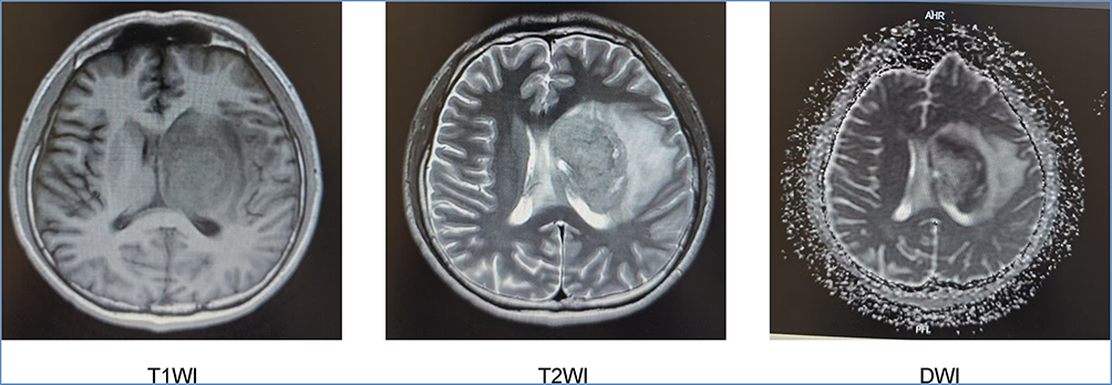 |
Figure 1 T1WI, T2WI and DWI axial views of the patient’s cranial MRI on February 19, 2025. Abbreviations: T1WI, T1-weighted imaging; T2WI, T2-weighted imaging; DWI: diffusion-weighted imaging. |
Following the aforementioned findings, the patient was admitted to our hospital on February 25, 2025. Physical examination on admission revealed the patient was conscious and alert with slurred speech and no facial asymmetry. The assessment showed grade 0/5 muscle strength and reduced tone in the right limbs while left-sided limbs exhibited normal muscle strength and tone. Skeletal muscle strength was measured by manual muscle testing with the Medical Research Council (MRC) grading scale. Bilateral pathological reflexes demonstrated no elicitability. HIV viral load was 6.61×10⁵ copies/mL and the CD4⁺ T cell counts was 41 /μL. CSF routine examination showed a negative Pandy test with a white blood cell (WBC) count of 5.0×10⁶/L. CSF biochemical analysis revealed total protein 2228.00 mg/L, β2-microglobulin 7.26 mg/L, lactate dehydrogenase 210.0 U/L, glucose 2.10 mmol/L, and microalbumin 1106.5 mg/L. No Cryptococcus neoformans was detected in CSF. Both serum Toxoplasma gondii antibody and CSF PCR for Toxoplasma deoxyribonucleic acid (DNA) were negative. CSF next-generation sequencing (NGS) returned negative results.
After 17 days of treatment with a standard antiretroviral regimen (Bictegravir 50mg/Emtricitabine 200mg/Tenofovir Alafenamide 25mg, one tablet orally once daily), intravenous mannitol (125mL, 1–2 times daily as needed for intracranial pressure control), and oral trimethoprim-sulfamethoxazole (160mg/800mg, one tablet every other day for secondary prophylaxis), the patient exhibited limited improvement in muscle strength and progressed to a state of superficial coma. A follow-up head computed tomography (CT) scan revealed a suspected neoplastic lesion in the left cerebral parenchyma with extensive perilesional edema and brain herniation. The patient was escalated to a higher dose of mannitol. However, the patient’s condition showed no significant improvement, with persistent superficial coma. The follow-up MRI of the brain on March 31, 2025, demonstrated lesions in the left cerebral hemisphere and brainstem, suggesting possible neoplastic or infectious processes with mass effect compressing the ventricular system, midline shift to the right, and suspected brain herniation (Figure 2). Compared to the prior external MRI, this examination revealed significant progression with expanded lesion extent, aggravated mass effect compressing the right cerebral hemisphere, and markedly worsened perilesional edema.
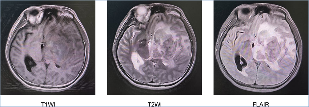 |
Figure 2 T1WI, T2WI and FLAIR axial views of the patient’s cranial MRI on March 31, 2025. Abbreviations: T1WI, T1-weighted imaging; T2WI, T2-weighted imaging; FLAIR, fluid-attenuated inversion recovery. |
Given the patient’s profound immunosuppression, the presence of a ring-enhancing lesion on MRI and continued clinical deterioration despite initial antiretroviral therapy and prophylaxis, empirical anti-Toxoplasma therapy was prioritized. This decision was made after thorough communication with the patient’s family, considering the high risks of brain biopsy in his critical condition and the lack of conclusive laboratory evidence. The patient received combined anti-Toxoplasma therapy (compound sulfamethoxazole tablets 1.44g three times daily, azithromycin dispersible tablets 0.5g once daily) starting from April 28, 2025. After one week of treatment, consciousness gradually regained with essentially normal speech clarity while right-sided muscle strength remained grade 0/5 and left-sided grade 5/5. Anti-Toxoplasma treatment was subsequently maintained for a total course of six weeks. Follow-up brain MRI on June 13 demonstrated interval reduction in lesion size with decreased surrounding edema compared to the prior scan (Figure 3). Following recovery of right-sided muscle strength to grade 2/5 and clinical improvement, the patient was discharged on June 22 with planned continuation of antiviral therapy post-discharge.
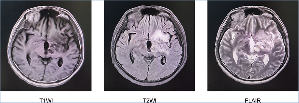 |
Figure 3 T1WI, T2WI and FLAIR axial views of the patient’s cranial MRI on June 13, 2025. Abbreviations: T1WI, T1-weighted imaging; T2WI, T2-weighted imaging; FLAIR, fluid-attenuated inversion recovery. |
Discussion
We reported a case with a large solitary ring-enhancing intracranial lesion causing marked mass effect and midline shift, with concurrent identification of newly diagnosed HIV infection. Given the absence of toxoplasma antibodies and undetectable toxoplasma deoxyribonucleic acid (DNA) in cerebrospinal fluid, initial clinical suspicion strongly favored a malignant tumor. The lack of response to initial HIV therapy and symptomatic progression despite maximal medical management presented a formidable diagnostic dilemma. The subsequent dramatic response to empirical anti-Toxoplasma therapy highlighted a critical clinical scenario and underscored the diagnostic complexities and potential pitfalls in managing single mass lesions in advanced HIV disease.
HIV-associated cerebral toxoplasmosis was frequently misdiagnosed as other neurological conditions such as neoplastic and vascular lesions, resulting in delayed therapeutic intervention. An African-origin woman with new-onset seizures and weight loss was initially misdiagnosed with a brain tumor due to neoplasm-mimicking neuroimaging, highlighting how cerebral toxoplasmosis in undiagnosed HIV can mimic malignancy and delay targeted antimicrobial therapy.8 HIV-associated cerebral toxoplasmosis in another 65-year-old female presenting as isolated hemiparesis, and delayed recognition might cause permanent disability even when eventual antimicrobial efficacy.7 A case of AIDS complicated with toxoplasma encephalopathy with rapid progressive memory loss as the initial symptom and misdiagnosed as multiple sclerosis.9 Therefore, we advocate that for HIV patients with CD4⁺ counts < 100/μL and solitary ring-enhancing lesions, empirical anti-Toxoplasma therapy should be initiated immediately as a diagnostic-therapeutic measure, even in seronegative cases, with radiological response serving as a surrogate diagnostic endpoint.
Despite providing diagnostic certainty for ring-enhancing lesions, brain biopsy carries significant morbidity and mortality risks such as intracranial bleeding and permanent neurological injury.10 Noninvasive laboratory examinations of Toxoplasma gondii infection primarily relied on serological assays and molecular testing (PCR and NGS), yet each method exhibited inherent limitations and could not serve as a standalone diagnostic criterion. Serological diagnosis of Toxoplasma gondii infection remained unreliable as IgM persistence in HIV patients obscured interpretation while antibody titer fluctuations lacked specificity. True reactivation occured only under profound immunosuppression and escaped serological detection in 5% of AIDS-related encephalitis cases.11,12 Molecular detection methods based on PCR could identify Toxoplasma gondii DNA fragments in various clinical specimens, including CSF, blood, bone marrow, amniotic fluid, and aqueous humor. The sensitivity of this approach was unaffected by the patient’s immune status, making it particularly suitable for diagnosing Toxoplasma gondii infection in immunocompromised patients such as those with AIDS. While demonstrating high specificity (96–100%), its sensitivity varied across sample types, with CSF specimens showing 35–72% sensitivity.13 This variation might stem from complex interactions among multiple factors, including the density of Toxoplasma gondii distribution, parasitic forms, and physicochemical properties of the specimens. Our patient’s profoundly low CD4⁺ count (41/μL) explained the absent humoral response. This case reinforced that negative serology cannot exclude TE in advanced immunosuppression. Metagenomic NGS (mNGS) was a very sensitive tool for detecting common opportunistic central nervous system pathogen in HIV-infected patients. CSF mNGS demonstrated high sensitivity (100%, 95% CI: 61%–100%) and specificity (100%, 95% CI: 95%–100%).14 However, given the current limited clinical experience with this method, its results required comprehensive evaluation alongside patients’ clinical manifestations and supplementary examinations. Therefore, in clinical practice, the possibility of seronegative TE should be strongly suspected in patients with advanced HIV disease and very low CD4+ counts, as the absence of a humoral immune response can lead to false-negative serological results. However, it is important to note that the evidence guiding the management of seronegative TE is primarily derived from case reports and small series. Hence, while empirical therapy is a rational approach in this high-risk setting, it should be initiated with the understanding that radiological and clinical response is the primary diagnostic endpoint, and close monitoring is essential.
Diagnostic imaging techniques, such as diffusion-weighted magnetic resonance imaging and MR spectroscopy, demonstrated potential for etiological discrimination among ring-enhancing lesions and should be considered in further workups. TE might present as single or multiple ring-enhancing lesions with surrounding edema on cranial CT or MRI, a finding once considered relatively specific for TE.15 Conversely, brain tumors such as PCNSL typically demonstrated space-occupying lesions with enhancement on contrast scans. Both TE and PCNSL predominantly affected the basal ganglia, exhibiting ring enhancement and perilesional edema. Although the “ring-enhancing lesion” was historically regarded as a characteristic feature of TE, PCNSL can mimic this pattern.16 In clinical practice, differentiating ring-enhancing PCNSL from infectious etiologies was critical yet challenging. Apparent diffusion coefficient (ADC) values might be helpful in differentiating toxoplasmosis from lymphoma in patients with AIDS. One study showed that diffusion-weighted MRI with ADC quantification demonstrated significantly higher diffusion in AIDS-associated toxoplasmosis lesions versus lymphoma, where ADC ratios greater than 1.6 were exclusively seen in toxoplasmosis.17
When biopsy was contraindicated (brainstem involvement or bleeding risk), a therapeutic trial with anti-Toxoplasma agents served as both diagnostic and therapeutic intervention. Empiric anti-Toxoplasma therapy constituted first-line management for advanced HIV patients with characteristic neuroimaging findings and seropositivity. The patient achieved near-complete radiological resolution within 6 weeks, consistent with current guidelines.18 This contrasted sharply with the natural history of tumors or infections like PCNSL, which would progress without targeted therapy.
This case report has several limitations that must be acknowledged. First, the diagnosis of TE remains presumptive, lacking gold-standard histopathological confirmation from a brain biopsy. While the dramatic response to anti-Toxoplasma therapy is strongly indicative of the diagnosis, we cannot definitively exclude other possibilities, such as a PCNSL or an infectious process that coincidentally responded to the anti-inflammatory effects of azithromycin. Second, our reliance on empirical diagnosis underscores a common clinical challenge. The imperfect sensitivity of available non-invasive tests like PCR and mNGS in this specific context. Finally, the initial and follow-up imaging findings, while suggestive, were not pathognomonic and exhibited significant overlap with neoplastic lesions like glioma or lymphoma. Advanced imaging techniques such as perfusion MRI and MR spectroscopy were not performed and could have provided additional diagnostic insights. These limitations are inherent to the real-world management of critically ill patients with contraindications to invasive procedures and highlight the diagnostic challenges our report aims to address.
Conclusions
In conclusion, this case demonstrated that initiating an empirical anti-Toxoplasma therapeutic trial in a newly diagnosed AIDS patient with ring-enhancing intracranial lesions may help mitigate fatal treatment delays, even after misdiagnosis as a tumor. These observations highlight the potential value of prioritizing empirical management of opportunistic infections over neoplasms in differential diagnosis. Based on our experience and the available evidence, empirical anti-Toxoplasma therapy should be considered a first-line diagnostic-therapeutic strategy for ring-enhancing masses in patients with advanced AIDS, especially when invasive diagnostics are not feasible. This approach may help mitigate fatal treatment delays. However, the decision must be individualized, and the role of biopsy for definitive diagnosis remains crucial in non-responding or atypical cases. Rapid initiation might reduce the risk of irreversible deficits. Future studies are needed to identify reliable predictors of seronegative toxoplasmosis and to refine this management strategy.
Data Sharing Statement
The datasets used and/or analysed during the current study are available from the corresponding author on reasonable request.
Ethics Approval and Informed Consent
Ethical approval for this case report (Approval Number: 2024136) was obtained from the Ethics Committee of Zigong First People’s Hospital, Sichuan, China and complied with the Declaration of Helsinki as revised in 2013.
Acknowledgments
We thank Jing Tang and Wei Deng from the Department of Infectious Diseases, Zigong First People’s Hospital for their contribution to the treatment of the participant.
Funding
This study was supported by the Key Science and Technology Projects of Zigong (2021ZC11).
Disclosure
The authors declare no competing interests.
References
1. Acquired Immunodeficiency Syndrome Professional Group Society of Infectious Diseases Chinese Medical Association, Chinese Center for Disease Control and Prevention. Chinese guidelines for the diagnosis and treatment of human immunodeficiency virus infection/acquired immunodeficiency syndrome (2024 edition). Chin Med J. 2024;137(22):2654–2680. doi:10.1097/CM9.0000000000003383
2. Porter SB, Sande MA. Toxoplasmosis of the central nervous system in the acquired immunodeficiency syndrome. N Engl J Med. 1992;327(23):1643–1648. doi:10.1056/NEJM199212033272306
3. Luft BJ, Remington JS. Toxoplasmic encephalitis in AIDS. Clin Infect Dis. 1992;15(2):211–222. doi:10.1093/clinids/15.2.211
4. Vidal JE. HIV-related cerebral toxoplasmosis revisited: current concepts and controversies of an old disease. J Int Assoc Provid AIDS Care. 2019;18:2325958219867315. doi:10.1177/2325958219867315
5. Maharani K, Dian S, Ganiem AR, et al. Clinical presentation, management, and outcome of suspected central nervous system infections in Indonesia: a prospective cohort study. Infection. 2024;52(2):583–595. doi:10.1007/s15010-023-02170-0
6. Wang ZD, Wang SC, Liu HH, et al. Prevalence and burden of Toxoplasma gondii infection in HIV-infected people: a systematic review and meta-analysis. Lancet HIV. 2017;4(4):e177–e188. doi:10.1016/S2352-3018(17)30005-X
7. Kang Y, Zhang X, Zhang L, Huang M, Tang C, Zhang L. Cerebral toxoplasmosis mimicking stroke in a woman living with undiagnosed HIV. J Infect Public Health. 2024;17(8):102477. doi:10.1016/j.jiph.2024.102477
8. Alves D, Sobrosa P, Morais Passos R, et al. Cerebral toxoplasmosis mimicking a brain neoplasm in an inaugural HIV-positive patient: the importance of early decision-making and background assessment in the emergency department. Cureus. 2025;17(1):e76936. doi:10.7759/cureus.76936
9. Wu J, Luo X, Huang N, Li Y, Luo Y. Misdiagnosis of HIV with toxoplasmosis encephalopathy with progressive memory loss as the initial symptom: a case report. Front Neurol. 2022;13:809811. doi:10.3389/fneur.2022.809811
10. Scott BJ, Douglas VC, Tihan T, Rubenstein JL, Josephson SA. A systematic approach to the diagnosis of suspected central nervous system lymphoma. JAMA Neurol. 2013;70(3):311–319. doi:10.1001/jamaneurol.2013.606
11. Kodym P, Malý M, Beran O, et al. Incidence, immunological and clinical characteristics of reactivation of latent Toxoplasma gondii infection in HIV-infected patients. Epidemiol Infect. 2015;143(3):600–607. doi:10.1017/S0950268814001253
12. Pereira-Chioccola VL, Vidal JE, Su C. Toxoplasma gondii infection and cerebral toxoplasmosis in HIV-infected patients. Future Microbiol. 2009;4(10):1363–1379. doi:10.2217/fmb.09.89
13. Robert-Gangneux F, Belaz S. Molecular diagnosis of toxoplasmosis in immunocompromised patients. Curr Opin Infect Dis. 2016;29(4):330–339. doi:10.1097/QCO.0000000000000275
14. Zhu Y, Zhao W, Yang X, et al. Metagenomic next-generation sequencing for identification of central nervous system pathogens in HIV-infected patients. Front Microbiol. 2022;13:1055996. doi:10.3389/fmicb.2022.1055996
15. Mahadevan A, Ramalingaiah AH, Parthasarathy S, Nath A, Ranga U, Krishna SS. Neuropathological correlate of the “concentric target sign” in MRI of HIV-associated cerebral toxoplasmosis. J Magn Reson Imaging. 2013;38(2):488–495. doi:10.1002/jmri.24036
16. Jittapiromsak N, Sriinkua P, Nimitpornsuko P, Jittapiromsak P. Diffusion-weighted imaging and susceptibility-weighted imaging in differentiating ring-enhancing primary central nervous system lymphoma from infections. Neuroradiol J. 2025;19714009251354883. doi:10.1177/19714009251354883
17. Camacho DL, Smith JK, Castillo M. Differentiation of toxoplasmosis and lymphoma in AIDS patients by using apparent diffusion coefficients. AJNR Am J Neuroradiol. 2003;24(4):633–637.
18. National Institutes of Health, HIV Medicine Association, Infectious Diseases Society of America. Guidelines for the prevention and treatment of opportunistic infections in adults and adolescents with HIV. 2025. Available from: https://clinicalinfo.hiv.gov/en/guidelines/hiv-clinical-guidelines-adult-and-adolescent-opportunistic-infections.
 © 2025 The Author(s). This work is published and licensed by Dove Medical Press Limited. The
full terms of this license are available at https://www.dovepress.com/terms
and incorporate the Creative Commons Attribution
- Non Commercial (unported, 4.0) License.
By accessing the work you hereby accept the Terms. Non-commercial uses of the work are permitted
without any further permission from Dove Medical Press Limited, provided the work is properly
attributed. For permission for commercial use of this work, please see paragraphs 4.2 and 5 of our Terms.
© 2025 The Author(s). This work is published and licensed by Dove Medical Press Limited. The
full terms of this license are available at https://www.dovepress.com/terms
and incorporate the Creative Commons Attribution
- Non Commercial (unported, 4.0) License.
By accessing the work you hereby accept the Terms. Non-commercial uses of the work are permitted
without any further permission from Dove Medical Press Limited, provided the work is properly
attributed. For permission for commercial use of this work, please see paragraphs 4.2 and 5 of our Terms.
Recommended articles
Confronting Dual Therapeutic Hurdles in Advanced HIV: A Case Report on INSTIs Resistance and Talaromyces marneffei Coinfection
Zou L, Diao Y, Huang Z, You C
International Medical Case Reports Journal 2025, 18:1071-1075
Published Date: 22 August 2025