
Back to Journals » Clinical Ophthalmology » Volume 20
Management of Traumatic Submacular Hemorrhage by Pneumatic Displacement: A Review
Authors Aljohani S 
Received 28 December 2025
Accepted for publication 11 March 2026
Published 17 March 2026 Volume 2026:20 592239
DOI https://doi.org/10.2147/OPTH.S592239
Checked for plagiarism Yes
Review by Single anonymous peer review
Peer reviewer comments 3
Editor who approved publication: Dr Yousef Fouad
Saud Aljohani
Department of Ophthalmology, Imam Abdulrahman Bin Faisal University, Dammam, Saudi Arabia
Correspondence: Saud Aljohani, Department of Ophthalmology, Imam Abdulrahman Bin Faisal University, Dammam, 34212, Saudi Arabia, Email [email protected]
Abstract: Traumatic submacular haemorrhage is a vision-threatening ocular emergency that can result in permanent visual loss if not treated promptly. Following blunt ocular trauma, rapid accumulation of blood beneath the macula leads to photoreceptor damage through iron-mediated oxidative stress, mechanical disruption, and impaired metabolic exchange. Pneumatic displacement, with or without intravitreal tissue plasminogen activator (tPA), has emerged as a minimally invasive therapeutic option; however, standardized clinical guidelines for its use in traumatic cases remain lacking. A narrative literature search was conducted across Scopus, Web of Science, Medline, Embase, PubMed, OpenAlex, and Dimensions up to November 24, 2025, supplemented by citation tracking. After duplicate removal and title/abstract screening, full-text assessment was performed based on predefined inclusion criteria focusing on traumatic submacular haemorrhage managed with pneumatic displacement. Of 177 identified records, four studies met the final eligibility criteria. The available evidence suggests that pneumatic displacement using intravitreal gas alone, most commonly 100% perfluoropropane (C3F8), achieved anatomical displacement in some cases but showed lower functional success, particularly in thick or dense haemorrhages. In contrast, intravitreal tPA combined with expansile gas was the most frequently employed minimally invasive approach and resulted in high rates of complete subfoveal blood displacement and clinically meaningful visual improvement, especially in younger patients. Pars plana vitrectomy with subretinal tPA was reserved for large or complex haemorrhages and produced favorable outcomes, albeit with higher surgical risk. Although the current evidence is limited, largely retrospective, and derived from small-sample studies, early treatment appears to be associated with favorable anatomical and functional outcomes. Intravitreal tPA combined with pneumatic displacement appears to be the most effective first-line therapy, while gas-only displacement may be considered when tPA is unavailable. Well-designed prospective studies with standardized outcome measures are needed to define optimal management strategies.
Keywords: traumatic submacular haemorrhage, pneumatic displacement, intravitreal gas, tissue plasminogen activator, perfluoropropane C3F8, sulfur hexafluoride SF6
Introduction
Traumatic submacular haemorrhage is considered an ophthalmic emergency that can result in significant and permanent loss of vision if not addressed promptly.1–5 After a blunt force trauma, submacular blood can collect rapidly in the eye, resulting in substantial injury to the photoreceptors because of iron toxicity, mechanical disruption, and impaired metabolic exchange.2,4,5 Irreversible photoreceptor damage can occur within the first 24 hours of the injury; therefore, timely intervention is critical in such cases. However, there is a lack of clear standardized treatment guidelines.2
Various therapeutic interventions have been reported for the management of submacular haemorrhage. These include pneumatic displacement either with gas alone or in combination with intravitreal tissue plasminogen activator (tPA), and pars plana vitrectomy with intravitreal or subretinal tPA.6–9 Of these modalities, pneumatic displacement has gained considerable popularity as it is minimally invasive and has promising results. It involves the use of an expansile gas bubble to displace the haemorrhage away from the fovea and potentially limit photoreceptor damage.1,10–13
Management of traumatic submacular haemorrhage differs from other causes of submacular haemorrhage, such as age-related macular degeneration (AMD)-related or polypoidal choroidal vasculopathy (PCV), in which anti-VEGF therapy is typically a core component to control neovascular activity, with pneumatic displacement or pars plana vitrectomy with subretinal tPA reserved for thick or large, fovea-threatening haemorrhage.2,4 The purpose of this review is to critically examine current knowledge regarding traumatic submacular hemorrhage with respect to its pathogenesis, diagnosis, and treatment. It aims to explore the efficacy and safety of pneumatic displacement techniques and compare them with other therapies. It will also discuss procedural considerations and possible complications, and highlight key factors that affect prognosis for visual outcomes.
Materials and Methods
A narrative review was conducted to study traumatic submacular haemorrhage, focusing on the therapeutic efficacy and outcome of pneumatic displacement. A comprehensive literature search was carried out for publications published up to November 24, 2025. The databases Scopus, Web of Science, Medline, Embase, PubMed, OpenAlex, and Dimensions were chosen for the literature search. Further studies were retrieved by backward and forward citation searching (snowballing) in the Incitesful.xyz tool. Duplicate entries were discarded before screening the publications. Titles and abstracts were screened for potential relevance, followed by full-text review for final inclusion. The keywords applied in the search strategy were as follows: “traumatic submacular haemorrhage” OR “traumatic submacular hemorrhage” OR “submacular hemorrhage AND trauma” AND “pneumatic displacement” OR “intravitreal gas” OR “perfluoropropane” OR “sulfur hexafluoride.” Further references were included from the bibliographies of the selected studies. Eligible studies examined traumatic submacular haemorrhage, its diagnostic features, vascular characteristics, and clinical implications, as well as the use of pneumatic displacement and its therapeutic efficacy. Case reports, conference abstracts, and non-English language articles were excluded.
Initially, 177 articles were identified and retrieved. After the removal of duplicates, 171 unique records remained. Screening the titles and abstracts of the publications resulted in the exclusion of an additional 131 articles, and 20 were deemed irrelevant. Five of the remaining records could not be retrieved because either the access to their full-text versions was unavailable or they were published in a language other than English. After full-text evaluation of the remaining 15 articles, 11 were excluded due to the absence of pneumatic displacement management, exclusive focus on non-traumatic causes such as AMD or PCV, insufficient reporting of clinical outcomes, or duplication of previously published patient populations. Only four articles fulfilled the selection criteria and were thus eligible for inclusion in this review.
Pathophysiology
Blunt ocular trauma can result in multiple posterior segment injuries, including choroidal rupture and subretinal haemorrhage. Choroidal rupture typically results from closed-globe injuries, often appearing parallel and to the side (temporal) of the optic disc. There is macular involvement in approximately 66% of the cases.2,17
Iron-Mediated Oxidative Stress
Breakdown of erythrocytes within the submacular clot releases free iron, which is highly cytotoxic to the retinal tissue.16,18 Iron-induced oxidative stress damages lipids, proteins, and nucleic acids by generating free radicals, particularly via the Fenton reaction. This ultimately leads to the destruction of the photoreceptors.2,19
Photoreceptor Damage and Mechanical Disruption
Accumulation of blood within the subretinal space leads to direct injury to both the photoreceptors and the retinal pigment epithelium, which is a single layer of pigmented cells behind the retina. As the clot contracts, it also exerts mechanical pressure on the photoreceptor outer segments, creating shear forces that may cause structural avulsion of these critical structures. This leads to further retinal disruption and dysfunction.1,13,16,21,22
Impaired Nutrient and Oxygen Exchange
The presence of a submacular clot creates a physical barrier that interferes with normal metabolic exchange between the retinal pigment epithelium and photoreceptors.13,16,21,23 A thick clot would also affect the diffusion of oxygen and essential metabolites and nutrients from the choriocapillaris. Photoreceptors have a very high metabolic demand and are very susceptible to any kind of delay in this exchange. Any prolonged shortage can quickly cause photoreceptor dysfunction and resultant cell death.16,23
Natural History of Untreated Submacular Haemorrhage
Without treatment, submacular haemorrhage often results in severe and permanent loss of vision.16,24 Available data suggest that photoreceptors are irreversibly damaged within the first 24 hours after an injury, with nearly complete loss of vision evident after 7 days of persistent haemorrhage.2,9,16 Early intervention within this seven-day window leads to better anatomical and visual outcomes than delayed care.9,25
Diagnostic Evaluation
Clinical Presentation
Patients often present with a sudden reduction in vision following a blunt force injury to the eye. It is often accompanied by a central scotoma, reflecting the disruption of foveal function caused by the underlying haemorrhage.21 On fundus examination, a reddish-brown subretinal discoloration centered in the macular region is commonly observed. Additional signs of ocular trauma, such as commotio retinae or choroidal rupture, may also be present.9,17
Imaging Modalities
Optical coherence tomography (OCT) (Figure 1) is a key diagnostic modality for defining the precise location and extent of the submacular haemorrhage in traumatic cases. It provides essential information to differentiate between subretinal, subretinal pigment epithelium, and intraretinal blood, and helps assess the integrity of the outer retinal segment. It also helps identify prognostic indicators, such as inner segment/outer segment disruption, foveal thickness, and the overall submacular haemorrhagic area.2,14,15 Increased baseline central macular thickness on OCT has been correlated with poorer visual outcomes.23 OCT also supports the grading of submacular haemorrhage size using disc-diameter (DD) criteria: small (1–4 DD), medium (≥4 DD but confined within the temporal arcade), and massive (extending beyond the arcade) [2]. The FLATCAPS classification system further refines structural characterization by evaluating foveal involvement, haemorrhage layer location (L0–L4), thickness categories (T0–T2), size grades (S0–S3), and duration of haemorrhage (A0–A2) [15]. OCT may additionally demonstrate the BALAD sign, a bacillary layer detachment with overlying hyperreflective material. The BALAD sign reflects intrabacillary haemorrhage and has been associated with poorer visual prognosis.14 Serial OCT imaging offers valuable follow-up information by tracking changes in the volume of the haemorrhage, documenting patterns of clot displacement after pneumatic intervention, and assessing restoration of the foveal architecture. All of these factors correlate with the potential for the recovery of vision.2 Fundus photography (Figure 2) also contributes significantly to the evaluation by providing wide-field documentation of the size of the haemorrhage and its configuration and spatial relationship to major retinal vessels, facilitating both baseline assessment and longitudinal monitoring.15 It may also highlight coexisting traumatic findings such as choroidal rupture, which typically appears as yellowish-white, curvilinear streaks in closed-globe injuries.3 Fluorescein angiography can be selectively utilized to investigate underlying choroidal neovascularization or vascular abnormalities obscured by the haemorrhage. However, it is generally not required in the routine assessment of traumatic submacular haemorrhage.2,17
 |
Figure 1 OCT of the right eye (same patient in Figure 2) showing a large dome-shaped subfoveal mixed hyperreflective–hyporeflective lesion with marked posterior shadowing, consistent with thick submacular haemorrhage and outer retinal disruption, with mild overlying vitreous haemorrhage. |
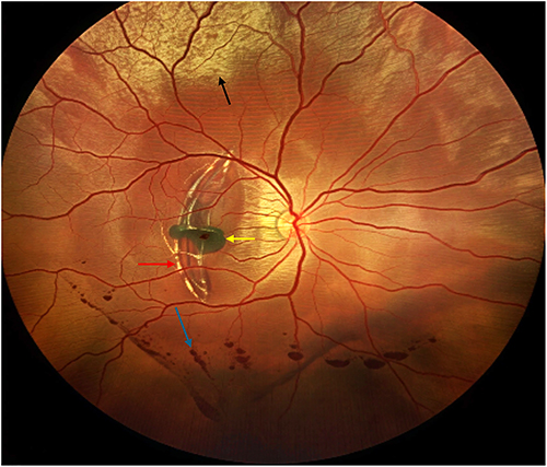 |
Figure 2 Color fundus right eye showing: Superior commotio retinae (black arrow), Choroidal rupture (red arrow), Subretinal hemorrhage involving the macula (yellow arrow), and Inferior vitreous hemorrhage (blue arrow), post-blunt ocular trauma. |
Techniques of Pneumatic Displacement for Traumatic Submacular Hemorrhage
Pneumatic displacement is a minimally invasive procedure used to manage submacular haemorrhage. It involves displacing the accumulated blood from the foveal region by using an expansile intravitreal gas bubble. This technique may be performed with gas alone or in combination with a fibrinolytic agent, most commonly tPA. This is followed by optimal positioning of the patient to maximize the displacement of the collected blood.9,14,19
Gas Types
The choice of intravitreal gas is a critical factor determining the success of the treatment. It also influences the duration of the tamponade and the magnitude of the buoyant force applied to the haemorrhage. The two most frequently used gaseous agents are sulfur hexafluoride (SF6) and perfluoropropane (C3F8).2,3,16
SF6 provides a moderate and relatively short-acting tamponade effect, typically lasting 1–2 weeks, which is generally enough for the effective displacement of the clot. It also reduces the need for prolonged postoperative positioning. This lowers the overall patient discomfort.26 In contrast, C3F8 expands more and remains in the vitreous cavity for approximately 6–8 weeks, offering a prolonged tamponade effect. This may be particularly advantageous in cases with a denser clot.27 However, its extended duration requires prolonged prone positioning and increases the risk of intraocular pressure-related complications.26
Adjunctive Use of Intravitreal tPA
Intravitreal tPA is commonly used as an adjunct to gas tamponade to help with the clot liquefaction. It catalyzes the conversion of plasminogen to plasmin, promoting fibrinolysis and enabling more effective displacement of haemorrhagic material by the gas bubble.2,16,19 tPA can be administered either prior to or following the gas injection. The time of administration depends on the protocol being used.16 Typical dosing ranges from 0.05–0.1 mL, corresponding to 25–50 µg delivered intravitreally.9,19,25 While tPA may enhance anatomical outcomes, its ability to penetrate the neurosensory retina in adequate concentrations to reach the subretinal space remains debated.16,28 Some studies support sufficient diffusion, while others raise concerns about limited permeability.10,19 Furthermore, tPA is not completely risk-free. Incidents of retinal toxicity and secondary haemorrhages after the use of tPA have been reported, highlighting the importance of cautious dosing and meticulous technique.2,29–31
Patient Positioning
The positioning of the patient plays a very important role in the success of the pneumatic displacement procedure. Most protocols recommend prone (face-down) positioning, allowing the gas bubble to exert upward pressure on the submacular clot, thereby shifting it away from the foveal center.2,4,16,27,31 The duration of positioning varies considerably in the literature, ranging from 24 hours to 7–14 days.2,16,19,22 In protocols involving tPA, prone positioning is temporarily delayed after the injection to allow the fibrinolytic enzyme to diffuse adequately before initiating the clot displacement.16
Procedure Steps
The procedure begins by selecting the appropriate type of gas and its required volume. Typically, 0.3–0.6 mL of undiluted SF6 or C3F8 is used.2,16,19,27,33 C3F8 is often preferred in cases requiring greater buoyancy, such as when haemorrhages are organized or thicker.27,33
After aseptic preparation and local anaesthetic, the gas is injected 3–4 mm posterior to the limbus using a 29 or 30-gauge needle. Anterior chamber paracentesis is also performed to prevent an acute increase in the intraocular pressure (IOP).2,7,16
Postoperatively, the patient is evaluated based on the best-corrected visual acuity, intraocular pressure, slit-lamp biomicroscopy, dilated fundus examination, and retinal imaging. These evaluations are essential for determining the efficacy of the treatment provided, identifying potential complications, and guiding the patient regarding further management.27,31
Indications and Contraindications
Timely intervention is essential in the management of such patients, as irreversible permanent damage to the retina can occur rapidly, sometimes within the first 24 hours following the accumulation of submacular haemorrhage.14
Pneumatic displacement is contraindicated if there is structural retinal damage present, such as retinal tears or retinal detachment. Similarly, large choroidal ruptures, particularly those involving the foveal region, significantly reduce the likelihood of a successful displacement and limit the potential for visual recovery. Furthermore, a dense vitreous haemorrhage can obstruct and prevent adequate visualization of the posterior chamber. It would make intraoperative management and postoperative assessment very difficult. Lastly, there would be patient-related limitations. Some patients may find it very difficult to follow the required postoperative positioning regimen, which could compromise both the safety and effectiveness of the procedure.2,23,31
Management Approaches for Submacular Haemorrhage
Following a comprehensive literature review, four studies were identified as being directly relevant to the management of traumatic submacular haemorrhage. These studies served as the foundation for evidence-based therapeutic recommendations9,14,19,25 (Table 1).
 |
Table 1 Main Findings in the Studies |
Overview of Available Therapies
Pneumatic Displacement with Gas Alone
Several studies have reported the use of gas-only pneumatic displacement; however, its use in traumatic cases is variable and often limited. Gujral et al reported that 4 out of 20 trauma-related eyes (20%) in their study were managed with 03 mL of 100% C3F8 gas alone.9 Rishi et al included only a single traumatic eye in their gas-only cohort25 Chen et al did not employ gas-only treatment for any trauma-related cases, opting instead for combined gas and tPA therapy in all three cases19 In Choudhary et al, 9 out of 20 eyes received gas alone; although trauma accounted for 50% of the cohort, the exact number of traumatic cases treated with gas-only was not specified14 Gas-only pneumatic displacement relies on the buoyant force of an expansile intravitreal gas bubble (typically 100% C3F8) to shift subretinal blood away from the fovea, assisted by strict prone positioning9,14,25 However, its efficacy appears reduced in cases with dense or thick haemorrhage In Gujral’s trauma cohort, gas-only outcomes were not analyzed separately, but the overall trauma group achieved 90% complete displacement and 80% visual improvement (≥2 Snellen lines) at six months.9 In contrast, Rishi et al reported 571% anatomical success and 429% functional improvement in their gas-only group These are considerably lower than the outcomes achieved with tPA-assisted approaches.25 Choudhary et al observed clinical improvements in the gas-only group, though trauma-specific results were not delineated14 Given these findings, gas-only pneumatic displacement is generally reserved for cases where tPA is unavailable or contraindicated Across all studies, its outcomes have been consistently inferior to those achieved with combination therapy, particularly in eyes with more extensive or organized haemorrhages.9,14,25
Intravitreal Gas Combined with tPA
The use of intravitreal tPA with expansile gas represents the most frequently applied minimally invasive strategy for managing patients with traumatic submacular haemorrhage. Across the included studies, this approach was used in 25 trauma-related eyes: 15 in Gujral et al,9 7 in Rishi et al,25 and 3 in Chen et al19 In Choudhary et al, tPA was administered subretinally during vitrectomy, rather than intravitreally14 Intravitreal tPA facilitates enzymatic fibrinolysis of the clot, promoting liquefaction, while the gas bubble displaces the liquefied blood with the help of appropriate patient positioning9,19,25 Dosing protocols varied across studies: Gujral et al used a higher concentration (100 mg/01 mL delivered in 0.05 mL), Rishi et al administered 50 µg/01 mL with C3F8, and Chen et al used 30–100 µg/0.1 mL with either SF6 or C3F8.9,19,25 Across studies, this combined approach yielded superior anatomical and functional outcomes. In Gujral’s trauma cohort, 16 of 20 eyes achieved ≥2-line visual improvement, and 18 of 20 achieved complete displacement.9 Rishi et al reported 84% anatomical success and 52% visual improvement, while Chen et al demonstrated 51% functional improvement at week 1, increasing to 64% at 12 months, with trauma cases performing comparably to the broader cohort.19,23 Although complications were infrequent, they included secondary glaucoma and macular hole formation (Gujral et al), as well as vitreous haemorrhage and retinal detachment (Rishi et al, Chen et al)9,19,25 Despite these risks, intravitreal tPA with gas remains the standard minimally invasive intervention for the treatment of traumatic submacular haemorrhage. Furthermore, it offers better outcomes than gas alone.
Pars Plana Vitrectomy with Subretinal tPA or Clot Evacuation
Pars plana vitrectomy (PPV) combined with subretinal tPA injection or clot evacuation is generally indicated for large, dense, or organized haemorrhages, or in eyes with coexisting structural damage that may reduce the success of less invasive techniques.9,14,25
Only a limited number of traumatic cases in the literature have been managed with a vitrectomy. Gujral et al reported one trauma case treated with PPV and subretinal tPA.9 Rishi et al included two traumatic eyes in their vitrectomy cohort.25 Choudhary et al treated 9 of 20 eyes with vitrectomy and subretinal tPA, though the exact number of trauma cases was not specified.14 Chen et al did not include a vitrectomy treatment option for traumatic haemorrhage.19
A vitrectomy generally resulted in favourable outcomes in these studies. In Gujral et al, the single vitrectomy-treated eye achieved both anatomical and functional success9 Rishi et al reported 929% anatomical success and 64.3% functional improvement in the vitrectomy group.25 Choudhary et al described notable visual gains and resolution of bacillary layer detachment (BALAD) in vitrectomy cases, reinforcing the utility of this approach in selected patients14 However, vitrectomy is associated with higher complication rates compared to the minimally invasive procedures. Reported adverse events include iatrogenic retinal breaks, retinal detachment, cataract progression, and secondary glaucoma, predominantly within mixed-etiology cohorts.14,25 Nevertheless, PPV with subretinal tPA remains a crucial surgical option for patients with thick haemorrhages, coexisting retinal tears or detachments, or vitreous haemorrhage that obscures the posterior segment and limits the use of non-surgical methods.
Gaps in the Current Evidence
Despite a growing interest in the management of traumatic submacular haemorrhage, there are still significant gaps in the current literature that need to be addressed. Notably, the lack of randomized controlled trials (RCTs) makes it difficult for practitioners to develop strong evidence-based guidelines and compare the available treatment modalities. Furthermore, variations in procedural protocols, with differences in the type, concentration, and volume of the expansile gas used, as well as the duration and angle of postoperative positioning, make it difficult to choose the best approach. While early intervention has been widely acknowledged as critical, the optimal therapeutic window for maximizing anatomical and functional recovery has not been clearly established. Moreover, inconsistent ways of reporting outcomes, such as different definitions of anatomical displacement and visual improvement, further complicate inter-study comparisons and make it difficult to develop standardized clinical practice guidelines.
Role of New Imaging Technologies
New imaging techniques, particularly optical coherence tomography angiography (OCTA), are beginning to influence how traumatic submacular haemorrhage is diagnosed and managed. OCTA offers high-resolution, non-invasive visualization of the retinal and choroidal microvasculature, allowing clinicians to identify subtle trauma-induced vascular abnormalities, such as choroidal rupture-associated neovascularization, which is not evident on conventional imaging. By showing blood flow dynamics and microvascular health of the vessels, OCTA provides important prognostic insights and may help guide decisions regarding treatment options. This is particularly important in cases where adjunctive therapy is being considered.34
Potential Innovations
Researchers have been investigating several innovations with an aim to improve both the safety and efficacy of treatment options for submacular haemorrhage. Using lighter and shorter-acting intravitreal gases may allow adequate clot displacement while reducing the need for prolonged postoperative positioning and decreasing the risk of developing complications, such as increased intraocular pressure and cataract progression associated with long-acting tamponade agents. Simultaneously, research into safer fibrinolytic agents seeks to identify alternatives to recombinant tPA with an aim of keeping its benefits while reducing the risks of retinal toxicity and secondary haemorrhage.
Limitations of This Review
This review is limited by certain considerations. First, only four studies specifically addressing traumatic submacular haemorrhage were included, resulting in a small sample size and limiting the generalizability of the findings. Additionally, a few of the included studies involved mixed-etiology cohorts in which trauma-specific outcomes were not fully separated. Although we attempted to extract and interpret trauma-related data based on the reported subgroup proportions, this may limit the precision of trauma-specific conclusions. Furthermore, the included studies differ in their designs, treatment protocols, tPA dosing regimens, and follow-up durations. The absence of standardized definitions for key outcomes makes it difficult to compare and also introduces a potential for interpretation bias. Finally, most studies did not have long-term follow-up, so it is difficult to evaluate the durability of treatment outcomes and the incidence of late-onset complications, particularly in a relatively young patient population commonly affected by trauma-related haemorrhage.
Conclusion
Intravitreal tPA combined with an expansile gas has emerged as the most effective and consistently employed approach among all available treatment modalities. It demonstrates high rates of clot displacement and provides clinically meaningful improvements in visual acuity. In contrast, gas-only displacement has been used in a limited number of cases and has yielded less consistent outcomes, making it a secondary option, reserved for scenarios where tPA is unavailable or contraindicated. However, the current evidence remains limited, predominantly small retrospective studies, and heterogeneous in terms of study design, treatment protocols, and outcome reporting. There is a need for more studies, preferably prospective randomized controlled trials, to provide more evidence and facilitate clinical decision-making. Lastly, there is a need for developing standardization of treatment protocols and outcome reporting guidelines, so that evidence-based management strategies aimed at optimizing visual recovery in patients with traumatic submacular haemorrhage can be developed.
Data Sharing Statement
All data generated or analyzed during this study are included in this article. Further inquiries can be directed to the corresponding author.
Acknowledgments
The author would like to thank Dr. Shakil Ahmad, expert librarian from the Directorate of Library Affairs at our university, for his assistance, guidance, and support in the methodology and results sections of this study.
Funding
No grant has supported this research.
Disclosure
The author reports no conflicts of interest in this work.
References
1. Pappas G, Vidakis N, Petousis M, Kounali V, Korlos A. An innovatory surgical technique for submacular hemorrhage displacement by means of a bioengineering perspective. Vision. 2021;5(2):23. doi:10.3390/vision5020023
2. Casini G, Loiudice P, Menchini M, et al. Traumatic submacular hemorrhage: available treatment options and synthesis of the literature Int J Retina Vitreous. 2019;5(1):48. doi:10.1186/s40942-019-0200-0. BioMed Central.
3. Wang WX, Getahun H, Ramamurthy S, et al. Pars plana vitrectomy with tissue plasminogen activator for traumatic submacular hemorrhage. Int J Ophthalmol. 2025;18(9):1797. doi:10.18240/ijo.2025.09.22
4. Jeong S, Park D-G, Sagong M. Management of a submacular hemorrhage secondary to age-related macular degeneration: a comparison of three treatment modalities. J Clin Med. 2020;9(10):3088. doi:10.3390/jcm9103088
5. Valášková J, Vesely P, Krasnik V, et al. Surgical treatment of hemorrhagic complications of age-related macular degeneration, predictive factors and outcomes. Bratislavské Lekárske Listy/Bratislava Med J. 2025;126(3):299. doi:10.1007/s44411-024-00014-3
6. Patikulsila D, Winaikosol P, Choovuthayakorn J, Watanachai N, Chaikitmongkol V, Kunavisarut P. Pars plana vitrectomy and subretinal tissue plasminogen activator for large exudative submacular hemorrhage: a case series. BMC Ophthalmol. 2022;22(1). doi:10.1186/s12886-022-02639-w
7. Szeto S, Tsang CW, Mohamed S, et al. Displacement of submacular hemorrhage using subretinal cocktail injection versus pneumatic displacement: a real-world comparative study. Ophthalmologica. 2024;247(2):118. doi:10.1159/000537953
8. Kim J-B, Nirwan RS, Kuriyan AE. Polypoidal Choroidal Vasculopathy. Curr Ophthalmol Reports. 2017;5(2):176. doi:10.1007/s40135-017-0137-0
9. Gujral GS, Agarwal M, Mayor R, Shroff D, Chhablani J, Shanmugam M. Clinical profile and management outcomes of traumatic submacular hemorrhage. J Curr Ophthalmol. 2019;31(4):411. doi:10.1016/j.joco.2019.09.001
10. Barzelay A, Daniels A, Cohen GY, Barak A, Schwartz S, Katz G. Pneumatic displacement with intravitreal tPA injection versus vitrectomy with sub retinal tPA injection in small and medium sub macular hemorrhages- a multicenter comparative study. BMC Ophthalmol. 2024;24(1):218. doi:10.1186/s12886-024-03468-9
11. Ohji M. Submacular hemorrhage: my personal journey to the goal. Graefe’s Arch Clin Experim Ophthalmol. 2025;263(4):901. doi:10.1007/s00417-024-06671-y. Springer Science+Business Media.
12. Wong CW, Alegre JCY, Yeo YSI, Cheung CMG. Submacular hemorrhage: treatment update and remaining challenges. Ann Eye Sci. 2018;2:3. doi:10.21037/aes.2016.12.06
13. Lu AQ, Prensky JG, Baker PS, Scott IU, Mahmoud TH, Todorich B. Update on medical and surgical management of submacular hemorrhage. Expert Rev Ophthalmol. 2020;15(1):43. doi:10.1080/17469899.2020.1725474
14. Choudhary A, Kapoor S, Sehgal G, et al. Characteristics of submacular hemorrhage with bacillary layer detachment and intrabacillary hemorrhage. Euro J Ophthalmol. 2024;35(4):1349. doi:10.1177/11206721241300204
15. Kimura T, Araki T, Yasukawa T, et al. Differences in clinical characteristics and treatment outcomes of submacular hemorrhage caused by age-related macular degeneration and retinal macroaneurysms: a multicenter survey from the Japan clinical retina study (J-CREST) group. PLoS One. 2022;17(9):e0274508. doi:10.1371/journal.pone.0274508
16. Stroman WR, Gross JG, Taylor R, Rodgers FL. Current treatment strategies for submacular hemorrhage. Expert Rev Ophthalmol. 2017;12(2):159. doi:10.1080/17469899.2017.1280395
17. Ozer O. A practical clinical approach to traumatic choroidal rupture. Med Hypothesis Discov Innov Optom. 2023;4(2):76. doi:10.51329/mehdioptometry176
18. Mao Z, Yu X, Tian X, et al. Therapeutic effect of subretinal injection of alteplase plus Conbercept for acute submacular hemorrhage secondary to polypoid choroidal vasculopathy. Res Square. 2023. doi:10.21203/rs.3.rs-2864932/v1
19. Chen CY, Hooper C, Chiu D, Chamberlain M, Karia N, Heriot WJ. Management of submacular hemorrhage with intravitreal injection of tissue plasminogen activator and expansile gas. Retina. 2007;27(3):321–10. doi:10.1097/01.iae.0000237586.48231.75
20. He X, Hahn P, Iacovelli J, et al. Iron homeostasis and toxicity in retinal degeneration. Progress Retinal Eye Res. 2007;26(6):649. doi:10.1016/j.preteyeres.2007.07.004
21. Ueda‐Consolvo T, Takahashi S, Oiwake T, et al. Assessment of retinal pigment epithelium tears in eyes with submacular hemorrhage secondary to age-related macular degeneration. Sci Rep. 2025;15(1):3606. doi:10.1038/s41598-025-88128-8
22. Ogata M, Oh H, Nakata A, et al. Displacement of submacular hemorrhage secondary to age-related macular degeneration with subretinal injection of air and tissue plasminogen activator. Sci Rep. 2022;12(1):22139. doi:10.1038/s41598-022-26289-6
23. Chen K, Sun M-H, Sun -C-C, et al. Traumatic maculopathy with massive subretinal hemorrhage after closed-globe injuries. Ophthalmol Retina. 2018;3(1):53. doi:10.1016/j.oret.2018.08.007
24. Hillenmayer A, Wertheimer CM, Hillenmayer M, et al. Comparison of different treatment options in submacular haemorrhage. BMC Ophthalmol. 2024;24(1):525. doi:10.1186/s12886-024-03794-y
25. Rishi E, Gopal L, Rishi P, Sengupta S, Sharma T. Submacular hemorrhage: a study amongst Indian eyes. Indian J Ophthalmol. 2012;60(6):521. doi:10.4103/0301-4738.103779
26. Hall SK, Williamson TH, Guillemaut J, Goddard T, Baumann AP, Hutter JC. Modeling the dynamics of tamponade multicomponent gases during retina reattachment surgery. AIChE J. 2017;63(9):3651. doi:10.1002/aic.15739
27. Bae K, Cho HJ, Yoon JM, Kang SW. Optical coherence tomographic features and prognosis of pneumatic displacement for submacular hemorrhage. PLoS One. 2016;11(12):e0168474. doi:10.1371/journal.pone.0168474
28. Sriram R, Manayath GJ. Subretinal injection of tissue plasminogen activator for submacular hemorrhage – when and how to do. Oman J Ophthalmol. 2025;18(3):284. doi:10.4103/ojo.ojo_98_25. Medknow.
29. Grohmann C, Dimopoulos S, Bartz-Schmidt KU, et al. Surgical management of submacular hemorrhage due to n-AMD: a comparison of three surgical methods. Int J Retina Vitreous. 2020;6(1):27. doi:10.1186/s40942-020-00228-x
30. Jiang D, Yang X, Shang Q-F, Wu R. Subretinal recombinant human tissue plasminogen activator injection using a 41G needle for the management of submacular hemorrhages: a 3-case report. Int J Ophthalmol. 2023;16(4):663. doi:10.18240/ijo.2023.04.24
31. Fang I-M, Lin Y, Yang C, Yang C, Ms C. Effects of intravitreal gas with or without tissue plasminogen activator on submacular haemorrhage in age-related macular degeneration. Eye. 2007;23(2):397. doi:10.1038/sj.eye.6703017
32. Confalonieri F, Stene-Johansen I, Lumi X, Petrovski G. Intravitreal r-tPA injection and pneumatic displacement for submacular retinal hemorrhage: a case series. Case Rep Ophthalmol. 2022;13(2):630. doi:10.1159/000526068
33. Çakır M, Çekıç O, Yılmaz Ö. Pneumatic displacement of acute submacular hemorrhage with and without the use of tissue plasminogen activator. Euro J Ophthalmol. 2010;20(3):565. doi:10.1177/112067211002000305
34. Preziosa C, Corvi F, Pellegrini M, Bochicchio S, Rosar AP, Staurenghi G. Optical coherence tomography angiography findings in a case of choroidal neovascularization secondary to traumatic choroidal rupture. Retinal Cases Brief Rep. 2020;14(4):339–342. doi:10.1097/ICB.0000000000000704
 © 2026 The Author(s). This work is published and licensed by Dove Medical Press Limited. The
full terms of this license are available at https://www.dovepress.com/terms
and incorporate the Creative Commons Attribution
- Non Commercial (unported, 4.0) License.
By accessing the work you hereby accept the Terms. Non-commercial uses of the work are permitted
without any further permission from Dove Medical Press Limited, provided the work is properly
attributed. For permission for commercial use of this work, please see paragraphs 4.2 and 5 of our Terms.
© 2026 The Author(s). This work is published and licensed by Dove Medical Press Limited. The
full terms of this license are available at https://www.dovepress.com/terms
and incorporate the Creative Commons Attribution
- Non Commercial (unported, 4.0) License.
By accessing the work you hereby accept the Terms. Non-commercial uses of the work are permitted
without any further permission from Dove Medical Press Limited, provided the work is properly
attributed. For permission for commercial use of this work, please see paragraphs 4.2 and 5 of our Terms.