
Back to Journals » International Journal of Nanomedicine » Volume 20
Magnetic Nanocomposite Hydrogels in Orthopedics: Applications and Perspectives
Authors Shi S, He S, Lin Q, Sun S
Received 9 May 2025
Accepted for publication 11 December 2025
Published 20 December 2025 Volume 2025:20 Pages 15379—15394
DOI https://doi.org/10.2147/IJN.S539423
Checked for plagiarism Yes
Review by Single anonymous peer review
Peer reviewer comments 2
Editor who approved publication: Dr Sachin Mali
Sifeng Shi,1 Siyuan He,2 Qiang Lin,1 Shaosong Sun1
1Department of Orthopedic Surgery, The Affiliated Hospital of Xuzhou Medical University, Xuzhou, People’s Republic of China; 2School of Clinical Medicine, Xuzhou Medical University, Xuzhou, People’s Republic of China
Correspondence: Shaosong Sun, Department of Orthopedics, The Affiliated Hospital of Xuzhou Medical University, Xuzhou, People’s Republic of China, Email [email protected] Sifeng Shi, Department of Orthopedics, The Affiliated Hospital of Xuzhou Medical University, Xuzhou, People’s Republic of China, Email [email protected]
Abstract: Orthopedic regenerative medicine faces significant challenges in treating critical-sized bone defects, infections, and achieving spatiotemporal therapeutic control. Traditional hydrogels, while providing a biocompatible three-dimensional (3D) environment, often lack the dynamic responsiveness and mechanical strength required for effective bone repair. The integration of magnetic nanoparticles (MNPs), particularly iron oxides (Fe3O4, γ-Fe2O3), into hydrogel matrices has emerged as a transformative strategy to overcome these limitations. These magnetic nanocomposite hydrogels (MNHs) leverage the unique superparamagnetic properties of MNPs to enable remote and non-invasive control over their structure and function via external magnetic fields. This review comprehensively explores the design principles, synthesis methodologies, and multifaceted applications of MNHs in orthopedics. Key advancements discussed include their role in enhancing targeted drug delivery (eg, on-demand antibiotic or growth factor release), facilitating cell-based therapies through magnetic retention and mechanostimulation of mesenchymal stem cells (MSCs), and serving as dynamic scaffolds for bone tissue engineering with improved osteogenic commitment. Furthermore, MNHs exhibit great promise in anti-infective therapies by leveraging magnetic hyperthermia to eradicate biofilms and in diagnostic monitoring as contrast agents for MR. Despite their immense potential, clinical translation is contingent upon addressing critical challenges such as long-term biocompatibility of MNPs, scalability of fabrication, and achieving precise in vivo control of magnetic fields. Future perspectives highlight the convergence of MNHs with 4D bioprinting and artificial intelligence (AI) for designing patient-specific, intelligent systems. This review concludes that MNHs represent a paradigm shift towards personalized and adaptive regenerative solutions, poised to redefine treatment strategies in orthopedics and beyond.
Keywords: magnetic nanoparticles, hydrogels, bone regeneration, drug delivery, orthopedic infection
Introduction
Background of Orthopedic Challenges
Orthopedic injuries and degenerative bone diseases (eg, fractures, osteoporosis, osteoarthritis) represent a significant global health burden (Figure 1). According to the World Health Organization (WHO), over 20 million patients worldwide require surgical interventions for bone defects annually,1 with aging populations and trauma cases further exacerbating this demand.2 Despite advances in medical technologies, current strategies for bone repair face persistent limitations.3 Slow healing and poor regeneration: Critical-sized bone defects (>2 cm) often fail to heal spontaneously due to inadequate vascularization and insufficient osteogenic activity.4 For instance, non-union rates in tibial fractures remain as high as 5–14% even with surgical stabilization.5 Infection risks: Orthopedic implants (eg, plates, screws) carry a 1–2% risk of infection for closed fractures, and can be as high as 25% to 30% for severe open injuries fractures.6 Bacterial biofilm formation (eg, Staphylococcus aureus) lead to implant failure and systemic sepsis.7 Mechanical mismatch: Conventional synthetic materials (eg, titanium alloys, PMMA bone cement) exhibit stiffness exceeding natural bone, causing stress shielding and secondary fractures.8
 |
Figure 1 Challenges in the field of orthopedics. |
Traditional therapies, including autografts (gold standard) and allografts, are constrained by donor site morbidity, immune rejection, and limited availability.9 Meanwhile, existing biomaterials often lack dynamic responsiveness to physiological changes (eg, pH, mechanical loading), hindering their ability to adapt to complex healing microenvironments. This unmet clinical need has driven the exploration of smart biomaterials capable of integrating structural support, bioactive signaling, and real-time therapeutic modulation.10
Overview of Hydrogels in Orthopedics
Hydrogels, three-dimensional hydrophilic polymer networks capable of absorbing large amounts of water, have emerged as a cornerstone material in modern orthopedics. Their unique physicochemical properties and biomimetic extracellular matrix (ECM) architecture make them ideal candidates for addressing complex bone and cartilage repair challenges (Figure 2).
 |
Figure 2 Applications of hydrogel in Orthopedics. |
Hydrogels have numerous properties driving orthopedic applications (Table 1). Biocompatibility and Bioactivity: Natural polymer-based hydrogels (eg, hyaluronic acid, collagen) exhibit intrinsic cell-adhesive motifs (eg, arginine-glycine-aspartic acid sequences), promoting osteoblast adhesion and mineralization.11 Synthetic variants (eg, PEG-based hydrogels) can be functionalized with bioactive peptides (eg, BMP-2) to enhance osteogenesis.12 Injectable and Minimally Invasive Delivery: Thermo-responsive hydrogels (eg, chitosan/β-glycerophosphate systems) undergo sol-gel transitions at body temperature, enabling precise filling of irregular bone defects through syringe injection.13 Tunable Drug Delivery: Hydrogel porosity and degradation kinetics allow sustained release of therapeutics. For example, Vancomycin/MP-2-loaded gelatin hydrogels can continuously release Vancomycin and BMP-2 for 6 weeks, and the result showed an effective anti-bacterial activity with no significant cytotoxicity. While almost all vancomycin in Pluronic F127 solution was rapidly released within 10 h, the release of vancomycin in ALG solution was significantly delayed (~ 27% after 36 h), exhibiting an initial burst release within the first 5 hours, followed by a linear release profile from hours 5 to 36.14
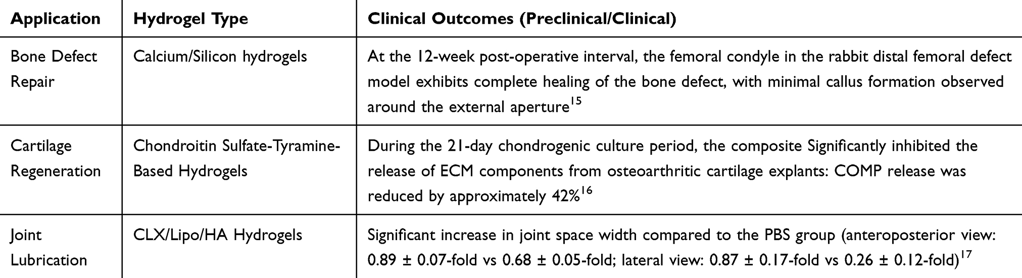 |
Table 1 Current Applications of Hydrogels in Orthopedics |
Despite their promise, traditional hydrogels face critical barriers including Mechanical Weakness, Passive Therapeutic Action and Limited Responsiveness. Most hydrogels (elastic modulus <1 MPa) cannot withstand physiological loads in weight-bearing bones (eg, femur requires >100 MPa).18 Drug release relies on diffusion/degradation, lacking spatiotemporal control over dosage19 and Inability to dynamically adapt to microenvironmental changes (eg, pH shifts in infected wounds, mechanical loading cycles).20 To address these inherent limitations, research has shifted toward stimuli-responsive “smart” hydrogels. These advanced materials can dynamically alter their properties or functions in response to external triggers—such as temperature, light, or magnetic fields—enabling spatiotemporal control that is crucial for advanced therapies. This paradigm shift paves the way for incorporating functional nanomaterials like magnetic nanoparticles (MNPs) to create actively controlled systems.21 The integration of functional nanomaterials—particularly magnetic nanoparticles (MNPs)—into hydrogel matrices has opened new frontiers. MNPs empower hydrogels with remote controllability, mechanical reinforcement, and on-demand therapeutic activation.
Emergence of Magnetic Nanoparticles (MNPs)
The advent of magnetic nanoparticles (MNPs), typically iron oxide-based (eg, Fe3O4, γ-Fe2O3) with diameters ranging from 10–100 nm, has revolutionized therapeutic strategies in orthopedics (Figure 3). MNPs uniquely combine superparamagnetism, high surface-to-volume ratio, and biocompatibility, enabling remote control and dynamic modulation of biological processes through external magnetic fields.22
 |
Figure 3 Applications of Magnetic Nanoparticles (MNPs) in orthopedics. |
There are three key Properties of MNPs for Orthopedic Applications (Table 2), including Remote Controllability Flexibility, Biofunctionalization and Real-Time Imaging. Superparamagnetic MNPs generate heat (magnetic hyperthermia) or mechanical forces (magnetomechanical actuation) under alternating magnetic fields (AMF),23 allowing spatiotemporal control over drug release or cell behavior.24 MNPs can be coated with polymers (eg, PEG, dextran) or conjugated with targeting ligands (eg, RGD peptides) to enhance bone-specific accumulation and reduce off-target effects.25 MNPs also serve as contrast agents for MRI, enabling non-invasive tracking of hydrogel degradation and bone regeneration progress.26
 |
Table 2 Current Applications of MNPs in Orthopedics |
Advantages Over Conventional Nanomaterials
Magnetic nanoparticles (MNPs) exhibit dynamic responsiveness by enabling on-demand activation mechanisms such as drug release triggered by alternating magnetic field (AMF) pulses. This capability overcomes the reliance on passive diffusion that limits traditional hydrogel-based systems.30 A key strength of MNPs lies in their multifunctionality, allowing them to perform simultaneous roles in therapy as drug carriers, in diagnostics as MRI contrast agents, and in tissue engineering as mechanical reinforcement elements.31 Furthermore, MNPs offer the advantage of deep tissue penetration. Since magnetic fields can penetrate bone tissue without significant attenuation, they enable non-invasive, remote control of implanted MNP-laden constructs.32
While MNPs alone exhibit remarkable capabilities, their integration into hydrogels creates synergistic systems that address both mechanical and biofunctional limitations. Overview of Hydrogels in Orthopedics explores how MNP-hydrogel hybrids achieve programmable mechanics, smart drug delivery, and magnetically guided tissue regeneration.
Design and Synthesis of Magnetic Nanocomposite Hydrogels
While traditional hydrogels have served as invaluable static scaffolds by providing a biocompatible, three-dimensional environment for cell growth, their inability to dynamically interact with surrounding tissues limits their regenerative potential. In contrast, stimuli-responsive or “smart” hydrogels represent a significant evolution, capable of altering their physical or chemical properties—such as swelling, stiffness, or shape—in response to specific external cues like temperature, light, or magnetic fields. This dynamic functionality enables precise, spatiotemporal control over biological processes, bridging the gap between passive support and active tissue regeneration.21 Among these advanced systems, magnetic nanocomposite hydrogels (MNHs), particularly those incorporating magnetic nanoparticles, have garnered significant attention. Their unique capacity to be remotely and non-invasively controlled by external magnetic fields offers unparalleled opportunities for targeted drug delivery, mechanical stimulation of cells, and the guidance of complex tissue morphogenesis, positioning them at the forefront of next-generation regenerative strategies.
Design Principles
The design of magnetic nanocomposite hydrogels follows several key principles to ensure optimal functionality. Iron oxide nanoparticles (Fe3O4, γ-Fe2O3) serve as the primary magnetic component due to their inherent biocompatibility and superparamagnetic properties.33 These nanoparticles are typically engineered with a size range of 10–50 nm and surface modifications such as citrate coating to enhance colloidal stability and promote uniform dispersion within the hydrogel matrix.34 The hydrogel matrix can be formulated using either natural polymers including collagen and hyaluronic acid, or synthetic polymers such as PEG and PLGA, with selection criteria based on required degradation kinetics, mechanical strength, and biological activity.35 Crosslinking strategies employ either physical interactions like hydrogen bonding or chemical methods including photo-crosslinking to achieve an optimal balance between structural integrity and nanoparticle mobility, as detailed in Table 3.36 A critical consideration is interfacial engineering between the nanoparticles and polymer network. This can be accomplished through covalent bonding using amine-carbodiimide chemistry or physical adsorption via electrostatic interactions, both serving to enhance composite stability and prevent nanoparticle aggregation under magnetic stimulation.37
 |
Table 3 Synthesis Strategies of Magnetic Nanocomposite Hydrogels |
Key Parameters for Performance Optimization (Table 4)
 |
Table 4 Current Research Advances in Magnetic Nanocomposite Hydrogels |
The performance of magnetic nanocomposite hydrogels is governed by several key parameters that require careful optimization. The concentration of magnetic nanoparticle (MNP) loading plays a critical role, as higher loading generally enhances magnetic responsiveness but may concurrently compromise hydrogel elasticity and biocompatibility.43 Surface functionalization strategies are essential for improving MNP stability and bioactivity. Approaches such as PEGylation or silica coating effectively reduce nanoparticle aggregation, while the conjugation of specific ligands like RGD peptides actively promotes cell adhesion and integration.44 Furthermore, the parameters of the applied magnetic field significantly influence system performance. The frequency (typically 50–500 kHz) and strength (usually 5–20 mT) of an alternating magnetic field (AMF) must be precisely tuned to effectively trigger targeted drug release or hyperthermia effects while ensuring no damage occurs to surrounding tissues.45
The tailored design of magnetic nanocomposite hydrogels lays the foundation for their application in dynamic bone regeneration. Section III will delve into how these systems enable spatiotemporally controlled drug delivery, mechanostimulation, and real-time monitoring of healing processes.
Applications in Orthopedics
Drug Delivery and Controlled Release
Magnetic nano-hydrogels (MNHs) exhibit revolutionary potential in orthopedic drug delivery due to their spatiotemporal controllability and targeted release capabilities.46 By embedding magnetic nanoparticles (MNPs) like Fe3O4 into hydrogel matrices, therapeutic agents such as bone morphogenetic protein-2 (BMP-2) and doxorubicin can be loaded and delivered with precision.47 For instance, a study introduced a multifunctional supramolecular hydrogel system based on dehydropeptide gels, incorporating phenylalanine-coated magnetic nanoparticles and PEGylated liposomes co-assembled with doxorubicin to achieve dual-responsive drug release (thermal and low-frequency alternating magnetic field triggers), while enhancing gelation kinetics and cytocompatibility, thereby providing a novel strategy for smart drug delivery systems.48
External magnetic fields enable triggered release mechanisms, allowing localized drug activation. The application of an external magnetic field causes the hybrid hydrogel to quickly transition from gel to sol.49 This dual functionality of MNPs—acting as both drug carriers and magnetic actuators—ensures minimal systemic toxicity and enhanced therapeutic efficacy.
Bone Tissue Engineering
MNHs can serve as dynamic scaffolds for osteoblast proliferation and differentiation.50 Their 3D porous structure mimics the natural extracellular matrix (ECM), providing mechanical support and bioactive cues.51 For example, Cetin et al investigated the bone-forming potential of a super porous hydrogel made from PHEMA and Gel, which was seeded with preosteoblastic MC3T3-E1 cells.52
The synergistic integration of additive manufacturing (3D printing) and advanced hydrogel science has emerged as a transformative paradigm in biomedical engineering, enabling the precise fabrication of intricate, hydrous constructs that closely mimic the native extracellular matrix (Figure 4).53 This powerful combination is propelling groundbreaking advancements across a spectrum of applications, from regenerative medicine and personalized drug delivery to the development of intelligent, responsive medical devices. The efficacy of this approach is critically dependent on the selection of appropriate printing modalities and tailored biomaterial inks, each offering distinct advantages and technical considerations. Regarding fabrication techniques, several key 3D printing technologies have been adapted to process hydrogel-based bioinks. Material extrusion, notably through direct ink writing (DIW), represents a cost-effective and widely accessible method that operates via the layer-by-layer deposition of viscous materials through a nozzle.54 Its success is heavily contingent on the ink’s rheological properties, which must exhibit shear-thinning behavior for extrudability and rapid shape fidelity post-deposition. In contrast, vat photopolymerization techniques, such as stereolithography (SLA) and digital light processing (DLP), utilize light energy to selectively crosslink liquid photopolymer resins. These methods are renowned for their superior printing speed and high resolution (typically 20–200 μm), but necessitate the use of photocrosslinkable polymers (eg, gelatin methacryloyl, GelMA).55 Pushing the boundaries of precision, high-definition (HD) printing methods like two-photon polymerization (TPP) achieve remarkable sub-micron resolution, facilitating the creation of highly complex microarchitectures. Alternatively, material jetting operates by precisely depositing droplets of bioink to generate constructs with defined cellular heterogeneity. While it offers high resolution, it typically requires low-viscosity inks and the jetting process itself (thermal or piezoelectric) can introduce stressors that challenge cell viability. The material composition of the bioink is equally paramount, dictating the biological and mechanical functionality of the printed construct. Nature-derived polymers —such as alginate, collagen, chitosan, and hyaluronic acid—are extensively utilized for their innate biocompatibility, bioactivity, and inherent cell-interaction motifs.56 Conversely, synthetic polymers like poly(ethylene glycol) (PEG) provide a platform with highly tunable mechanical and chemical properties.57 A particularly innovative frontier is the advent of 4D printing using stimuli-responsive hydrogels (SRHs). These “intelligent” materials can undergo predictable changes in shape, volume, or functionality in response to external cues such as temperature (eg, poly(N-isopropylacrylamide), PNIPAM), pH, or magnetic fields. For instance, the strategic incorporation of magnetic nanoparticles (eg, Fe3O4) within the hydrogel matrix creates magneto-responsive scaffolds.58 These composites can be non-invasively actuated or manipulated under an applied magnetic field, enabling advanced applications such as untethered soft robotic devices for targeted object manipulation, dynamic tissue scaffolds, and spatially controlled drug delivery systems.
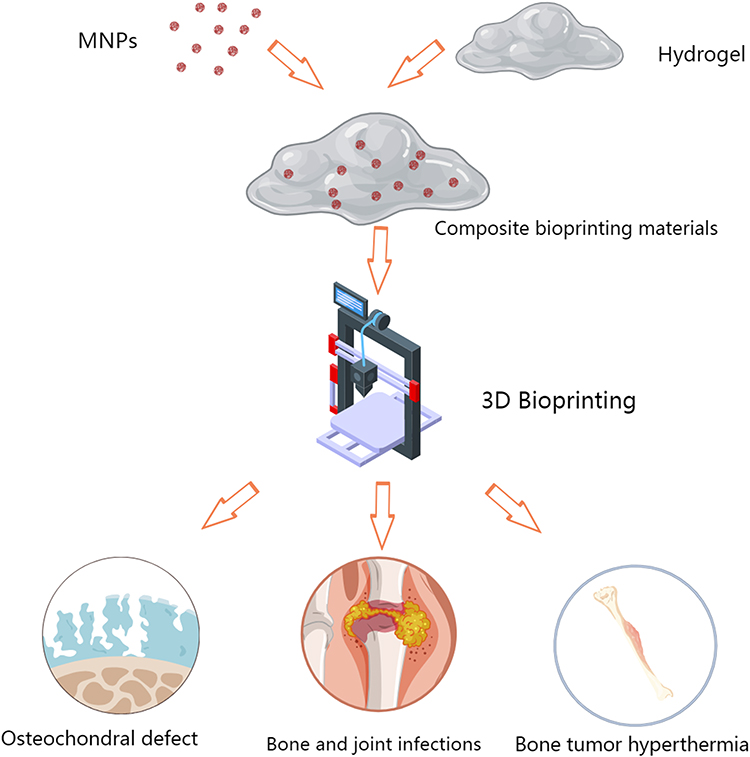 |
Figure 4 3D biological printing Magnetic Nanocomposite Hydrogels and its application in orthopedics. |
Magnetic Nanoparticles for Enhanced Cell Delivery and Function The integration of magnetic nanoparticles (MNPs) into hydrogel scaffolds provides a powerful, remote-controlled strategy to enhance cell-based therapies in bone tissue engineering.59 This approach, known as magnetic cell targeting, fundamentally improves upon passive cell delivery methods. When therapeutic cells (eg, mesenchymal stem cells - MSCs) are labeled with MNPs or encapsulated within MNP-laden hydrogels, the application of an external static magnetic field (SMF) can actively guide and retain these cells at the specific defect site. This significantly increases cell localization and engraftment efficiency, overcoming a major limitation of systemic or passive local delivery where cell dispersion leads to poor therapeutic outcomes.60 Beyond targeted delivery, MNPs exert significant positive effects on cell viability and proliferation. The presence of MNPs, particularly under SMF exposure, has been shown to upregulate key integrins and enhance cell-scaffold interactions, leading to improved adhesion and spreading.61 Furthermore, studies indicate that MNP-induced SMF conditions can stimulate mitochondrial activity and promote the secretion of autocrine and paracrine growth factors, creating a more favorable microenvironment that boosts metabolic activity and proliferation.62 Crucially, MNPs can directly influence osteogenic commitment. The mechanical forces exerted by MNPs under a magnetic field activate critical mechanotransduction pathways, such as the RhoA/ROCK signaling cascade.63 This mechanical stimulation can synergize with biochemical cues to promote the expression of osteogenic markers and enhance extracellular matrix mineralization. Therefore, MNPs act not merely as delivery vehicles but as active biophysical stimulators that enhance cell survival, proliferation, and differentiation, ultimately leading to more robust and accelerated bone regeneration.
Anti-Infective Therapies
Orthopedic infections, particularly biofilm-associated ones, pose significant challenges. MNHs integrate magnetic hyperthermia to eradicate biofilms. Under alternating magnetic fields (AMF), Fe3O4 nanoparticles generate localized heat (42–45°C), disrupting biofilm integrity and enhancing antibiotic penetration. Elena et al explored a novel combination therapy using magnetic-responsive nanocarriers for treating biofilm-associated infections, which involves antibiotic-loaded mesoporous silica nanoparticles capped with a thermo-responsive polymer and decorated with superparamagnetic iron oxide nanoparticles. Upon application of an alternating magnetic field, the nanoparticles generate heat, triggering antibiotic release and significantly reducing bacterial viability in E. coli biofilms by 4 log10 units compared to controls.64
Lu et al designed poly (β-amino ester) (PBAE) hydrogel that combined antibacterial and osteogenic functions, dual release of vancomycin (VAN) and total flavonoids of Rhizoma Drynariae (TFRD), demonstrating effective antibacterial properties against S. aureus and E. coli, as well as enhanced osteogenic differentiation capabilities.65
This dual-action strategy—thermal ablation and sustained antimicrobial release—addresses both acute and chronic orthopedic infections.
Mechanical Reinforcement and Lubrication
Hydrogels usually have less mechanical strength even compared to normal cartilage.66
The inclusion of Fe3O4 nanoparticles in the Hydrogels provides magnetic responsive properties and also boosts the mechanical strength and toughness of the hydrogels, making them suitable for load-bearing applications. For instance, in Lee research, the Fe3O4 nanoparticles significantly enhance the mechanical properties of the magneto-responsive nanocomposite hydrogels (MR_NCHs) by contributing to the formation of a robust network structure through physical interactions with laponite nanoparticles and poly(DMAAm) (PDMAAm) chains. This network structure increases the crosslinking density, leading to improved toughness, elasticity, and overall mechanical strength of the hydrogels. Making the hydrogels suitable for biomedical applications.67 Magnetic alignment techniques create anisotropic structures that mimic natural tissue mechanics. Hydrogel with magnetically oriented Fe3O4-MMT hybrids demonstrated a low friction coefficient, rivaling natural synovial fluid.30 Such lubricious properties are critical for joint repair, reducing wear and preventing secondary inflammation.
Diagnostic and Monitoring Tools
Combining cutting-edge advancements in nanotechnology and biomedical imaging, magnetic nanoparticles (MNPs) have emerged as pivotal tools for non-invasive tracking of hydrogel degradation and tissue regeneration. MNPs serve as contrast agents in MRI, enabling non-invasive tracking of hydrogel degradation and tissue regeneration. For example, superparamagnetic Fe3O4 in scaffolds allowed real-time visualization of subchondral bone repair, correlating MRI signal attenuation with new bone formation.68 Superparamagnetic iron oxide nanoparticles (SPIONs, eg, Fe3O4) with sizes of 10–50 nm exhibit high T2 relaxation enhancement due to their magnetic moment fluctuations, which disrupt local magnetic fields. This property is critical for visualizing hydrogel degradation and bone regeneration in scaffolds like GMHA (gelatin-methacryloyl/hyaluronic acid). MSC-recruiting hydrogels, and AI-enhanced systems are transforming MNPs from passive imaging tools into dynamic theranostic platforms. These theranostic systems provide feedback on scaffold integration and therapeutic efficacy, essential for personalized orthopedics. These advancements hold promise for personalized orthopedics, oncology, and regenerative medicine, bridging the gap between lab research and clinical translation.69
Challenges and Limitations
Despite the immense potential of magnetic nanocomposite hydrogels in biomedical and engineering applications, their practical implementation faces several critical challenges.
Biocompatibility and Long-Term Safety of MNPs
The clinical translation of magnetic nanoparticles (MNPs) is hindered by unresolved concerns about their biocompatibility and long-term biological effects.70,71 Prolonged retention of MNPs (eg, Fe3O4) in vivo may lead to iron ion release, causing oxidative stress or inflammatory responses.72 Poor interfacial bonding between MNPs and hydrogel matrices can also result in particle leakage, compromising stability.73 While MNPs like Fe3O4 are generally considered safe, their accumulation in organs (eg, liver, lung) raises risks of localized toxicity and chronic inflammation.74 For instance, Doxorubicin-Loaded Iron Oxide Nanoparticles in cancer chemotherapy may trigger unintended immune reactions or oxidative stress, potentially damaging healthy tissues.75 Because doxorubicin itself is a cytotoxic drug, being carried by nanoparticles may further exacerbate its damage to normal tissues. Research by Huang et al revealed that when iron oxide nanoparticles break down inside cells, they release ferric (Fe3+) ions, potentially causing cytotoxic effects.76 Furthermore, the lack of long-term safety data in large animal models (eg, non-human primates) and human trials limits confidence in their clinical use.77 Biodegradable MNP design and surface functionalization may help address this issue. Core-shell structures like Fe3O4@SiO2 delay iron ion release and enable macrophage-mediated clearance.78 Coating MNPs with Dextran reduces immunogenicity and improved biocompatibility of nanoparticles.79 Recent studies also highlight the potential for MNPs to interfere with cellular processes, such as promoting carcinogenic clonal evolution by enhancing pinocytosis in cancer cells. Comprehensive toxicological profiling, including biodegradation kinetics and immunogenicity assessments, is urgently needed to address these gaps.
Scalability and Stability of Hydrogel-MNP Systems
The manufacturing scalability and structural stability of hydrogel-MNP composites remain critical bottlenecks.80 Key challenges include: Batch-to-batch variability: Heterogeneity in MNP size, shape, and polymer coatings (eg, PEG, dextran) can alter hydrogel mechanical properties and drug-release profiles.81,82 Aggregation of MNPs via physical mixing methods weakens magnetic responsiveness and mechanical integrity is another problem that needs to be solved. Directly synthesizing MNPs within hydrogel networks (eg, co-precipitation of Fe3+/Fe2+ clusters) ensures monodispersity.67 Magnetic field dependency: Hydrogel-MNP systems often require sustained external magnetic fields for targeted delivery, but field strength decays rapidly in vivo, especially in obese patients, leading to inconsistent MNP retention and dispersion.83 Storage stability: Hydrogels loaded with MNPs may undergo phase separation or oxidation during long-term storage, compromising their therapeutic efficacy.84 Advanced quality control protocols, such as microfluidic synthesis and lyophilization techniques, are being explored to improve reproducibility and shelf life.85,86
Precision Control of Magnetic Fields in vivo
Achieving spatiotemporal precision in magnetic field control under in vivo conditions presents a formidable challenge, primarily due to several key factors. A fundamental limitation arises from Earnshaw’s theorem, which dictates that static magnetic fields cannot stably trap magnetic nanoparticles (MNPs) at deep tissue targets.87 This necessitates the implementation of dynamic field adjustments and feedback-controlled systems for effective targeting.88 Further complexity is introduced by the requirement for dynamic adaptability within complex tissue environments. Implanted constructs may face structural failure or functional inactivation when exposed to dynamic microenvironments, such as those involving wound contraction or bacterial infection.89 Real-time imaging constraints also pose a significant hurdle, as current MRI-based tracking technologies lack the necessary resolution and speed for real-time MNP guidance, often leading to off-target accumulation.90 Finally, inherent physiological variability presents a major challenge. Factors including blood flow, tissue density, and local metabolic activity can significantly disrupt intended MNP trajectories. Addressing this variability requires the development of adaptive control algorithms, such as PID control, to continuously optimize magnetic field parameters in response to the changing biological milieu.91
Emerging solutions include closed-loop systems combining magnetic particle imaging (MPI) and AI-driven predictive models to enhance targeting accuracy.92 Addressing these challenges demands interdisciplinary innovation, from nanoparticle engineering to AI-enhanced control systems. While MNPs hold transformative potential in orthopedics, their safe and effective translation hinges on resolving biocompatibility concerns, optimizing scalable fabrication, refining magnetic targeting, and navigating regulatory complexities. Future research should prioritize large-scale human trials and degradable MNP designs to mitigate long-term risks.
Future Perspectives
Advanced Fabrication Techniques: 4D Printing and AI-Driven Design
The development of next-generation hydrogel systems increasingly leverages advanced fabrication technologies such as 4D printing and artificial intelligence (AI) to construct dynamic, adaptive architectures with unprecedented precision.93 A prominent direction involves the creation of 4D-printed reconfigurable hydrogels. Emerging techniques, including femtosecond laser bimodal processing that combines additive and removal modes, allow for the fabrication of intricate microchannels and anisotropic structures within hydrogels like GelMA at submicron resolution.94–96 These engineered systems possess the capability to dynamically alter their shape or porosity in response to external stimuli such as pH or temperature changes, thereby mimicking natural tissue remodeling processes. For instance, the use of photocontrolled metallopolymer adhesives enables the reversible assembly of basic hydrogel units into complex, reconfigurable structures, showing great potential for applications in soft robotics or adaptive implants.96–98 Concurrently, AI-driven optimization is being integrated into the hydrogel design pipeline. Machine learning algorithms are employed to predict key performance metrics, including mechanical properties, drug-release kinetics, and biodegradation rates, thereby accelerating the development of tailored hydrogel materials.99,100
Multifunctional Hydrogels: Synergistic Integration of MNPs and Nanomaterials
Future hydrogel systems are evolving towards multifunctional “all-in-one” platforms through the synergistic integration of magnetic nanoparticles (MNPs) with diverse nanomaterials such as carbon nanotubes and quantum dots.101–104 These advanced composites demonstrate remarkable capabilities for biomedical applications. A prominent development involves self-healing conductive hydrogels created by integrating MNPs with conductive polymers like PEDOT: PSS or graphene oxide. These hybrid networks achieve dual functionality, combining magnetic targeting with real-time electrical signaling capabilities that show particular promise for neural and cardiac tissue repair applications.105–107 Another significant advancement emerges in theranostic nanocomposites, where core-shell structured MNPs (eg, Fe3O4@Au) embedded within hydrogel matrices can simultaneously function as MRI contrast agents, photothermal actuators, and controlled drug release carriers.108–111 Further innovation includes pH-responsive hydrogel systems loaded with MNPs that enable localized chemo-photothermal therapy while effectively evading immune detection, representing a sophisticated approach to targeted cancer treatment.112–114
Integration with Wearable Devices for Personalized Medicine
Hydrogels are poised to revolutionize wearable health technologies by enabling seamless integration with the human body for real-time diagnostics and therapy.115,116 This integration manifests through several key applications. Smart wound dressings represent a significant advancement, where 3D-printed hydrogel patches loaded with engineered extracellular vesicles (EVs) and magnetic nanoparticles (MNPs) can monitor wound parameters including pH, temperature, and bacterial load while releasing antimicrobial agents on demand.117,118 These systems are further enhanced by AI algorithms that analyze data from embedded biosensors to dynamically adjust drug-release rates.119,120 Closed-loop implantable systems constitute another promising direction. Hydrogel-based wearables interfacing with soft robotic micromachines, such as ECM-mimetic actuators, could autonomously regulate drug delivery or provide mechanical support.121,122 A specific example includes glucose-responsive hydrogels combined with MNPs and insulin reservoirs, potentially forming self-regulating implants for diabetes management.123,124 Remote patient monitoring applications are also emerging, where hydrogel sensors integrated into wearables like smart insoles or epidermal patches can transmit biomechanical data (eg, joint pressure) and biochemical data (eg, cytokine levels) to cloud platforms for AI-driven health predictions.125 The convergence of advanced fabrication, multifunctional nanomaterials, and wearable integration is driving hydrogels toward intelligent, patient-specific solutions. While key challenges remain—including scaling AI-designed hydrogels for clinical use and ensuring long-term biocompatibility of hybrid systems—these are being addressed through interdisciplinary collaboration. With ongoing innovations in 4D-printed adaptive scaffolds and closed-loop theranostic wearables, hydrogels are positioned to redefine personalized orthopedics and regenerative medicine. Current research continues to advance AI-driven hydrogel design, multifunctional nanocomposites, and wearable health technologies.126–128
Conclusion
The integration of magnetic nanoparticles into hydrogel matrices represents a significant advancement in smart biomaterials for orthopedic applications. This review has detailed how magnetic nanocomposite hydrogels overcome the limitations of traditional systems by enabling remote spatiotemporal control over therapeutic actions through external magnetic fields. These systems demonstrate multifunctional capabilities, serving as mechanically reinforced scaffolds that promote bone regeneration while simultaneously functioning as theranostic platforms for diagnosis and treatment. The convergence with additive manufacturing facilitates the fabrication of patient-specific constructs with predefined responsiveness. Clinical translation, however, depends on addressing several critical challenges. Long-term biocompatibility and biodegradation of MNPs require thorough investigation, while scalable fabrication processes must be developed to ensure consistency. Achieving precise magnetic field control in deep tissues remains an engineering hurdle that may require closed-loop systems with real-time feedback. Future research should focus on developing next-generation MNPs with biodegradable coatings and hybrid nanostructures to enhance functionality and safety. Intelligent system integration involving artificial intelligence and wearable devices could enable predictive design and adaptive therapy. Ultimately, high-quality clinical studies are essential to validate the efficacy and safety of these promising systems.
In summary, magnetic nanocomposite hydrogels represent a paradigm shift toward personalized orthopedic regeneration. By addressing existing translational barriers through interdisciplinary collaboration, these advanced systems are poised to redefine standards of care in bone repair and broader regenerative medicine applications.
Acknowledgments
All figures in the manuscript were Created with MedPeer (medpeer.cn). The authors would thank Fan Bo from Xuzhou Medical University for her help in the figure drawing.
Disclosure
The authors declare no competing interests.
References
1. Habibovic P. Strategic directions in osteoinduction and biomimetics. Tissue Eng Part A. 2017;23(23–24):1295. doi:10.1089/ten.tea.2017.0430
2. Lin C, Wang Y, Huang Z, et al. Advances in filament structure of 3D bioprinted biodegradable bone repair scaffolds. Int J Bioprint. 2021;7(4):426. doi:10.18063/ijb.v7i4.426
3. Pigeot S, Bourgine PE, Claude J, et al. Orthotopic bone formation by streamlined engineering and devitalization of human hypertrophic cartilage. Int J Mol Sci. 2020;21(19):7233. doi:10.3390/ijms21197233
4. Liu X, Chen W, Zhang C, et al. Co-seeding human endothelial cells with human-induced pluripotent stem cell-derived mesenchymal stem cells on calcium phosphate scaffold enhances osteogenesis and vascularization in rats. Tissue Eng Part A. 2017;23(11–12):546. doi:10.1089/ten.tea.2016.0485
5. Warmerdam E, Orth M, Pohlemann T, Ganse B. Gait analysis to monitor fracture healing of the lower leg. Bioengineering. 2023;10(2):255. doi:10.3390/bioengineering10020255
6. Vanvelk N, Chen B, Van Lieshout EMM, et al. Duration of perioperative antibiotic prophylaxis in open fractures: a systematic review and critical appraisal. Antibiotics. 2022;11(3):293. doi:10.3390/antibiotics11030293
7. Lu Y, Cai WJ, Ren Z, Han P. The role of staphylococcal biofilm on the surface of implants in orthopedic infection. Microorganisms. 2022;10(10):1909. doi:10.3390/microorganisms10101909
8. Nasr Azadani M, Zahedi A, Bowoto OK, Oladapo BI. A review of current challenges and prospects of magnesium and its alloy for bone implant applications. Prog Biomater. 2022;11(1):1. doi:10.1007/s40204-022-00182-x
9. Kohli N, Sharma V, Orera A, et al. Pro-angiogenic and osteogenic composite scaffolds of fibrin, alginate and calcium phosphate for bone tissue engineering. J Tissue Eng. 2021;12:20417314211005610. doi:10.1177/20417314211005610
10. Dai X, Yao X, Zhang W, et al. The osteogenic role of barium titanate/polylactic acid piezoelectric composite membranes as guiding membranes for bone tissue regeneration. Int J Nanomed. 2022;17:4339. doi:10.2147/IJN.S378422
11. Li J, Zhang T, Pan M, et al. Nanofiber/hydrogel core-shell scaffolds with three-dimensional multilayer patterned structure for accelerating diabetic wound healing. J Nanobiotechnology. 2022;20(1):28. doi:10.1186/s12951-021-01208-5
12. van Houdt CIA, Koolen MKE, Lopez-Perez PM, et al. Regenerating critical size rat segmental bone defects with a self-healing hybrid nanocomposite hydrogel: effect of bone condition and BMP-2 incorporation. Macromol Biosci. 2021;21(8):e2100088. doi:10.1002/mabi.202100088
13. Zhang K, Xue K, Loh XJ. Thermo-responsive hydrogels: from recent progress to biomedical applications. Gels. 2021;7(3):77. doi:10.3390/gels7030077
14. Jung SW, Oh SH, Lee IS, Byun JH, Lee JH. In situ gelling hydrogel with anti-bacterial activity and bone healing property for treatment of osteomyelitis. Tissue Eng Regen Med. 2019;16(5):479. doi:10.1007/s13770-019-00206-x
15. Guo C, Qi J, Liu J, et al. The ability of biodegradable thermosensitive hydrogel composite calcium-silicon-based bioactive bone cement in promoting osteogenesis and repairing rabbit distal femoral defects. Polymers. 2022;14(18):3852. doi:10.3390/polym14183852
16. Uzieliene I, Bironaite D, Pachaleva J, et al. Chondroitin sulfate-tyramine-based hydrogels for cartilage tissue repair. Int J Mol Sci. 2023;24(4):3451. doi:10.3390/ijms24043451
17. Lei Y, Wang X, Liao J, et al. Shear-responsive boundary-lubricated hydrogels attenuate osteoarthritis. Bioact Mater. 2022;16:472. doi:10.1016/j.bioactmat.2022.02.016
18. Enache AC, Cojocaru C, Samoila P, et al. Evaluation of physically and/or chemically modified chitosan hydrogels for proficient release of insoluble nystatin in simulated fluids. Gels. 2022;8(8):495. doi:10.3390/gels8080495
19. Mikusova V, Ferkova J, Zigrayova D, Krchnak D, Mikus P. Comparative study of polysaccharide-based hydrogels: rheological and texture properties and ibuprofen release. Gels. 2022;8(3):168. doi:10.3390/gels8030168
20. Tang S, Wang L, Zhang Y, Zhang F. A biomimetic platelet-rich plasma-based interpenetrating network printable hydrogel for bone regeneration. Front Bioeng Biotechnol. 2022;10:887454. doi:10.3389/fbioe.2022.887454
21. Neumann M, Di Marco G, Iudin D, et al. Stimuli-responsive hydrogels: the dynamic smart biomaterials of tomorrow. Macromolecules. 2023;56(21):8377. doi:10.1021/acs.macromol.3c00967
22. Wang T, Zhao H, Jing S, et al. Magnetofection of miR-21 promoted by electromagnetic field and iron oxide nanoparticles via the p38 MAPK pathway contributes to osteogenesis and angiogenesis for intervertebral fusion. J Nanobiotechnology. 2023;21(1):27. doi:10.1186/s12951-023-01789-3
23. Yang Y, Huang M, Qian J, Gao D, Liang X. Tunable Fe(3)O(4) nanorods for enhanced magnetic hyperthermia performance. Sci Rep. 2020;10(1):8331. doi:10.1038/s41598-020-65095-w
24. Ruan L, Chen W, Wang R, Lu J, Zink JI. Magnetically stimulated drug release using nanoparticles capped by self-assembling peptides. ACS Appl Mater Interfaces. 2019;11(47):43835. doi:10.1021/acsami.9b13614
25. Krasia-Christoforou T, Socoliuc V, Knudsen KD, Tombacz E, Turcu R, Vekas L. From single-core nanoparticles in ferrofluids to multi-core magnetic nanocomposites: assembly strategies, structure, and magnetic behavior. Nanomaterials. 2020;10(11):2178. doi:10.3390/nano10112178
26. Baki A, Remmo A, Lowa N, Wiekhorst F, Bleul R. Albumin-coated single-core iron oxide nanoparticles for enhanced molecular magnetic imaging (MRI/MPI). Int J Mol Sci. 2021;22(12):6235. doi:10.3390/ijms22126235
27. Medina-Moreno A, El-Hammadi MM, Martinez-Soler GI, Ramos JG, Garcia-Garcia G, Arias JL. Magnetic and pH-responsive magnetite/chitosan (core/shell) nanoparticles for dual-targeted methotrexate delivery in cancer therapy. Drug Deliv Transl Res. 2025;15(5):1646. doi:10.1007/s13346-024-01701-y
28. Armijo LM, Wawrzyniec SJ, Kopciuch M, et al. Antibacterial activity of iron oxide, iron nitride, and tobramycin conjugated nanoparticles against Pseudomonas aeruginosa biofilms. J Nanobiotechnology. 2020;18(1):35. doi:10.1186/s12951-020-0588-6
29. Ge J, Asmatulu R, Zhu B, Zhang Q, Yang SY. Synthesis and properties of magnetic Fe(3)O(4)/PCL porous biocomposite scaffolds with different sizes and quantities of Fe(3)O(4) particles. Bioengineering. 2022;9(7):278. doi:10.3390/bioengineering9070278
30. Estelrich J, Escribano E, Queralt J, Busquets MA. Iron oxide nanoparticles for magnetically-guided and magnetically-responsive drug delivery. Int J Mol Sci. 2015;16(4):8070. doi:10.3390/ijms16048070
31. Monteserin M, Larumbe S, Martinez AV, Burgui S, Francisco Martin L. Recent advances in the development of magnetic nanoparticles for biomedical applications. J Nanosci Nanotechnol. 2021;21(5):2705. doi:10.1166/jnn.2021.19062
32. Rotundo S, Brizi D, Flori A, Giovannetti G, Menichetti L, Monorchio A. Shaping and focusing magnetic field in the human body: state-of-the art and promising technologies. Sensors. 2022;22(14):5132. doi:10.3390/s22145132
33. Maier A, van Oossanen R, van Rhoon GC, et al. From structure to function: understanding synthetic conditions in relation to magnetic properties of hybrid Pd/Fe-Oxide nanoparticles. Nanomaterials. 2022;12(20):3649. doi:10.3390/nano12203649
34. Poller WC, Lowa N, Schleicher M, et al. Initial interaction of citrate-coated iron oxide nanoparticles with the glycocalyx of THP-1 monocytes assessed by real-time magnetic particle spectroscopy and electron microscopy. Sci Rep. 2020;10(1):3591. doi:10.1038/s41598-020-60162-8
35. Chopra H, Singh I, Kumar S, et al. A comprehensive review on hydrogels. Curr Drug Deliv. 2022;19(6):658. doi:10.2174/1567201818666210601155558
36. GhavamiNejad A, Ashammakhi N, Wu XY, Khademhosseini A. Crosslinking strategies for 3D bioprinting of polymeric hydrogels. Small. 2020;16(35):e2002931. doi:10.1002/smll.202002931
37. Selzer L, Odenbach S. Empirical law for the magnetorheological effect of nanocomposite hydrogels with magnetite microparticles. Gels. 2023;9(3):182.
38. Shi L, Zeng Y, Zhao Y, et al. Biocompatible injectable magnetic hydrogel formed by dynamic coordination network. ACS Appl Mater Interfaces. 2019;11(49):46233. doi:10.1021/acsami.9b17627
39. Chen S, Jang TS, Pan HM, et al. 3D freeform printing of nanocomposite hydrogels through in situ precipitation in reactive viscous fluid. Int J Bioprint. 2020;6(2):258. doi:10.18063/ijb.v6i2.258
40. Cao Z, Wang D, Li Y, et al. Effect of nanoheat stimulation mediated by magnetic nanocomposite hydrogel on the osteogenic differentiation of mesenchymal stem cells. Sci China Life Sci. 2018;61(4):448. doi:10.1007/s11427-017-9287-8
41. Brady MA, Talvard L, Vella A, Ethier CR. Bio-inspired design of a magnetically active trilayered scaffold for cartilage tissue engineering. J Tissue Eng Regen Med. 2017;11(4):1298. doi:10.1002/term.2106
42. Betsch M, Cristian C, Lin YY, et al. Incorporating 4D into bioprinting: real-time magnetically directed collagen fiber alignment for generating complex multilayered tissues. Adv Healthc Mater. 2018;7(21):e1800894. doi:10.1002/adhm.201800894
43. Qian H, Shan Y, Gong R, et al. Fibroblasts in scar formation: biology and clinical translation. Oxid Med Cell Longev. 2022;2022:4586569. doi:10.1155/2022/4586569
44. Narayanaswamy V, Al-Omari IA, Kamzin AS, et al. Specific absorption rate dependency on the Co(2+) distribution and magnetic properties in Co(x)Mn(1-x)Fe(2)O(4) nanoparticles. Nanomaterials. 2021;11(5):1231. doi:10.3390/nano11051231
45. Narayanaswamy V, Al-Omari IA, Kamzin AS, Issa B, Obaidat IM. Tailoring interfacial exchange anisotropy in hard-soft core-shell ferrite nanoparticles for magnetic hyperthermia applications. Nanomaterials. 2022;12(2):262. doi:10.3390/nano12020262
46. Veloso SRS, Silva JFG, Hilliou L, et al. Impact of citrate and lipid-functionalized magnetic nanoparticles in dehydropeptide supramolecular magnetogels: properties, design and drug release. Nanomaterials. 2020;11(1):16. doi:10.3390/nano11010016
47. Veloso SRS, Andrade RGD, Castanheira EMS. Review on the advancements of magnetic gels: towards multifunctional magnetic liposome-hydrogel composites for biomedical applications. Adv Colloid Interface Sci. 2021;288:102351. doi:10.1016/j.cis.2020.102351
48. Veloso SRS, Tiryaki E, Spuch C, et al. Tuning the drug multimodal release through a co-assembly strategy based on magnetic gels. Nanoscale. 2022;14(14):5488. doi:10.1039/D1NR08158F
49. Singh U, Morya V, Datta B, Ghoroi C, Bhatia D. Stimuli responsive, programmable DNA nanodevices for biomedical applications. Front Chem. 2021;9:704234. doi:10.3389/fchem.2021.704234
50. Han X, Xu H, Che L, et al. Application of inorganic nanocomposite hydrogels in bone tissue engineering. iScience. 2020;23(12):101845. doi:10.1016/j.isci.2020.101845
51. Rehman SRU, Augustine R, Zahid AA, Ahmed R, Tariq M, Hasan A. Reduced graphene oxide incorporated GelMA hydrogel promotes angiogenesis for wound healing applications. Int J Nanomed. 2019;14:9603. doi:10.2147/IJN.S218120
52. Tabatabaee S, Baheiraei N, Salehnia M. Fabrication and characterization of PHEMA-gelatin scaffold enriched with graphene oxide for bone tissue engineering. J Orthop Surg Res. 2022;17(1):216. doi:10.1186/s13018-022-03122-4
53. Barcena AJR, Dhal K, Patel P, Ravi P, Kundu S, Tappa K. Current biomedical applications of 3D-printed hydrogels. Gels. 2023;10(1):8. doi:10.3390/gels10010008
54. Advincula RC, Dizon JRC, Caldona EB, et al. On the progress of 3D-printed hydrogels for tissue engineering. MRS Commun. 2021;11(5):539. doi:10.1557/s43579-021-00069-1
55. Alexander AE, Wake N, Chepelev L, Brantner P, Ryan J, Wang KC. A guideline for 3D printing terminology in biomedical research utilizing ISO/ASTM standards. 3D Print Med. 2021;7(1):8. doi:10.1186/s41205-021-00098-5
56. Liao W, Lu J, Wang Q, et al. Osteogenesis of iron oxide nanoparticles-labeled human precartilaginous stem cells in interpenetrating network printable hydrogel. Front Bioeng Biotechnol. 2022;10:872149. doi:10.3389/fbioe.2022.872149
57. Kaliaraj GS, Shanmugam DK, Dasan A, Mosas KKA. Hydrogels-A promising materials for 3D printing technology. Gels. 2023;9(3):260. doi:10.3390/gels9030260
58. Anada T, Pan CC, Stahl AM, et al. Vascularized bone-mimetic hydrogel constructs by 3D bioprinting to promote osteogenesis and angiogenesis. Int J Mol Sci. 2019;20(5):1096. doi:10.3390/ijms20051096
59. Liu Z, Liu J, Cui X, Wang X, Zhang L, Tang P. Recent advances on magnetic sensitive hydrogels in tissue engineering. Front Chem. 2020;8:124. doi:10.3389/fchem.2020.00124
60. Bolinas DKM, Barcena AJR, Mishra A, et al. Mesenchymal stem cells loaded in injectable alginate hydrogels promote liver growth and attenuate liver fibrosis in cirrhotic rats. Gels. 2025;11(4):250. doi:10.3390/gels11040250
61. Hou R, Zhang G, Du G, et al. Magnetic nanohydroxyapatite/PVA composite hydrogels for promoted osteoblast adhesion and proliferation. Colloids Surf B Biointerfaces. 2013;103:318. doi:10.1016/j.colsurfb.2012.10.067
62. Hermenegildo B, Ribeiro C, Perez-Alvarez L, et al. Hydrogel-based magnetoelectric microenvironments for tissue stimulation. Colloids Surf B Biointerfaces. 2019;181:1041. doi:10.1016/j.colsurfb.2019.06.023
63. Deng Z, Jia Y, Liu H, et al. RhoA/ROCK pathway: implication in osteoarthritis and therapeutic targets. Am J Transl Res. 2019;11(9):5324.
64. Alvarez E, Estevez M, Gallo-Cordova A, et al. Superparamagnetic iron oxide nanoparticles decorated mesoporous silica nanosystem for combined antibiofilm therapy. Pharmaceutics. 2022;14(1):163. doi:10.3390/pharmaceutics14010163
65. Lv L, Cheng W, Wang S, et al. Poly(beta-amino ester) dual-drug-loaded hydrogels with antibacterial and osteogenic properties for bone repair. ACS Biomater Sci Eng. 2023;9(4):1976. doi:10.1021/acsbiomaterials.2c01524
66. Chen C, Huang S, Chen Z, et al. Kartogenin (KGN)/synthetic melanin nanoparticles (SMNP) loaded theranostic hydrogel scaffold system for multiparametric magnetic resonance imaging guided cartilage regeneration. Bioeng Transl Med. 2023;8(1):e10364. doi:10.1002/btm2.10364
67. Lee JH, Han WJ, Jang HS, Choi HJ. Highly tough, biocompatible, and magneto-responsive Fe(3)O(4)/Laponite/PDMAAm nanocomposite hydrogels. Sci Rep. 2019;9(1):15024.
68. Fu Y, Zoetebier B, Both S, Dijkstra PJ, Karperien M. Engineering of optimized hydrogel formulations for cartilage repair. Polymers. 2021;13(9):1526. doi:10.3390/polym13091526
69. Popa EG, Santo VE, Rodrigues MT, Gomes ME. Magnetically-responsive hydrogels for modulation of chondrogenic commitment of human adipose-derived stem cells. Polymers. 2016;8(2):28. doi:10.3390/polym8020028
70. Miri A, Najafzadeh H, Darroudi M, Miri MJ, Kouhbanani MAJ, Sarani M. Iron oxide nanoparticles: biosynthesis, magnetic behavior, cytotoxic effect. ChemistryOpen. 2021;10(3):327.
71. Saafane A, Girard D. Interaction between iron oxide nanoparticles (Fe(3)O(4) NPs) and human neutrophils: evidence that Fe(3)O(4) NPs possess some pro-inflammatory activities. Chem Biol Interact. 2022;365:110053.
72. Hu F, Wang D, Ma X, et al. Concurrent dual-contrast enhancement using Fe(3)O(4) nanoparticles to achieve a CEST signal controllability. ACS Omega. 2023;8(27):24153. doi:10.1021/acsomega.2c07000
73. Keller T, Trinks N, Brand J, et al. Design of nanohydrogels for targeted intracellular drug transport to the trans-golgi network. Adv Healthc Mater. 2023;12(13):e2201794. doi:10.1002/adhm.202201794
74. Yousef MI, Abuzreda AA, Kamel MA. Cardiotoxicity and lung toxicity in male rats induced by long-term exposure to iron oxide and silver nanoparticles. Exp Ther Med. 2019;18(6):4329. doi:10.3892/etm.2019.8108
75. Hernandes EP, Lazarin-Bidoia D, Bini RD, Nakamura CV, Cotica LF, de Oliveira Silva Lautenschlager S. Doxorubicin-loaded iron oxide nanoparticles induce oxidative stress and cell cycle arrest in breast cancer cells. Antioxidants. 2023;12(2):237. doi:10.3390/antiox12020237
76. Dechsupa N, Kosintarajit P, Kamkan K, et al. Iron(III)-quercetin complexes’ safety for MRI cell tracking in cell therapy applications: cytotoxic and genotoxic assessment. Nanomaterials. 2022;12(16):2776. doi:10.3390/nano12162776
77. Zhang S, Wu S, Shen Y, Xiao Y, Gao L, Shi S. Cytotoxicity studies of Fe(3)O(4) nanoparticles in chicken macrophage cells. R Soc Open Sci. 2020;7(4):191561. doi:10.1098/rsos.191561
78. Wang S, Liu Y, Yang A, et al. Xanthate-modified magnetic Fe(3)O(4)@SiO(2)-based polyvinyl alcohol/chitosan composite material for efficient removal of heavy metal ions from water. Polymers. 2022;14(6):1107.
79. Chircov C, Stefan RE, Dolete G, et al. Dextran-coated iron oxide nanoparticles loaded with curcumin for antimicrobial therapies. Pharmaceutics. 2022;14(5):1057. doi:10.3390/pharmaceutics14051057
80. Quazi MZ, Park N. Nanohydrogels: advanced polymeric nanomaterials in the era of nanotechnology for robust functionalization and cumulative applications. Int J Mol Sci. 2022;23(4). doi:10.3390/ijms23041943
81. Jiang H, Xu Q, Wang X, et al. Preparation of antibacterial, arginine-modified Ag nanoclusters in the hydrogel used for promoting diabetic, infected wound healing. ACS Omega. 2023;8(14):12653. doi:10.1021/acsomega.2c07266
82. Gutierrez AM, Frazar EM, X Klaus MV, Paul P, Hilt JZ. Hydrogels and hydrogel nanocomposites: enhancing healthcare through human and environmental treatment. Adv Healthc Mater. 2022;11(7):e2101820. doi:10.1002/adhm.202101820
83. Schambra HM. Repetitive transcranial magnetic stimulation for upper extremity motor recovery: does it help? Curr Neurol Neurosci Rep. 2018;18(12):97. doi:10.1007/s11910-018-0913-8
84. Casas-Forero N, Trujillo-Mayol I, Zuniga RN, Petzold G, Orellana-Palma P. Effects of cryoconcentrated blueberry juice as functional ingredient for preparation of commercial confectionary hydrogels. Gels. 2022;8(4):217. doi:10.3390/gels8040217
85. Nastruzzi A, Pitingolo G, Luca G, Nastruzzi C. DoE analysis of approaches for hydrogel microbeads’ preparation by millifluidic methods. Micromachines. 2020;11(11):1007. doi:10.3390/mi11111007
86. Grenier J, Duval H, Barou F, Lv P, David B, Letourneur D. Mechanisms of pore formation in hydrogel scaffolds textured by freeze-drying. Acta Biomater. 2019;94:195. doi:10.1016/j.actbio.2019.05.070
87. Tombacz E, Turcu R, Socoliuc V, Vekas L. Magnetic iron oxide nanoparticles: recent trends in design and synthesis of magnetoresponsive nanosystems. Biochem Biophys Res Commun. 2015;468(3):442. doi:10.1016/j.bbrc.2015.08.030
88. Ashkin A, Gordon JP. Stability of radiation-pressure particle traps: an optical Earnshaw theorem. Opt Lett. 1983;8(10):511. doi:10.1364/OL.8.000511
89. Jia B, Li G, Cao E, Luo J, Zhao X, Huang H. Recent progress of antibacterial hydrogels in wound dressings. Mater Today Bio. 2023;19:100582. doi:10.1016/j.mtbio.2023.100582
90. Kecik K, Mitura A. Theoretical and experimental investigations of a pseudo-magnetic levitation system for energy harvesting. Sensors. 2020;20(6):1623. doi:10.3390/s20061623
91. Tomitaka A, Arami H, Ahmadivand A, et al. Magneto-plasmonic nanostars for image-guided and NIR-triggered drug delivery. Sci Rep. 2020;10(1):10115. doi:10.1038/s41598-020-66706-2
92. Zhou H, Mayorga-Martinez CC, Pane S, Zhang L, Pumera M. Magnetically driven micro and nanorobots. Chem Rev. 2021;121(8):4999. doi:10.1021/acs.chemrev.0c01234
93. Tran TS, Balu R, Mettu S, Roy Choudhury N, Dutta NK. 4D printing of hydrogels: innovation in material design and emerging smart systems for drug delivery. Pharmaceuticals. 2022;15(10):1282. doi:10.3390/ph15101282
94. Qu G, Huang J, Li Z, et al. 4D-printed bilayer hydrogel with adjustable bending degree for enteroatmospheric fistula closure. Mater Today Bio. 2022;16:100363. doi:10.1016/j.mtbio.2022.100363
95. Kim SH, Seo YB, Yeon YK, et al. 4D-bioprinted silk hydrogels for tissue engineering. Biomaterials. 2020;260:120281. doi:10.1016/j.biomaterials.2020.120281
96. Zhuo S, Geever LM, Halligan E, Tie BSH, Breheny C. A development of new material for 4D printing and the material properties comparison between the conventional and stereolithography polymerised NVCL hydrogels. J Funct Biomater. 2022;13(4):262. doi:10.3390/jfb13040262
97. Tan L, Lee H, Fang L, Cappelleri DJ. A power compensation strategy for achieving homogeneous microstructures for 4D printing shape-adaptive PNIPAM hydrogels: out-of-plane variations. Gels. 2022;8(12):828. doi:10.3390/gels8120828
98. Aronsson C, Jury M, Naeimipour S, et al. Dynamic peptide-folding mediated biofunctionalization and modulation of hydrogels for 4D bioprinting. Biofabrication. 2020;12(3):035031. doi:10.1088/1758-5090/ab9490
99. Fu Z, Angeline V, Sun W. Evaluation of printing parameters on 3D extrusion printing of pluronic hydrogels and machine learning guided parameter recommendation. Int J Bioprint. 2021;7(4):434. doi:10.18063/ijb.v7i4.434
100. Al-Kharusi G, Dunne NJ, Little S, Levingstone TJ. The role of machine learning and design of experiments in the advancement of biomaterial and tissue engineering research. Bioengineering. 2022;9(10):561. doi:10.3390/bioengineering9100561
101. Barrett-Catton E, Ross ML, Asuri P. Multifunctional hydrogel nanocomposites for biomedical applications. Polymers. 2021;13(6):856. doi:10.3390/polym13060856
102. Yin Y, Hu B, Yuan X, Cai L, Gao H, Yang Q. Nanogel: a versatile nano-delivery system for biomedical applications. Pharmaceutics. 2020;12(3):290. doi:10.3390/pharmaceutics12030290
103. Paresishvili T, Janelidze M, Kakabadze Z. Multifunctional hybrid hydrogel for the prevention of post-surgery tumor recurrence. Am J Cancer Res. 2023;13(6):2504.
104. Xin W, Gao Y, Yue B. Recent advances in multifunctional hydrogels for the treatment of osteomyelitis. Front Bioeng Biotechnol. 2022;10:865250. doi:10.3389/fbioe.2022.865250
105. Lee M, Kim MC, Lee JY. Nanomaterial-based electrically conductive hydrogels for cardiac tissue repair. Int J Nanomed. 2022;17:6181. doi:10.2147/IJN.S386763
106. Liang Y, Qiao L, Qiao B, Guo B. Conductive hydrogels for tissue repair. Chem Sci. 2023;14(12):3091. doi:10.1039/D3SC00145H
107. Xu J, Tsai YL, Hsu SH. Design strategies of conductive hydrogel for biomedical applications. Molecules. 2020;25(22):5296. doi:10.3390/molecules25225296
108. Siqueira ERL, Pinheiro WO, Aquino VRR, et al. Engineering gold shelled nanomagnets for pre-setting the operating temperature for magnetic hyperthermia. Nanomaterials. 2022;12(16):2760. doi:10.3390/nano12162760
109. Al-Musawi S, Albukhaty S, Al-Karagoly H, Almalki F. Design and synthesis of multi-functional superparamagnetic core-gold shell coated with chitosan and folate nanoparticles for targeted antitumor therapy. Nanomaterials. 2020;11(1):32. doi:10.3390/nano11010032
110. Tomitaka A, Ota S, Nishimoto K, Arami H, Takemura Y, Nair M. Dynamic magnetic characterization and magnetic particle imaging enhancement of magnetic-gold core-shell nanoparticles. Nanoscale. 2019;11(13):6489. doi:10.1039/C9NR00242A
111. Campbell S, Maitland D, Hoare T. Enhanced pulsatile drug release from injectable magnetic hydrogels with embedded thermosensitive microgels. ACS Macro Lett. 2015;4(3):312. doi:10.1021/acsmacrolett.5b00057
112. Thambi T, Jung JM, Lee DS. Recent strategies to develop pH-sensitive injectable hydrogels. Biomater Sci. 2023;11(6):1948. doi:10.1039/D2BM01519F
113. Ding F, Zhang L, Chen X, Yin W, Ni L, Wang M. Photothermal nanohybrid hydrogels for biomedical applications. Front Bioeng Biotechnol. 2022;10:1066617. doi:10.3389/fbioe.2022.1066617
114. Ji Y, Wang C. Magnetic iron oxide nanoparticle-loaded hydrogels for photothermal therapy of cancer cells. Front Bioeng Biotechnol. 2023;11:1130523. doi:10.3389/fbioe.2023.1130523
115. Li J, Ding Q, Wang H, et al. Engineering smart composite hydrogels for wearable disease monitoring. Nanomicro Lett. 2023;15(1):105. doi:10.1007/s40820-023-01079-5
116. Li S, Zhou X, Dong Y, Li J. Flexible self-repairing materials for wearable sensing applications: elastomers and hydrogels. Macromol Rapid Commun. 2020;41(23):e2000444. doi:10.1002/marc.202000444
117. Thet NT, Alves DR, Bean JE, et al. Prototype development of the intelligent hydrogel wound dressing and its efficacy in the detection of model pathogenic wound biofilms. ACS Appl Mater Interfaces. 2016;8(24):14909. doi:10.1021/acsami.5b07372
118. Liu L, Li X, Nagao M, Elias AL, Narain R, Chung HJ. A pH-indicating colorimetric tough hydrogel patch towards applications in a substrate for smart wound dressings. Polymers. 2017;9(11):558. doi:10.3390/polym9110558
119. Ganesh KN, Hillmyer M, Kaplan D, et al. From biosensors to drug delivery and tissue engineering: open biomaterials research. ACS Omega. 2022;7(8):6437. doi:10.1021/acsomega.2c00787
120. Wu H, Du P, Kokate R, Wang JX. A semi-analytical solution and AI-based reconstruction algorithms for magnetic particle tracking. PLoS One. 2021;16(7):e0254051. doi:10.1371/journal.pone.0254051
121. Liang Q, Xia X, Sun X, et al. Highly stretchable hydrogels as wearable and implantable sensors for recording physiological and brain neural signals. Adv Sci. 2022;9(16):e2201059. doi:10.1002/advs.202201059
122. Bian S, Zhu B, Rong G, Sawan M. Towards wearable and implantable continuous drug monitoring: a review. J Pharm Anal. 2021;11(1):1. doi:10.1016/j.jpha.2020.08.001
123. Volpatti LR, Facklam AL, Cortinas AB, et al. Microgel encapsulated nanoparticles for glucose-responsive insulin delivery. Biomaterials. 2021;267:120458. doi:10.1016/j.biomaterials.2020.120458
124. Mohanty AR, Ravikumar A, Peppas NA. Recent advances in glucose-responsive insulin delivery systems: novel hydrogels and future applications. Regen Biomater. 2022;9:rbac056. doi:10.1093/rb/rbac056
125. Puiggali-Jou A, Babeli I, Roa JJ, et al. Remote spatiotemporal control of a magnetic and electroconductive hydrogel network via magnetic fields for soft electronic applications. ACS Appl Mater Interfaces. 2021;13(36):42486. doi:10.1021/acsami.1c12458
126. Heidarian P, Kouzani AZ, Kaynak A, Zolfagharian A, Yousefi H. Dynamic mussel-inspired chitin nanocomposite hydrogels for wearable strain sensors. Polymers. 2020;12(6):1416. doi:10.3390/polym12061416
127. Aliakbar Ahovan Z, Esmaeili Z, Eftekhari BS, et al. Antibacterial smart hydrogels: new hope for infectious wound management. Mater Today Bio. 2022;17:100499. doi:10.1016/j.mtbio.2022.100499
128. Ren M, Yi PH. Artificial intelligence in orthopedic implant model classification: a systematic review. Skeletal Radiol. 2022;51(2):407. doi:10.1007/s00256-021-03884-8
 © 2025 The Author(s). This work is published and licensed by Dove Medical Press Limited. The
full terms of this license are available at https://www.dovepress.com/terms
and incorporate the Creative Commons Attribution
- Non Commercial (unported, 4.0) License.
By accessing the work you hereby accept the Terms. Non-commercial uses of the work are permitted
without any further permission from Dove Medical Press Limited, provided the work is properly
attributed. For permission for commercial use of this work, please see paragraphs 4.2 and 5 of our Terms.
© 2025 The Author(s). This work is published and licensed by Dove Medical Press Limited. The
full terms of this license are available at https://www.dovepress.com/terms
and incorporate the Creative Commons Attribution
- Non Commercial (unported, 4.0) License.
By accessing the work you hereby accept the Terms. Non-commercial uses of the work are permitted
without any further permission from Dove Medical Press Limited, provided the work is properly
attributed. For permission for commercial use of this work, please see paragraphs 4.2 and 5 of our Terms.