Back to Journals » International Journal of Nanomedicine » Volume 15
Low-Frequency versus High-Frequency Ultrasound-Mediated Transdermal Delivery of Agomelatine-Loaded Invasomes: Development, Optimization and in-vivo Pharmacokinetic Assessment
Authors Tawfik MA, Tadros MI ![]() , Mohamed MI, Nageeb El-Helaly S
, Mohamed MI, Nageeb El-Helaly S ![]()
Received 25 September 2020
Accepted for publication 21 October 2020
Published 12 November 2020 Volume 2020:15 Pages 8893—8910
DOI https://doi.org/10.2147/IJN.S283911
Checked for plagiarism Yes
Review by Single anonymous peer review
Peer reviewer comments 3
Editor who approved publication: Dr Thomas Webster
Mai Ahmed Tawfik, Mina Ibrahim Tadros, Magdy Ibrahim Mohamed, Sara Nageeb El-Helaly
Department of Pharmaceutics and Industrial Pharmacy, Faculty of Pharmacy, Cairo University, Cairo, Egypt
Correspondence: Mina Ibrahim Tadros
Cairo University, Kasr El-Aini Street, Cairo 11562, Egypt
Tel +20 1223620458
Fax +20 223628426
Email [email protected]
Aim: Agomelatine (AGM) is the first melatonergic antidepressant. It suffers from low oral bioavailability (< 5%) due to extensive hepatic metabolism. The current work aimed to develop an alternative AGM-loaded invasomes to enhance transdermal drug bioavailability.
Methodology: AGM-loaded invasomes were developed using two drug: lipid ratios (1:10 or 1:7.5), four terpene types (limonene, cineole, fenchone or citral) and two terpene concentrations (0.75% or 1.5%, w/v). They were characterized for drug entrapment efficiency (EE%), particle size (PS), zeta potential (ZP) and drug released percentages after 0.5h (Q0.5h) and 8h (Q8h). The optimum invasomes (I1, I2 and I4) were evaluated for morphology, drug-crystallinity, and ex-vivo drug flux. The variables influencing sonophoresis of the best achieved invasomal gel system (I2) were optimized including, ultrasound frequency (low, LFU or high, HFU), mode (pulsed or continuous), application period (10 min or 15 min) and duty cycle (50% or 100%). AGM pharmacokinetics were evaluated in rabbits following transdermal application of I2-LFU-C4 system, relative to AGM oral dispersion.
Results: The superiority of I2 invasomes [comprising AGM and phosphatidylcholine (1:10) and limonene (1.5% w/v)] was statistically revealed with respect to EE% (78.6%), PS (313 nm), ZP (− 64 mV), Q0.5h (30.1%), Q8h (92%), flux (10.79 μg/cm2/h) and enhancement ratio (4.83). The optimum sonophoresis conditions involved application of LFU in the continuous mode for 15 min at a 100% duty cycle (I2-LFU-C4 system). The latter system showed significantly higher Cmax, and relative bioavailability (≈ 7.25 folds) and a similar Tmax (0.5 h).
Conclusion: I2-LFU-C4 is a promising transdermal system for AGM.
Keywords: agomelatine, invasomes, transdermal, sonophoresis, low frequency ultrasound
Introduction
The American psychiatric association defines depression as a serious medical illness that negatively influences how you act, how you feel, and how you think. Depression affects the person’s quality of life due to its impact on mood, behavior, sleep and appetite. The emotional and physical complications could decrease the personal ability to function properly.1 Agomelatine (AGM) is the first-in-class melatonergic antidepressant drug. It acts by dual mechanism; melatonin receptors (MT1 & MT2) agonist and serotonin receptor (5-HT2C) antagonist.2 In a network meta-analysis of 21 antidepressants for the acute treatment of major depressive disorder, AGM demonstrated the highest acceptability among patients.3
Following oral administration, AGM is highly and rapidly absorbed (>75%) from the gastrointestinal tract. Unfortunately, the absolute bioavailability of AGM is <5%. This could be attributed to the extensive first pass metabolism, mainly by the hepatic cytochrome P4501A2.4 AGM possesses an intermediate molecular weight (243.3 g/mol) and promising lipophilic properties (log P value, 2.83). In view of the aforementioned, few studies were conducted to enhance the transdermal delivery of AGM via the development of microemulsion gels,4 liquid nanocrystals5 and polymeric nanoparticles6 to bypass the first pass metabolism and enhance drug bioavailability.
The diffusion of any drug via the skin is hampered by the stratum corneum (SC).7 Therefore, various passive and active permeation techniques were adopted to enhance the percutaneous drug absorption. The passive approaches could influence the drug–vehicle interactions (eutectic systems, supersaturated systems, prodrugs and ion pairing), promote the optimization of formulation (patch technology, nano-vesicular systems, and microemulsions) and modification of the stratum corneum (chemical enhancers and hydration).8 The active enhancement techniques involve the use of external energy and hence, could be classified into three classes; electrically-assisted methods (magnetophoresis, iontophoresis, radiofrequency, and electroporation), mechanical methods (skin stretching, abrasion, skin puncture, microneedles, and perforation), and physical methods (thermophoresis, laser radiation and sonophoresis).8 The combination of techniques was recently explored to improve the transdermal drug permeation.9,10
Invasomes are novel elastic nano-vesicular systems comprising phospholipids, ethanol and one or mixture of terpenes.11,12 PC constitutes the invasomal bilayer matrix. Terpenes, along with ethanol, have a synergistic effect on the percutaneous drug absorption by imparting fluidity or flexibility to the phospholipid bilayers, disrupting the lipids of SC and improving the partitioning of drug and/or nanovesicles into the skin.13,14
Sonophoresis is a promising technique that temporarily increases the skin permeability via application of ultrasound waves, so that drugs can be delivered in a non-invasive way.15 The enhanced skin permeability could be correlated to the acoustic streaming, the cavitation forces, the thermal effect of the ultrasound energy and the disruption of the arrangement of the skin lipid bilayers.7,16 Two application protocols of ultrasound waves were investigated. The simultaneous protocol involves the mixing of the drug-loaded system with the coupling medium and applying the ultrasound waves. On the other hand, the pre-treatment protocol involves the application of the ultrasound waves on the coupling medium, prior to the passive delivery of the drug-loaded system. The latter protocol is preferred since it minimizes the possibility of drug degradation following the application of ultrasound waves during the treatment period.17 Sonophoresis involves the use of low, intermediate or high frequency ultrasound waves.7 Low frequency ultrasound (LFU) waves were previously applied to enhance the transdermal delivery of various drugs like caffeine, fentanyl and insulin.18,19 On the other hand, the transdermal permeation of other drugs like salicylic acid20 and ondansetron hydrochloride10 was promoted following the application of high frequency ultrasound (HFU) waves. Aldwaikat et al21 reported that drug enhancement ratio is inversely correlated to the ultrasound frequency. LFU produces more cavitational forces and promotes higher drug permeation efficiency rather than HFU.7,22
In the current work, a dual tackling approach based on passive permeation (invasomes) and active permeation (sonophoresis; LFU or HFU) techniques was explored to enhance the transdermal delivery of AGM. AGM-loaded invasomes were developed according to 22.41 full factorial design. The variables influencing the application of sonophoresis on the best achieved invasomal gel systems were optimized. The pharmacokinetics of AGM following the application of the optimum sonophoresis conditions to the best achieved invasomal gel system was evaluated in rabbits.
Materials and Methods
Materials
Agomelatine (AGM) and Clonazepam (internal standard; IS) were kindly provided by Hikma Pharmaceutical Co. (Cairo, Egypt) and Amoun Pharmaceutical Co. (Cairo, Egypt), respectively. Phosphatidylcholine (PC; L-α-Lecthin granular from soybean oil) was acquired from Acros Organics (Geel, Belgium). Limonene, cineole, fenchone, citral and acetonitrile (HPLC grade) were purchased from Sigma-Aldrich® (St. Louis, MO). Hydroxypropyl methylcellulose K4M (HPMC) was purchased from Dow Chemical Company (Midland, US). Chloroform, methanol, ethanol, disodium hydrogen phosphate, potassium dihydrogen phosphate and sodium chloride were supplied by El-Nasr Pharmaceutical Chemicals Co. (Cairo, Egypt). Semi-permeable membrane tubing was purchased from Spectrum Laboratories Inc. (Spectra Por© Molecular weight cut off 12–14 kD, California, USA). Other chemicals (analytical grade) were used as received.
Preparation of AGM-Loaded Invasomes
Sixteen AGM-loaded invasomes comprising lipid (phosphatidylcholine, PC), terpene (limonene, cineole, fenchone or citral), and ethanol were prepared by using the thin film hydration technique.14,23 The composition of the developed invasomes is shown in Table 1. The investigated variables were (i) drug: lipid ratio (1:10 or 1:7.5), (ii) terpene type (limonene, cineole, fenchone or citral), and (iii) terpene concentration (0.75% or 1.5%, w/v). Briefly, AGM (50 mg), PC and terpene were accurately weighed and dissolved in a 2:1 mixture of chloroform and methanol in a dry, clean, round bottom flask, using a bath sonicator (Crest ultrasonics corp., Trenton, USA). The organic solvent was evaporated by rotating the flask under reduced pressure at 120 rpm for 15 min at 60 °C in a rotatory evaporator (Heidolph VV 2000, Burladingen, Germany) until a clear thin film is formed on the inner walls of the flask. The film was left under vacuum to get rid of the organic solvents, traces. Following, the film was hydrated with 10 mL of 30:70 (v/v) mixture of ethanol and phosphate buffer saline (PBS, pH 7.4) by rotating the flask under normal pressure at 120 rpm for 1 hour at room temperature, so that a final AGM concentration of 5 mg/mL was achieved. The invasomes were sonicated (Crest ultrasonics corp., Trenton, USA) for 5 min to promote the development of fine dispersions, and left to equilibrate overnight at 4 °C.
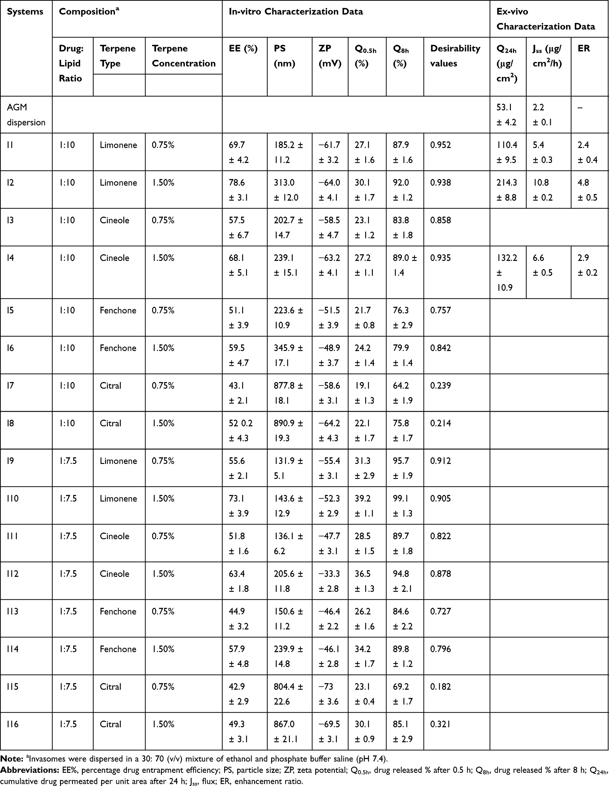 |
Table 1 The Composition, in-vitro and ex-vivo Characterization Data (Mean ± S.D., n = 3) of the Developed Agomelatine Invasomes, in Comparison to AGM Aqueous Dispersion |
In-vitro Characterization of AGM-Loaded Invasomes
Determination of AGM Entrapment Efficiency (EE%)
The EE% of the drug was estimated, in triplicates, according to the direct technique. Briefly, 1 mL of each invasomal dispersion was ultra-centrifuged at 22000 rpm (Sigma 3–30 KS, Sigma Laborzentrifugen GmbH, Germany) for 1 h at 4 °C. The supernatant, containing the unentrapped drug, was discarded, while the drug-loaded pellet was dissolved in ethanol by sonication. The AGM concentration was measured spectrophotometrically (Shimadzu, model UV-1601 PC, Kyoto, Japan) after appropriate dilution at λmax 276 nm.24
Parallel studies, using drug-free invasomes, were conducted as controls. The EE% of AGM was calculated according to the following equation,

Determination of Particle Size (PS), Polydispersity Index (PDI) and Zeta Potential (ZP)
The mean PS (Z-Average) and PDI of AGM-loaded invasomes were determined at 25 ± 1°C, in triplicates, via dynamic light scattering (DLS) analysis (Malvern Zetasizer Nano ZS; Worcestershire, UK). Prior to measurements, each invasomal dispersion was properly diluted (10 folds) with deionized water to ensure appropriate light scattering intensity. DLS analyzes the fluctuation of intensity of a scattered laser beam (caused by the Brownian random motion of vesicles), at an angle of 90º, as function of time.25 The derived data were needed to estimate the diffusion coefficient and hence, the Z-Average of vesicles according to Stoke-Einstein equation.26 Low PDI values (<0.3) could indicate homogenous particle size distributions.27
A laser doppler anemometer, coupled with the Zetasizer Nano ZS, was utilized to determine the electrophoretic mobility and hence, the zeta potential of the charged vesicles.
In-vitro AGM Release Studies
The dialysis bag method was selected for carrying out the in-vitro drug release studies.28,29 Briefly, aliquot samples of AGM-loaded invasomes (containing AGM equivalent to 5 mg) were filled in pre-conditioned semi-permeable membrane tubings, tightly sealed from both sides, and placed in stoppered glass bottles containing phosphate buffer saline (pH 7.4, 100 mL). The whole set ups were placed in an incubated shaking water bath (Unimax, IKA, Germany). The latter was maintained at 32 ± 0.5 ºC, and continuously shaken at 100 strokes per min. At pre-determined time points (0.5, 1, 2, 3, 4, 6 and 8 h), aliquots of 3 mL were withdrawn from the receptor compartment and analyzed spectrophotometrically at λmax 276 nm. To maintain constant volumes and sink conditions, equivalent volumes of fresh medium were added to the bottles at each sampling point.
The drug released percentages (mean ± SD) were plotted versus time. For comparison, AGM released percentages after 0.5 h (Q0.5h) and 8 h (Q8h) were assessed. Parallel in-vitro drug release studies were similarly conducted for drug-free invasomes to act as controls and for AGM aqueous dispersion (5 mg/mL) to check that the dialysis membrane would not represent an obstacle for the release of AGM in this medium.29
Optimization of AGM-Loaded Invasomes via 22.41 Full Factorial Design
A 22.41 full factorial design was carried out to study the effect of the different formulation variables on the characteristics of the developed AGM-loaded invasomes using Design-Expert® software (Stat-Ease, Inc., Minneapolis, MN). The independent variables were, X1: drug: lipid ratio (1:10 or 1:7.5), X2: terpene type (limonene, cineole, fenchone or citral), and X3: terpene concentration (0.75% or 1.5%, w/v). The drug EE% (Y1), PS (Y2), ZP (Y3), Q0.5h (Y4) and Q08h (Y5) were chosen as the dependent variables (responses). The desirability formula was adjusted to minimize PS & Q0.5h and maximize EE%, ZP (as absolute value) and Q8h. One-way ANOVA and Duncan test were attempted to compare the results of the selected responses using SPSS software 17.0 (SPSS Inc., Chicago, USA). A P value <0.05 was considered statistically significant. The optimum AGM-loaded invasomes were selected.
Characterization of the Optimum AGM-Loaded Invasomes
Transmission Electron Microscopy (TEM)
The morphologic and topographic characteristics of the optimum AGM-loaded invasomes were visualized using Joel JEM 1230 transmission electron microscope (Tokyo, Japan). A drop of each invasomal dispersion was stratified on a carbon-coated copper grid, negatively stained with a drop of 1% (w/v) phosphotungstic acid, air-dried at room temperature, and finally examined at an accelerating voltage of 80 kV.30
Lyophilization of the Optimum AGM-Loaded Invasomes
The freshly prepared optimum AGM-loaded invasomes were frozen and lyophilized (Novalyphe-NL 500 lyophilizer; Savant Instruments; NY, USA) under a vacuum of 7 × 10⁻² mbar at -45 °C for 24 h. The lyophilized invasomes passed through the following solid-state investigations.
Differential Scanning Calorimetry (DSC) Studies
The thermal properties and phase transition behavior of AGM, AGM-PC physical mixture and the optimum lyophilized invasomes were assessed by using Shimadzu differential scanning calorimeter (DSC-60 Shimadzu, Kyoto, Japan). Samples (≈ 3–4 mg) were heated in standard aluminum pans at a heating rate of 10°C/min, over a temperature range of 30–300 °C, under nitrogen flow (30 mL/min). Indium (99.9%) was used as a reference.25
Powder X-Ray Diffraction (PXRD) Studies
PXRD studies of AGM, AGM-PC physical mixture and the optimum lyophilized invasomes were assessed at room temperature (PANalytical X’Pert PRO diffractometer; Almelo, Netherlands). The samples were prepared using nickel filtered Cu Kα radiation (λ = 1.542 Å, 45 kV, and 35 mA). The results were presented as intensity versus 2θ (5–80°).31
Ex-vivo Drug Permeation Studies
The protocol of the ex-vivo studies was approved by the institutional review board; Research Ethics Committee-Faculty of Pharmacy, Cairo University (Approval No. PI 2605). The ex-vivo studies were conducted, in accordance with EU Directive 2010/63/EU for animal experiments. Newly born male Wistar rats (weighing 70 ± 20 g) were euthanized and the dorsal hair was shaved.4,5 The full-thickness skin was carefully removed and inspected for any abnormalities, bites, or scratches. The epidermal layers were separated from the underlying subcutaneous fats and stored at -20 °C until use within 3 days. Prior to conducting the ex-vivo drug permeation studies, the epidermal layers were left to thaw, and equilibrate in phosphate buffer saline (pH 7.4, 24 h) at room temperature so that a trans-epidermal hydration gradient could be maintained. The hydrated epidermal layers were carefully mounted in modified Franz diffusion cells, between the donor and the receptor compartments, so that the stratum corneum side up. The drug permeation surface area was 3 cm2. AGM-loaded samples of the optimum invasomes or the aqueous AGM dispersion (containing the equivalent of 5 mg of AMG) were added to the donor compartments while the receptor compartments were loaded with a 50:50 (v/v) mixture of ethanol and phosphate buffer saline (pH 7.4, 10 mL) to maintain sink conditions.4,5 The stirring rate was set at 100 rpm, and the temperature was adjusted at 32 ± 0.5 °C.13–32 Aliquots (1 mL) of the receptor compartments were collected and immediately replaced with fresh medium at 0.5, 1, 2, 4, 6, 8 and 24 hours. The withdrawn samples were analyzed using a previously validated HPLC method.4,5 The drug permeation studies were conducted in triplicates and the results were presented as mean ± SD. The cumulative permeated drug amounts through the skin per unit area (μg/cm2) were plotted against time (h). The flux (Jss) was calculated from the slope of the curve at steady state. The enhancement ratio (ER) was calculated via dividing the flux value of invasomes by that of the aqueous dispersion,13 according to the following equations33


HPLC Determination of AGM
The AGM concentration in the withdrawn samples was analyzed, at 25 ± 1 °C, using a validated HPLC method at 230 nm with minor modifications.4,5 HPLC system (Shimadzu, Kyoto, Japan) equipped with an LC-10 AD isocratic pump and a SPD-10 A UV/VIS detector connected to a C-R6A chromatopac integrator was utilized. A reversed phase C18 column (4.6 mm×250 mm, particle size 5 μm; Teknokroma, Barcelona, Spain) was used. The mobile phase was composed of a 1:1 mixture of acetonitrile and potassium dihydrogen phosphate buffer (15 mMol). The pH was adjusted at 3.5 with orthophosphoric acid, and the flow rate was set at 1.2 mL/min.34 Under the investigated conditions, the retention time for AGM was 5.5 min.
Selection of the Best Achieved Invasomes
The selection of the best achieved invasomes was performed using Design-Expert software where, the ex-vivo drug permeation parameters, cumulative permeated AGM amount per unit area after 24 hours (Q24h; μg/cm2), flux (Jss; μg/cm2/h) and ER were chosen as the dependent variables.
Development of AGM-Loaded Invasomal Gel Systems
HPMC-based coupling gel for sonophoresis was prepared (0.5% w/v) and kept in a refrigerator, overnight, to get air bubbles-free clear hydrogels. AGM-loaded invasomal gel systems were developed via the incorporation of the best achieved AGM-loaded invasomes (I2) into the coupling gels at a ratio of 1:3.
Ex-vivo Drug Permeation Studies
Further ex-vivo drug permeation studies were conducted to explore the effect of sonophoresis on enhancing the drug permeation from the developed invasomal gel systems, relative to the passive permeation of the aqueous drug dispersion or the best achieved AGM-loaded invasomes (I2).
The pre-treatment protocol for the application of ultrasound waves was adopted, as proposed by Ammar et al.10 Briefly, the hydrated epidermal layers of the rat skin were carefully placed on Petri dish, taking into consideration that the epidermis is facing upwards. The drug-free coupling gel was placed on the epidermis and the ultrasound horn for LFU (Cole-Parmer, Trenton, USA) or HFU (Radium skin massager, Tokyo, Japan) was fixed and placed directly over the coupling gel.10,17 The investigated variables were the ultrasound frequency (LFU or HFU), ultrasound mode (pulsed or continuous), the application period (10 or 15 min) and the duty cycle (50% or 100%). A 24 full factorial design was adopted to explore the influences of these variables, Table 2.
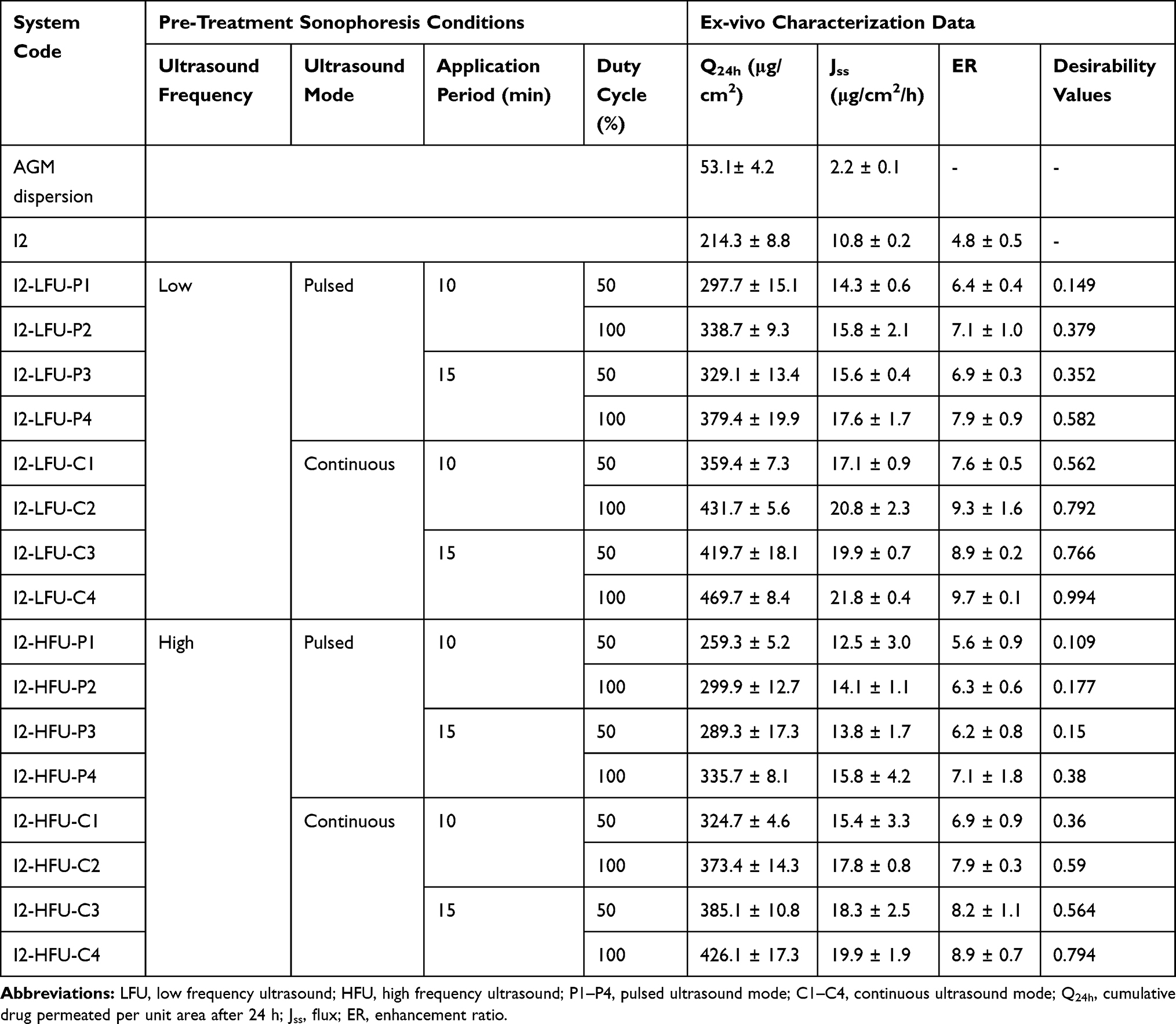 |
Table 2 Pre-Treatment Sonophoresis Conditions and ex-vivo Characterization Data (Mean ± S.D., n = 3) of the Developed Agomelatine-Loaded I2-Invasomal Gel Systems, in Comparison to the Untreated AGM Dispersion and I2 Invasomes |
After ultrasound pre-treatment, the skin samples were carefully wiped and mounted on the modified Franz diffusion cells. The ex-vivo drug permeation studies for AGM-loaded invasomal gel system were conducted, as previously mentioned.
In-vivo Estimation of AGM Pharmacokinetics in Rabbits
Study Design
The in-vivo studies were conducted, in accordance with EU Directive 2010/63/EU for animal experiments, to compare the pharmacokinetics of AGM following the transdermal application of I2-LFU-C4 system (test treatment) and the oral administration of AGM aqueous dispersion (reference treatment). The studies were performed using a non-blind, two-treatment, randomized, crossover design. The protocol of the in-vivo studies was approved (PI–2605) by the Research Ethics Committee at the Faculty of Pharmacy at Cairo University, Cairo, Egypt.
Animals
Adult male New Zealand albino rabbits (weighing 2.0–2.5 kg each) were derived from the animal house (Faculty of Pharmacy, Cairo University, Egypt). They were housed under optimum environmental conditions with respect to room temperature (25 °C), humidity (50%), ventilation (15–20 AC/h), and adoption of alternating 12 h light/dark cycles. The animals were accommodated as two rabbits per cage, fed with standard dry food and had free access to water.
Administration of Treatments and Blood Collection
The animals were randomly assigned to one of two groups, each consisting of 3 rabbits. They were cannulated (under mild anesthesia) in the marginal ear vein. The dorsal hair was shaved to ensure proper transdermal application. On the study day, a dose of 1 mg from the test treatment4,5 was applied on the dorsal skin of each rabbit of the first group. On the other hand, an equivalent dose of the reference treatment was administered orally to the second group (phase I). Following a wash out period of one week, the reverse of randomization of the treatments took place (phase II). In either phase, the blood samples were derived from the marginal ear vein at 0 (pre-dose), 0.25, 0.5, 1, 2, 4, 8 and 12 h following drug administration or application, transferred to EDTA-treated plastic tubes, and centrifuged at 5000 rpm for 15 min. Clear plasma samples were separated, transferred into polyethylene tubes, and kept at −20 °C until analysis.
Estimation of AGM Concentration in Rabbit Plasma
The concentration of AGM in the thawed plasma samples was analyzed using a Triple Quadrupole LC/MS/MS mass spectrometer (Micromass, Manchester, United Kingdom), according to the method developed by Said et al.4,5 Briefly, 100 µL of clonazepam solution (internal standard; 1.6 µg/mL) was vortexed with 0.5 mL of plasma samples. The drug and the internal standard were extracted with ethyl acetate (4 mL), and vortexed for 1 min. The organic layer was separated by centrifugation (Eppendorf centrifuge 5804 R, Hamburg, Germany) at 4000 rpm for 10 min at 4 ºC, and finally dried using a vacuum concentrator (Eppendorf 5301, Hamburg, Germany). The dried sample was reconstituted in 200 µL of the mobile phase, prior to analysis.
For analysis, the samples (20 µL) were injected into the mass spectrometer. The elutes were separated on Waters SunFire C18 column (4.6 mm X 50 mm, 100Å, 5 µm) using an isocratic mobile phase consisting of a mixture of acetonitrile and 0.01 M ammonium formate (80:20, v/v), at a flow rate 1 mL/min. The LC/MS/MS mass spectrometer was equipped with an electrospray ionization (ESI) source that was adjusted to operate in the positive ion mode. The multiple reaction monitoring mode was employed to detect the transitions of the m/z 244.0 and 315.96 precursor ions to the m/z 185.30 and 270.00 product ions for AGM and IS, respectively. A linear calibration curve (r2 = 0.9974) was constructed between AGM plasma concentrations (ng/mL) and AGM/IS peak area ratios over AGM concentration range of 0.25–200 ng/mL. Under the investigated conditions, the retention times for AGM and IS were 0.73 and 0.69 min, respectively.
Pharmacokinetic and Statistical Analyses
The mean AGM plasma concentrations (± SD) of both treatments were plotted versus time. The pharmacokinetic parameters; peak AGM plasma concentration (Cmax, ng/mL), the time to reach Cmax (Tmax, h), the mean residence time (MRT0–∞, h), the elimination half-life (Tel), the area under the curve from zero to the last sampling point (AUC0–12h, ng.h/mL) and from zero to infinity (AUC0–∞, ng.h/mL) were estimated by application of non-compartmental analysis using WinNonlin software Ver. 8.2 (New Jersey, USA). The AUC (0–12h) and AUC (0–∞) values were used for calculating the relative bioavailability of the test treatment. Results were expressed as mean (±SD) except for Tmax and MRT which were expressed as median (range) values.
Results and Discussion
Development of AGM Invasomes by Thin Film Hydration Technique
Invasomes are nano-vesicular elastic drug carriers, which show high flexibility, high drug entrapment efficiency, enhanced drug permeability and satisfactory stability. They are mainly composed of phospholipids, ethanol, and terpenes.11,12 Herein, sixteen AGM-loaded invasomes were successfully prepared via the thin film hydration technique using AGM, PC (at a drug: lipid ratio of 1:10 or 1:7.5), ethanol (30%) and terpene (limonene, cineole, fenchone or citral; 0.75% or 1.5%, w/v). Terpenes, along with ethanol, exert synergistic effects and potentiate the percutaneous drug absorption.13,14,35
In-vitro Characterization of the Developed AGM-Loaded Invasomes
A 22.41 full factorial design was employed to estimate the significance of the direct and/or interactive contribution of the formulation variables on the characteristics of the developed AGM-loaded invasomes. Two numerical factors, drug: lipid ratio (A) and concentration of terpene (C) and one categorical factor (terpene type, B) were investigated. The results were statistically analyzed and the final equations for the 5 responses (PS, ZP, EE%, Q0.5h and Q8h) were derived in terms of coded factors, as follows
Y1 (PS) = 372.94–38.07 A - 179.52 B1 – 177.12 B2 – 132.94 B3 + 32.67 C (4)
Y2 (ZP) = −55.89 + 2.94 A - 2.46 B1 + 5.21 B2 + 7.69 B3 + 0.71 C + 1.56 AB1 + 7.24 AB2 −0.94 AB3 + 1.96 AC (5)
Y3 (EE%) = 57.39–2.53 A + 11.86 B1 + 2.79 B2 - 4.06 B3 + 5.34 C (6)
Y4 (Q0.5h) = 29.08 + 3.46 A + 3.02 B1 + 1.42 B2 + 0.89 B3 + 3.02 C (7)
Y5 (Q8h) = 84.92 + 3.68 A + 8.98 B1 + 4.41 B2 - 2.02 B3 + 3.24 C - 1.64 B1C - 0.67 B2C −1.29 B3C (8)
where, B [1], B [2], and B [3] are the coefficients of multi-level categorical factor.
AGM Entrapment Efficiency %
The AGM EE% varied from 42.9 ± 2.9% (I15) to 78.6 ± 3.1% (I2); Table 1. ANOVA results pointed out that all independent variables (X1: drug: lipid ratio, X2: terpene type and X3: terpene concentration) had significant effects on EE% (Y1) with respective p values of <0.01, <0.0001 and <0.0001.
A direct correlation was observed between PC concentration (X1) and AGM EE%. This finding could be correlated to the surface-active properties of PC which allows the embedding of the drug and the formation of coherent lipid layers covering the vesicles and thus, minimizing drug leakage.13 This finding was supported by Fatouh et al36 who attributed the high drug EE% to the preferential localization of the drug in the lipid matrix, rather than in the surrounding medium, due to its lipophilic nature (log P = 2.8) and low aqueous solubility (0.358 mg/mL). The higher dispersion viscosity, at higher PC concentration, would favor drug entrapment and retards external drug diffusion.30 It is worth to note that ethanol has a positive contribution on AGM EE% due to the enhanced solubility of the drug in ethanol.13
The terpene type (X2) had a significant effect on drug EE%. According to post hoc analysis (Duncan test), the drug EE percentages of the investigated terpene-based invasomes were significantly different and could be arranged in the following descending order; limonene-, cineole-, fenchone- and citral-based systems. This could be explained with reference to the lipophilicity of each terpene; limonene (log P = 4.83), cineole (log P = 2.82), fenchone (log P = 2.13), and citral (log P = 2.02).37 The higher the terpene’s lipophilicity, the higher the AGM solubilization, and the higher the drug EE%.38 The high drug EE% of limonene-based invasomes was revealed with El-Nabarawi et al37 for dapsone invasomes, Prasanthi et al38 for finasteride invasomes and Lakshmi et al39 for curcumin invasomes.
A significant improvement in AGM EE% was revealed upon increasing the terpene concentration (X3) from 0.75% w/v to 1.5% w/v. This could be attributed to the enhanced solubilization of AGM in the more lipophilic vesicles. Similar patterns were noted for vardenafil hydrochloride invasomes upon increasing the terpene concentration from 0.5% to 2%.13
Particle Size (PS), Polydispersity Index (PDI) and Zeta Potential (ZP)
The PS of the developed AGM-loaded invasomes range from 131.9 ± 5.1 nm (I9) to 890.9 ± 19.3 nm (I8), Table 1. The PS was significantly affected by (i) drug: lipid ratio (P < 0.001), (ii) terpene type (P < 0.0001) and (iii) terpene concentration (P < 0.01).
Invasomes comprising high lipid content were significantly larger in size than the corresponding vesicles prepared at a drug: lipid ratio of 1:7.5. The higher viscosities of the invasomal dispersions, at the higher lipid concentration, would lead to higher mass transfer resistances, lower sonication efficiencies and larger particles.30,40
The terpene type had a significant influence on PS. An inverse correlation could be established between PS and terpene’s lipophilicity. Compared to other invasomes, limonene-based ones possessed the lowest PS. As noted earlier, limonene has the greatest lipophilic properties (log P = 4.83). Considering the concept of “like dissolves like”, the small PS of limonene-based invasomes could be correlated to the matched lipophilic properties of AGM, PC and limonene. Similar findings were reported by Saffari et al41 who showed that limonene-based invasomes were smaller in size than cineole-based ones.
A significant increase in PS was observed upon increasing the terpene concentration from 0.75% w/v to 1.5% w/v. This could be referred to the ability of invasomes to accommodate more drug at a higher terpene concentration. These findings were supported by the work of Dragicevic-Curic et al42 for temoporfin-loaded invasomes.
The PDI values of the developed invasomes varied from 0.14 ± 0.05 (I9) to 0.65 ± 0.19 (I8), data not shown. A direct correlation was observed between the particle size and the PDI. Except for citral-based invasomes, all systems had PDI values < 0.3, confirming the development of uniform dispersions.
Zeta potential is a critical measure that estimates the degree of repulsion between the adjacent vesicles and hence, the physical stability of the developed dispersions. High magnitude of zeta potential values (≥30 mV) promotes higher system stability and lower tendency towards aggregation.30 The ZP values of the developed AGM-loaded invasomes ranged from −33.3 ± 2.8 mV (I12) to −73 ± 3.6 mV (I15); Table 1. The high magnitude of negative values could be attributed to ethanol and PC. Ethanol produces a modification of the net surface charge thereby, promoting the stabilization of the vesicular charges via electrostatic repulsion, and minimizing their subsequent de-aggregation.13,27,37 In a parallel line, the negatively charged phosphatidyl group of PC could be positioned to the outside, while the positively charged choline group of PC would be oriented to the inside, resulting in a high magnitude of negative charges.43
In-vitro AGM Release Studies
The in-vitro drug release profiles from AGM-loaded invasomes prepared at a drug: lipid ratio of 1:10 (I1 – I8) and at a drug: lipid ratio of 1:7.5 (I9 – I16), in comparison to AGM aqueous dispersion, are displayed in Figure 1A and B, respectively. The drug release from the aqueous dispersion was almost complete within 3 hours, confirming that dialysis membrane did not hinder the diffusion of AGM. On the other hand, AGM-loaded invasomes showed biphasic drug release profiles characterized with burst drug release phases, and subsequent more prolonged drug release ones. The former phase might be attributed to the weakly bound or adsorbed drug fractions near the surfaces of the vesicles, while the more prolonged drug release phase could be described with reference to the partitioning of the deeply entrapped drug fractions into the external phase. Practically, the burst drug release is needed to rapidly achieve the minimum effective drug concentration, while the more prolonged drug release is required to maintain the therapeutic drug response for an extended period.44 The drug release profile discriminators (Q0.5h and Q8h; Y4 & Y5) of AGM-loaded invasomes are listed in Table 1. Statistical analysis of data confirmed that the drug: lipid ratio (X1), terpene type (X2) and terpene concentration (X3) had significant impacts on Q0.5h and Q8h.
 |
Figure 1 (A) In-vitro drug release profiles from AGM-loaded invasomes prepared at a drug: lipid ratio of 1: 10, in comparison to AGM aqueous dispersion, in phosphate buffer saline (pH 7.4) at 32 ± 0.5 °C (mean ± S.D., n = 3). (B) In-vitro drug release profiles from AGM-loaded invasomes prepared at a drug: lipid ratio of 1: 7.5, in comparison to AGM aqueous dispersion, in phosphate buffer saline (pH 7.4) at 32 ± 0.5 °C (mean ± S.D., n = 3). |
The Q0.5h and Q8h of AGM-loaded invasomes prepared at a drug: lipid ratio of 1:10 were significantly (P < 0.0001) lower than the corresponding values of AGM-loaded invasomes prepared at a drug: lipid ratio of 1:7.5. This could be referred to the higher mass transfer resistance, higher EE% and greater PS.36 Similar findings were reported by Tawfik et al30 who attributed the lowered drug diffusion rates to the lipid matrix. Lipid coating was suggested to hinder the drug diffusion and consequently, the drug release rate.30
Compared to other invasomes, limonene-based ones had significantly higher Q0.5h (P < 0.001) and Q8h (P < 0.0001). The Q0.5h and Q8h of AGM-loaded invasomes could be arranged in the following descending order, limonene-based, cineole-based, fenchone-based and citral-based invasomes. Previous reports showed that this order could be inversely correlated to the boiling points of terpenes.37 Limonene possesses the lowest boiling point (176 °C) while citral has the highest one (229 °C). The higher the boiling point of the terpene, the stronger the cohesiveness (self-association) of the molecules.37
The terpene concentration had a significant effect on Q0.5h and Q8h (P < 0.0001). The higher the terpene concentration, the higher the drug EE% and consequently, the higher the drug concentration gradient between invasomes and dissolution medium. This driving force would be expected to enhance drug diffusion and release into the medium.
Selection of the Optimum AGM-Loaded Invasomes
As noted earlier, the optimization criteria for AGM-loaded invasomes were set to minimize PS & Q0.5h, and maximize EE%, ZP (as absolute value) and Q8h. The highest desirability values (0.952, 0.938 and 0.935) were achieved with I1, I2 and I4, respectively (Table 1). Hence, these systems were promoted for further investigations.
Characterization of the Optimum AGM-Loaded Invasomes
Morphological Examination
TEM micrographs of a representative invasomal system (I2) revealed the development of non-aggregated spherical vesicles, Figure 2. These vesicles had comparable diameters to what was estimated via DLS, Table 1. The non-aggregated nature of the vesicles could be possibly related to the electrostatic stabilization caused by the possession of high magnitude negative zeta potential.
 |
Figure 2 Transmission electron micrographs of a representative AGM-loaded invasomal system (I2). |
Differential Scanning Calorimetry (DSC) Studies
DSC is a thermotropic technique that is used to assess the drug crystallinity in the developed systems and to annotate its possible interactions with other ingredients. The DSC thermograms of pure AGM, AGM-PC physical mixture and I2 lyophilized invasomes were portrayed in Figure 3A. AGM is a crystalline drug, showing a sharp endothermic peak at 102 °C, corresponding to its melting point.36 The permanence of the characteristic peak of AGM in the physical mixture, and the disappearance of the same peak in the DSC thermogram of I2 lyophilized invasomes could illustrate that the drug is molecularly dispersed or present in the amorphous state within invasomes.
 |
Figure 3 (A) DSC thermograms of drug (a), drug: phosphatidylcholine physical mixture (b) and I2 lyophilized invasomes (c). (B) X-ray diffractograms of drug (a), drug: phosphatidylcholine physical mixture (b) and I2 lyophilized invasomes (c). |
Powder X-Ray Diffraction Studies (PXRD)
The powder X-Ray diffraction studies were conducted to confirm the results of DSC. PXRD of AGM, AGM-PC physical mixture and I2 lyophilized invasomes were plotted in Figure 3B. The PXRD of AGM showed sharp intense diffraction peaks at 2θ (9.29, 17.22, 18.62, 20.16 and 22.16), proving its crystalline nature. Like DSC, the PXRD of I2 lyophilized invasomes showed no distinct diffraction peaks. This could confirm the presence of AGM in the amorphous state within invasomes.
Ex-vivo Drug Permeation of the Optimum AGM-Loaded Invasomes
Ex-vivo drug permeation studies are highly correlated to the in-vivo behavior of transdermal drug delivery systems.31 The ex-vivo drug permeation studies from the optimum invasomes (I1, I2 & I4) were compared to that of AGM aqueous dispersion, as a reference treatment, due to the lack of commercially available transdermal AGM products, Figure 4.
 |
Figure 4 Ex-vivo drug permeation profiles from the optimum AGM-loaded invasomes (I1, I2 & I4), in comparison to AGM aqueous dispersion, in phosphate buffer saline (pH 7.4) at 32 ± 0.5 °C (mean ± SD; n = 3). |
The ex-vivo drug permeation parameters (Q24h, Jss and ER) of the optimum AGM-loaded invasomes (I1, I2 and I4) and AGM aqueous dispersion is listed in Table 1. The superiority of AGM-loaded invasomes over AGM aqueous dispersion was revealed. This pattern could be explained with reference to the synergistic effect of ethanol/terpene on drug permeation, possibly due to the lipid extraction from SC, macroscopic barrier perturbation, and the enhancement in drug partitioning to SC.13,45 A comparable attitude was proved for many drugs like dapsone,37 temoporfin42 and propranolol hydrochloride.45 I2 invasomes had significantly (P < 0.01) higher Q24h (214.3 μg/cm2), Jss (10.79 μg/cm2/h) and ER (4.8) than either I1 invasomes (containing a lower limonene concentration) or I4 invasomes (containing cineole at a similar concentration). Hence, I2 invasomes were promoted for further studies.
Ex-vivo Drug Permeation Studies of I2 Invasomal Gel Systems Following Pre-Treatment with Ultrasound Waves
Sonophoresis is a non-invasive, transdermal technique that temporarily increases the skin permeability via application of ultrasound waves. The conditions of sonophoresis were optimized via 24 full factorial design. Two categorical factors, the ultrasound frequency (A) and ultrasound mode (B), and two numerical factors, the application period (C) and the duty cycle (D), were investigated, each at two levels. The results were statistically analyzed and the final equations for the ex-vivo drug permeation parameters (Q24h, Jss and ER) were derived in terms of coded factors, as follows
Y6 (Q24h) = +357.44 −20.74 A +41.29 B +21.83 C +24.40 D (9)
Y7 (Jss) = 16.93 −0.95 A +1.96 B +0.93 C +1.06 D (10)
Y8 (ER) = +7.57 −0.42 A + 0.88 B + 0.42 C + 0.47 D (11)
The pre-treatment sonophoresis conditions, and the ex-vivo drug permeation parameters (Q24, Jss & ER) of AGM-loaded I2 invasomal gel systems following active permeation via ultrasound waves are summarized in Table 2.
Significantly (P < 0.0001) higher Q24, Jss and ER were revealed with the pre-treated I2 invasomal gel systems. The enhanced skin permeability due to ultrasound application could be described with different mechanisms including, the acoustic streaming, the rectified diffusion and the cavitation forces.7 Hydrogels are commonly used as coupling media to transfer LFU or HFU waves from the ultrasound source to the skin.10,46 Herein, preliminary studies were conducted to explore the optimum invasomes: coupling gel ratio. Three ratios (1:2 and 1:3 and 1:4) were investigated to explore their influence on Q24, Jss & ER. Compared to invasomal gel systems prepared at 1:2 ratio, significantly (P < 0.01) higher ex-vivo drug permeation parameters were achieved with the corresponding systems prepared at 1:3 ratios. Interestingly, no significant (P > 0.05) difference was observed between the results following the utilization of either 1:3 or 1:4 ratios. Hence, an invasomes: coupling gel ratio of 1:3 was selected and adopted in the current studies. Similar findings were reported by Ammar et al10 for ondansetron hydrochloride-loaded bilosomal gel systems prepared at bilosomes: coupling gel ratios of 1:2 and 1:3.
The Influence of the Frequency of the Ultrasound Waves
Sonophoresis could be achieved via the application of low-, intermediate- or high-frequency ultrasound waves. Herein, the influence of LFU and HFU on the ex-vivo drug permeation parameters is investigated, Figure 5. Significantly (P < 0.0001) higher Q24, Jss and ER were achieved following LFU pre-treatment. It is worth to note that the cavitation describes the formation of bubbles in the coupling medium. The oscillation and collapsing of these microbubbles over the SC would enhance the transdermal flux and skin permeability.7 Aldwaikat et al21 reported that ER is inversely correlated to the applied US frequency. In other words, more cavitation forces are produced with LFU rather than with HFU.
 |
Figure 5 (A) Ex-vivo drug permeation profiles from the LFU-pretreated I2 invasomal gel systems, in comparison to untreated I2 and AGM aqueous dispersion, in phosphate buffer saline (pH 7.4) at 32 ± 0.5 °C (mean ± SD; n = 3). (B) Ex-vivo drug permeation profiles from the HFU-pretreated I2 invasomal gel systems, in comparison to untreated I2 and AGM aqueous dispersion, in phosphate buffer saline (pH 7.4) at 32 ± 0.5 °C (mean ± SD; n = 3). |
The Influence of the Mode of the Ultrasound Waves
The effect of ultrasound mode (pulsed or continuous) was assessed. Significantly (P < 0.0001) higher Q24, Jss and ER were revealed following the application of the continuous mode. This can be referred to the effect of acoustic streaming.10 The transient increase in the temperature of the coupling media as a result of application of ultrasound energy is expected to improve the drug diffusion and skin permeability.7 These influences are faster and more intense with the continuous mode. Similar findings were reported for the enhanced transdermal delivery of diclofenac-loaded dendrimers following sonophoresis.47
The Influence of the Application Period
Two application periods (10 and 15 minutes) were investigated. A positive (P < 0.0001) correlation was detected between the application period and Q24, Jss and ER. According to Huang et al,47 the skin permeation is directly proportional to the ultrasound-application period. This could be related to the generation of higher cavitation forces. As reported,10 significantly higher cumulative permeated amounts of ondansetron hydrochloride were achieved upon increasing the application period of HFU from 5 to 10 minutes.
The Influence of Duty Cycle of the Ultrasound Waves
The duty cycle could be defined as the ratio between the “ON”/“OFF” time periods of the device within the ultrasound application period. The Q24, Jss and ER of AGM was directly proportional to the duty cycle. Higher cavitation forces and greater acoustic intensities would be expected upon decreasing the “OFF” time during the application period.10 Similar results were obtained by Huang et al47 who reported a significant (P = 0.0054) increase in the permeation of diclofenac following the use of 100%, instead of 50%, duty cycle. In a parallel line, Lee and Zhou,48 reported that the effectiveness of sonophoresis of drugs and macromolecules was significantly improved upon increasing the duty cycle from 20% to 100%.
In view of the aforementioned, the optimized pre-treatment sonophoresis conditions for I2 invasomal gel systems involved the application of LFU, in a continuous mode, for 15 min at 100% duty cycle. I2-LFU-C4 system showed the highest ex-vivo drug permeation parameters, Q24h (469.7 μg/cm2), Jss (21.8 μg/cm2/h) and ER (9.7) and possessed the highest desirability value (0.994), Table 2. Consequently, the pre-treated I2-invasomal gel system with the optimized LFU conditions (system code; I2-LFU-C4) was promoted for further in-vivo studies.
In-vivo Pharmacokinetic Studies of AGM in Rabbits
The AGM plasma concentration-time profiles following the transdermal application of AGM-loaded I2-LFU-C4 system (test treatment) and the oral administration of AGM aqueous dispersion (reference treatment) in rabbits are displayed in Figure 6. The derived AGM pharmacokinetic parameters were summarized in Table 3.
 |
Table 3 The Pharmacokinetic Parameters and the Relative Bioavailability Percentages of Agomelatine Following the Transdermal Application of Agomelatine-Loaded I2-LFU-C4 System and the Oral Administration of Agomelatine Aqueous Dispersion in Rabbits (Mean ± S.D., n = 6) |
 |
Figure 6 AGM plasma concentration-time profiles following the transdermal application of AGM-loaded I2-LFU-C4 system and the oral administration of AGM aqueous dispersion in rabbits (mean ± S.D., n = 6). |
A significant difference (P < 0.01) was detected between the Cmax values of AGM following the oral administration of the reference treatment (27.94 ± 5.50 ng/mL) and the test treatment (178.93 ± 20.20 ng/mL). Interestingly, both treatments had a median Tmax of 0.5 h. The rapidly achieved Tmax value of AGM aqueous dispersion may be correlated to the fast-oral absorption of AGM from the gastrointestinal tract into the blood. On the other hand, the attainment of the maximum drug plasma concentration of the test treatment after a similar period, regardless of the skin barrier, would highlight the potentiality of this system to allow rapid drug diffusion into the blood. As proposed by Dragicevic-Curic et al42 to describe the permeation enhancing ability of invasomes, some of the developed invasomes are fragmented during their permeation via the SC, while the smaller invasomes are able to permeate the deeper SC layers intact.
Upon fragmentation of the vesicles, the terpenes and phospholipids would permeate the intercellular lipid matrices, in a molecularly dispersed manner, where they mix with the SC lipids and hence, modify the lipid lamellae. The positive contribution of ethanol in disturbing the arrangement of the bilayer structures of the SC intercellular lipid matrices should be also considered. Herein, these concomitant processes in the skin were potentiated by LFU and hence, the maximum drug plasma concentration was achieved at a median Tmax of 0.5 h only.
In a parallel line, the higher flexibility of the smaller invasomes as well as the involvement of the trans-epidermal osmotic gradient would facilitate the permeation of the intact vesicles into the SC, resulting in a subsequent prolonged drug permeation into the deeper SC layers. These findings were in line with those reported for ondansetron hydrochloride following the transdermal application of HFU-pretreated bilosomal gel systems and the oral administration of an aqueous drug solution.10
The mean residence time (MRT0-∞) and the elimination half-life (Tel) of AGM following the oral administration of the reference treatment (3.09 h and 2.40 h, respectively) and the transdermal application of the test treatment (2.73 h and 2.57 h, respectively) were non-significantly different (P > 0.05).
Taking into consideration the low oral bioavailability of AGM (<5%),4,5 a significant (P < 0.01) improvement in the bioavailability of AGM was noted following the transdermal application of the test treatment. The relative bioavailability of AGM was 7.25-folds higher (with respect to the mean AUC0-12h values) and 7.29-folds higher (with respect to the mean AUC0-∞ values), Table 3. The improved AGM bioavailability could be attributed to several factors including, (i) the lipophilic nature of AGM and the possession of an optimum log P value (2.83) for transdermal drug delivery, (ii) the avoidance of the first pass metabolism, (iii) the high surface area to volume ratio of the nano-vesicular systems (invasomes) which promotes the intimate drug contact with the skin, (iv) the permeation enhancement effect of invasomes due to the synergistic effects of PC, ethanol and limonene, and (v) the influence of sonophoresis which temporarily increases the skin permeability in a non-invasive manner.
Conclusion
AGM invasomes were successfully developed by thin-film hydration technique according to 22.41 full factorial design. The investigated variables included, drug: lipid ratio, terpene type and terpene concentration. Considering the drug EE%, particle size, zeta potential, Q0.5h and Q8h, the highest desirability values were achieved with I1, I2 and I4 invasomes. The superiority of I2 invasomes was revealed with respect to the ex-vivo drug permeation parameters, Q24, Jss and ER. Pre-treatment of I2 invasomal gel system with the optimized sonophoresis conditions including, the application of LFU, in a continuous mode, for 15 min at 100% duty cycle (system code: I2-LFU-C4) improved the bioavailability of AGM by ≈ 7.25 folds, relative AGM aqueous dispersion. I2-LFU-C4 system would be expected to rapidly initiate and maintain the therapeutic response of AGM for a sufficient period. Further clinical studies are needed to confirm these findings.
Disclosure
The authors report no conflicts of interest for this work.
References
1. Du T, Rao S, Wu L, et al. An association study of the m6A genes with major depressive disorder in Chinese Han population. J Affect Disord. 2015;183:279–286. doi:10.1016/j.jad.2015.05.025
2. Millan MJ, Gobert A, Lejeune F, et al. The novel melatonin agonist agomelatine (S20098) is an antagonist at 5-hydroxytryptamine 2C receptors, blockade of which enhances the activity of frontocortical dopaminergic and adrenergic pathways. J Pharmacol Exp Ther. 2003;306(3):954–964. doi:10.1124/jpet.103.051797
3. Cipriani A, Furukawa TA, Salanti G, et al. Comparative efficacy and acceptability of 21 antidepressant drugs for the acute treatment of adults with major depressive disorder: a systematic review and network meta-analysis. Lancet. 2018;391(10128):1357–1366. doi:10.1016/S0140-6736(17)32802-7
4. Said M, Elsayed I, Aboelwafa AA, Elshafeey AH. Transdermal agomelatine microemulsion gel: pyramidal screening, statistical optimization and in vivo bioavailability. Drug Deliv. 2017;24(1):1159–1169. doi:10.1080/10717544.2017.1365392
5. Said M, Elsayed I, Aboelwafa AA, Elshafeey AH. A novel concept of overcoming the skin barrier using augmented liquid nanocrystals: Box-Behnken optimization, ex vivo and in vivo evaluation. Colloids Surf B Biointerfaces. 2018;170:258–265. doi:10.1016/j.colsurfb.2018.06.025
6. Shinde M, Bali N, Rathod S, Karemore M, Salve P. Effect of binary combinations of solvent systems on permeability profiling of pure agomelatine across rat skin: a comparative study with statistically optimized polymeric nanoparticles. Drug Dev Ind Pharm. 2020;46(5):826–845. doi:10.1080/03639045.2020.1757697
7. Ita K. Recent progress in transdermal sonophoresis. Pharm Dev Technol. 2015;22(4):458–466. doi:10.3109/10837450.2015.1116566
8. Zorec B, Préat V, Miklavčič D, Pavšelj N. Active enhancement methods for intra- and transdermal drug delivery: a review. Zdrav Vestn. 2013;82:339–356.
9. Nair A, Vyas H, Shah J, Kumar A. Effect of permeation enhancers on the iontophoretic transport of metoprolol tartrate and the drug retention in skin. Drug Deliv. 2011;18(1):19–25. doi:10.3109/10717544.2010.509361
10. Ammar HO, Mohamed MI, Tadros MI, Fouly AA. High frequency ultrasound mediated transdermal delivery of ondansetron hydrochloride employing bilosomal gel systems: ex-vivo and in-vivo characterization studies. J Pharm Investig. 2020;50(6):613–624. doi:10.1007/s40005-020-00491-y
11. Lakshmi PK, Kalpana B, Prasanthi D. Invasomes-novel vesicular carriers for enhanced skin permeation. Syst Rev Pharm. 2013;4(1):26–30. doi:10.4103/0975-8453.135837
12. Babaie S, Del Bakhshayesh AR, Ha JW, Hamishehkar H, Kim KH. Invasome: a novel nanocarrier for transdermal drug delivery. Nanomaterials. 2020;10(2):341. doi:10.3390/nano10020341
13. Ammar HO, Tadros MI, Salama NM, Ghoneim AM. Ethosome-derived invasomes as a potential transdermal delivery system for vardenafil hydrochloride: development, optimization and application of physiologically based pharmacokinetic modeling in adults and geriatrics. Int J Nanomedicine. 2020;15:5671–5685. doi:10.2147/IJN.S261764
14. Qadri GR, Ahad A, Aqil M, Imam SS, Ali A. Invasomes of isradipine for enhanced transdermal delivery against hypertension: formulation, characterization, and in vivo pharmacodynamic study. Artif Cells Nanomed Biotechnol. 2017;45(1):139–145. doi:10.3109/21691401.2016
15. Park D, Park H, Seo J, Lee S. Sonophoresis in transdermal drug delivery. Ultrasonics. 2014;54(1):56–65. doi:10.1016/j.ultras.2013.07.007
16. Alexander A, Dwivedi S, Giri TK, et al. Approaches for breaking the barriers of drug permeation through transdermal drug delivery. J Control Release. 2012;164(1):26–40. doi:10.1016/j.jconrel.2012.09.017
17. Polat BE, Blankschtein D, Langer R. Low-frequency sonophoresis: application to the transdermal delivery of macromolecules and hydrophilic drugs. Expert Opin Drug Deliv. 2010;7(12):1415–1432. doi:10.1517/17425247.2010.538679
18. Boucaud A, Machet L, Arbeille B, et al. In vitro study of low-frequency ultrasound-enhanced transdermal transport of fentanyl and caffeine across human and hairless rat skin. Int J Pharm. 2001;228(1–2):69–77. doi:10.1016/S0378-5173(01)00820-1
19. Boucaud A, Tessier L, Machet L, Vaillant L, Patat F. Transdermal delivery of insulin using low frequency ultrasound.
20. Langer M, Lewis S, Fleshman S, Lewis G. “SonoBandage” a transdermal ultrasound drug delivery system for peripheral neuropathy. Proc Meet Acoust. 2013;19(1). doi:10.1121/1.4801417
21. Aldwaikat M, Alarjah M. Investigating the sonophoresis effect on the permeation of diclofenac sodium using 3D skin equivalent. Ultrason Sonochem. 2015;22:580–587. doi:10.1016/j.ultsonch.2014.02.017
22. Polat BE, Hart D, Langer R, Blankschtein D. Ultrasound-mediated transdermal drug delivery: mechanisms, scope, and emerging trends. J Control Release. 2011;152(3):330–348. doi:10.1016/j.jconrel.2011
23. Chen M, Liu X, Fahr A. Skin penetration and deposition of carboxyfluorescein and temoporfin from different lipid vesicular systems: in vitro study with finite and infinite dosage application. Int J Pharm. 2011;408(1–2):223–234. doi:10.1016/j.ijpharm.2011.02.006
24. Fatouh AM, Elshafeey AH, Abdelbary A. Agomelatine-based in situ gels for brain targeting via the nasal route: statistical optimization, in vitro, and in vivo evaluation. Drug Deliv. 2017;24(1):1077–1085. doi:10.1080/10717544.2017.1357148
25. Abd-Elbary A, Makky AM, Tadros MI, Alaa-Eldin AA. Laminated sponges as challenging solid hydrophilic matrices for the buccal delivery of carvedilol microemulsion systems: development and proof of concept via mucoadhesion and pharmacokinetic assessments in healthy human volunteers. Eur J Pharm Sci. 2016;82:31–44. doi:10.1016/j.ejps.2015.11.006
26. Yeap SP, Lim J, Ngang HP, Ooi BS, Ahmad AL. Role of particle-particle interaction towards effective interpretation of Z-average and particle size distributions from dynamic light scattering (DLS) analysis. J Nanosci Nanotechnol. 2018;18(10):6957–6964. doi:10.1166/jnn.2018.15458
27. Dragicevic-Curic N, Scheglmann D, Albrecht V, Fahr A. Development of different temoporfin-loaded invasomes-novel nanocarriers of temoporfin: characterization, stability and in vitro skin penetration studies. Colloids Surf B Biointerfaces. 2009;70(2):198–206. doi:10.1016/j.colsurfb.2008
28. Balzus B, Colombo M, Sahle FF, Zoubari G, Staufenbiel S, Bodmeier R. Comparison of different in vitro release methods used to investigate nanocarriers intended for dermal application. Int J Pharm. 2016;513(1–2):247–254. doi:10.1016/j.ijpharm.2016.09.033
29. Duarah S, Durai RD, Narayanan VB. Nanoparticle-in-gel system for delivery of vitamin c for topical application. Drug Deliv Transl Res. 2017;7(5):750–760. doi:10.1007/s13346-017-0398-z
30. Tawfik MA, Tadros MI, Mohamed MI. Lipomers (lipid-polymer hybrid particles) of vardenafil hydrochloride: a promising dual platform for modifying the drug release rate and enhancing its oral bioavailability. AAPS PharmSciTech. 2018;19(8):3650–3660. doi:10.1208/s12249-018-1191-0
31. Ammar HO, Mohamed MI, Tadros MI, Fouly AA. Transdermal delivery of ondansetron hydrochloride via bilosomal systems: in vitro, ex vivo, and in vivo characterization studies. AAPS PharmSciTech. 2018;19(5):2276–2287. doi:10.1208/s12249-018-1019-y
32. Skelly JP, Shah VP, Maibach HI, et al. FDA and AAPS report of the workshop on principles and practices of in vitro percutaneous penetration studies: relevance to bioavailability and bioequivalence. Pharm Res. 1987;4(3):265–267. doi:10.1023/A:1016428716506
33. Shinde M, Salve P, Rathod S. Development and evaluation of nanoparticles based transdermal patch of agomelatine for the treatment of depression. J Drug Deliv Ther. 2019;9(Suppl 4):126–144. doi:10.22270/jddt.v9i4-s.3229
34. Liu Y, Chen L, Ji Y. Quantification and structural elucidation of potential impurities in agomelatine active pharmaceutical ingredient. J Pharm Biomed Anal. 2013;81–82:193–201. doi:10.1016/j.jpba.2013.04.016
35. Chen J, Jiang QD, Chai YP, Zhang H, Peng P, Yang XX. Natural terpenes as penetration enhancers for transdermal drug delivery. Molecules. 2016;21(12):1709. doi:10.3390/molecules21121709
36. Fatouh AM, Elshafeey AH, Abdelbary A. Intranasal agomelatine solid lipid nanoparticles to enhance brain delivery: formulation, optimization and in vivo pharmacokinetics. Drug Des Devel Ther. 2017;11:1815–1825. doi:10.2147/DDDT.S102500
37. El-Nabarawi MA, Shamma RN, Farouk F, Nasralla SM. Dapsone-loaded invasomes as a potential treatment of acne: preparation, characterization, and in vivo skin deposition assay. AAPS PharmSciTech. 2018;19(5):2174–2184. doi:10.1208/s12249-018-1025-0
38. Prasanthi D, Lakshmi PK. Iontophoretic transdermal delivery of finasteride in vesicular invasomal carriers. Pharm Nanotechnol. 2013;1(2):136–150. doi:10.2174/2211738511301020009
39. Lakshmi PK, Mounica V, Manoj Kumar Y, Prasanthi D. Preparation and evaluation of curcumin invasomes. Int J Drug Deliv. 2014;6(2):113–120.
40. Patil RR, Gaikwad RV, Samad A, Devarajan PV. Role of lipids in enhancing splenic uptake of polymer-lipid (LIPOMER) nanoparticles. J Biomed Nanotechnol. 2008;4(3):359–366. doi:10.1166/jbn.2008.320
41. Saffari M, Shirazi FH, Moghimi HR. Terpene-loaded liposomes and isopropyl myristate as chemical permeation enhancers toward liposomal gene delivery in lung cancer cells; a comparative study. Iran J Pharm Res. 2016;15(3):261–267.
42. Dragicevic-Curic N, Scheglmann D, Albrecht V, Fahr A. Temoporfin-loaded invasomes: development, characterization and in vitro skin penetration studies. J Control Release. 2008;127(1):59–69. doi:10.1016/j.jconrel.2007.12.013
43. Makino K, Yamada T, Kimura M, Oka T, Ohshima H, Kondo T. Temperature- and ionic strength-induced conformational changes in the lipid head group region of liposomes as suggested by zeta potential data. Biophys Chem. 1991;41(2):175–183. doi:10.1016/0301-4622(91)80017-l
44. Ghoneim AM, Tadros MI, Alaa-Eldin AA. Spray-dried silica xerogel nanoparticles as a promising gastroretentive carrier system for the management of chemotherapy-induced nausea and vomiting. Int J Nanomedicine. 2019;14:9619–9630. doi:10.2147/IJN.S232841
45. Zhao K, Singh J. In Vitro percutaneous absorption enhancement of propranolol hydrochloride through porcine epidermis by terpenes/ethanol. J Control Release. 1999;62(3):359–366. doi:10.1016/s0168-3659(99)00171-6
46. Pereira TA, Ramos DN, Lopez RFV. Hydrogel increases localized transport regions and skin permeability during low frequency ultrasound treatment. Sci Rep. 2017;7:44236. doi:10.1038/srep44236
47. Huang B, Dong WJ, Yang GY, Wang W, Ji CH, Zhou FN. Dendrimer-coupled sonophoresis-mediated transdermal drug-delivery system for diclofenac. Drug Des Devel Ther. 2015;9:3867–3876.
48. Lee KL, Zhou Y. Quantitative evaluation of sonophoresis efficiency and its dependence on sonication parameters and particle size. J Ultrasound Med. 2015;34(3):519–526. doi:10.7863/ultra.34.3.519
 © 2020 The Author(s). This work is published and licensed by Dove Medical Press Limited. The
full terms of this license are available at https://www.dovepress.com/terms
and incorporate the Creative Commons Attribution
- Non Commercial (unported, 3.0) License.
By accessing the work you hereby accept the Terms. Non-commercial uses of the work are permitted
without any further permission from Dove Medical Press Limited, provided the work is properly
attributed. For permission for commercial use of this work, please see paragraphs 4.2 and 5 of our Terms.
© 2020 The Author(s). This work is published and licensed by Dove Medical Press Limited. The
full terms of this license are available at https://www.dovepress.com/terms
and incorporate the Creative Commons Attribution
- Non Commercial (unported, 3.0) License.
By accessing the work you hereby accept the Terms. Non-commercial uses of the work are permitted
without any further permission from Dove Medical Press Limited, provided the work is properly
attributed. For permission for commercial use of this work, please see paragraphs 4.2 and 5 of our Terms.