
Back to Journals » Clinical Ophthalmology » Volume 20
Long Term Visual Outcomes Following Implantation of a New Hydrophobic IOL in a Real-World Clinical Setting
Authors Agarwal S  , Chang LYL, Thornell E
Received 11 December 2025
Accepted for publication 12 March 2026
Published 26 March 2026 Volume 2026:20 588225
DOI https://doi.org/10.2147/OPTH.S588225
Checked for plagiarism Yes
Review by Single anonymous peer review
Peer reviewer comments 2
Editor who approved publication: Dr Bharat Gurnani
Smita Agarwal,1– 3 Lily Yu-Li Chang,1 Erin Thornell1
1Wollongong Eye Specialists, Wollongong, NSW, Australia; 2Graduate School of Medicine, University of Wollongong, Wollongong, NSW, Australia; 3Central Clinical School, Sydney University, Sydney, NSW, Australia
Correspondence: Smita Agarwal, Wollongong Eye Specialists, Level 2, 2 Victoria St, Wollongong, NSW, Australia, Tel +61242276388, Email [email protected]
Purpose: To assess long-term visual and patient-reported outcomes of the CNWTTx (Clareon® PanOptix®) trifocal intraocular lens in a real-world clinical setting.
Methods: A retrospective, non-comparative, real-world observational study performed on 70 eyes from 44 patients who underwent standard phacoemulsification and implantation of CNWTT0-T4 trifocal intraocular lens (IOLs) (Alcon Labs, Ft Worth, TX, USA). Postoperative testing was performed at 4– 6 weeks following surgery and included monocular and binocular uncorrected distance (UDVA), intermediate (UIVA) and near (UNVA) visual acuity, distance-corrected intermediate (DCIVA) and near (DCNVA) visual acuity, manifest refraction, best corrected distance visual acuity (BCVA), stereoacuity, spectacle independence and monocular and binocular mesopic contrast sensitivity (CS). Visual symptoms were recorded using a subjective scale (nil, tolerable, bothersome) and satisfaction was recorded using a scale of 1 (least satisfied) to 10 (most satisfied). 6– 12 months postoperative VA data on a subset of patients (N = 35 eyes) was available to monitor continued vision improvement.
Results: Manifest refraction spherical equivalent (MRSE) was − 0.12 ± 0.39 D (mean ± SD) with 88% achieving within ± 0.5 D of the refractive target. Postoperative monocular UDVA and BCVA (mean ± SD) were 0.05 ± 0.10 LogMAR and − 0.01 ± 0.07 LogMAR, with 61% and 90% achieving 0.0 LogMAR or better, respectively. Sixty percent of eyes monocularly achieved N5 (approximately 0.18 LogMAR) or better unaided at near, 77% achieved N6 (approximately 0.2 LogMAR) or better at near, and 68.0% achieved N8 (approximately 0.1 to 0.2 LogMAR) or better at intermediate. Mean postoperative binocular CS was 1.67 ± 0.26 and mean stereoacuity was 124.63 ± 124.35 sec arc. Mean subjective satisfaction score was 8.74 ± 1.12. Bothersome glare and halos were reported by 10% and 4.3% of patients, respectively. Full spectacle independence was reported by 92.3% of bilaterally implanted patients.
Conclusion: The CNWTTx IOL provides good vision at all ranges with the majority of patients not requiring spectacles for daily tasks.
Keywords: multifocal IOL, chord mu, cataract
Introduction
With an ageing population, increasing use of digital technology, and patients’ active lifestyles, excellent vision and uncompromised visual quality following lens replacement and cataract surgery have been in high demand. Premium intraocular lenses (IOLs) such as extended depth of focus and multifocal IOLs provide a greater range of vision compared to monofocal IOLs, but higher incidence of visual disturbances such as glare and halos have been reported in IOLs with diffractive optics.1
The TFNT00 IOL (AcrySof® IQ PanOptix®; Alcon Labs, Ft Worth, TX, USA) is a non-apodised, aspheric IOL with a 4.5 mm central diffractive area with 15 diffractive rings composed of an acrylate/methacrylate copolymer. It is based on a quadrifocal design and uses a proprietary optical technology to redistribute the focal point at 120 cm to the distance focal point for amplified performance. Light is split into three foci (distance, intermediate at 60 cm, and near at 40 cm), with a smoother transition between the zones.2 It has low dependence on pupil size3 and transmits 88% of incoming light to the retina,3 compared to 80–82% which has been reported for other trifocal designs.4 The TFNT00 IOL has been used extensively in cataract and presbyopic patients, achieving good visual and refractive outcomes, high rates of patient satisfaction and spectacle independence.5 Since the release of the TFNT00 IOL, a new innovative hydrophobic acrylate-hydroxyethyl methacrylate copolymer material has been developed for use in more recent IOL models.
The CNA0T0 monofocal IOL was first introduced in 2018 and combined the optic design of the SN60WF with a newer cross-linked acrylic optic biomaterial that features a hydrophilic polymer (2-hydroxyethyl-methacrylate) and a hydrophobic component (phenylethyl acrylate) (Clareon®; Alcon Labs, Ft Worth, TX, USA). The combination of the newer biomaterial, which features a higher water content (ie. 1.5% instead of 0.5% in the acrylate/methacrylate copolymer), and the precision edge design6 of the CNA0T0 IOL are intended to improve visual quality by reducing glistenings and surface haze7 and mitigating the risks of positive dysphotopsias and surface light scattering.8 Direct comparisons between clinical outcomes of the materials is scarce, however, Agarwal and Thornell9 reported a higher proportion of patients reporting improved clarity in the eye that received CNA0T0 IOLs compared to SN60WF IOLs.
In 2023, the TNFT00 optic technology was combined with the CNA0T0 material and its precision edge design to produce the CNWTTx IOL. Specifications of the CNWTTx IOL are outlined in Table 1. It has been reported that the CNWTTx IOL has superior contrast sensitivity (CS) and lower incidence of photopsia compared to TFNT00.10 However, to the authors’ knowledge, reports of patient-reported outcomes and real-world clinical populations of the CNWTTx IOL are limited.
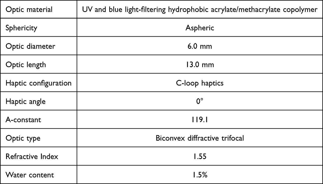 |
Table 1 Specifications of the CNWTTx Lens |
This current study reports visual and refractive outcomes of the CNWTTx IOL, with additional focus on patient-reported quality of vision and satisfaction, which has not been reported in the literature to date. We also conducted correlation analyses to understand the role of chord mu on visual outcomes and visual disturbance profile, with the goal to provide clinical recommendations on potential cut-off values.
Methods
Study Design and Inclusion/Exclusion Criteria
This was a real-world retrospective observational study, including 70 eyes from 44 patients that underwent phacoemulsification and implantation of the CNWTTx (CNWTT0 - CNWTT4) trifocal IOL (Alcon, Ft Worth, TX, USA) for the treatment of cataract (37 patients) or refractive lens exchange (RLE) (7 patients). Data was excluded if there was significant underlying ocular pathology with the potential to compromise visual outcomes, history of kerato-refractive procedures or a non-plano target. A large chord mu (ie. the distance between the pupil centre and visual axis) can cause visual disturbances if the visual axis does not align with the central diffractive zone, and as such eyes with preoperative chord mu >0.7 mm were not deemed eligible for implantation of the CNWTTx IOL. As this was a real-world study, patients with mild and well-controlled ocular comorbidities were implanted with CNWTT0 - CNWTT4, and their data included in the analysis, based on the surgeon’s clinical discretion. Twenty-six patients (59%) underwent bilateral implantation of CNWTTx and had data included for both eyes; the remaining patients had data included for one eye only. At the time of postoperative testing, 51.4% were pseudophakic in the fellow eye, 38.6% had cataract in the fellow eye and 10% were phakic in the fellow eye with no cataract. This study followed the tenets of the Declaration of Helsinki and received ethics approval from the University of Wollongong Human Research Ethics Committee (2024/280).
Preoperative and Postoperative Measurements
All procedures were performed by the same surgeon (SA). Biometry was performed preoperatively (IOLMaster700, Zeiss, Germany) and lens power was calculated using the Barrett True TK Universal II formula aiming for a target of ±0.25 D. Eyes with preoperative astigmatism >0.75 D received CNWTTx toric IOL (CNWTT2-4). Standard phacoemulsification and aspiration of lens material was performed through a 2.3 mm main incision located temporally. Following this, the IOL was implanted and the posterior capsule was polished. Postoperatively, patients complied with the following drop regime; 1 drop each of ofloxacin (Allergan, Dublin, Ireland) and prednisolone acetate/phenylephrine hydrochloride (Allergan, Dublin, Ireland); 2 hourly on the day of surgery and then 4 times a day for 2 weeks and 4 weeks, respectively. Patients were advised to commence ketorolac tromethamine (Allergan, Dublin, Ireland) eye drops, one drop four times a day for 1 week starting after 7 days postoperatively.
Postoperative testing was performed 4–6 weeks following surgery. Monocular uncorrected distance (UDVA), intermediate (UIVA) and near (UNVA) visual acuity were measured under photopic conditions using a ETDRS chart placed at 6 metres and a reading chart placed at the patients’ preferred intermediate (60–80 cm) and near distances (40 cm). Manifest refraction and best corrected distance visual acuity (BCVA) were measured using a phoropter and a Snellen chart placed at 6 metres under photopic conditions (DMD-FVD-24 DMD Vista Vision Wide VA Tester, DMD Computers, Turin, Italy). Distance-corrected intermediate (DCIVA) and near (DCNVA) visual acuity were recorded using a reading chart placed at the patients’ preferred intermediate (60–80 cm) and near distances (40 cm). Binocular UDVA, UIVA and UNVA were measured for 26 patients that had undergone bilateral IOL implantation ie. patients that received CNWTTx IOLs in each eye and were recruited for the study for both eyes. Monocular and binocular (if bilaterally implanted) CS was measured using the Pelli-Robson chart under mesopic conditions, within the DMD-FVD-24 DMD Vista Vision Wide VA Tester (DMD Computers, Turin, Italy), and corrected stereoacuity was measured for bilaterally implanted patients using the Titmus Fly Test under photopic conditions. Patients were prompted to report subjective symptoms, spectacle usage, and satisfaction using standardised interview questions; patient-reported incidence of glare and halos were recorded using a subjective scale (nil, tolerable, bothersome), and subjective satisfaction was measured using a 1 to 10 scale whereby 1 denotes highly dissatisfied and 10 denotes highly satisfied. Patient-reported spectacle usage (distance/driving, intermediate/computer work, near/reading, or never used) was translated into level of spectacle independence. Modulation transfer function (MTF) was measured using an automated aberrometer (iTrace; Tracey technologies) for 63 eyes. Visual acuity, stereoacuity, CS and manifest refraction were measured again at 6–12 months for a subset of 35 eyes out of the original cohort of 70 eyes.
Statistical Analysis
Correlation between preoperative chord mu, measured using biometry (IOLMaster700, Zeiss, Germany), and UDVA, UIVA and UNVA were assessed using a Pearson correlation test (Excel version 2406; Microsoft, Washington, USA). Correlation between chord mu and incidence of glare and halos was then assessed using a point-biserial correlation test (https://www.socscistatistics.com/tests/biserial/default.aspx). A P-value of ≤0.05 was considered statistically significant.
Results
Data from a total of 70 eyes from 44 patients was included in the analysis. Patient demographics for the total cohort and the 6–12 month cohort are outlined in Table 2. Toric IOLs were implanted in 25 eyes (12 eyes received CNWTT2, 11 eyes received CNWTT3 and 2 eyes received CNWTT4 IOLs) and 45 eyes received CNWTT0 non-toric IOLs. A total of 35 eyes from 19 patients returned for 6–12 month follow up with the remaining patients lost to follow up. Of these eyes, 2 eyes received CNWTT2 IOLs, 6 eyes received CNWTT3 IOLs and 1 eye received CNWTT4 IOLs, with the remaining 26 eyes receiving non toric CNWTT0 IOLs. Based on patient information collected during the preoperative baseline consultation, mild ocular comorbidities were present in 41% of the patients, including dry eyes (N = 15 patients), previous corneal ulcer due to Herpes Zoster Ophthalmicus (N = 1), mild dry ARMD (N = 1), and well-controlled primary open angle glaucoma (N = 1 patient). No intra- or postoperative complications were reported.
 |
Table 2 Preoperative Patient Demographics for the Total Cohort and 6–12 Month Testing Cohort |
Postoperative outcomes are outlined in Table 3. Mean MRSE at 4–6 weeks was −0.12 ± 0.39 D with 88% and 98.6% of eyes achieving within ±0.5 D and ±1.0 D of the refractive target, respectively (Figure 1A). Average postoperative astigmatism was −0.41 ± 0.41 D with 73% of eyes achieving 0.5 D of astigmatism or less (Figure 1B). At 6–12 months average SE and astigmatism remained stable at −0.03 ± 0.30 D and −0.39 ± 0.32 D, respectively.
 |
Table 3 Postoperative Outcomes Measured 4–6 Weeks Following Implantation of CNWTTx Lenses |
 |
Figure 1 (A) Deviation from target spherical equivalent target and (B) postoperative residual astigmatism measured 4–6 weeks following implantation of CNWTTx lenses. Abbreviation: MRSE, manifest refractive spherical equivalent. |
Average postoperative monocular UDVA and BCVA at 4–6 weeks were 0.05 ± 0.10 LogMAR and −0.01 ± 0.07 LogMAR with 61% and 90% achieving 0.0 LogMAR or better, respectively (Figure 2A). Postoperative UDVA was the same as postoperative BCVA in 54% of eyes with 31% of the eyes experienced 1 line improvement in BCVA compared to UDVA, which was likely due to residual refractive error during the immediate post-operative period (6 weeks). (Figure 1B). At 6–12 months, UDVA and BCVA remained stable at 0.02 ± 0.08 LogMAR and −0.03 ± 0.05 LogMAR, respectively.
 |
Figure 2 Cumulative visual acuity measured with and without distance correction at (A) 6 metres, (DVA), (B) 60 cm (IVA) and (C) 40 cm (NVA) at 4–6 weeks following implantation of CNWTTx lenses. Abbreviations: BCVA, best corrected distance visual acuity; DCIVA, distance corrected intermediate visual acuity; DCNVA, distance corrected near visual acuity; UDVA, uncorrected distance visual acuity; UIVA, uncorrected intermediate visual acuity; UNVA, uncorrected near visual acuity. |
At the 4–6 weeks postoperative visit, 68.0% achieved N8 (approximately 0.1–0.2 LogMAR) or better unaided at intermediate (Figure 2B), and 60% of eyes achieved N5 (approximately 0.18 LogMAR) or better unaided at near (Figure 2C). More eyes achieved near vision of N6 (approximately 0.2 LogMAR) or better without distance correction compared to with distance correction (77% versus 67%) (Figure 2C). This was likely attributed to the slightly myopic MRSE found in the patient cohort. At 6–12 months, UIVA and UNVA remained stable and were consistent with the vision assessed during the initial 4–6 week postoperative period.
For those patients that underwent bilateral implantation, average postoperative binocular UDVA, UIVA and UNVA were −0.02 ± 0.06 LogMAR, N8 (approximately 0.1–0.2 LogMAR) at 60–80 cm and N6 (approximately 0.2 LogMAR) at 40 cm, respectively. There was no correlation between chord mu and UDVA (r = 0.143, P = 0.239), UIVA (r = −0.006, P = 0.960) or UNVA (r = 0.047, P = 0.699).
Average postoperative uncorrected monocular CS for all eyes implanted with CNWTTx IOL was 1.35 ± 0.21 at 4–6 weeks and 1.48 ± 0.27 at 6–12 months. For the subgroup of 26 patients who had undergone bilateral implantation of CNWTTx IOLs, average binocular mesopic CS at 4–6 weeks was 1.67 ± 0.26, and average stereoacuity was 124.63 ± 124.35 sec arc. At 6–12 months, stereoacuity had improved to 70.77 ± 25.97 sec arc. A mean MTF of 0.5 was maintained up to a spatial frequency of 10 cycles per degree (cpd; Figure 3). Average subjective satisfaction rated out of 10 was 8.74 ± 1.12 for the total cohort. For bilaterally implanted patients, average satisfaction increased by 4.93% from 8.7 ± 1.1 after the first eye to 9.0 ± 0.8 after the second eye, however this change was not significant (P = 0.26). At 6–12 months, average subjective satisfaction score was 9.5 ± 0.58.
 |
Figure 3 Mean modulation transfer function as a function of spatial frequency measured 4–6 weeks following implantation CNWTTx lenses. |
Bothersome glare was reported for 7 eyes (10%) with 17 eyes (24.3%) reporting mild glare and 46 eyes (65.7%) reporting no glare. Bothersome halos were reported for 3 eyes (4.3%) with 27 eyes (38.6%) reporting mild halos and 40 eyes (57.1%) reporting no halos. For bilaterally implanted patients, the incidence of mild and bothersome glare increased from 33.3% to 38.9% and 5.5% to 11.1% after the first and second eye, respectively. The incidence of mild halos increased from 44.4% to 72.2% after the first and second eye, while the incidence of bothersome halos remained unchanged (5.5%). There was no correlation between chord mu and the incidence of either glare (r = −0.027, P = 0.823) or halos (r = 0.112, P = 0.357).
Full spectacle independence was reported by 78.6% of total patients with 15.7% of patients reported requiring spectacles for near vision tasks only, such as small-print reading. When only bilaterally implanted patients were assessed, 92.3% reported full spectacle independence while a single patient (3.8%) reported requiring spectacles for near tasks only, and one patient (3.8%) reported requiring spectacles for near tasks performed under low light.
Discussion
The CNWTTx IOL (CNWTT0-T4) combines the proven trifocal optic in TFNT00 with the innovative material used in CNA0T0 and became commercially available in Australia in 2023. This study presents real-world clinical outcomes of patients who underwent standard phacoemulsification and implantation of CNWTTx over the last 1–2 years. To our knowledge, this study is the first to report CNWTTx visual outcomes in a real-world patient cohort with documented mild ocular comorbidities at baseline.
This study reports high refractive accuracy, with 88% of eyes achieving within ±0.5 D of the SE target, consistent with previous literature (90–100%).11,12 Average postoperative SE was slightly myopic (ie. −0.12 D) and correcting this resulted in a slight decrease in near visual acuity. While tolerance to defocus was not tested as part of this study, data provided by the lens manufacturer claims that distance visual acuity of 6/7.5 or better is maintained from a range of +0.5 D to −2.5 D of blur,13 while independent studies suggest a range of +0.5 D to −3.0.10,14 Hence, it may be of benefit to aim for a slightly myopic target or first myopic target when selecting the IOL dioptric power, for example −0.25 D, as this may help improve near visual acuity without compromising distance visual acuity.15,16
At 4–6 weeks, the current study reported 61% of eyes achieved UDVA 0.0 LogMAR or better. This compares favourably to what was previously reported in the literature; Mendicute et al11 reported 34% of eyes while Garzon et al12 reported 68% of eyes achieving 0.0 LogMAR or better, and Khoramnia et al14 reported 59%. Average monocular UDVA reported in this study (0.05 LogMAR) is similar to that reported by Tuuminen and Jeon17 (0.03 LogMAR), Khoramnia et al14 (0.05 LogMAR) and Mendicute et al11 (0.07 LogMAR). Intermediate and near visual outcomes, such as distance corrected and unaided visual acuities, have not been consistently reported in the aforementioned publications. However, the reported VAs ranged from 0.03 to 0.13 LogMAR for UNVA and −0.01 to 0.01 LogMAR for UIVA. In this current study, intermediate and near visual acuities were measured in N notation and hence are not directly comparable to previous studies that assessed visual acuities using LogMAR notation. Nevertheless, based on the optical principles of the TFNT00 optic, it is expected that intermediate and near visual potential would be similar to that achieved at infinity. Hence, it is plausible to expect distance-corrected intermediate and near visual acuities would be similar to the postoperative BCVA of −0.01 ± 0.07 LogMAR in our patient cohort. However, the use of N-notation near chart was a limitation inherent to the real-world nature of the study. For future investigations, it will be beneficial to implement LogMAR VA charts at intermediate and near, which will allow accurate assessment of threshold visual acuities and direct comparison with previously published studies. Another limitation to our intermediate and near assessment was the non-standardised working distance. Previous multifocal IOL designs have used 80 cm as an intermediate focal point, while the optic of both CNWT and TFNT platforms have their intermediate focal point at 60 cm, reportedly a more “natural” focal distance aligned with modern-day patients’ technology-heavy lifestyles. In fact, Mencucci et al18 reported better intermediate vision outcomes for the TFNT00 IOL when measured at 60 cm compared to 80 cm. Previous reports of the CNWTTx IOL generally recorded intermediate vision at 60–66 cm, and near vision at 40 or 33 cm. Due to the retrospective nature of this study and the real-world clinical testing conditions, patients were asked to hold the reading chart “approximately 60 cm”, “approximately arm’s length” or “computer distance” away for intermediate vision, and “about ruler length” or “phone distance” for near vision. As this distance was not tightly controlled, it is likely that the exact distance varied slightly between individuals and could more closely reflect the individual’s preferred working distances rather than the distances optimised to the IOL optic.
In terms of continued visual improvement from the 4–6 week immediate postoperative period to the more extended 6–12 month assessment, it was interesting to note that there was no significant change in UDVA, BCVA, intermediate, and near VAs over this period. Of all the visual parameters, stereoacuity was the only measure that had clinically significant change from the 4–6 week (124.63 ± 124.35 sec arc) to 6–12 month period (70.77 ± 25.97 sec arc). Patient-reported subjective satisfaction score also increased from 8.74 ± 1.12 at 4–6 weeks to 9.5 ± 0.58 at 6–12 months. These findings may be due to a combination of factors including the resolution of postoperative dry eye and neuroadaptation, which has been shown to take up to 6 months following multifocal IOL implantation.19 Considering only a small subset of eyes was assessed at 6–12 months, longer-term follow up with a larger cohort would help elucidate long-term outcomes, and may give an insight into the role neuroadaptation plays in visual recovery and patient satisfaction.
In the current study, 92.3% of bilaterally implanted patients reported not requiring spectacles at all while 3.8% reported only requiring spectacles for near tasks. Spectacle independence is higher than what was reported following bilateral implantation of TFNT00 (83%)20 and for AT Lisa (82.1%),21 as well as what has been reported for the CNWTTx IOL (88–89%).22,23 However, spectacle independence can vary substantially between studies, depending on factors such as follow up time and methodologies in measuring patient reported outcomes. In this current study, the vast majority of patients reported not requiring spectacles for daily tasks, and considering this clinical parameter was obtained at 4–6 weeks postoperatively, it has potential to further improve with neuroadaptation and ongoing postoperative recovery. Even for unilaterally operated patients, spectacle independence rates were 70.4% with the majority only requiring spectacles for near tasks. This is consistent with the overall high satisfaction rate (ie. 8.74 out of 10).
The higher light utility of the CNWTTx optic is designed to optimise CS outcomes compared to other multifocal IOLs.24 Even in comparison to its predecessor TFNT00, CNWTTx has been reported to have superior CS which the authors attributed to the lower objective scatter index (OSI) of the newly innovated material.10 Normative values for CS measured with Pelli-Robson for the healthy 60-year old population has been reported as 1.68.25 The average binocular CS reported in this current study was 1.67 ± 0.26, which was comparable to the age-matched population normal and the CS range of 1.6426 to 1.9227 in different multifocal IOL models. While CS has been reported to remain within age normal limits following CNWTTx implantation, there may be some decrease at higher spatial frequencies.11,14 Due to the use of Pelli Robson as our clinical standard of care, this study did not utilise other subjective CS assessments at different spatial frequencies. However, ray tracing aberrometry was used (ie. iTrace) to measure MTF, which may be used as supplementary data to objectively assess CS. As shown in this current study (Figure 3) MTF of ≥0.5 was maintained up to a spatial frequency of 10 cpd. MTF findings can be difficult to compare between reports due to the wide variety of testing and reporting methods used. However, Labuz et al28 reported findings of a bench study that compared MTF for five presbyopia-correcting IOLs; Triumph, AT Lisa, Synergy, TFNT00 and Trinova Pro C. When measured using polychromatic light source, a 3 mm pupil and a corneal model that simulated the population average of SA, MTF values at the equivalent of 10 cpd ranged from approximately 0.25–0.4, with TFNT00 achieving approximately 0.27. The hydroxyethylmethacrylate material of the CNWTTx IOL has a higher water content which is designed to minimise glistenings and improve clarity of the IOL, and together with the ENLIGHTEN technology of the optic which theoretically maximises light transmission to the retina, may help improve MTF and subjective CS. While the current study reported good MTF for this cohort (ie. ≥0.5 up to a spatial frequency of 10 cpd), interpretation of differences between the CNWTTx material and other material is limited without a comparison group. Future research that includes direct comparison with TFNT00 IOLs will help confirm results reported by Lee et al,10 and elucidate the part the new biomaterial plays in ensuring premium visual quality, as the similar optic design will remove confounding factors. Automated methods of measuring image quality such as the MTF using iTrace, may also minimise patient errors and serve as supplementary information to subjective CS results. Additionally, it may help differentiate between the optical quality of the IOL itself and that of the optical system.
The low incidence of bothersome photic symptoms reported in this study is consistent with previous investigations of TFNT00 and CNWTTx IOLs, although variations in testing methods were observed due to the nature of different study designs. Similarly, the incidence of severe photic phenomena reported here was similar to what has been reported by Lee et al, who measured patients’ response utilising simulation images ranging from mild to severe10 Hovanesian et al22 who used the PRO questionnaire (Research InSight LLC, Laguna Beach, CA, USA) reported only 7% of eyes reported bothersome symptoms, while Mendicute et al11 reported approximately 20% using the QUVID questionnaire. These variations may be due to differences in the testing methods. The current study used a subjective scale without simulation images and may have the benefit of more closely reflecting standard of care in real-world clinical settings. It is also likely that patient responses varied by lifestyle (eg. those that do little or no night driving) and how questions were phrased; the use of a validated questionnaire would help make these results more standardised and less affected by subjective perception.29 However, each testing method has its own merits and is fit-for-purpose depending on the nature of the study. While a subjective scale without simulation images may be appropriate for real-world situations, the use of a validated questionnaire may be more informative when the primary purpose is to evaluate the IOL’s optimal performance and safety profile.
Preoperative chord mu is often used as an indicator for suitability of multifocal IOLs. A large chord mu, which is easily identified preoperatively using biometry, increases the likelihood of IOL decentration and poor visual outcomes as the visual axis may not be well aligned with the central diffractive zone. A recent review article recommended surgeons avoid implanting multifocal IOLs in eyes with preoperative chord mu ≥0.5 mm.30 This current study did not have specific inclusion or exclusion criteria regarding chord mu; all eyes considered suitable for multifocal IOLs were eligible for inclusion, and as such, reflects a real-world cohort. A total of 9 eyes had preoperative chord mu ≥0.5 mm. Of these eyes, only one eye experienced halos and no eyes experienced glare. Further investigation showed no correlation between visual outcomes at any distance, or incidence of glare or halos, and overall incidence of photic phenomenon was very low. To clarify the tolerance of the IOL to large chord mu values, these exploratory findings need to be verified via larger prospective studies, as the limited sample size in this current study is a limiting factor on statistical power. The edge design of the CNA0T0 and CNWTTx IOLs have been modified to reduce the risk of photic phenomena. A bench study suggested that the edge curvature and fully functional optic of the CNA0T0 IOL contributed to having lower glare-type phenomena6 when compared to other monofocal IOLs. The low incidence and good tolerance of photic symptoms reported in this study support these claims. The majority of symptoms, 38.9% for glare and 72.2% for halos, were reported as mild or tolerable. This, combined with improved CS and increased spectacle independence, suggests that the prevalence of photic symptoms was not clinically significant in the context of overall improved visual function.
Due to the retrospective nature of this study, there were certain limitations that may have affected the reporting of study results. The use of an N-notation reading chart held at variable distances, in addition to the inability to measure below N4, or approximately 0.1 LogMAR, may have led to inconsistency in reported near and intermediate visual outcomes. Although reflecting a real-world outcome, the practice of measuring near and intermediate vision at each patient’s preferred distance introduced variation that complicates the interpretation of results. Both patients undergoing lens exchange for cataract or refractive error were considered eligible and not analysed separately, and each eye was treated as an independent observation for patients who underwent bilateral implantation thereby introducing error due to inter-eye correlation. Longer term 6–12 month data was only available for some eyes (N = 35 eyes) due to the majority of routine cataract patients not requiring follow ups beyond 4–6 weeks post-operatively, resulting in missing values that impact data quality. Although preoperative parameters were similar between the cohorts, the absence of complete data for the 6–12 month postoperative period limits interpretation of longer-term outcomes. Furthermore, the use of interview-style questions as opposed to validated questionnaires and the inclusion of some unilaterally implanted eyes may have also affected visual and patient-reported outcomes eg. spectacle independence and satisfaction. However, this study reported clinical outcomes in a real-world patient cohort with mild ocular comorbidities, using clinical methodologies that were widely available in a standard, clinical ophthalmology setting, and closely reflected modern-day patients’ work and lifestyle habits. This study also suggests that stereoacuity and patient satisfaction improve in the long-term with neuroadaptation as seen in the subset of eyes at 6–12 months.
In conclusion, results from this retrospective, non-comparative, real-world observational study suggest that the CNWTTx IOL can provide exceptional visual outcomes with a high rate of patient satisfaction and spectacle independence in a real-world patient population. The visual acuity and visual quality achieved at distance, intermediate, and near will support modern cataract and RLE patients in achieving their lifestyle and vocational demands. Further prospective, standardised studies (ie. validated questionnaires to assess subjective outcomes, consistent testing periods, and standardised testing distance) will help establish relevance to the larger population. In particular, longitudinal studies that assess long term stability and clarity of the lens (ie. posterior capsular opacification and glistenings) will be of significant benefit.
Data Sharing Statement
The authors do not wish to share the data included in this study due to patient privacy and confidentiality.
Ethics Approval
This study received ethics approval from the University of Wollongong Human Research Ethics Committee (2024/280) and was conducted according to the Tenets of the Declaration of Helsinki. Informed written consent was acquired from all participants prior to inclusion in the study.
Author Contributions
All authors made a significant contribution to the work reported, whether that is in the conception, study design, execution, acquisition of data, analysis and interpretation, or in all these areas; took part in drafting, revising or critically reviewing the article; gave final approval of the version to be published; have agreed on the journal to which the article has been submitted; and agree to be accountable for all aspects of the work.
Funding
No funding was received.
Disclosure
Smita Agarwal is a member of the Alcon Advisory Committee. Lily Yu-Li Chang was employed by Alcon Laboratories Australia Pty Ltd in the Medical Affairs function until 15 August 2025. Medical Affairs is a non-commercial function that supports clinical research and clinical evidence validation. The authors report no other conflicts of interest in this work.
References
1. Cao K, Friedman DS, Jin S, et al. Multifocal versus monofocal intraocular lenses for age-related cataract patients: a system review and meta-analysis based on randomised controlled trials. Surv Ophthalmol. 2019;64:647–11.
2. Alio JL, Plaza-Puche AB, Alio Del Barrio J, et al. Clinical outcomes with a diffractive trifocal intraocular lens. Eur J Ophthalmol. 2018;28:419–424. doi:10.1177/1120672118762231
3. Kohnen T. First implantation of a diffractive quadrafocal (trifocal) intraocular lens. J Cataract Refract Surg. 2015;41:2330–2332. doi:10.1016/j.jcrs.2015.11.012
4. Breyer DRH, Kaymak H, Ax T, Kretz FTA, Auffarth GU, Hagen PR. Multifocal intraocular lenses and extended depth of focus intraocular lenses. Asia Pac J Ophthalmol. 2017;6:339–349.
5. Nicula CA, Popescu R, Rednik AM, Nicula D, Bulboaca AE, Stanescu I. Refractive lens exchange in hyperopic presbyopes with the AcrySof IQ PanOptix intraocular lens: one-year results and analysis of the literature. Ther Clin Risk Manag. 2020;16:1125–1137. doi:10.2147/TCRM.S279065
6. Das KK, Werner L, Collins S, Hong X. In vitro and schematic model eye assessment of glare or positive dysphotopsia-type photic phenomena: comparison of a new material IOL to other monofocal IOLs. J Cataract Refract Surg. 2019;45:219–227.
7. Werner L, Thatthamla I, Ong M, et al. Evaluation of clarity characteristics in a new hydrophobic acrylic IOL in comparison to commercially available IOLs. J Cataract Refract Surg. 2019;45:1490–1497. doi:10.1016/j.jcrs.2019.05.017
8. Oshika T, Fujita Y, Inamura M, Miyata K. Mid-term and long-term clinical assessments of a new 1-piece hydrophobic acrylic IOL with hydroxyethyl methacrylate. J Cataract Refract Surg. 2020;46:682–687. doi:10.1097/j.jcrs.0000000000000142
9. Agarwal S, Thornell E. Comparison of visual outcomes and patient satisfaction following cataract surgery with two monofocal intraocular lenses: clareon and AcrySof IQ monofocal. Open Ophthalmol J. 2021;15:144–150. doi:10.2174/1874364102115010144
10. Lee YW, Choi CY, Moon K, et al. Clinical outcomes of a new multifocal intraocular lenses with hydroxyl methacrylate and comparative results of contrast sensitivity, objective scatter, and subjective photic phenomena. BMC Ophthalmol. 2022;22:379. doi:10.1186/s12886-022-02600-x
11. Mendicute J, Lauzirika G, Illarramendi I, Martine-Soroa I. Visual, refractive, functional and patient satisfaction outcomes after implantation of a new trifocal diffractive intraocular lens. Clin Ophthalmol. 2024;18:27785. doi:10.2147/OPTH.S480311
12. Garzon N, Garcia-Bella J, Arriola-Villalobos P, Martinez-de-la-Casa JM, Villanueva C, Garcia-Feijoo J. Rotational stability, centration and patient satisfaction outcomes with a trifocal toric intraocular lens. J Cataract Refract Surg. 2025;51:762–768. doi:10.1097/j.jcrs.0000000000001687
13. Alcon. Clareon PanOptix Trifocal Hydrophobic Acrylic IOL Model CNWTT0; 2021.
14. Khoramnia R, Naujokaitis T, Baur ID, et al. Functional outcomes after refractive lens exchange with implantation of a glistening-free diffractive trifocal intraocular lens. Am J Ophthalmol. 2024;268:296–305. doi:10.1016/j.ajo.2024.07.037
15. Hayashi K, Sato T, Igarashi C, Yoshida M. Effect of spherical equivalent error on visual acuity at various distances in eyes with a trifocal intraocular lens. J Refract Surg. 2019;35:274–279.
16. Jeon S, Moon K, Kwon H. Long-term clinical outcomes after trifocal intraocular lens implantation: a retrospective observational study. J Refract Surg. 2023;39:236–241.
17. Tuuminen R, Jeon S. Comparison of two-year clinical and patient-reported outcome measures between AcrySof IQ and clareon PanOptix multifocal intraocular lenses. Am J Ophthalmol. 2025;274:264–275.
18. Mencucci R, Favuzza E, Caporossi O, Savastano A, Rizzo S. Comparative analysis of visual outcomes, reading skills, contrast sensitivity, and patient satisfaction with two models of trifocal diffractive intraocular lenses and an extended range of vision intraocular lens. Graefe’s Arch Clin Exp Ophthalmol. 2018;256:1913–1922. doi:10.1007/s00417-018-4052-3
19. Zhang L, Lin D, Wang Y, et al. Comparison of visual neuroadaptations after multifocal and monofocal intraocular lens implantation. Front Neurosci. 2021;15:648863. doi:10.3389/fnins.2021.648863
20. Hovanesian JA, Jones M, Allen Q. The PanOptix trifocal IOL vs the ReSTOR 2.5 active focus and ReSTOR 3.0-ADD multifocal lenses: a study of patient satisfaction, visual disturbances, and uncorrected visual performance. Clin Ophthalmol. 2021;15:983–990. doi:10.2147/OPTH.S285628
21. Maurino V, Allan BD, Rubin GS, Bunce C, Xing W, Findl O; Moorfields IOL Study Group. Quality of vision after bilateral multifocal intraocular lens implantation: a randomised trial-AT LISA 809M versus AcrySof ReSTOR SN6AD1. Ophthalmology. 2015;122:700–710. doi:10.1016/j.ophtha.2014.10.002
22. Hovanesian JA, Jones M, Allen Q. The Clareon vs AcrySof PanOptix trifocal IOL: a comparative study of patient satisfaction and visual performance. Clin Ophthalmol. 2024;18:2977–2984. doi:10.2147/OPTH.S476666
23. Scheepers MA, Pasin NM, Hall B. Visual outcomes, quality of vision, and patient satisfaction of a trifocal intraocular lens. Canadian J Ophthalmol. 2025;60:e530–535. doi:10.1016/j.jcjo.2025.01.018
24. Sudhir RR, Dey A, Bhattacharrya S, Bahulayan A. AcrySof IQ PanOptix intraocular lens versus extended depth of focus intraocular lens and trifocal intraocular lens: a clinical overview. Asia Pac J Ophthalmol. 2019;8:335–349. doi:10.1097/APO.0000000000000253
25. Mantyjarvi M, Laitinen T. Normal values for Pelli-Robson contrast sensitivity test. J Cataract Refract Surg. 2001;27:261–266. doi:10.1016/S0886-3350(00)00562-9
26. Altemir-Gomez I, Millan MS, Vega F, et al. Comparison of visual and optical quality of monofocal versus multifocal intraocular lenses. Eur J Ophthalmol. 2019;30:299–306.
27. Gupta G, Anand N, Khurana A, Gupta J. Study of visual functions with multifocal versus monofocal intraocular lenses after phacoemulsification in patients with age-related cataract. Ind J Clin Exp Ophthalmol. 2020;6:511–517.
28. Labuz G, Yan W, Baur ID, Khoramnia R. Comparison of five presbyopia-correcting intraocular lenses: optical-bench assessment with visual-acuity simulation. J Clin Med. 2023;12:2523.
29. Ison M, Scott J, A J, Apel A. Patient expectation, satisfaction and clinical outcomes with a new multifocal intraocular lens. Clin Ophthalmol. 2021;15:4131–4140.
30. Montrimas A, Zemaitiene R, Yao K, Grzybowski A. Chord mu and chord alpha as postoperative predictors in multifocal intraocular lens implantation. Graefe’s Arch Clin Exp Ophthalmol. 2023;262:367–380. doi:10.1007/s00417-023-06098-x
 © 2026 The Author(s). This work is published and licensed by Dove Medical Press Limited. The
full terms of this license are available at https://www.dovepress.com/terms
and incorporate the Creative Commons Attribution
- Non Commercial (unported, 4.0) License.
By accessing the work you hereby accept the Terms. Non-commercial uses of the work are permitted
without any further permission from Dove Medical Press Limited, provided the work is properly
attributed. For permission for commercial use of this work, please see paragraphs 4.2 and 5 of our Terms.
© 2026 The Author(s). This work is published and licensed by Dove Medical Press Limited. The
full terms of this license are available at https://www.dovepress.com/terms
and incorporate the Creative Commons Attribution
- Non Commercial (unported, 4.0) License.
By accessing the work you hereby accept the Terms. Non-commercial uses of the work are permitted
without any further permission from Dove Medical Press Limited, provided the work is properly
attributed. For permission for commercial use of this work, please see paragraphs 4.2 and 5 of our Terms.