Back to Journals » International Journal of Nanomedicine » Volume 21
Janus Membranes as Multifunctional Nanostructures for Bone Defect Repair: Advances, Challenges, and Clinical Translation
Authors Liu J, Liu S, Tan R, Chang W, Chen Z, Wang Y, Zhou P, Sun H ![]() , Xu Z
, Xu Z
Received 25 August 2025
Accepted for publication 19 January 2026
Published 29 January 2026 Volume 2026:21 563011
DOI https://doi.org/10.2147/IJN.S563011
Checked for plagiarism Yes
Review by Single anonymous peer review
Peer reviewer comments 3
Editor who approved publication: Dr Yan Shen
Jiachen Liu,1,* Shuyan Liu,1,* Rui Tan,1,* Wenliao Chang,1 Zhuo Chen,1 Yimin Wang,1 Peng Zhou,1 Han Sun,1 Zhonghua Xu2
1Articular Orthopaedics, Third Affiliated Hospital of Soochow University, Changzhou, People’s Republic of China; 2Department of Orthopedics, Affiliated Jintan Hospital of Jiangsu University, Changzhou, People’s Republic of China
*These authors contributed equally to this work
Correspondence: Han Sun, Email [email protected] Zhonghua Xu, Email [email protected]
Abstract: Bone defect repair, particularly for critically sized defects, remains a formidable clinical challenge owing to the limitations of conventional bone grafts and single-functional scaffolds. Janus membranes, with their asymmetric structure and capacity for multi-functional integration, offer an innovative “all-in-one” solution for bone tissue engineering. This review systematically outlines recent advancements in Janus membranes for bone repair, focusing on three core aspects: nanofabrication strategies for achieving asymmetric properties (eg wettability, charge, and porosity), multi-functional designs that mimic the natural periosteum for guided bone regeneration, antibacterial activity, soft-tissue integration, and haemostasis, and clinical translation potential along with future perspectives. This review summarises the progress in the use of Janus membranes as multi-functional nanostructures for bone regeneration. It systematically introduces the manufacturing techniques for Janus membranes, such as the layer-by-layer method and ion-modification approaches. Furthermore, it focuses on the dual-functional and multi-functional designs of Janus membranes and their integration with nanotechnology. In addition, we delve into the current challenges (eg complexity in manufacturing processes and limited research on material mechanical properties) and propose potential solutions, such as intelligent manufacturing (eg 3D printing) and dynamic response systems. This review provides a comprehensive reference for the field and promotes the clinical translation of Janus membranes.
Keywords: bone defect repair, Janus membranes, osteogenesis, bone tissue engineering, multi-functional integration
Clinical Challenges in Bone Defect Repair
Bones perform critical physiological functions, including physical mobility, protection of internal organs, and regulation of the haematopoietic microenvironment. This renewal occurs throughout people’s lives. Bone tissue possesses significant regenerative capacity and can heal minor trauma.1,2 However, in clinical practice, bone defects occur due to severe trauma, tumours, and pathological conditions (such as osteoporosis, osteogenesis imperfecta, and congenital joint disorders). Bone defects cannot heal through repair mechanisms when they reach a critical size. This is due to factors such as disrupted blood supply, insufficient osteoblasts, poor mechanical stability, lack of growth factors, and impaired bone conduction and induction (Figure 1). 3–6 Therefore, bone defects represent a common and clinically challenging problem in fields such as orthopaedics, oral and maxillofacial surgery, and neurosurgery. Bone grafting is the clinical “gold standard” for treating bone defects. According to the latest data, bone grafting is the second most common tissue transplantation procedure worldwide, after blood transfusion.7 Globally, four million people require bone grafts or bone replacement surgery each year. In the United States, the number of patients with age-related bone diseases is projected to increase from 2.1 million in 2005 to 3.0 million by 2025. With an ageing population in Europe, fracture cases are estimated to increase by approximately 28% between 2010 and 2025. Furthermore, substantial treatment costs and loss of patients’ labour capacity impose a heavy socioeconomic burden. Although the “gold standard” of clinical treatment is still based on autologous bone grafts, both autologous and allogeneic bone transplantation have drawbacks. Among patients undergoing autologous bone grafting, 20.6% of the cases will have problems, including complications, limited availability, longer operation time, additional surgeries, scars and pain at the donor site, and extended hospital stay.8,9 Allogeneic grafts also present significant challenges, including limited availability, immunogenicity, risk of disease transmission, and high cost, complicating clinical management.10–13
 |
Figure 1 Clinical challenges of bone defects. |
Meanwhile, when bone tissue encounters defects, the periosteum is often accompanied by inevitable damage.14 As an integral component enveloping the bone, the periosteum serves critical functions. It is a composite structure used for physical bone protection. This structure can serve as a mechanical sensor for sensing mechanical loads, a biochemical molecular library for initiating cascade signal transduction, an osteoblast ecological site for bone formation and repair, and an “umbilical cord” for nourishing bone tissue.15,16 The dense collagen fibre network of the periosteum itself provides a three-dimensional scaffold for adhesion, migration, and proliferation of the migrating cells. Meanwhile, periosteal coverage can prevent the growth of surrounding soft tissues and interfere with bony healing.17 In bone defects of critical size, the destruction of the periosteum is complete, and the recovery is difficult, which is also an important problem in bone defect repair.18,19
Clinical Application of Bone Tissue Engineering in Bone Defect Repair
Bone tissue engineering (BTE) is an alternative to bone grafting. It aims to promote the regeneration of natural tissues and cells by providing a temporary artificial scaffold/microenvironment.20,21 This graft contains all three components necessary for tissue regeneration, including bone-conduction scaffolds, bone-inducing signalling molecules, and osteoblasts.22,23 BTE provides a scaffold/microenvironment conducive to bone tissue growth/regeneration by simulating the extracellular matrix of bone tissue, supporting the formation of a new bone. These scaffolds degrade during the formation of new bone.4,24,25 Good BTE usually requires good biocompatibility, osteogenic biological activity, a three-dimensional porous structure, excellent mechanical properties, degradability, and processability.26–29
However, traditional BTE scaffolds often have only a single function, making it difficult to address complex clinical scenarios. For instance, hydroxyapatite (HA) is an important material in BTE.30 It is the main inorganic component of bone tissue and has good biocompatibility and bone-inducing properties. However, the excellent cytocompatibility of hydroxyapatite promotes the adhesion and growth of most cell types on its surface. Fibroblasts migrate and proliferate to bone defects and form a fibrous envelope, affecting the healing of bone defects.31,32
As mentioned earlier, the periosteum, an important bone structure, plays a significant role in bone repair.33–35 It comprises an outer fibrous layer and an inner cambium layer. The outer fibrous layer contains numerous fibroblasts, forming a dense collagen fibre network that serves as a mechanical barrier, tension maintenance, and physical barrier. The inner cambium mainly comprises numerous osteogenesis-related cells and matrix components that play a role in bone generation and repair.36,37 Traditional BTE often uses a single-material scaffold, such as HA ceramics and poly L-lactic acid (PLLA) porous bodies, which only provide static bone-conduction scaffolds. It is difficult to simulate the exquisite structure and highly complex dynamic functions of the natural periosteum.38,39 Therefore, new types of bone tissue-engineered scaffolds need to be developed.
Overview of Janus Membranes
The word “Janus” originated from the name of an ancient Roman god with two opposite faces, one looking forward to the past and the other to the future.40 Janus materials are a distinct class of composites characterised by an asymmetric chemical composition and spatially segregated functionalities. Janus membranes are an emerging concept in membrane science. Membrane materials exhibit asymmetry in morphology, structure, or chemical composition between the two sides. This implies that the membranes exhibit anisotropy in these properties across their two surfaces.41,42 Janus membranes were first introduced by Cheng and Wiersma in 1982. Since then, research on the manufacture of Janus membranes and improvement methods has increased rapidly.43 Currently, common Janus membranes are primarily categorised into two types. The first type is wettability differential membranes, characterised by one super hydrophilic/hydrophilic surface and one super hydrophobic/hydrophobic surface.44 Another type is the positively charged-negatively charged Janus membrane (charge differential Janus membrane), in which the two sides of the membrane have opposite surface charges.45 Other types of Janus membranes include those with chemical activity and porosity. This article reviews the preparation of Janus membranes and their applications in bone defect treatment.
Design and Fabrication Methods of Janus Membranes
Wettability-Differentiated Janus Membranes
Primary fabrication methods for wettability-differentiated Janus membranes include “A-on-B”, “A-off-B”, and “A-and-B” approaches.42
“A-on-B” refers to the single-side modification of a base material using unidirectional deposition, spraying, light-driven modification, vacuum filtration, vapour modification, electrospinning coating, layer-by-layer (LbL) assembly, or plasma treatment.42,46–48
“A-off-B” refers to modify one side, then reverse to modify the opposite side using similar methods to achieve significant wettability contrast.42,49
“A-and-B” refers to the sequential fabrication of hydrophobic/hydrophilic layers, typically via electrospinning/spraying. Although simple and efficient, this method carries the risk of layer delamination during long-term use owing to the inherent asymmetry in wettability.42,50,51 Figure 2 shows the electrospun fabrication of wettability-differentiated Janus membranes. Innovative techniques include femtosecond laser micromachining for etching microgrooves/pores on pre-stretched silicone, as well as monolithic gradient structures formed through hydrophilicity/hydrophobicity monomer gradients, which enable the one-step fabrication of hydrogel membranes with graded pore channels.52–54
 |
Figure 2 Schematic of layer-by-layer electrospinning of wettability-differentiated Janus membranes. |
In addition, plasma surface modification technology provides another important route for the direct and efficient construction of Janus interfaces with wettability contrast.55 As a “top-down” dry process, plasma treatment (eg, using radio-frequency oxygen plasma) bombards the material surface with high-energy particles. This process enables cleaning, etching at the nanoscale, and the introduction of polar functional groups, such as hydroxyl (-OH) and carboxyl (-COOH) groups, allowing precise and controllable alteration of the surface chemical composition and wettability of the material. The core advantage of this technique is that it can perform a unilateral treatment on a single-material membrane to achieve wettability reversal from hydrophobic to super-hydrophilic without the need to prelaminate two materials with different properties, thus simplifying the Janus membrane fabrication process. Concurrently, ion-modification technology can adjust the porosity and mechanical properties of a material, representing an efficient preparation pathway.56–58
Charge-Differentiated Janus Membranes
Membranes with opposite surface charges represent another important category, enabling directional ion transport and efficient separation in ion sieving, wastewater treatment, and battery separators.57 Naturally, cell membranes exhibit a typical Janus structure with a positively charged extracellular side, and a negatively charged cytoplasmic side, facilitating asymmetric ion transport.45,59,60 Common artificial charge-differentiated Janus membranes are bipolar membranes that combine anion-exchange membranes (AEM) and cation-exchange membranes (CEM) via lamination, forming bipolar interfaces. The fabrication methods include polyelectrolyte LbL assembly and membrane charge density modulation.61,62
Other Asymmetric Janus Membranes
Janus membranes exhibit opposing properties on their two sides. Besides wettability and charge, they include membranes with asymmetric conductivity, chemical activity, and porosity gradients. These three categories of Janus membranes have distinct preparation methods.63–66 For porosity/pore gradient asymmetry, spin coating-phase separation combined with electrospinning-gradient deposition are typically used. Chemically asymmetric versions utilise hydrogel in situ polymerisation along with a dual-sided functionalised coating, whereas electrical conductivity variations rely on coating/phase inversion paired with spray deposition of conductive materials.43
Janus membrane fabrication relies on diverse materials and processing techniques. The main polymer materials, manufacturing technologies, and characteristics commonly used in bone repair Janus membranes are summarised in Table 1.
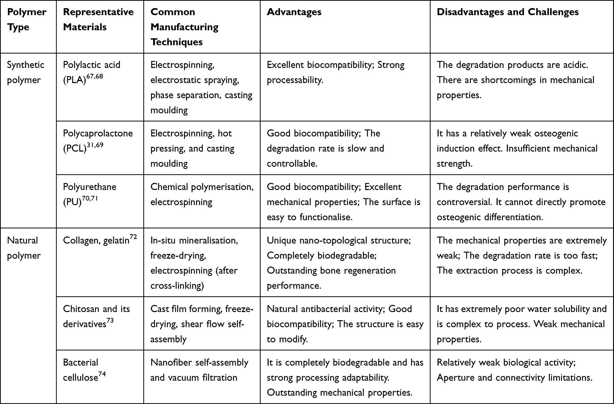 |
Table 1 Comparison of Main Polymer Materials, Manufacturing Techniques, and Characteristics for Janus Bone Repair Membranes |
Advantages of Janus Membranes in Bone Defect Repair
In regenerative medicine for bone defects, guided bone tissue regeneration (GBR) relies on the spatial maintenance and tissue-selective guidance functions of barrier membranes.75 In clinical practice, barrier membranes for guided bone regeneration are primarily classified as non-resorbable and resorbable.76,77 Non-resorbable membranes (such as polytetrafluoroethylene membranes) require secondary surgical removal and carry the risk of early membrane exposure and infection. However, owing to their exceptional mechanical and barrier properties, they remain an indispensable option for treating large, non-contained, or vertical bone defects.77,78 The advantage of resorbable barrier membranes over non-resorbable membranes is the elimination of the need for subsequent surgical removal. They are classified as natural polymers (collagen) and synthetic polymers (aliphatic polyesters). Among the available resorbable membranes, collagen membranes play a dominant role in clinical practice because of their solid scientific foundation and extensive clinical validation.79,80
However, current mainstream clinical barrier membranes (such as collagen and polytetrafluoroethylene membranes) are mostly homogeneous or simple bilayer structures, exhibiting significant limitations: their passive isolation properties make it difficult to dynamically regulate the bone repair microenvironment, and they lack the capacity for active intervention in osteoblast activity, angiogenesis, and the immune response;76 their antibacterial efficacy is limited, often requiring additional loading of antimicrobial agents; their capacity for immunomodulation is restricted, relying solely on the inherent properties of the material; and they lack proangiogenic effects. Scaffold materials widely used in BTE, such as hydroxyapatite and polylactic acid-based polymers, although possessing structurally biomimetic characteristics, lack effective physical barrier functions and cannot achieve precise separation between soft tissue and bone regeneration areas.68 Conventional electrospun nanofiber membranes can provide a physical barrier, but their single-component and homogeneous structures struggle to simultaneously integrate bioactive functions such as osteogenesis promotion, antibacterial activity, and immunomodulation.81
Leveraging their asymmetric structure and multi-functional integration characteristics, Janus membranes, through the synergistic design of materials science and biology, can combine multiple functions into a single entity, offering a breakthrough solution for bone defect treatment.49 Their core advantage stems from the meticulous selection of materials and structural design, which integrates a variety of seemingly contradictory functions, enabling active and precise regulation of the bone repair process (Figure 3).
 |
Figure 3 Janus Membrane Dual-Functional Mechanism: Simultaneous Barrier Isolation and Osteogenic Microenvironment Regulation. |
Core Advantages: From Multi-Functional Integration to Biomimetic Design
Therefore, its value for clinical translation is highlighted by the following three core advantages: Multi-functional integration, simultaneous achievement of a physical barrier, local delivery of antimicrobial agents, and immunomodulation, significantly reducing the risk of postoperative infection recurrence; Enhanced Regenerative Efficacy for Complex Bone Defects: The sustained release of osteogenic factors from the functional layer can accelerate mineralised matrix deposition and shorten the healing period; Biomimetic Replacement of Natural Periosteum Function: The bilayer structure mimics the outer layer (dense fibrous barrier) and inner layer (osteogenic cambium layer) of the periosteum, providing a biomimetic repair template for bone defects (Figure 4). For example, in the dual-functional synergy of barrier and osteogenesis, a dense hydrophobic layer (such as PCL and PLA) effectively blocks fibroblast infiltration, and the dense PCL layer developed by Pan et al31 can isolate soft-tissue interference and provide a stable environment for bone regeneration. Functionally, bone regeneration is promoted by loading osteogenic active substances (such as HAp and strontium apatite). The functional side of the HAp/PLA membrane designed by Ma et al67 significantly induced osteogenic differentiation of human adipose-derived mesenchymal stem cells (hADSC). Sun et al82 fabricated an antibacterial PVP/ZIF-8 layer based on a previous study. It can rapidly release Zn2+ in the acidic environment of an infection to achieve precise local sterilisation, realising antibacterial, osteogenic, and barrier functions. Zhang et al’s PLA-metal-phenolic network (MPN) membrane has a dense surface that simulates the fibrous layer to block soft tissues, and the porous surface promotes vascularisation and osteogenesis through the Cu2⁺ -monic acid network.68 This design achieves the reproduction of “anatomy - function”. The double-layer design precisely replicated the functions of the outer and inner osteogenic layers of periosteum. Lai et al reported that the ferroelectric Janus membrane of Lai et al83 regulates the microenvironment through polarisation. The negative electrode surface promotes the osteogenic differentiation of BMSCs, whereas the positive electrode surface enhances the proliferation of fibroblasts and synchronises the repair of soft and hard tissues. In addition, Janus membranes integrate haemostasis and osteogenesis. The carboxymethyl chitin/HAP membrane features a dense B-side layer that rapidly absorbs water and concentrates coagulation factors.21 Simultaneously, the porous A-side layer provides an osteoconductive microenvironment for bone formation. This dual-function design addresses the problem of synergy between intraoperative bleeding control and bone regeneration.
 |
Figure 4 Functional schematic of the Janus membranes. |
Therefore, with its multidimensional functional integration, dynamic microenvironment regulation, and biomimetic structural design, the Janus membrane has emerged as a new-generation candidate material driving the advancement of GBR technology towards intelligence and precision, demonstrating its irreplaceable application potential in the field of complex bone defect repair.
Intrinsic Mechanism: The Internal Link Between Nanoscale Features and Biological Effects
The immense potential of Janus membranes for bone defect repair originates from the intrinsic link between their precisely designed nanoscale features and biological effects.84 By asymmetrically constructing the wettability, charge, porosity, surface topography, and chemical composition on the two sides of the membrane, Janus membranes create two biological interfaces at the nanoscale, achieving active “spatial programming” of cell behaviour and tissue regeneration (Table 2). These physicochemical properties act synergistically to form the theoretical basis for achieving the core function of “preferential selection of bone cells over fibroblasts”.85
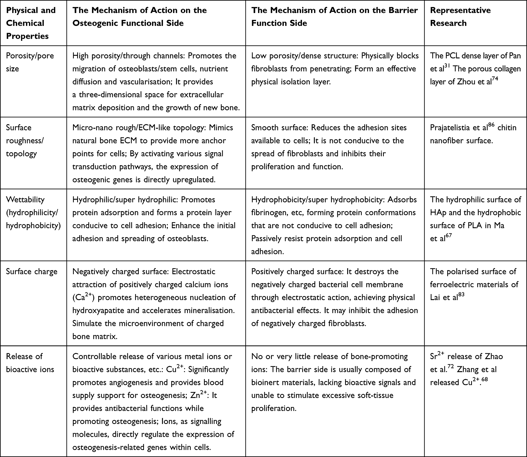 |
Table 2 Key Material Properties Regulating the Osteogenesis and Barrier Functions of Janus Membranes |
First, from the perspective of physical topography, the nanofibrous structure of the membrane surface can precisely mimic the physical environment of the natural extracellular matrix. By providing a smooth nano-topography, the dense side effectively inhibited the adhesion and migration of fibroblasts, functioning as a reliable physical barrier. Conversely, the porous or rough side provides critical anchoring points for osteoblasts by simulating the micro/nanomorphology of the bone matrix. This activates signalling pathways, such as those involving integrins, guiding osteoblasts to home, spread, and differentiate directionally towards the bone defect area.69,74 Furthermore, their controllable physical structure is more conducive to the loading and sustained release of drugs and various growth factors, making Janus membranes more comprehensive and better suited for addressing the complex bone defect environment.31
Building on this, the surface chemistry and charge characteristics further endow Janus membranes with intelligent cell-selective capabilities. The difference in hydrophilicity/hydrophobicity determines the directional transport of liquids and selectively regulates the adhesion behaviour of different cells by influencing the adsorption conformation of biological molecules, such as fibronectin.69,74 In addition, imparting opposite surface charges on both sides of the membrane can mimic the asymmetric charge distribution of the cell membrane. This design not only directly disrupts the negatively charged bacterial membrane through electrostatic interactions to achieve efficient physical antibacterial effects but also actively attracts positively charged calcium ions in vivo, creating favourable conditions for the heterogeneous nucleation of hydroxyapatite, significantly accelerating the osseointegration process.87
More importantly, loading functional nanomaterials, such as hydroxyapatite and strontium-apatite nanobelts, as active carriers onto specific functional layers is a core strategy for achieving multi-functional synergy and manipulating the microenvironment. These nanoscale components can continuously and controllably release bioactive ions (eg, Ca2⁺, Sr2⁺, Zn2⁺) during degradation. These ions act as key chemical signalling molecules that regulate the intracellular environment. By activating core osteogenic signalling pathways, such as Wnt/β-catenin and BMP/Smad, and simultaneously modulating the immune microenvironment, they precisely intervene in the entire bone regeneration process at the chemical level.72,74
Compared with traditional homogeneous or simple bilayer bone defect repair membranes (such as collagen or polytetrafluoroethylene membranes), the core advantages of Janus membranes are their asymmetric nanostructures and high degree of multi-functional integration. Traditional membranes primarily rely on a physical barrier for passive isolation, offering singular functionality, and struggle to simultaneously meet complex clinical needs, such as antibacterial activity, pro-angiogenesis, and immunomodulation. In contrast, Janus membranes achieve a paradigm shift from “passive barrier” to “active regulation” by constructing distinct physicochemical properties (eg wettability, charge, and porosity) on the two sides of a single membrane. This enables synchronous guidance of bone regeneration, inhibition of infection, and promotion of soft-tissue integration, providing a more precise and efficient “all-in-one” solution for repairing complex and infected bone defects. The design philosophy of Janus membranes begins at the nanoscale, integrating physical barriers, chemical signals, and bioactive factors into an asymmetric system, realising the “spatial programming” of the complex biological process of bone repair.
Current Applications of Janus Membranes in Bone Defects
Osteogenic and Barrier Functions
As the most fundamental and core functional design of Janus membranes, the “barrier-osteogenesis” dual-functional synergy serves as the cornerstone for achieving GBR.88 Fibroblast invasion during bone repair impedes osteogenesis. A barrier-covering defect is essential as a physical barrier and space maintainer.76,89,90 This section focuses on the design of Janus membranes that do not possess inherent active antimicrobial capabilities, primarily relying on their physicochemical properties to achieve barrier and osteogenic functions, and elucidates the fundamental working principle of functional barrier membranes. Table 3 summarises the core design strategies of current Janus membranes for barrier and osteogenesis purposes.
 |
Table 3 Janus Membrane Design Strategies for Barrier/Osteogenesis |
Ma et al67 successfully prepared a different-impregnated Janus membrane composed of hydroxyapatite (HAp) nanoribbons/polylactic acid (PLA) using the pouring method followed by osmotic evaporation. HAp is the primary inorganic component of bone tissue. Owing to its excellent biocompatibility and bone-inducing properties, it is the most important material used for bone repair and regeneration. Its excellent cytocompatibility facilitates the adhesion and growth of most cell types on the surface of HAp. Polylactic acid (PLA) is a biocompatible and biodegradable polymer. Its natural hydrophobicity may help prevent adhesion between the defect area and surrounding tissues. Therefore, the HAp/PLA Janus membrane can be used as a bone-inducing/barrier structure for BTE. The A-side (PLA) is hydrophobic and acts as a barrier to reduce the influence of soft tissues on osteogenesis. Its B-side (HAp) is hydrophilic and has high cytocompatibility, which can induce osteogenic differentiation of human adipose-derived mesenchymal stem cells (hADSC) without requiring additional growth factors.
Prajatelistia et al86 fabricated an impregnated differential Janus GBR membrane composed of a chitin nanofiber side (ChN) and a phosphorylcholine polymer-containing side (PMT). The surface of chitin nanofibers exhibits bone-inducing properties that can induce bone regeneration, and the nanostructure mimics the extracellular matrix to promote cell adhesion, proliferation, and migration. 2-Methacryloyloxyethyl phosphorylcholine (MPC) contains amphoteric groups that simulate the hydrophilic head groups of biological membranes and can resist protein adsorption and cell adhesion induced by amphoteric ions. The MPC monomer can be copolymerised with the 3-(trimethoxysilyl) propyl methacrylate (TSMA) monomer via free-radical copolymerisation to obtain cross-linking sites. This copolymer is then anchored to chitin amines and hydroxyl groups to form a phosphorylcholine-containing polymer (PMT). The A-side (PMT side) inhibits the migration of fibroblasts or epithelial cells, accelerating the healing of the bone and injury site. In contrast, B-side (ChN) promotes bone injury repair by facilitating osteoblast adhesion and mimicking the extracellular matrix.
Pan et al31 efficiently and feasibly prepared Janus GBR membranes with asymmetric functional structures using a multi-layer solution-casting method. The membrane structure was adjusted by controlling the rate of solvent evaporation and the kinetics of bilayer solute self-assembly. This achieved the effect of isolating the soft tissue on one side and promoting osteogenesis on the other. Surface A comprised polycaprolactone (PCL). By taking advantage of its smooth, dense, hydrophobic, and bioinert surface, it mechanically supports and barriers soft tissues and is used as an isolation layer. The B-side comprised a composite layer of nano-hydroxyapatite (HAn)/PCL/polyethylene glycol (PEG) (B layer). Exploiting its rough surface, porosity, hydrophilicity, biocompatibility, and biological activity can guide the regeneration and mineralisation of bone tissues. The continuous release of calcium and phosphate ions during degradation promotes osteogenesis.
Zhao et al66 fabricated Janus GBR membranes with different porosities using the liquid permeation method. Based on the work of Pan et al,31 they innovatively introduced to promote bone regeneration. Polycaprolactone methacrylate (PCLMA) exhibits good biocompatibility and relatively slow biodegradation. It is often used as a scaffold material to provide mechanical support in BTE. PCLMA undergoes free-radical polymerisation under blue light and changes from liquid to solid.91,92 Meanwhile, collagen fibres can undergo intramural mineralisation with various minerals in vitro. Intrafibrillar mineralisation has been proven to be a better scaffold for BTE, featuring a unique nanoscale topological structure, bone-like mechanical properties, and excellent bone regeneration performance.93 Zhao et al mineralised collagen using intramural strontium apatite to form the precursor of the membrane and then added PCLMA. In the uncured viscous liquid state, it penetrates and occupies the gaps between collagen fibres. Subsequently, when crosslinked with blue light, a nano-interlocking interface was formed. The resulting membrane had two sides, A-and-B. The A-side is a dense PCLMA layer that acts as a barrier to prevent fibroblast invasion. The B-side is a porous collagen layer that promotes osteoblast growth and releases strontium ions. Sr is an essential trace element in humans. It exerts a dual effect on bone metabolism by promoting bone formation and inhibiting bone resorption, promoting bone formation.
Zhou et al developed a simple strategy of vacuum-assisted filtration and etching to fabricate bacterial cellulose (BC)/Ti3C2Tx (MXene) Janus GBR membranes with different porosity. Similarly, Zhou et al introduced metallic titanium to promote bone repair.74 Ti is hydrophilic and highly biocompatible and promotes bone repair. Moreover, Ti3C2Tx MXenes are a new type of two-dimensional nanomaterial. Negatively charged functional groups promote osteogenic differentiation, generating a charged microenvironment suitable for bone healing. However, it is brittle and exhibits a rapid degradation rate, which is not conducive to its application in the field of GRB. BC can form hydrogen bonds with the functional groups on the active surface of MXenes and has good biocompatibility. Therefore, this combination enhanced the mechanic al strength and slowed the degradation rate. Zhou et al prepared a mixed dispersion of BC and MXene and added CaCO3 as a pore-forming agent. A porous layer was formed by vacuum filtration and drying. For the porous layer, the same method was used without the addition of CaCO3 to prepare bilayer membranes with different porosities. Therefore, the BC/MXene Janus GBR membrane has a dense surface on the A-side, which acts as a reliable physical barrier to prevent fibroblast infiltration, and a porous layer on the B-side, which promotes osteogenesis and osteoblast adhesion.
Osteogenic, Antibacterial, and Barrier Functions
Infection problems faced in bone defect repair are common in clinical treatment and remain a major problem to be solved for bone regeneration or delayed healing.94–96 As a multi-functional Janus membrane, it has certain advantages in promoting bone repair and exerting antibacterial effects. Several studies have been conducted on Janus membranes with bone-promoting and antibacterial barrier properties. The main categories, classified according to their antibacterial components, are listed in (Table 4).
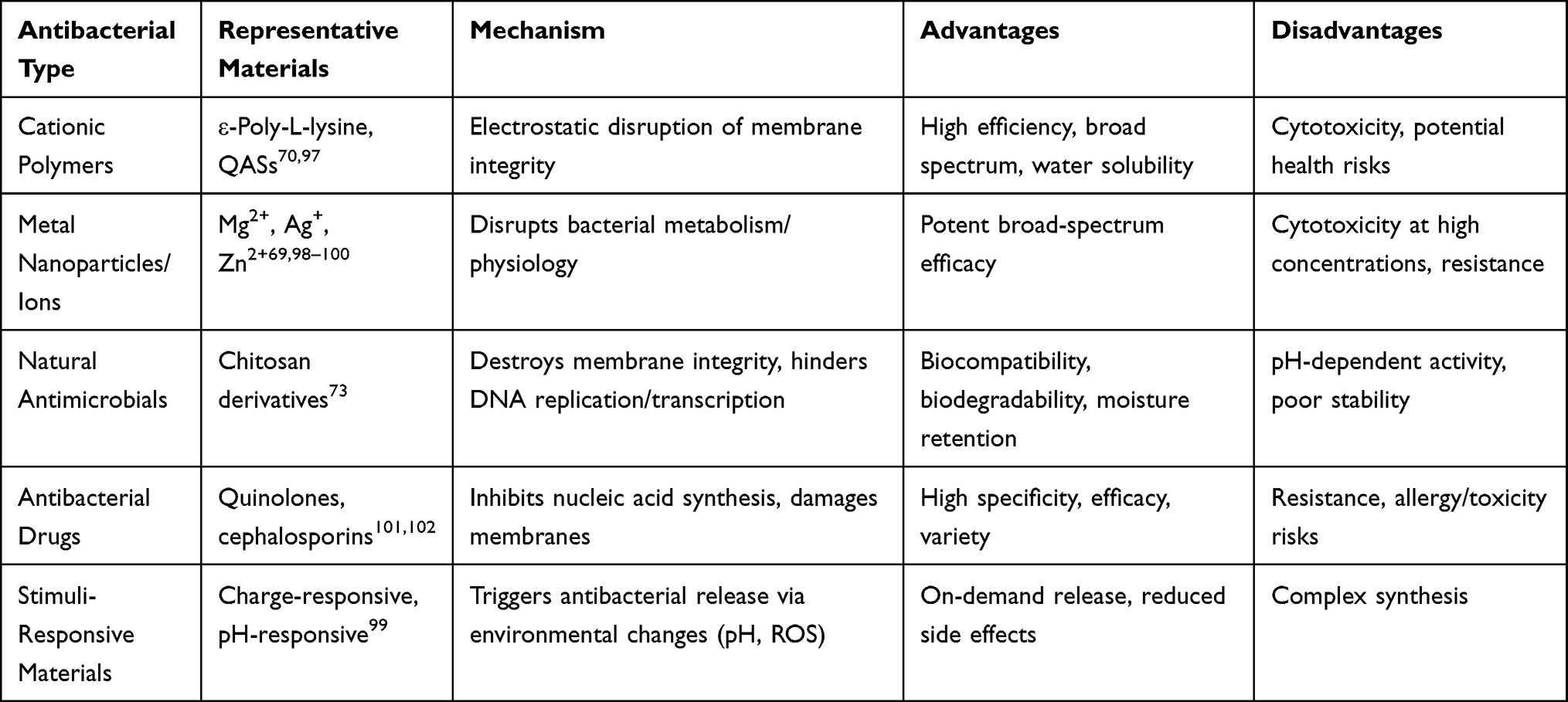 |
Table 4 Classification and Properties of Antibacterial Components in Janus Membranes |
Natural Antibacterial Agents
Ma et al,73 based on the studies of Pan et al and Zhao et al, added antibacterial effects to surface A. Ma et al utilised a shear flow-driven LbL self-assembly method. This method is a coating fabrication technique based on the a-LbL method. This triggers the reconfiguration of the stretched polymer through negative pressure attractors and promotes the effective adhesion of the adsorption material. Airflow drying was performed on the surface of the substrate to improve the structural characteristics and uniformity.103 This method enabled the simple and efficient fabrication of the Janus GBR membrane. Side A comprised chitosan and polyacrylic acid (CHI/PAA). Chitosan is a natural antibacterial agent with good antibacterial properties. On side B, an in situ mineralisation composite with calcium-phosphate-collagen/polyethylene glycol (CaP@COL/PEG) layers mimicked the natural composition of bone tissue. This composite promoted osteogenic differentiation and enhanced the mechanical strength of the membrane.
Cationic Polymers
Zhang et al97 constructed a Janus fibrous membrane by electrospinning the classic amphiphilic block copolymer methoxy polyethylene glycol block-polylactic acid-glycolic acid (mPEG-b-PLGA).104 Through the secondary assembly of amphiphilic block copolymers and the photo-cross-linking of methacrylic gelatin (GelMA), a Janus GBR membrane was obtained. This membrane exhibits both wetting differences and different porosities. ε-Poly-L-lysine (ε-PL) is a natural cationic polypeptide with inherent stability, water solubility, biodegradability. Crucially, it demonstrated good antibacterial activity, effectively inhibiting bacterial proliferation during bone repair. Niobium pentoxide (Nb2O5) has excellent biocompatibility, promotes osteogenesis, and increases hydroxyapatite deposition, exhibiting advantages in bone repair. In addition, it promotes the activation of anti-inflammatory macrophages, creating a favourable osteoimmune environment for osteogenic differentiation. Zhang et al added Nb2O5 particles to an mPEG-b-PLGA solution and performed ultrasonic treatment and electrospinning to form a loose, porous, and hydrophobic A-layer. They also added ε-PL and GelMA to the mPEG-b-PLGA solution, mixed them, and electrospun the B layer using the layer-layer method. Photo-cross-linking was then performed to form a dense and hydrophilic layer B. Thus, resulting in the fabrication of a double-layer Janus GBR membrane. One layer (layer A) promoted osteoblast adhesion and osteogenesis. The other layer (layer B) has biological barriers and antibacterial effects.
Lin et al70 and He et al71 introduced quaternary ammonium salts (QASs) and bioactive dopamine (DA) into polyurethane (PU) to develop Janus GBR films with different surface roughnesses and hydrophilicities. QASs are bactericides with broad antibacterial spectra, good stability, and high antibacterial activities. Therefore, it is difficult for these bacteria to develop resistance. PU exhibits good biocompatibility and excellent mechanical properties. The introduction of dopamine fragments is a relatively simple and effective method for enhancing the biological activity. Substances inspired by mussel adhesion proteins endow materials with strong adhesion, promoting cell adhesion. Simultaneously, dopamine can endow the material with excellent mineralisation and antioxidant capabilities. He et al utilised chemical polymerisation to place QAS with antibacterial properties on the upper surface and DA with rich biological activity on the bottom. Because of the difference in the number of nitrogen-containing groups, the upper surface was smooth, whereas the lower surface was rough and porous.104,105 Therefore, surface A (QASs) had an antibacterial and cell adhesion reduction barrier effect. The B-side (DA) has a multi-functional Janus membrane that promotes osteoblast adhesion, antioxidant properties, and mineralisation.
Metal Nanoparticles
Wang et al69 fabricated Janus GRB composite films with different porosities of dimagnesium/polycaprolactone (Mg-MgO/PCL) using electrospinning. Magnesium exhibits mechanical adaptability, biodegradability, and antibacterial properties. Mg2⁺ activates transient receptor potential (TRP) cation channels and Wnt signalling pathways. Consequently, it regulates the bone immune environment, promotes apatite nucleation, and induces biomineralisation. However, Mg degradation causes chronic cytotoxicity and generates hydrogen simultaneously. Rapid and excessive decomposition can cause the formation of cavities in the body, delaying bone healing. MgO (Magnesium Oxide) nanoparticles mitigate hydrogen-related tissue damage by releasing antibacterial and reactive oxygen species. PCL has good biocompatibility and a relatively slow biodegradation rate. This is common in BTE and can slow the penetration of corrosive ions and premature corrosion of Mg. Wang et al first fabricated Mg/PCL composite membranes by hot pressing embedded Mg sheets. Subsequently, electrospinning was used to deposit an MgO NP-PCL mixture onto the composites, producing Mg-MgO/PCL Janus GBR membranes. The A-side of the membrane was a dense casting layer, and the Mg sheets and MgO/PCL layers enhanced protection against fibroblast penetration. It also exhibits antibacterial effects. The B-side is a porous layer of PCL containing Mg2+, which promotes the adhesion of osteoblasts and simultaneously induces biological mineralisation.
Cheng et al98 and Li et al99 developed double-layer piezoelectric PLLA–zinc oxide (ZnO) composite Janus GBR films with different charges to promote bone formation during periodontal surgery. ZnO nanoparticles exhibit excellent biocompatibility, strong antibacterial properties, and potential to enhance piezoelectric performance. Cheng et al prepared PLLA and PLLA layers containing ZnO via electrospinning, and bonded these membranes with type I collagen. Thus, a PLLA-ZnO Janus GBR membrane was obtained. Side A is a non-piezoelectric PLLA that is uncharged to prevent cell growth and acts as a barrier to prevent fibroblast infiltration. The B-side is ZnO-PLLA, a piezoelectric PLLA with a surface charge that can promote bone regeneration. In addition, Zn2⁺ is released. The released Zn2⁺, combined with the piezoelectric response, synergistically enhanced the antibacterial effect.
Huang et al100 prepared Janus barrier membranes with different bilateral roughness values based on chitin hydrogel for periodontal defect repair. Chitin is commonly used in the manufacture of biomaterial scaffolds in tissue engineering and has good biocompatibility. Dopamine (PDA) nanoparticles can effectively enhance bone regeneration by promoting cell adhesion and proliferation, regulating osteoblast and osteoclast differentiation, and influencing cell polarisation. Copper, a trace element crucial to human health, inhibits osteoclast activity by regulating immune cell function and plays an important role in enhancing osteogenesis. Hydroxyapatite (HAP) exhibits excellent biocompatibility and osteogenic effects. As a coating for implants, it has been proven to significantly promote bone regeneration. Huang et al first fabricated a chitin film and then used the rapid deposition technology of a CuSO4/H2O2 catalytic system to rapidly coat the surface with PDA nanoparticles.106 This system also allowed for further polymerisation with polysulfobetaine methacrylate (PSBMA) with an anti-adhesion ability to produce a smooth and anti-fouling surface. This process incorporates Cu ions into the hydrogel membrane. Finally, the gelatin-HAP solution was added to one side of the membrane to produce a Janus hydrogel. Surface A was a smooth PDA-PSBMA layer containing copper ions, which has antibacterial properties and reduces adhesion. Surface B is a rough gelatin-HAP layer that promotes cell adhesion and bone regeneration.
Sun et al82 developed a Janus membrane with different impregnations using uniaxial electrospinning. Polycaprolactone (PCL) is a biodegradable polymer with excellent mechanical characteristics. Its hydrophobicity enables the continuous release of the carrier. Polyvinylpyrrolidone (PVP) is a hydrophilic polymer used as a rapid-dissolution matrix carrier in various drug delivery systems. Zeolitic Imidazolate Framework-8 (ZIF-8) nanoparticles (ZIF-8 NP) are biocompatible metal-organic frameworks composed of zinc ions (Zn2+) and 2-methylimidazole (MIN). They decompose rapidly in acidic solutions and exert antibacterial effects by releasing antibacterial Zn2+ and MIN into the acidic microenvironment of bacterial infections. Tacrolimus (FK506) enhances osteoblast differentiation and mineralisation in vitro and in vivo by activating the BMP signalling pathway. Sun et al prepared mixed solutions of PCL and FK506 (solution A) and PVP and ZIF-8 NPs (solution B). Solutions A-and-B (1:1 v/v) were then transferred to syringes and left undisturbed to form layers. Subsequently, electrospinning was used to fabricate the PCL/PVP/ZIF-8/FK506 Janus nanofibers. The A-side is hydrophobic PCL and FK506, which can achieve continuous drug release and provide continuous osteogenic stimulation. The B-side comprises PVP and ZIF-8 NPs, which can achieve rapid release in the acidic environment of bacterial infection to prevent bacterial infection. The small pore size of the electrospun nanofibers effectively prevented cell migration across the membrane barrier.
Loading Antibacterial Drugs
Moxifloxacin (MXF) is hydrophilic. It is a fourth-generation quinolone drug with broad-spectrum antibacterial activity, a long half-life, and few side effects. Icariin (ICA) is hydrophobic. As the main active compound in Epimedium plant species, it promotes the growth and proliferation of osteoblasts. Huang et al101 prepared nanofibers by separately blending hydrophilic MXF with PCL and hydrophobic ICA with gelatin. Subsequently, Janus GBR membranes with different wettabilities were assembled using layer-wise electrospinning. The A-side is a hydrophilic MXF, which has antibacterial properties and can prevent early infection and inflammation after membrane implantation. The B-side is a hydrophobic ICA that is released from the bottom layer in direct contact with the damaged bone and shows excellent performance in promoting bone regeneration. Furthermore, due to the significant hydrophilic difference between MXF and ICA, the Janus membrane showed rapid MXF release (≈80% at 21 days) but slow ICA release (≈30% at 21 days). This release profile enables early anti-infective effects and continuous promotion of bone regeneration in bone defects.
Xia et al102 prepared a Janus membrane with integrated fluid barrier, antibacterial, and osteogenic functions by electrospinning. It is an infiltrative differential membrane with a multi-layer structure used to treat cranial bone defects. Both dopamine (PDA) and calcium polyphosphate (CPP) promote the upregulation of multiple osteogenic genes, and PDA can chelate calcium ions to promote the formation of mineralisation layers. Xia et al prepared calcium polyphosphate nanoparticles on the surface of polydopamine (PCPP NPs) and obtained nanoparticles with good biocompatibility and osteogenic ability. Cefazolin (CFZ) is the preferred drug for preventing neurosurgical infections. Xia et al prepared an osteogenic layer by electrospinning a composite solution of PCL, gelatin, and PCPP NPs. It has a hydrophilic structure, promotes osteoblast adhesion, and exhibits osteogenic properties. Subsequently, an antibacterial and fluid barrier layer was fabricated based on the osteogenic LbL. The antibacterial and fluid barrier layer comprises three layers: upper, middle, and lower. The upper and lower layers comprised the PCL. The central layer was an antibacterial layer injected with cefazolin (PCL-CFZ), which was also prepared by electrospinning. It is hydrophobic and prevents cerebrospinal fluid leakage. CFZ was continuously released to inhibit bacterial infections.
Dynamic Response Materials
Li et al99 used the “A-and-B” method to fabricate poly (L-lactide) (PLLA) (P)/(PLLA/gelatin) (PG) Janus membranes with charge differences to treat alveolar bone loss induced by periodontitis. This membrane is a piezoelectric material that can generate charges in response to mechanical strain.107 PLLA is a biodegradable, FDA-approved polymer designed to have piezoelectric properties. Gelatin has good biodegradability, and its degradation products are nontoxic to organisms. Li et al fabricated PLLA using electrospinning. PLLA and gelatin were dissolved in trifluoroethanol (TFE) and electrospun to produce PG using the same parameters as those used for manufacturing PLLA films. The two films were stacked and placed in a tablet press to obtain double-layer P/PG films through physical compression. Corona polarisation was performed to positively charge the P-side and negatively charge the PG side. The A-side of the PLLA/PG Janus membrane was positively charged (P) and exhibited strong antibacterial activity through electrostatic interactions with negatively charged bacterial membranes. Meanwhile, the B-side is negatively charged (PG), which is conducive to the adsorption of cations such as Ca2+, promotes biological mineralisation, and facilitates the influx of Ca2+ into the cells, accelerating osteogenesis. Furthermore, given that teeth in the oral cavity are often subjected to chewing forces, the piezoelectric Janus membrane can generate a significant electrical output under these conditions.
The Role of Janus Membranes in Promoting Bone and Soft-Tissue Repair
Severe bone defects are often accompanied by soft-tissue defects, and poor soft-tissue recovery can affect the prognosis. For example, the loss of soft-tissue barrier function increases the risk of infection. The reduction in blood supply and nutrients required for bone regeneration leads to the failure of bone regeneration or delayed healing.108,109 Therefore, a Janus membrane that can both promote osteogenesis and soft-tissue repair has emerged as a requirement.
Lai et al83 incorporated the function of promoting soft-tissue repair on surface A by forming a barrier layer. Lai et al first prepared ferroelectric P(VDF-TrFE) films using the solution-casting technology. Subsequently, these films were polarised between the electrodes and spray-coated with a (HAn) (P)/(PLLA/gelatin)/PCL/polyethylene glycol gold layer to enhance the electrical conductivity. Therefore, Janus GBR membranes with charge differences (Janus electro-microenvironments (JEM)) were fabricated. The negatively polarised JEM (-) membrane effectively promotes osteogenic differentiation in bone marrow mesenchymal stem cells (BMSCs). This effect has been experimentally linked to the stimulation of mitochondrial autophagy. Mitochondrial autophagy triggers calcium-phosphate deposition. In contrast, the positively polarised JEM (+) membrane effectively enhanced fibroblast proliferation, migration, and differentiation. This effect was associated with increased mitochondrial oxidative phosphorylation.
Han et al87 fabricated a bone-inducing/barrier structure Janus GBR film with different specific surface roughnesses using a magnesium alloy and calcium hydrogen phosphate dihydrate (CaHPO4·2H2O)/magnesium fluoride (MgF2) by the “A-on-B” chemical transformation and deposition method. Magnesium ions (Mg2+) are closely associated with the healing of soft tissues. They improve the migration of human skin fibroblasts (HSF) and Haut Cancer Transformed (HaCaT) cells and promote the transformation of HSF into myofibroblasts, accelerating soft-tissue healing. DCPD [dicalcium phosphate dihydrate (CaHPO4·2H2O, denoted as DCPD)] has a relatively high solubility and can release more Ca2+ and PO43-, inducing osteogenic differentiation of stem cells, production of collagen-rich (osteoid) matrices, and matrix mineralisation. In addition, DCPD allows direct osteogenesis in the humoral environment. Han et al first immersed Mg sheets in hydrofluoric acid to generate a smooth MgF2 transition layer. Subsequently, polycarbonate was sprayed onto the outer surface as a protective mask, and a rough dicalcium phosphate dihydrate (DCPD) coating was chemically deposited. Finally, dichloromethane was used to dissolve and remove the polycarbonate layer on one side, exposing the MgF2 surface on side A and promoting fibroblast recruitment and adhesion. The B-side (DCPD) creates a favourable microenvironment for the adhesion, proliferation, and differentiation of osteoblasts to promote bone formation.
The Osteogenic and Haemostatic Effects of Janus Membranes
Bone defects often lead to bone haemorrhage, hindering surgery and causing serious postoperative complications. Therefore, it is important to design bone repair materials with haemostatic effects. Chitosan exhibits good solubility and biodegradability, making it a suitable biomaterial. Hydroxyapatite is the main inorganic component of bone tissue and has good biocompatibility and bone-induction performance. Based on this, Lv et al32 cast hydroxyapatite nanoparticles (HAP) on one side of Carboxymethyl chitin porous membrane (CPM) and conveniently fabricated (CPM-HAP) Janus GBR membranes with different porosities and no cross-linking agent. Surface A was a porous CPM layer containing HAP, providing a microenvironment for osteogenesis. The dense CPM-HAP layer on Side B served as a physical barrier that prevented epithelial cells and fibroblasts from infiltrating bone defects. Simultaneously, its exceptional water-absorption capacity enables rapid fluid uptake from the blood, concentrating coagulation factors to achieve a combined barrier function and haemostatic promotion.
Multi-Functional Integration of Janus Membranes
Clinically, bone defects are often complex and involve anatomical disruption, dysregulation of the defect microenvironment, biomechanical imbalance, and other issues.110 Consequently, bone defect repair frequently requires multi-functional materials to address these complexities. Janus membranes with multiple integrated functionalities are required for this purpose. Current research on multi-functional Janus membranes is highly active. The key advancements are summarised below, with representative studies detailed in Table 5.
 |
Table 5 Multi-Functional Integration Strategies for Janus Membranes |
Integration of Metal Ions and Natural Compounds. Zhang et al68 fabricated a Janus membrane with different porosities using the LbL method, which integrates barrier, osteogenesis, anti-inflammatory, and angiogenic stimulation. Polylactic acid (PLA) has the advantages of low cost, ease of processing, and biocompatibility. MPNs are complex supramolecular structures formed by the coordination of metal ions and phenolic ligands. They have been widely studied as bioactive nano-coatings for implant modification, drug release, and stem cell differentiation. Metal ions, such as copper (Cu2+), are trace elements found in the human body. Tannic acid (TA), a naturally occurring phenolic molecule, promotes tissue healing by stimulating angiogenesis and stem cell differentiation. It also exhibits anti-inflammatory and osteogenic properties. Zhang et al fabricated porous polylactic acid films (PLAM) via evaporative phase separation. Subsequently, sequential immersion in TA and copper ion (Cu2⁺) solutions enabled the LbL assembly of an MPN nano-interface on the PLAM substrate. Surface A is a dense PLAM-MPN layer that serves as a barrier for bone regeneration, reducing soft-tissue invasion. Meanwhile, the surface B features porous PLAM with unidirectional evaporation-induced pores. This architecture enables the MPN coating to simultaneously exert osteogenic activity, stimulate angiogenesis, and exert anti-inflammatory effects at the porous interface.
Integration of Drugs and Bioactive Substances. Lu et al111 prepared a porosity-differentiated multi-functional Janus membrane via freeze-drying and cross-linking. Poly (lactic-co-glycolic acid) (PLGA) and polycaprolactone (PCL) possess excellent mechanical strength and biocompatibility, rendering them ideal scaffolds for tissue engineering and clinical applications. These copolymers form a dense structural barrier that restricts fibroblast infiltration into bone defects. Chlorhexidine (CHX) exhibits broad-spectrum antibacterial activity against both gram-positive and gram-negative bacteria. Epidermal growth factor (EGF) accelerates wound healing by stimulating fibroblast proliferation, migration, and re-epithelialisation. Basic fibroblast growth factor (bFGF) plays a key role in cell proliferation and promotes wound healing and fracture repair by stimulating vascular cell growth, tissue repair and angiogenesis. Lu et al electrospun a PLGA/PCL copolymer into a membrane (PP membrane) and incorporated CHX, EGF, and bFGF into a chitosan solution. This solution was freeze-dried and crosslinked onto a PP membrane to form a porous chitosan sponge (widely used for drug delivery and tissue engineering), yielding an integrated multi-functional Janus membrane. Side A (dense PP layer) acts as a barrier against fibroblasts.113 Side B (chitosan sponge loaded with CHX/EGF/bFGF) provides antibacterial action, promotes bone repair, stimulates angiogenesis, and facilitates soft-tissue regeneration.
Integration of Metals and Bioactive Ions. Cheng et al113 fabricated a roughness/wettability-differentiated Janus fibrous membrane using LbL-assisted electrospinning for GBR. Bone morphogenetic protein-2 (BMP-2) and acidic fibroblast growth factor (aFGF) synergistically induce bone regeneration and angiogenesis by modulating oxidative phosphorylation/glycolysis and upregulating osteogenic/angiogenic gene expression. Zinc oxide (ZnO) nanoparticles exert bactericidal effects by disrupting bacterial membranes and inhibiting cellular respiration and DNA replication. Cheng et al electrospun negatively charged gelatin@BMP-2-PLLA fibrous membranes alternately immersed in chitosan (CS, positively charged) and aFGF (negatively charged) solutions. The LbL process formed aFGF-CS multilayers on the surface, protecting its bioactivity and enabling sustained release. Directionally aligned PCL/PLGA@ZnO fibres were electrospun as the outer layer. The resulting Janus membrane featured Side A (outer layer) with hydrophobic, directionally aligned PCL/PLGA@ZnO fibres that prevented epithelial invasion and bacterial infection. Side B (inner layer) comprised randomly oriented gelatin@PLLA nanofibers, enabling the sustained release of aFGF/BMP-2, which significantly promoted vasculogenesis and bone regeneration.
Analysis of the Current State of Mechanical Properties of Janus Membranes
Bone is a composite material with excellent mechanical properties, whose strength and modulus vary depending on the type and location, related to differences in geometry, the boundary conditions of mechanical testing, or different anatomical sites.114 As a reference, from a mechanical performance perspective, the Young’s modulus of scaffolds should fall within the ranges of 15 to 20 GPa and 0.1 to 2 GPa for cortical bone and cancellous bone, respectively; the compressive strength of cortical and cancellous bone should be between 100 to 200 MPa and 2 to 20 MPa, respectively.115–118 This significant mechanical disparity implies that an ideal bone repair material must possess mechanical properties matched to the target repair site: materials for load-bearing cortical bone defects require sufficient strength and modulus to replace its function, whereas for non-load-bearing regions or cancellous bone defects, the requirements for mechanical performance are relatively relaxed, with greater emphasis placed on biological activity.
Surveying the studies discussed in this chapter, the current research frontier for Janus membranes is primarily focused on multi-functional integration and regulation of biological activity. However, their mechanical properties, which are key indicators for their application in load-bearing bone defect repair, have rarely been sufficiently characterised or discussed. This status quo precisely reflects a stage in the field’s development where researchers seem to prioritise verifying the feasibility of their complex functionalities. A minority of researchers have not conducted corresponding investigations into the mechanical properties, whereas most of the remaining researchers focus on reporting the tensile properties of materials (such as tensile strength and elastic modulus) and severely lack a systematic evaluation of their compressive properties (such as compressive strength and modulus). Only Lu et al111 tested compressive strength and reported a value of 1.7 MPa, which is suitable for a non-load-bearing oral environment.
This imbalance in characterisation reflects the initial positioning of the Janus membrane within the current research community, primarily as a functionalised “barrier membrane”. However, bone primarily bears compressive loads in physiological environments. For Janus membrane designs that aim to address the challenge of load-bearing bone repair, compressive strength is the core indicator for maintaining the repair space and resisting internal pressure without collapse. The systematic characterisation and reporting of the compressive properties of Janus membranes are crucial for accurately assessing their load-bearing potential and establishing a complete structure–property–application database.
Selective Cell Adhesion and Osteogenic Differentiation Regulation of Janus Membranes
In BTE, scaffold materials are not only physical supports but also regulators of cell adhesion, proliferation, and differentiation. As described in the Chapter (Advantages of Janus Membranes in Bone Defect Repair), Janus membranes can achieve the selective recruitment and behavioural regulation of different cell types through their asymmetric physicochemical properties. This is the biological basis for their functional advantages. This section systematically summarises the cell types commonly used in Janus membrane research and their behavioural responses to Janus membranes, providing a cellular perspective for understanding their osteogenic promotion mechanisms.
Cell Types Commonly Used in Janus Membrane Research and Their Selection Basis
In related studies on Janus membranes, the following cell types have been widely used, each possessing different characteristics and research advantages.
Human mesenchymal stem cells are ideal seed cells for BTE because of their powerful self-renewal and multidirectional differentiation potential. Among them, BMSCs are the most extensively studied cell type, regarded as the classic cellular model for osteogenic differentiation, and are commonly used to assess the most fundamental osteoinductive potential of materials.86 Adipose-derived stem cells are highly favoured for their ease of acquisition and wide availability. For example, Ma et al67 used hADSCs to verify the osteoinductive capability of their HAp/PLA Janus membrane, proving that it could induce osteogenic differentiation of hADSCs without growth factors.
Periodontal ligament stem cells are commonly used in periodontal bone defect repair studies. hPDLSCs have become the preferred cell model because of their key role in periodontal tissue regeneration.67 The use of these cells can more accurately simulate the specific microenvironment of periodontal regeneration.
Mouse preosteoblast cell line (MC3T3-E1). This cell line is often used as a preliminary screening model for preclinical mechanistic research because of its stable phenotype and ease of culture. For example, Prajatelistia et al86 and Zhao et al66 used MC3T3 cells to provide preliminary and efficient evidence of the osteogenic activity of Janus membranes, although their results require further validation using human cell models.
Cell Seeding, Biocompatibility, and Differentiation Efficacy Assessment
The fate of cells on Janus membranes directly determines the repair efficacy of the material. Existing research using a standard biological evaluation system has confirmed the biocompatibility and active osteogenic regulation of Janus membranes. Cells are usually dripped directly or seeded onto the functional layer of the Janus membrane (ie hydrophilic, porous, or negatively charged osteogenic surface) at a certain density for culturing. Cell viability is usually assessed using live/dead cell staining or kits such as CCK-8. Most studies have reported that the functional side of Janus membranes supports high cell viability and sustained proliferation, demonstrating their excellent biocompatibility.
The asymmetric design of Janus membranes effectively guides the spatial distribution of cells, leading to selective adhesion and spatial distribution of cells. For example, Prajatelistia et al86 showed that osteoblasts preferentially adhered to the chitin side with a nanofiber structure, whereas fibroblast adhesion was significantly inhibited on the phosphorylcholine-containing polymer side. This inherent cell selectivity is the cytological basis for Janus membranes to achieve barrier-osteogenesis separation. Janus membranes exhibit osteogenic differentiation and regulatory mechanisms. Research has shown that, compared to homogeneous membranes or material blank control groups, stem cells seeded on the functional side of Janus membranes exhibit a higher osteogenic differentiation rate. This was confirmed by early osteogenic markers, such as a significant increase in alkaline phosphatase (ALP) activity. RT-qPCR analysis revealed that the expression levels of core osteogenic genes (eg Runx2, OPN, and OCN) were significantly upregulated.68,107 Late-stage mineralised nodule formation, such as quantitative analysis through Alizarin Red staining, showed a significant increase in extracellular matrix calcium deposition.83
The underlying regulatory mechanisms are closely related to the physicochemical signals described in Chapter (Advantages of Janus Membranes in Bone Defect Repair). The porous/rough topological structure of Janus membranes promotes osteogenic differentiation by activating the integrin-FAK signalling pathway. The bioactive ions (such as Sr2⁺, Cu2⁺, and Zn2⁺) released from their functional layers drive the osteogenic program at the genetic level by activating core signalling pathways such as Wnt/β-catenin and BMP/Smad.66,69
Janus membranes are not passive carriers of cells but are active platforms for regulating cell behaviour. Through the precise design of their asymmetric structure, they can selectively support the adhesion and proliferation of osteogenesis-related cells (such as MSCs), efficiently promoting their differentiation into osteoblasts while inhibiting fibroblast infiltration, ensuring smooth progress of the bone regeneration process at the cellular level.
Current Challenges, Clinical Translation, and Future Perspectives
Although Janus membranes have numerous advantages in the treatment of bone defects, they still face challenges.
First, the Janus membrane manufacturing process often involves multiple complex steps. Processing methods such as electrospinning and electro-spraying require expensive equipment. Moreover, precise control of the membrane thickness remains challenging. Membrane thickness plays an important role and varies depending on the specific clinical application and defect site. Simultaneously, the moisture content, porosity, and surface charge of the membrane are often significantly related to the selected material and are difficult to control and tailor precisely. Currently, simple, efficient, fast, and controllable manufacturing processes are lacking.
Second, Janus membranes with different wettabilities are widely used in clinical practice. However, the inherent difference in wettability between the two sides of the Janus membrane compromises the interlayer adhesion strength. This weak interfacial bonding frequently causes delamination during surgery, ultimately diminishing the functional efficacy. Simultaneously, hydrophilic materials may expand during use, further aggravating delamination. Delamination can compromise the structural integrity and functionality of membranes.
The working environment of the Janus membrane in vivo is more complex than that in vitro, and its working mode in vivo requires further investigation. For example, different materials on both sides of a Janus membrane typically exhibit different degradation rates. Can this difference in degradation rate affect bone regeneration and the repair of the surrounding soft tissues? There are more cell types in the body, and the conditions of cell metabolism, apoptosis, and regeneration are complex. Under such conditions, will the pores of Janus membranes with different porosities, commonly used in clinical practice, become clogged by metabolic products? Can the charge difference between Janus membranes with different surface charges diminish over time, affecting their therapeutic effects? However, these issues require further investigation.
Although Janus membranes demonstrate significant potential for functional integration, their ultimate feasibility for clinical applications, particularly in the repair of load-bearing bone defects, is limited by their mechanical properties. As intricate mechanical structures, cortical and cancellous bones exhibit an order-of-magnitude difference in modulus and strength. This necessitates that repair materials possess mechanical properties that are compatible with specific implantation sites. Based on current research, the mechanical investigation of Janus membranes remains insufficient and lacks critical data essential for their application as load-bearing materials.
In addition, given the current manufacturing process for Janus membranes, their main application is in maxillofacial surgery, where bone defects are relatively small. It is less involved in the treatment of complex, large, and infectious bone defects.
Janus membranes, as rising stars in bone defect repair, have great potential in the treatment of bone defects. The authors’ outlook on the preparation and design of Janus membranes is presented below.
The fabrication processes for Janus membranes are complex and diverse. If Janus membranes are to be applied in clinical practice, large-scale production will pose a significant challenge, along with the issue of quality consistency between different batches. Therefore, the primary objective of this study was to develop a simple, efficient, and low-cost manufacturing process capable of large-scale production to precisely fabricate reliable Janus membranes.
Leveraging 3D printing technology enables precise anatomical matching of defect locations, sizes, and geometries. This capability facilitates the application of Janus membranes across diverse complex bone defects, enabling personalised targeted therapies.
Future advancements are anticipated in the manufacturing of multi-layer Janus membranes (≥3 layers) for enhanced multi-functional integration. Such architectures, including trilayer structures featuring hydrophilic external surfaces with an intermediate hydrophobic core, can address complex clinical scenarios.
Janus membranes have not yet been clinically translated. The successful translation of Janus membranes depends on rigorous preclinical validation and a step-by-step clinical trial evaluation process, as well as the early clarification of their regulatory pathways as combination products, paving the way for future clinical applications.
Finally, future Janus membranes should prioritise multimodal dynamic responsiveness by integrating physical, chemical, and biological signalling modalities. This entails the combination of electrical/pressure stimuli, controlled ion/drug release, and cytokine signalling to precisely regulate bone defect microenvironments.
Conclusions
The complexity of bone defect repair necessitates the use of repair materials with multi-functional synergy and spatiotemporal regulatory capabilities. This review systematically recounts the research progress on Janus membranes in this field and clarifies their immense potential as next-generation bone repair materials. Core advancements are primarily reflected in three aspects. First, in the design strategy, the use of asymmetric fabrication techniques, such as wettability contrast, charge distribution, and porosity gradients, has enabled the precise construction of membrane structures transitioning from isotropy to anisotropy. Second, owing to material layering and the spatial arrangement of active components, the successful integration of various functions, including physical barriers, osteogenesis promotion, antibacterial activity, immunomodulation, and pro-angiogenesis, has been achieved, overcoming the limitations of traditional single-function materials. Most crucially, their bilayer structure—“dense layer-porous layer” or “barrier layer-functional layer”—achieves a high degree of biomimicry of the natural periosteum at both the anatomical structure and physiological function levels, providing an ideal biomimetic microenvironment for bone regeneration.
These characteristics endow Janus membranes with profound clinical significance. Regarding infection control, localised and precise release of antimicrobial factors (eg metal ions and antibiotics) effectively reduces the risk of post-implantation infections. Regarding osteogenic efficacy, sustained provision of osteogenic active signals (eg Sr2⁺ and BMP-2) from the functional side significantly accelerated the healing process of bone defects. Furthermore, the regulation of the immune microenvironment, achieved through functions such as modulating macrophage polarisation, elevates bone repair from simple “cell-material” interactions to a higher dimension of “material-immune-osteogenesis” synergistic interplay.
Looking ahead, Janus membranes represent far more than a simple superposition of multiple functions; they signify a novel “active” bone repair paradigm. They bridge innovative laboratory research with practical clinical applications, heralding the evolution of BTE from static, passive scaffold materials to dynamic, intelligent, next-generation nanomedical scaffolds capable of deep informational interactions with living systems. Although challenges remain in areas such as scalable production and long-term safety assessment, their integrated and biomimetic design philosophy indicates a promising direction for overcoming the clinical challenges associated with complex bone defects.
Abbreviations
AEM, Anion-exchange membranes; ALP, Alkaline phosphatase; BC, Bacterial cellulose; BMP, Bone morphogenetic protein; BTE, Bone tissue engineering; CEM, Cation-exchange membranes; CFZ, Cefazolin; ChN, Chitin nanofiber; CHX, Chlorhexidine; CPM, Carboxymethyl chitin porous membrane; CPP, Calcium polyphosphate; CS, Chitosan; DA, Dopamine; DCPD, Dicalcium phosphate dihydrate (CaHPO4·2H2O, denoted as DCPD); ECM, Extracellular matrix; EGF, Epidermal growth factor; GBR, Guided bone tissue regeneration; GelMA, Gelatin methacryloyl; GRB, Guided bone regeneration; HA, Hydroxyapatite; HAP, Hydroxyapatite; HSF, Human skin fibroblasts; ICA, Icariin; JEM, Janus electro-microenvironments; LbL, Layer-by-layer; MgO, Magnesium Oxide; MIN, 2-Methylimidazole; MPC, 2-Methacryloyloxyethyl phosphorylcholine; MPN, Metal-phenolic network; MXF, Moxifloxacin; OCN, Osteocalcin; PCL, Polycaprolactone; PCLMA, Polycaprolactone methacrylate; PDA, Dopamine; PG, PLLA/gelatin; PLA, Polylactic acid; PLAM, Porous polylactic acid membrane; PLGA, Poly (lactic-co-glycolic acid); PLLA, Poly L-lactic acid; PMT, Phosphorylcholine-containing polymer; PSBMA, Polysulfobetaine methacrylate; PU, Polyurethane; PVP, Polyvinylpyrrolidone; QAS, Quaternary ammonium salts; TA, Tannic acid; TRP, Transient receptor potential; ZnO, Zinc oxide.
Data Sharing Statement
No data was used for the research described in the article.
Funding
This work was supported by the National Nature Science Foundation of China (32371408), the China Postdoctoral Science Foundation (2025T180622, 2023M740374), Changzhou Sci&Tech Program (CJ20241105, CJ20230058), Suzhou Medical College Academic Degree Master’s Cross Innovation Project (20244235009).
Disclosure
Jiachen Liu, Shuyan Liu, and Rui Tan are co-first authors for this study. The authors report no conflicts of interest in this work.
References
1. Dimitriou R, Jones E, McGonagle D, Giannoudis PV. Bone regeneration: current concepts and future directions. BMC Med. 2011;9:66. doi:10.1186/1741-7015-9-66
2. Hart NH, Newton RU, Tan J, et al. Biological basis of bone strength: anatomy, physiology and measurement. J Musculoskelet Neuronal Interact. 2020;20(3):347–23.
3. El-Rashidy AA, Roether JA, Harhaus L, Kneser U, Boccaccini AR. Regenerating bone with bioactive glass scaffolds: a review of in vivo studies in bone defect models. Acta Biomater. 2017;62:1–28. doi:10.1016/j.actbio.2017.08.030
4. Preethi Soundarya S, Haritha Menon A, Viji Chandran S, Selvamurugan N. Bone tissue engineering: scaffold preparation using chitosan and other biomaterials with different design and fabrication techniques. Int J Biol Macromol. 2018;119:1228–1239. doi:10.1016/j.ijbiomac.2018.08.056
5. Yang N, Liu Y. The role of the immune microenvironment in bone regeneration. Int J Med Sci. 2021;18(16):3697–3707. doi:10.7150/ijms.61080
6. Stahl A, Yang YP. Regenerative approaches for the treatment of large bone defects. Tissue Eng B. 2021;27(6):539–547. doi:10.1089/ten.TEB.2020.0281
7. Campana V, Milano G, Pagano E, et al. Bone substitutes in orthopaedic surgery: from basic science to clinical practice. J Mater Sci Mater Med. 2014;25(10):2445–2461. doi:10.1007/s10856-014-5240-2
8. Anker CJ, Holdridge SP, Baird B, Cohen H, Damron TA. Ultraporous beta-tricalcium phosphate is well incorporated in small cavitary defects. Clin Orthop Relat Res. 2005;434(434):251–257. doi:10.1097/01.blo.0000153991.94765.1b
9. Sohn H-S, Oh J-K. Review of bone graft and bone substitutes with an emphasis on fracture surgeries. Biomater Res. 2019;23:9. doi:10.1186/s40824-019-0157-y
10. Suvarnapathaki S, Wu X, Zhang T, et al. Oxygen generating scaffolds regenerate critical size bone defects. Bioact Mater. 2022;13:64–81. doi:10.1016/j.bioactmat.2021.11.002
11. Nauth A, Schemitsch E, Norris B, Nollin Z, Watson JT. Critical-size bone defects: is there a consensus for diagnosis and treatment? J Orthop Trauma. 2018;32(suppl 1):S7–S11. doi:10.1097/bot.0000000000001115
12. Sivakumar PM, Yetisgin AA, Sahin SB, Demir E, Cetinel S. Bone tissue engineering: anionic polysaccharides as promising scaffolds. Carbohydr Polym. 2022;283:119142. doi:10.1016/j.carbpol.2022.119142
13. Baldwin P, Li DJ, Auston DA, Mir HS, Yoon RS, Koval KJ. Autograft, allograft, and bone graft substitutes: clinical evidence and indications for use in the setting of orthopaedic trauma surgery. J Orthop Trauma. 2019;33(4):203–213. doi:10.1097/bot.0000000000001420
14. Laijun L, Yu Z, Chaojing L, Jifu M, Fujun W, Lu W. An enhanced periosteum structure/function dual mimicking membrane forin-siturestorations of periosteum and bone. Biofabrication. 2021;13(3). doi:10.1088/1758-5090/abf9b0
15. Li C, Fennessy P. The periosteum: a simple tissue with many faces, with special reference to the antler-lineage periostea. Biol Direct. 2021;16(1):17. doi:10.1186/s13062-021-00310-w
16. Sun H, Shang Y, Guo J, et al. Artificial periosteum with oriented surface nanotopography and high tissue adherent property. ACS Appl Mater Interfaces. 2023;15(39):45549–45560. doi:10.1021/acsami.3c07561
17. Colnot C, Zhang X, Knothe Tate ML. Current insights on the regenerative potential of the periosteum: molecular, cellular, and endogenous engineering approaches. J Orthop Res. 2012;30(12):1869–1878. doi:10.1002/jor.22181
18. Zhou Z, Liu Y, Li W, et al. A self-adaptive biomimetic periosteum employing nitric oxide release for augmenting angiogenesis in bone defect regeneration. Adv Healthc Mater. 2024;13(3):e2302153. doi:10.1002/adhm.202302153
19. Alford AI, Nicolaou D, Hake M, McBride-Gagyi S. Masquelet’s induced membrane technique: review of current concepts and future directions. J Orthop Res. 2021;39(4):707–718. doi:10.1002/jor.24978
20. Wubneh A, Tsekoura EK, Ayranci C, Uludağ H. Current state of fabrication technologies and materials for bone tissue engineering. Acta Biomater. 2018;80:1–30. doi:10.1016/j.actbio.2018.09.031
21. Cao G-D, Pei Y-Q, Liu J, Li P, Li P, Liu X-S. Research progress on bone defect repair materials. Zhongguo Gu Shang. 2021;34(4):382–388. doi:10.12200/j.issn.1003-0034.2021.04.018
22. Valtanen RS, Yang YP, Gurtner GC, Maloney WJ, Lowenberg DW. Synthetic and bone tissue engineering graft substitutes: what is the future? Injury. 2021;52(suppl 2):S72–S77. doi:10.1016/j.injury.2020.07.040
23. Ng J, Spiller K, Bernhard J, Vunjak-Novakovic G. Biomimetic approaches for bone tissue engineering. Tissue Eng B. 2017;23(5):480–493. doi:10.1089/ten.TEB.2016.0289
24. Guo L, Liang Z, Yang L, et al. The role of natural polymers in bone tissue engineering. J Control Release. 2021;338:571–582. doi:10.1016/j.jconrel.2021.08.055
25. Zenebe CG. A review on the role of wollastonite biomaterial in bone tissue engineering. BioMed Res Int. 2022;2022:4996530. doi:10.1155/2022/4996530
26. Niranjan R, Koushik C, Saravanan S, Moorthi A, Vairamani M, Selvamurugan N. A novel injectable temperature-sensitive zinc doped chitosan/β-glycerophosphate hydrogel for bone tissue engineering. Int J Biol Macromol. 2013;54:24–29. doi:10.1016/j.ijbiomac.2012.11.026
27. Shadjou N, Hasanzadeh M, Khalilzadeh B. Graphene based scaffolds on bone tissue engineering. Bioengineered. 2018;9(1):38–47. doi:10.1080/21655979.2017.1373539
28. Lewns FK, Tsigkou O, Cox LR, Wildman RD, Grover LM, Poologasundarampillai G. Hydrogels and bioprinting in bone tissue engineering: creating artificial stem-cell niches for in vitro models. Adv Mater. 2023;35(52):e2301670. doi:10.1002/adma.202301670
29. Sun H, Xu J, Wang Y, et al. Bone microenvironment regulative hydrogels with ROS scavenging and prolonged oxygen-generating for enhancing bone repair. Bioact Mater. 2023;24:477–496. doi:10.1016/j.bioactmat.2022.12.021
30. Subramaniam S, Fang Y-H, Sivasubramanian S, Lin F-H, Lin CP. Hydroxyapatite-calcium sulfate-hyaluronic acid composite encapsulated with collagenase as bone substitute for alveolar bone regeneration. Biomaterials. 2016;74:99–108. doi:10.1016/j.biomaterials.2015.09.044
31. Pan P, Wang J, Wang X, et al. Barrier membrane with Janus function and structure for guided bone regeneration. ACS Appl Mater Interfaces. 2024;16(36):47178–47191. doi:10.1021/acsami.4c08737
32. Lv S, Yuan X, Xiao J, Jiang X. Hemostasis-osteogenesis integrated Janus carboxymethyl chitin/hydroxyapatite porous membrane for bone defect repair. Carbohydr Polym. 2023;313:120888. doi:10.1016/j.carbpol.2023.120888
33. Li N, Song J, Zhu G, et al. Periosteum tissue engineering-a review. Biomater Sci. 2016;4(11):1554–1561. doi:10.1039/c6bm00481d
34. Yang Y, Rao J, Liu H, et al. Biomimicking design of artificial periosteum for promoting bone healing. J Orthop Translat. 2022;36:18–32. doi:10.1016/j.jot.2022.05.013
35. Sun H, Dong J, Wang Y, et al. Polydopamine-coated poly(l-lactide) nanofibers with controlled release of VEGF and BMP-2 as a regenerative periosteum. ACS Biomater Sci Eng. 2021;7(10):4883–4897. doi:10.1021/acsbiomaterials.1c00246
36. Zhang X, Deng C, Qi S. Periosteum containing implicit stem cells: a progressive source of inspiration for bone tissue regeneration. Int J Mol Sci. 2024;25(4):2162. doi:10.3390/ijms25042162
37. Okoturo E. Review of the literature on the current state of periosteum-mediated craniofacial bone regeneration. Craniomaxillofac Trauma Reconstr. 2024;17(3):253–262. doi:10.1177/19433875231214068
38. Zhu J, Li M, Yang S, Zou Y, Lv Y. Multifunctional electrospinning periosteum: development status and prospect. J Biomater Appl. 2025;39(9):996–1013. doi:10.1177/08853282251315186
39. Xue N, Ding X, Huang R, et al. Bone tissue engineering in the treatment of bone defects. Pharmaceuticals (Basel). 2022;15(7):879. doi:10.3390/ph15070879
40. Yang H-C, Hou J, Chen V, Xu Z-K. Janus Membranes: exploring duality for advanced separation. Angew Chem Int Ed Engl. 2016;55(43):13398–13407. doi:10.1002/anie.201601589
41. Peng Z, Huang J, Guo Z. Anisotropic Janus materials: from micro-/nanostructures to applications. Nanoscale. 2021;13(45):18839–18864. doi:10.1039/d1nr05499f
42. Meng L, Shi W, Li Y, Tong X, Li X, Wang Z. Janus membranes at the water-energy nexus: a critical review. Adv Colloid Interface Sci. 2023;318:102937. doi:10.1016/j.cis.2023.102937
43. Afsari M, Shon HK, Tijing LD. Janus membranes for membrane distillation: recent advances and challenges. Adv Colloid Interface Sci. 2021;289:102362. doi:10.1016/j.cis.2021.102362
44. Wang HX, Ding J, Dai LM, Wang XG, Lin T. Directional water-transfer through fabrics induced by asymmetric wettability. J Mater Chem. 2010;20(37):7938–7940. doi:10.1039/c0jm02364g
45. Yeung T, Gilbert GE, Shi J, Silvius J, Kapus A, Grinstein S. Membrane phosphatidylserine regulates surface charge and protein localization. Science. 2008;319(5860):210–213. doi:10.1126/science.1152066
46. Waldman RZ, Yang H-C, Mandia DJ, Nealey PF, Elam JW, Darling SB. Janus Membranes via diffusion-controlled atomic layer deposition. Adv Mater Interfaces. 2018;5(15). doi:10.1002/admi.201800658
47. Vanangamudi A, Dumée LF, Duke MC, Yang X. Nanofiber composite membrane with intrinsic Janus Surface for reversed-protein-fouling ultrafiltration. ACS Appl Mater Interfaces. 2017;9(21):18328–18337. doi:10.1021/acsami.7b02382
48. Yang H-C, Zhong WW, Hou JW, Chen V, Xu Z-K. Janus hollow fiber membrane with a mussel-inspired coating on the lumen surface for direct contact membrane distillation. J Membr Sci. 2017;523:1–7. doi:10.1016/j.memsci.2016.09.044
49. Yang D, Xu Z, Huang D, et al. Immunomodulatory multifunctional Janus collagen-based membrane for advanced bone regeneration. Nat Commun. 2025;16(1):4264. doi:10.1038/s41467-025-59651-z
50. Yang H-C, Xie Y, Hou J, Cheetham AK, Chen V. Darling SB Janus Membranes: creating asymmetry for energy efficiency. Adv Mater. 2018;30(43):e1801495. doi:10.1002/adma.201801495
51. Cheng C, Wei Z, Gu J, Wu Z, Zhao Y. Rational design of Janus nanofibrous membranes with novel under-oil superhydrophilic/superhydrophobic asymmetric wettability for water-in-diesel emulsion separation. J Colloid Interface Sci. 2022;606(2):1563–1571. doi:10.1016/j.jcis.2021.08.164
52. Wu Z, Yin K, Wu J, Zhu Z, Duan J-A, He J. Recent advances in femtosecond laser-structured Janus membranes with asymmetric surface wettability. Nanoscale. 2021;13(4):2209–2226. doi:10.1039/d0nr06639g
53. Cui Z, Zhang Y, Zhang Z, et al. Durable Janus membrane with on-demand mode switching fabricated by femtosecond laser. Nat Commun. 2024;15(1):1443. doi:10.1038/s41467-024-45926-4
54. Guo C, Qian Y, Liu P, et al. One-step construction of the positively/negatively charged ultrathin Janus nanofiltration membrane for the separation of Li+ and Mg2. ACS Appl Mater Interfaces. 2023;15(3):4814–4825. doi:10.1021/acsami.2c19956
55. Dufour T. From basics to frontiers: a comprehensive review of plasma-modified and plasma-synthesized polymer films. Polymers. 2023;15(17):3607. doi:10.3390/polym15173607
56. Morelli A, Hawker MJ. Utilizing radio frequency plasma treatment to modify polymeric materials for biomedical applications. ACS Biomater Sci Eng. 2023;9(7):3760–3777. doi:10.1021/acsbiomaterials.0c01673
57. Qiu Y, Wang C, Li R, et al. Dual-ion permeation Janus membrane-assisted element reconstitution system enables fluorosilicate-oriented recovery from fluoride-rich and silica-rich wastewaters. Water Res. 2025;268(B):122785. doi:10.1016/j.watres.2024.122785
58. Ziblim Z, Thapsukhon B, Choommongkol V, et al. Collagen‐cellulose‐poly (l‐lactide) scaffold by electrospinning and plasma‐assisting fabrication for bone tissue engineering applications. Plasma Process Polym. 2024;21(6):2300209. doi:10.1002/ppap.202300209
59. Bunea A-I, Harloff-Helleberg S, Taboryski R, Nielsen HM. Membrane interactions in drug delivery: model cell membranes and orthogonal techniques. Adv Colloid Interface Sci. 2020;281:102177. doi:10.1016/j.cis.2020.102177
60. Casares D, Escribá PV, Rosselló CA. Membrane lipid composition: effect on membrane and organelle structure, function and compartmentalization and therapeutic avenues. Int J Mol Sci. 2019;20(9):2167. doi:10.3390/ijms20092167
61. Strathmann H, Krol JJ, Rapp H-J, Eigenberger G. Limiting current density and water dissociation in bipolar membranes. J Membr Sci. 1997;125(1):123–142. doi:10.1016/S0376-7388(96)00185-8
62. Bazinet L, Lamarche F, Ippersiel D. Bipolar-membrane electrodialysis: applications of electrodialysis in the food industry. Trends Food Sci Technol. 1998;9(3):107–113. doi:10.1016/S0924-2244(98)00026-0
63. Qiu Z, Chu C, Wang K, et al. Sequential anodic oxidation and cathodic electro-Fenton in the Janus electrified membrane for reagent-free degradation of pollutants. Water Res. 2023;246:120674. doi:10.1016/j.watres.2023.120674
64. Zhao Y, Sun M, Zhao Y, Wang L, Lu D, Ma J. Electrified ceramic membrane actuates non-radical mediated peroxymonosulfate activation for highly efficient water decontamination. Water Res. 2022;225:119140. doi:10.1016/j.watres.2022.119140
65. Zhao Z, Gao W, Chang Y, et al. Asymmetric triple-functional Janus membrane for blood oxygenation. Adv Healthc Mater. 2024;13(5):e2302708. doi:10.1002/adhm.202302708
66. Zhou Y, Liu Y, Zhang M, Feng Z, Yu D-G, Wang K. Electrospun nanofiber membranes for air filtration: a review. Nanomaterials. 2022;12(7):1077. doi:10.3390/nano12071077
67. Ma B, Han J, Zhang S, et al. Hydroxyapatite nanobelt/polylactic acid Janus membrane with osteoinduction/barrier dual functions for precise bone defect repair. Acta Biomater. 2018;71:108–117. doi:10.1016/j.actbio.2018.02.033
68. Zhang Y, Chen Y, Ding T, et al. Janus porous polylactic acid membranes with versatile metal-phenolic interface for biomimetic periodontal bone regeneration. NPJ Regen Med. 2023;8(1):28. doi:10.1038/s41536-023-00305-3
69. Wang XL, Shen PQ, Gu NN, et al. Dual Mg-reinforced PCL membrane with a Janus structure for vascularized bone regeneration and bacterial elimination. ACS Biomater Sci Eng. 2023;10(1):537–549. doi:10.1021/acsbiomaterials.3c01360
70. Lin J, He Y, He Y, et al. Janus functional electrospun polyurethane fibrous membranes for periodontal tissue regeneration. J Mater Chem B. 2023;11(38):9223–9236. doi:10.1039/d3tb01407j
71. He Y, Wang X, He Y, et al. A bioinspired Janus polyurethane membrane for potential periodontal tissue regeneration. J Mater Chem B. 2022;10(14):2602–2616. doi:10.1039/d1tb02068d
72. Zhao Y, Sun W, Wu X, et al. Janus Membrane with Intrafibrillarly strontium-Apatite-mineralized collagen for guided bone regeneration. ACS Nano. 2024;18(9):7204–7222. doi:10.1021/acsnano.3c12403
73. Ma ZQ, Hu XT, Li XM, et al. Shear flow-assembled Janus membrane with bifunctional osteogenic and antibacterial effects for guided bone regeneration. ACS Biomater Sci Eng. 2024;10(6):3984–3993. doi:10.1021/acsbiomaterials.4c00420
74. Zhou H, Zhao Y, Zha X, et al. A Janus, robust, biodegradable bacterial cellulose/Ti3C2Tx MXene bilayer membranes for guided bone regeneration. Biomater Adv. 2024;161:213892. doi:10.1016/j.bioadv.2024.213892
75. Mizraji G, Davidzohn A, Gursoy M, Gursoy U, Shapira L, Wilensky A. Membrane barriers for guided bone regeneration: an overview of available biomaterials. Periodontol 2000. 2023;93(1):56–76. doi:10.1111/prd.12502
76. Ren Y, Fan L, Alkildani S, et al. Barrier membranes for guided bone regeneration (GBR): a focus on recent advances in collagen membranes. Int J Mol Sci. 2022;23(23):14987. doi:10.3390/ijms232314987
77. Sacks D, Baxter B, From the American Association of Neurological Surgeons (AANS), American Society of Neuroradiology (ASNR), Cardiovascular and Interventional Radiology Society of Europe (CIRSE), Canadian Interventional Radiology Association (CIRA), Congress of Neurological Surgeons (CNS), European Society of Minimally Invasive Neurological Therapy (ESMINT), European Society of Neuroradiology (ESNR), European Stroke Organization (ESO), Society for Cardiovascular Angiography and Interventions (SCAI), Society of Interventional Radiology (SIR), Society of NeuroInterventional Surgery (SNIS), and World Stroke Organization (WSO); et al. Multisociety consensus quality improvement revised consensus statement for endovascular therapy of acute ischemic stroke. Int J Stroke. 2018;13(6):612–632. doi:10.1177/1747493018778713.
78. Rubert A, Bagán L, Bagán JV. Retraction: oral leukoplakia, a clinical-histopathological study in 412 patients. J Clin Exp Dent. 2021;13(5):e426–e432. doi:10.4317/jced.532746
79. Sheikh Z, Qureshi J, Alshahrani AM, et al. Collagen based barrier membranes for periodontal guided bone regeneration applications. Odontology. 2017;105(1):1–12. doi:10.1007/s10266-016-0267-0
80. Cooperman L, Michaeli D. The immunogenicity of injectable collagen. I. A 1-year prospective study. J Am Acad Dermatol. 1984;10(4):638–646. doi:10.1016/s0190-9622(84)80271-6
81. Zhang W, Wang N, Yang M, et al. Periosteum and development of the tissue-engineered periosteum for guided bone regeneration. J Orthop Translat. 2022;33:41–54. doi:10.1016/j.jot.2022.01.002
82. Sun M, Liu Y, Jiao K, et al. A periodontal tissue regeneration strategy via biphasic release of zeolitic imidazolate framework-8 and FK506 using a uniaxial electrospun Janus nanofiber. J Mater Chem B. 2022;10(5):765–778. doi:10.1039/d1tb02174e
83. Lai C, Cheng M, Ning C, et al. Janus electro-microenvironment membrane with surface-selective osteogenesis/gingival healing ability for guided bone regeneration. Mater Today Bio. 2022;17:100491. doi:10.1016/j.mtbio.2022.100491
84. Glotzer SC, Solomon MJ. Anisotropy of building blocks and their assembly into complex structures. Nat Mater. 2007;6(8):557–562. doi:10.1038/nmat1949
85. Wang Q, Zhou R, Sun J, Liu J, Zhu Q. Naturally derived Janus cellulose nanomaterials: anisotropic cellulose nanomaterial building blocks and their assembly into asymmetric structures. ACS Nano. 2022;16(9):13468–13491. doi:10.1021/acsnano.2c04883
86. Prajatelistia E, Sanandiya ND, Nurrochman A, Marseli F, Choy S, Hwang DS. Biomimetic Janus chitin nanofiber membrane for potential guided bone regeneration application. Carbohydr Polym. 2021;251:117032. doi:10.1016/j.carbpol.2020.117032
87. Han Y, Wang X, Wei P, et al. Biodegradable magnesium alloy Janus membrane with surface-selective osteoinduction and soft tissue healing properties in guided bone regeneration. Acta Biomater. 2025;195:582–598. doi:10.1016/j.actbio.2025.02.001
88. Xu W, Yang Y, Li N, Hua J. Interaction between mesenchymal stem cells and immune cells during bone injury repair. Int J Mol Sci. 2023;24(19):14484. doi:10.3390/ijms241914484
89. Bee S-L, Hamid ZAA. Asymmetric resorbable-based dental barrier membrane for periodontal guided tissue regeneration and guided bone regeneration: a review. J Biomed Mater Res B. 2022;110(9):2157–2182. doi:10.1002/jbm.b.35060
90. Kim K, Su Y, Kucine AJ, Cheng K, Zhu D. Guided bone regeneration using barrier membrane in dental applications. ACS Biomater Sci Eng. 2023;9(10):5457–5478. doi:10.1021/acsbiomaterials.3c00690
91. Wang F, Xia D, Wang S, et al. Photocrosslinkable Col/PCL/Mg composite membrane providing spatiotemporal maintenance and positive osteogenetic effects during guided bone regeneration. Bioact Mater. 2022;13:53–63. doi:10.1016/j.bioactmat.2021.10.019
92. Elomaa L, Lindner M, Leben R, Niesner R, Weinhart M. In vitrovascularization of hydrogel-based tissue constructs via a combined approach of cell sheet engineering and dynamic perfusion cell culture. Biofabrication. 2022;15(1):015004. doi:10.1088/1758-5090/ac9433
93. Liu Y, Liu SA, Luo D, et al. Hierarchically staggered nanostructure of mineralized collagen as a bone-grafting scaffold. Adv Mater. 2016;28(39):8740–8748. doi:10.1002/adma.201602628
94. Johnson CT, García AJ. Scaffold-based anti-infection strategies in bone repair. Ann Biomed Eng. 2015;43(3):515–528. doi:10.1007/s10439-014-1205-3
95. Guo X, Song P, Li F, et al. Research progress of design drugs and composite biomaterials in bone tissue engineering. Int J Nanomed. 2023;18:3595–3622. doi:10.2147/ijn.S415666
96. Nair MB, Kretlow JD, Mikos AG, Kasper FK. Infection and tissue engineering in segmental bone defects—a mini review. Curr Opin Biotechnol. 2011;22(5):721–725. doi:10.1016/j.copbio.2011.02.005
97. Zhang T, Yang J, Lu Y, et al. Synergistic functions of the Janus fibrous membrane for enhanced bone repair. ACS Appl Mater Interfaces. 2025;17(10):14873–14887. doi:10.1021/acsami.4c18965
98. Cheng J, Wu L, Fu H, et al. Biodegradable piezoelectric Janus membrane with enhanced antibacterial and osteoinductive properties for periodontitis therapy. Adv Healthc Mater. 2025;14(12):e2500543. doi:10.1002/adhm.202500543
99. Li K, Song J, Lu Y, et al. Biodegradable piezoelectric Janus membrane enabling dual antibacterial and osteogenic functions for periodontitis therapy. ACS Appl Mater Interfaces. 2025;17(16):23707–23721. doi:10.1021/acsami.5c02557
100. Huang L, Wu T, Sun J, et al. Biocompatible chitin-based Janus hydrogel membranes for periodontal repair. Acta Biomater. 2024;190:219–232. doi:10.1016/j.actbio.2024.10.038
101. Huang C, Chi C, Zhao Y, et al. A periosteum-bioinspired electrospun Janus membrane with antibacterial and osteogenic dual function. Macromol Biosci. 2024;24(6):e2300501. doi:10.1002/mabi.202300501
102. Xia X, Yang Z, Wang H, et al. Janus radial nanofiber patch with leak-proof, antimicrobial, and osteogenic integration for skull base reconstruction. Biomater Adv. 2025;168:214122. doi:10.1016/j.bioadv.2024.214122
103. He C, Ye T, Teng W, et al. Bioinspired shear-flow-driven layer-by-layer in situ self-assembly. ACS Nano. 2019;13(2):1910–1922. doi:10.1021/acsnano.8b08151
104. Li Y, Zhang X, Zhang J, et al. Synthesis of a biodegradable branched copolymer mPEG-b-PLGA-g-OCol and its pH-sensitive micelle. Mater Sci Eng C Mater Biol Appl. 2020;108:110455. doi:10.1016/j.msec.2019.110455
105. Zhang Y, He W, Li J, et al. Gemini quaternary ammonium salt waterborne biodegradable polyurethanes with antibacterial and biocompatible properties. Mater Chem Front. 2017;1(2):361–368. doi:10.1039/C6QM00039H
106. Zhang C, Ou Y, Lei W-X, Wan L-S, Ji J, Xu Z-K. CuSO4/H2O2-induced rapid deposition of polydopamine coatings with high uniformity and enhanced stability. Angew Chem Int Ed Engl. 2016;55(9):3054–3057. doi:10.1002/anie.201510724
107. Yang X, Yang Z, Wang X, et al. Piezoelectric nanomaterials for antibacterial strategies. Appl Mater Today. 2024;40:102419. doi:10.1016/j.apmt.2024.102419
108. Elgali I, Omar O, Dahlin C, Thomsen P. Guided bone regeneration: materials and biological mechanisms revisited. Eur J Oral Sci. 2017;125(5):315–337. doi:10.1111/eos.12364
109. Duan Q, Shao H, Luo N, et al. Research progress in three-dimensional-printed bone scaffolds combined with vascularized tissue flaps for segmental bone defect reconstruction. Zhongguo Xiu Fu Chong Jian Wai Ke Za Zhi. 2025;39(5):639–646. doi:10.7507/1002-1892.202503081
110. Ying D, Zhang TS, Qi ML, Han B, Dong B. Artificial bone materials for infected bone defects: advances in antimicrobial functions. ACS Biomater Sci Eng. 2025;11(4):2008–2036. doi:10.1021/acsbiomaterials.4c01940
111. Lu X, Sun S, Li N, et al. Janus sponge/electrospun fibre composite combined with EGF/bFGF/CHX promotes reconstruction in oral tissue regeneration. Colloids Surf B Biointerfaces. 2024;243:114117. doi:10.1016/j.colsurfb.2024.114117
112. Cheng XT, Xu N, Wu H, et al. A coordinated cascade therapy-based Janus fibrous membrane drives bone regeneration through mediating the transformation of energy metabolism pathway. Adv Funct Mater. 2025;35(23). doi:10.1002/adfm.202423212
113. Denkbas EB, Odabasi M. Chitosan microspheres and sponges: preparation and characterization. J Appl Polym Sci. 2000;76(11):1637–1643.
114. Fleps I, Bahaloo H, Zysset PK, Ferguson SJ, Pálsson H, Helgason B. Empirical relationships between bone density and ultimate strength: a literature review. J Mech Behav Biomed Mater. 2020;110:103866. doi:10.1016/j.jmbbm.2020.103866
115. Gibson LJ. The mechanical behaviour of cancellous bone. J Biomech. 1985;18(5):317–328. doi:10.1016/0021-9290(85)90287-8
116. Rho JY, Kuhn-Spearing L, Zioupos P. Mechanical properties and the hierarchical structure of bone. Med Eng Phys. 1998;20(2):92–102. doi:10.1016/S1350-4533(98)00007-1
117. Goulet RW, Goldstein SA, Ciarelli MJ, Kuhn JL, Brown MB, Feldkamp LA. The relationship between the structural and orthogonal compressive properties of trabecular bone. J Biomech. 1994;27(4):375–389. doi:10.1016/0021-9290(94)90014-0
118. Bharadwaz A, Jayasuriya AC. Recent trends in the application of widely used natural and synthetic polymer nanocomposites in bone tissue regeneration. Mater Sci Eng C Mater Biol Appl. 2020;110:110698. doi:10.1016/j.msec.2020.110698
 © 2026 The Author(s). This work is published and licensed by Dove Medical Press Limited. The
full terms of this license are available at https://www.dovepress.com/terms
and incorporate the Creative Commons Attribution
- Non Commercial (unported, 4.0) License.
By accessing the work you hereby accept the Terms. Non-commercial uses of the work are permitted
without any further permission from Dove Medical Press Limited, provided the work is properly
attributed. For permission for commercial use of this work, please see paragraphs 4.2 and 5 of our Terms.
© 2026 The Author(s). This work is published and licensed by Dove Medical Press Limited. The
full terms of this license are available at https://www.dovepress.com/terms
and incorporate the Creative Commons Attribution
- Non Commercial (unported, 4.0) License.
By accessing the work you hereby accept the Terms. Non-commercial uses of the work are permitted
without any further permission from Dove Medical Press Limited, provided the work is properly
attributed. For permission for commercial use of this work, please see paragraphs 4.2 and 5 of our Terms.