Back to Journals » Clinical, Cosmetic and Investigational Dentistry » Volume 11
Is buffalo enamel a suitable substrate for bond strength tests?
Authors Baia JCP, Ribeiro MES ![]() , Nogueira BCL, Lima RR
, Nogueira BCL, Lima RR ![]() , Silva e Souza Júnior MH, Loretto SC
, Silva e Souza Júnior MH, Loretto SC ![]()
Received 11 November 2018
Accepted for publication 10 January 2019
Published 10 April 2019 Volume 2019:11 Pages 81—87
DOI https://doi.org/10.2147/CCIDE.S194201
Checked for plagiarism Yes
Review by Single anonymous peer review
Peer reviewer comments 2
Editor who approved publication: Professor Christopher E. Okunseri
Juliana Costa Pereira Baia,1 Mara Eliane Soares Ribeiro,1 Bárbara Catarina Lima Nogueira,1 Rafael Rodrigues Lima,2 Mário Honorato da Silva e Souza Júnior,1 Sandro Cordeiro Loretto1
1Department of Restorative Dentistry, UFPA – Federal University of Pará, Belém, PA, Brazil; 2Laboratory of Structural and Functional Biology, UFPA – Federal University of Pará, Belém, PA, Brazil
Aim: The aim of this study was to compare the bonding strength of dental materials in buffalo (Bubalus bubalis var. kerebau), bovine, and human enamel and the susceptibility of these substrates in acid etching.
Materials and methods: A total of 20 human third molars, 20 bovine incisors, and 20 buffalo incisors were used in a mechanical assay of microshear. The substrates were demineralized via conditioning with 37% phosphoric acid, and the ultra-morphological pattern of each substrate was analyzed by scanning electron microscopy.
Results: The results showed that there was no significant difference in adhesive bonding strength values between buffalo and human enamel (P≥0.05), with a fracture pattern of mixed type for all experimental groups.
Conclusion: The results indicate that buffalo enamel is similar to human dental substrate in tests of adhesive bonding strength and also show a similar behavior in the acid conditioning of the enamel.
Keywords: dental enamel, shear strength, dental acid etching, scanning electron microscopy
Introduction
The adhesive performance of dental materials can be estimated from primary in vitro studies data.1,2 Bond strength tests conducted in the laboratory, such as tests of microshear and microtension, provide valuable information regarding dental substrate characteristics that can help to define guidelines for their application.3,4
Human teeth have been used as a first choice in most laboratory adhesive studies; however, finding an alternative substrate would be useful.5 The use of animal teeth in dental studies has become increasingly necessary due to the difficulty in collecting the required numbers of sound teeth in specific age ranges and tooth types. The animal processing industry may provide standardized biological materials on a large scale and in adequate quantities for research projects.6 Bovine, swine, and sheep teeth are among the animal teeth mostly used in dental research.7,8
Often following encouragement by research ethics committees, many researchers have used animal teeth as substitutes for adhesive bond strength tests.9,10 The testing protocol normally includes acid etching, the effects of which lead to changes in enamel ultramorphology, and it is mainly responsible for adhesive infiltration and micromechanical retention.11 Although the enamel conditioning patterns are not the only factor required for appropriate bonding between substrate and composite, these patterns are associated with the clinical longevity of restorations.1–12
Buffalo dental enamel is similar to human and bovine enamel when hardness, mineral composition, and morphology are compared. Buffalos are mainly found in the northern parts of Brazil, and since they are used in animal product industries, it is possible to extract large amounts of healthy teeth to be used in a variety of laboratory tests.13 Bovine and buffalo enamel present a similar dental composition, ultrastructure, and favorable characteristics for primary studies, including a wide dental surface, low rate of cavities and the same accessibility in terms of collection method.7,14 However, there are no previous reports on adhesion behavior in buffalo enamel and whether this substrate can be used to replace human dental tissues for routine laboratory studies.
Therefore, the aims of this in vitro study were to compare the adhesive bond strength of buffalo, human, and bovine enamel using a microshear test and to study, through scanning electron microscopy (SEM), the ultra-morphological patterns after acid etching of each substrate.
Materials and methods
Ethical considerations, collection, storage, and cleaning of the teeth
This study was analyzed and approved by the Research Ethics Committee for Test Animals (protocol number: 84-2015) and Research Ethics Committee for Humans (protocol number: 1.504-270) of the Federal University of Pará. A total of 20 incisors and five molars were obtained from Bos taurus indicus (bovine), 20 incisors and five molars from Bubalus bubalis var. kerebau (buffalo), and 20 molars and five premolars from humans. Patients whose teeth were extracted for clinical indications (orthodontic treatment and/or poorly positioned in the oral cavity) signed a free informed consent term agreeing to donate their extracted teeth for use in this research.
The inclusion criteria for human teeth were: permanent teeth, presented as fully erupted with healthy crowns, and a complete root formation. The animal teeth were initially evaluated to detect cracks, fractures, and anatomical anomalies. Following extraction, the teeth were disinfected (0.1% thymol solution) for 1 week. Subsequently, they were washed in running water to remove any sign of blood and tissue fragments before being analyzed at 40× magnification to certify their structural integrity. Teeth with cracks or fractures were excluded. Before sample preparation, the teeth were stored in distilled water at 4°C.15
Specimen preparation for microshear test and SEM
The 20 buffalo incisors, 20 bovine incisors, and 20 human molars were used for microshear sample preparation. Their roots were sectioned at the cement–enamel junction using a double-sided diamond disc (KG Sorensen, Cotia, SP, Brazil). The crowns were pumiced with rubber points for 10 seconds. Each crown was embedded in polyvinyl chloride rings with fast set acrylic resin and the buccal surface positioned slightly beyond the ring limits. After 24 hours, the buccal surfaces of the samples were sequentially ground wet with #180, #400, and #600 silicon-carbide discs.
The morphological and ultrastructural analysis of acid-etching patterns (AEP) under SEM used 15 teeth: ie, five bovine molars, five buffalo molars, and five human premolars noted above. The teeth were sectioned longitudinally in a mesio-distal direction using a double-sided diamond disc (KG Sorensen) under irrigation, until 5×5 mm enamel blocks were obtained. The enamel surfaces of these blocks were first ground wet using #2000 then #2500 silicon carbide sandpaper followed by the use of polishing paste (Diamond Excel, FGM, Joinville, SC, Brazil) with felt discs. The blocks were then washed in an ultrasonic bath with distilled water for 2 minutes.
The flatted enamel surfaces of the three substrates were divided into three groups (Table 1).
 | Table 1 Division of the experimental groups Abbreviation: AEP, acid-etching pattern. |
Adhesive protocol and mechanical microshear test
On each surface, a bonding area was masked to 0.8 mm diameter using a perforated double-sided acid-resistant adhesive tape (Tectape, Manaus, AM, Brazil). The Single Bond Universal Adhesive System (3M Espe, Sumaré, SP, Brazil) was applied following the total acid technique: 37% phosphoric acid (Condac 37, FGM, Joinville, SC, Brazil) was applied for 15 seconds, washed with a water stream for 30 seconds, and air-dried. The adhesive was rubbed for 2 seconds, exposed to a gentle airflow for 5 seconds, and then photo-activated with an LED device (Bluephase, Ivoclar Vivadent, Liechtenstein, Áustria) for 10 seconds.
After the adhesive procedures were performed on the enamel surface, the first layer of the tape was removed and cylinders of composite resin were assembled using Tygon® tubing (0.8×0.5 mm). Two cylinders were placed on each enamel block. The tubes were filled with Filtek Z350 XT composite resin (3M Espe) and photo-activated for 20 seconds.10 The specimens were stored for 24 hours before microshear tests, and prior to testing, the Tygon tubes were removed using a #15 surgical blade.
The specimens were positioned in a universal testing machine (Kratos KE, Cotia, SP, Brazil), and microshear testing was performed using a 0.2 mm diameter metallic wire at a crosshead speed of 0.5 mm/min until fracture (Figure 1).
 | Figure 1 Laboratory procedures depicting the adhesive protocol and microshear assay. Notes: (A) Vestibular face of the free tooth to receive the restorative treatment. (B) Double-sided acid resistant tape setting. (C) 37% acid etching. (D) Rubbing the adhesive monomers for 20 seconds. (E) Light curing the adhesive system for 10 seconds. (F) Tygon-tube setting on the double-sided tape. (G–H) Assembly of composite resin cylinders. (I) Specimen set to a universal testing machine for microshear assay. |
Analysis of fracture patterns after microshear testing
The fracture patterns of all specimens were observed and recorded using a Leica M205A stereomicroscope at 35× magnification (Leica Microsystems, Wetzlar, Germany) and LAS software (Leica Microsystems). For this, the fractured specimens were washed in distilled water and immersed in 4% methylene blue for 10 minutes for better visualization. The images were captured and the fracture patterns were classified as adhesive, cohesive (in enamel or resin), or mixed.
Analysis of AEP with SEM
All samples (enamel blocks) were immersed in sodium hypochlorite (1% NaOCl) for 5 minutes, followed by an ultrasonic bath in distilled water for 30 seconds. The samples were then soaked in EDTA solution for 10 seconds to remove debris from the grinding process and then immersed again in an ultrasonic water bath for 1 minute. The samples were dehydrated by soaking them in an ascending concentration of alcohol solution baths (50%, 80%, 90%, and 100%) for 5 minutes at each concentration and then dried at room temperature.10
Acid etching was performed on the cleaned and dehydrated enamel blocks with 37% phosphoric acid (Condac 37, FGM) for 15 seconds, before washing with air–water spray for 20 seconds. The conditioned blocks were then assembled, metallized, and observed in a Scanning Electron Microscope (LEO-1430; Carl Zeiss Meditec AG, Jena, Germany). Electron micrographs were taken at 2,000× magnification, and the ultra-morphological responses to acid etching were classified as proposed by Silverstone.11,12,16
Statistical analysis
Data from the microshear test were evaluated by one-way ANOVA (P≤0.05). The fracture pattern was analyzed and calculated by percentage. It is important to state that it is normal for some composite cylinders to detach before testing (pretest debonding). Therefore, 40 cylinders were built, two for each enamel block. So, before the microSBS, 20 cylinders were randomly assigned to each treatment. The morphological and ultrastructural pattern of enamel acid etching obtained by SEM was analyzed qualitatively.
Results
Microshear
There were no significant differences (P=0.1747) in microshear values among groups: G1 (human) (20.046±1.71 MPa), G2 (bovine) (19.353±0.75 MPa), and G3 (buffalo) (19.595±0.80 MPa) (Table 2).
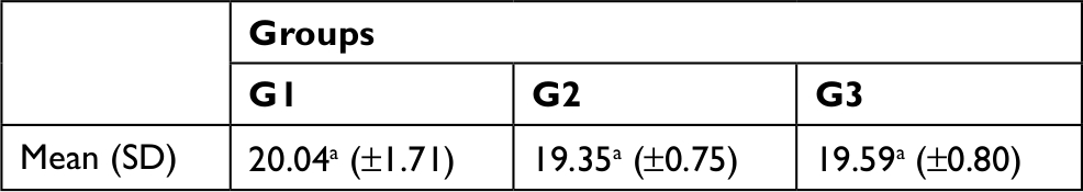 | Table 2 Difference between the mean (and SD) of the microshear test data (MPa), in the adhesive strategy of total acid etching, for the different substrates evaluated Notes: One-way ANOVA with Tukey posttest, adopting α level of significance (P≤0.05). G1, human enamel; G2, bovine enamel; G3, buffalo enamel. Different letters indicate statistical difference to 5%. ano statistical difference between the groups. |
Analysis of fracture pattern
The predominantly observed failure mode was a combination of types (adhesive/cohesive) for all experimental groups, corresponding to 57% of the total specimens analyzed (Figure 2).
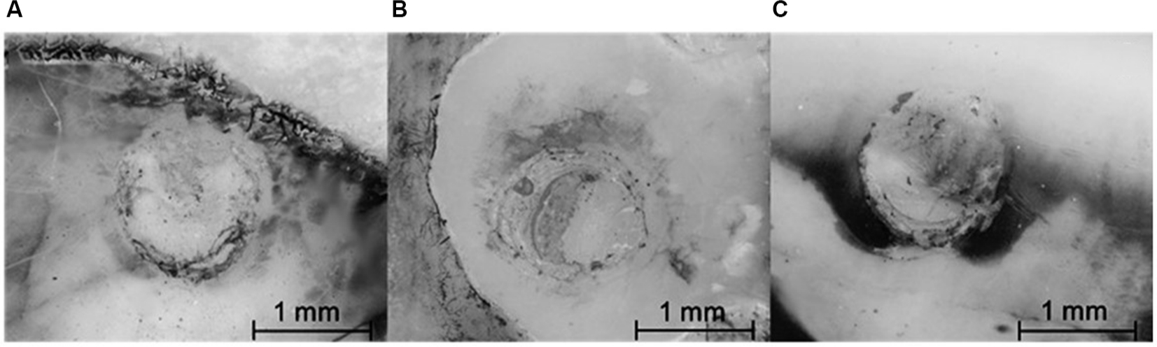 | Figure 2 Images of the prevalent fracture pattern obtained by Leica® stereomicroscope (35× magnification). Notes: G1-mixed-type fracture (A). G2-mixed-type fracture (B). G3-mixed-type fracture (C). |
SEM
The three substrates analyzed showed a type II Silverstone demineralization pattern, in which peripheral areas of the rods were removed and the nuclei were maintained, showing a retentive aspect (Figure 3).
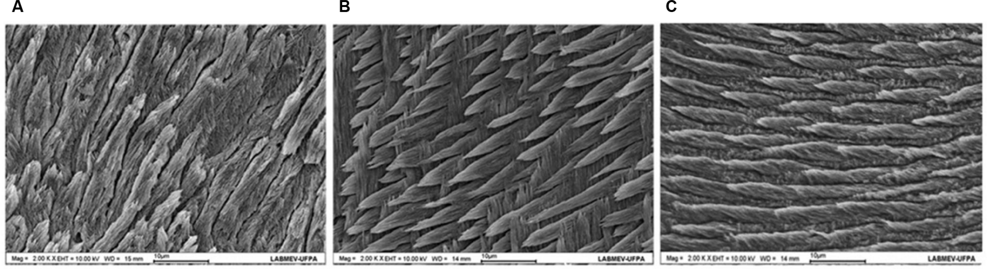 | Figure 3 Micrographs showing the similarity between human enamel (A), bovine enamel (B), and buffalo enamel (C). Notes: Images with similar cutting orientations and with 2,000× magnifications. The surfaces were treated with the purpose of removing the interprismatic enamel to favor the observation and characterization of the prisms. The similarity between the enamel prims, with characteristics of type II pattern of Silverstone, was observed. |
Discussion
For the first time in the literature, buffalo enamel was found as a replacement biological substrate for human enamel in in vitro tests. The two substrates (buffalo and human) showed similarities regarding bond strength and sensitivity to acid etching.
The first step for choosing tooth substrates from different animals for laboratory tests is to verify their ultrastructure and physicochemical properties, where the specific particularities of each species must be considered. SEM studies using swine teeth showed that their enamel is approximately half as thick as human, despite similarities in their susceptibility to acid etching. Thus, swine enamel is not indicated as a replacement for human substrate in in vitro adhesive tests.12,17 In this context, buffalo teeth showed histomorphological characteristics similar to human ones and are easily acquired as well. Therefore, they can be considered appropriate for laboratory studies.
Previous studies have confirmed that human and bovine substrates, both from mammals, show similarities regarding their enamel microstructure, histochemical, and morphological features, which enable bovine teeth for dental in vitro investigations.8,13,18 The G1 and G2 groups (human and bovine substrates, respectively) showed similar adhesive bond strength, consistent with the literature.16,19,20 Buffalo enamel also showed a similar behavior, as was shown in this study.
Due to its rod-like morphology, enamel has an anisotropic behavior, ie, directionally dependent properties. The rod-like organization is responsible for directing masticatory forces into underlying dental tissues, such as dentin.21,22 This orientation has a direct effect on bond strength; thus, adhesive resin restorations may have a better clinical performance if the enamel rod arrangement presents a transverse orientation. In this scenario, the adhesive strength may achieve values between 18 and 19 MPa.23 The arrangement of human, bovine, and buffalo enamel rods was previously analyzed, and a similar parallel ordering was observed.13
The effect of phosphoric acid on the surface of the experimental groups (bovine and buffalo enamel or G2 and G3) was similar to that already described by Silverstone.16 Although the most accepted acid etching pattern with suitable adhesion is type I Silverstone, the analysis of AEP in this study showed dissolution of the peripheral region of the enamel rods with preservation of the nuclei, that is, a type II Silverstone pattern in all experimental groups (Figure 3). Etched buffalo enamel showed the dissociation of inorganic components characterized by irregularities (Figure 3) that promoted the penetration of the adhesive system, allowing substrate hybridization and supporting adhesion. This pattern has previously been observed in bovine enamel subjected to acid etching.11,12
Bond strength values around 20–25 MPa can be considered adequate to assure a good clinical performance.24 In the present study, the mean values for human enamel bond strength (G1) was 20.04 MPa; this result did not differ significantly from the mean value observed for bovine enamel (G2) (19.35 MPa) and buffalo enamel (G3) (19.59 MPa). These results are consistent with other studies in the literature, whose mean values for human enamel bond strength was 22–25.4 MPa, similar to the bond strength to bovine enamel (15–21.2 MPa).10,17,25
Stresses that naturally occur in the tooth–restoration interface are considered complex but can be determined by tensile or shear stress tests.2,4 The adhesive strength measured through the microshear test, and the subsequent analysis of the post-fracture characteristics are well-established methods in the scientific literature to evaluate adhesive materials.15,25 Microshear is a preferable measured parameter than conventional shear. By using a reduced adhesive bonding area, it is possible to multiply the number of specimens in a single tooth, reducing the sample size needed to achieve adequate statistical power.3,21
The method used in this study to capture the images of the predominant adhesive fracture pattern was previously used to describe the internal anatomy of deciduous swine teeth.6 It has become possible to identify different components (bonding agent, composite, and dental structure) in the area of the fracture using this type of microscopy by observing the contrast obtained through the use of pigment agents on the enamel surface.
Mixed type fractures, predominant in all animal substrates analyzed in this study (Figure 1), occurred mainly at the adhesive interface, including some areas in which restorative material was present. Adhesive fractures and the prevalent type of fracture in the present study (mixed) are associated with satisfactory bond strength values that help extend the longevity of the restorations, as previously reported.9,14 Although the comparability of adhesive bond strength test values is dubious due to the variability of methods and devices used, and although these tests are not sufficient to determine the clinical success of a particular product or technique, evidence has shown that the performance of the materials can be initially evaluated through such in vitro tests. The rapid technological progress in adhesive materials makes their evaluation in primary studies essential, so they can progress to clinical trials and use. For example, if a restorative material does not prove to be effective upon controlled laboratory conditions, it might not work well when exposed to the more complex oral environment.3,17
A recent systematic review and meta-analysis compared the bond strength values obtained from human and bovine teeth in vitro studies and found that, despite the heterogeneity of the selected studies, the bovine substrate provided similar and comparable results to human enamel. The authors concluded that bovine substrate could be considered an adequate substitute in this type of analysis.5 For more than 30 years, bovine enamel was recognized as a substitute for human enamel in adhesive bonding strength tests. Although there are inherent peculiarities to each animal species, it is a fact that there is a great similarity between the two substrates, for instance in composition, due to similar percentage of calcium and phosphorus (by weight), as well as some mechanical properties such as hardness and chemical properties after acid etching.8,18,19
Buffalo enamel also showed similarities to human enamel regarding their hardness and chemical compositions. The chemical elements present in the human hydroxyapatite were also found in the enamel of buffalo.13 Buffalo and bovine teeth present similarities in size and provide enough space to assemble test specimens.5,7 Thereby, although the use of human teeth for bond strength studies is preferable, this biological substrate has become more and more scarce due to the minimally invasive approaches used in restorative dentistry and the reduction in tooth decay over recent decades. Thus, establishing a dental substrate analogous to human enamel is a significant contribution to the development of dental research.4,20,25
Conclusion
The results of this research showed that buffalo enamel is a suitable substitute for human enamel in bond strength tests and also presents similar behavior after etching.
Acknowledgment
This study was supported by PROPESP (Pró-Reitoria de Pesquisa e Pós-Graduação da UFPA) and the CAPES (Coordenação de Aperfeiçoamento de Pessoal de Nível Superior) of Brazil.
Disclosure
The authors report no conflicts of interest in this work.
References
Braga RR, Meira JBC, Boaro LC, Xavier TA. Adhesion to tooth structure: a critical review of “macro” test methods. Dent Mater. 2010;26(2):e38–e49. | ||
Sirisha K, Rambabu T, Shankar Y, Ravikumar P. Validity of bond strength tests: a critical review: Part I. J Conserv Dent. 2014;17(4):305–311. | ||
Placido E, Meira JB, Lima RG, Muench A, de Souza RM, Ballester RY. Shear versus micro-shear bond strength test: a finite element stress analysis. Dent Mater. 2007;23(9):1086–1092. | ||
Krifka S, Börzsönyi A, Koch A, Hiller KA, Schmalz G, Friedl KH. Bond strength of adhesive systems to dentin and enamel-human vs bovine primary teeth in vitro. Dent Mater. 2008;24(7):888–894. | ||
Soares FZ, Follak A, da Rosa LS, Montagner AF, Lenzi TL, Rocha RO. Bovine tooth is a substitute for human tooth on bond strength studies: a systematic review and meta-analysis of in vitro studies. Dent Mater. 2016;32(11):1385–1393. | ||
Fagundes NC, Cardoso MA, Miranda MS, et al. Morphological aspects and physical properties of enamel and dentine of Sus domesticus: a tooth model in laboratory research. Ann Anatomy. 2015;202:71–77. | ||
Santana LNS, Luz MS, Carneiro NCM, et al. Ultrastructure of buffalo tooth enamel: a possible replacement for human teeth in laboratory research. Braz J Oral Sci. 2011;10(3):163–166. | ||
Teruel JdeD, Alcolea A, Hernández A, Ruiz AJ. Comparison of chemical composition of enamel and dentine in human, bovine, porcine and ovine teeth. Arch Oral Biol. 2015;60(5):768–775. | ||
Sabatini C. Effect of phosphoric acid etching on the shear bond strength of two self-etch adhesives. J Appl Oral Sci. 2013;21(1):56–62. | ||
Aguiar JD, Medeiros IS, Souza Junior MHSE, Loretto SC. Influence of the extended use of desensitizing toothpastes on dentin bonding, microhardness and roughness. Braz Dent J. 2017;28(3):346–353. | ||
Zhu JJ, Tang AT, Matinlinna JP, Hägg U. Acid etching of human enamel in clinical applications: a systematic review. J Prosthet Dent. 2014;112(2):122–135. | ||
Lopes FM, Markarian RA, Sendyk CL, Duarte CP, Arana-Chavez VE. Swine teeth as potential substitutes for in vitro studies in tooth adhesion: a SEM observation. Arch Oral Biol. 2006;51(7):548–551. | ||
Nogueira BCL, Fernandes PM, Paiva ACJ, et al. Benchmarking of the ultrastructure and physical properties of bovine, buffalo and human enamel. Pesqui Vet Bras. 2014;34:485–490. | ||
Scherrer SS, Cesar PF, Swain MV. Direct comparison of the bond strength results of the different test methods: a critical literature review. Dent Mater. 2010;26(2):e78–e93. | ||
Dental Materials – Testing of Adhesion to Tooth Structure. Second ed. Switzerland; 2003. Technical specification ISO/TS 11405. Available from: https://www.sis.se/api/document/preview/903457/. Accessed 11 November, 2018. | ||
Reis AF, Giannini M, Kavaguchi A, Soares CJ, Line SR. Comparison of microtensile bond strength to enamel and dentin of human, bovine and porcine teeth. J Adhes Dent. 2004;6(2):117–121. | ||
Silverstone LM, Saxton CA, Dogon IL, Fejerskov O. Variation in the pattern of acid etching of human dental enamel examined by scanning electron microscopy. Caries Res. 1975;9(5):373–387. | ||
Fonseca RB, Haiter-Neto F, Carlo HL, et al. Radiodensity and hardness of enamel and dentin of human and bovine teeth, varying bovine teeth age. Arch Oral Biol. 2008;53(11):1023–1029. | ||
Nakamichi I, Iwaku M, Fusayama T. Bovine teeth as possible substitutes in the adhesion test. J Dent Res. 1983;62(10):1076–1081. | ||
Yassen GH, Platt JA, Hara AT. Bovine teeth as substitute for human teeth in dental research: a review of literature. J Oral Sci. 2011;53(3):273–282. | ||
De Las Casas EB, Cornacchia TP, Gouvêa PH, Cimini CA Jr. Abfraction and anisotropy-effects of prism orientation on stress distribution. Comput Methods Biomech Biomed Engin. 2003;6(1):65–73. | ||
Sirisha K, Rambabu T, Ravishankar Y, Ravikumar P. Validity of bond strength tests: a critical review-Part II. J Conserv Dent. 2014;17(5):420–426. | ||
Carvalho RM, Santiago SL, Fernandes CA, Suh BI, Pashley DH. Effects of prism orientation on tensile strength of enamel. J Adhes Dent. 2002;2:251–257. | ||
Usha C, Ramarao S, John BM, Rajesh P, Swatha S. Evaluation of the shear bond strength of composite resin to wet and dry enamel using dentin bonding agents containing various solvents. J Clin Diagn Res. 2017;11(1):ZC41–ZC44. | ||
Rüttermann S, Braun A, Janda R. Shear bond strength and fracture analysis of human vs. bovine teeth. PLoS One. 2013;8(3):e59181. |
 © 2019 The Author(s). This work is published and licensed by Dove Medical Press Limited. The
full terms of this license are available at https://www.dovepress.com/terms
and incorporate the Creative Commons Attribution
- Non Commercial (unported, 3.0) License.
By accessing the work you hereby accept the Terms. Non-commercial uses of the work are permitted
without any further permission from Dove Medical Press Limited, provided the work is properly
attributed. For permission for commercial use of this work, please see paragraphs 4.2 and 5 of our Terms.
© 2019 The Author(s). This work is published and licensed by Dove Medical Press Limited. The
full terms of this license are available at https://www.dovepress.com/terms
and incorporate the Creative Commons Attribution
- Non Commercial (unported, 3.0) License.
By accessing the work you hereby accept the Terms. Non-commercial uses of the work are permitted
without any further permission from Dove Medical Press Limited, provided the work is properly
attributed. For permission for commercial use of this work, please see paragraphs 4.2 and 5 of our Terms.