
Back to Journals » Orthopedic Research and Reviews » Volume 18
Intelligent Rehabilitation: Advances in Artificial Intelligence for Musculoskeletal Rehabilitation: A Narrative Review
Authors Hao J, Sun S, Dou T, Deng J, Li W, Ma C, Zhang Y, Yao L 
Received 9 March 2026
Accepted for publication 14 May 2026
Published 18 July 2026 Volume 2026:18 607912
DOI https://doi.org/10.2147/ORR.S607912
Checked for plagiarism Yes
Review by Single anonymous peer review
Peer reviewer comments 3
Editor who approved publication: Professor Qian Chen
Jiayue Hao,1,* Shibo Sun,2,* Tianxu Dou,3,* Jin Deng,1 Wen Li,1 Chicheng Ma,1 Yejia Zhang,4 Lutian Yao1
1Department of Orthopaedics, The First Hospital of China Medical University, Shenyang, Liaoning, People’s Republic of China; 2Shu Dong Yun Yi (Beijing) Technology Co., Ltd, Beijing, People’s Republic of China; 3Department of Sports Medicine, Shenyang Orthopedic Hospital, Shenyang, Liaoning, People’s Republic of China; 4Department of Physical Medicine & Rehabilitation, Perelman School of Medicine, University of Pennsylvania, Philadelphia, PA, USA
*These authors contributed equally to this work
Correspondence: Lutian Yao, Email [email protected] Yejia Zhang, Email [email protected]
Abstract: Musculoskeletal diseases, such as osteoarthritis and joint trauma, significantly impact patient mobility, independence, and quality of life. With the rising demand for effective and accessible rehabilitation strategies, artificial intelligence (AI) has emerged as a powerful tool to support diagnosis, surgical planning, and personalized rehabilitation. This narrative review summarizes recent advances in the application of AI in musculoskeletal disease management, with a particular emphasis on postoperative and conservative rehabilitation. We outline the foundational concepts of AI, including machine learning, deep learning, computer vision, and natural language processing, and discuss their roles in clinical decision-making and recovery monitoring. Furthermore, we examine emerging AI-assisted rehabilitation tools, including mobile applications, robotic exoskeletons, gamified platforms, and markerless motion tracking systems, which collectively enhance treatment precision, patient adherence, and remote care capabilities. Despite promising outcomes, current limitations include insufficient personalization, limited multimodal data integration, and inadequate clinical validation. Future developments should focus on improving model interpretability, integrating real-time biosensing, and optimizing user interface design to support clinically feasible and patient-centered musculoskeletal rehabilitation.
Keywords: artificial intelligence, musculoskeletal rehabilitation, computer vision, wearable devices, tele-rehabilitation
Introduction
Musculoskeletal diseases, including osteoarthritis, traumatic joint injuries, tendon and ligament disorders, and postoperative functional impairment, represent a major and growing health burden worldwide.1,2 These conditions are closely associated with pain, reduced mobility, impaired independence, and decreased quality of life, particularly in aging populations. For many patients, treatment does not end with diagnosis or surgery; rather, long-term functional recovery depends heavily on effective rehabilitation strategies.
Conventional musculoskeletal rehabilitation usually includes therapeutic exercise, physical modalities, activity modification, and staged postoperative training. Although these approaches are clinically important, they remain limited by several practical challenges.3 Rehabilitation protocols are often standardized rather than truly individualized, patient progress is commonly evaluated during intermittent clinical visits, and feedback on exercise quality or functional recovery is frequently delayed. These limitations are especially evident in chronic diseases such as osteoarthritis and in postoperative recovery after arthroscopy or joint replacement, where rehabilitation requires continuous adjustment according to pain, range of motion, muscle strength, gait pattern, and patient adherence.4
Artificial intelligence (AI) may help address these limitations by analyzing diverse clinical data and providing information that can support assessment, decision-making, and follow-up. Through machine learning, deep learning, computer vision, natural language processing, and sensor-based analytics, AI can support disease classification, risk prediction, treatment planning, movement assessment, and remote monitoring.4 In orthopedics, AI has been increasingly applied to image recognition, risk prediction, and clinical decision support.5 For example, Atik et al reported that optimized deep learning models achieved 74% accuracy in five-class classification and 93.75% accuracy in binary classification of knee osteoarthritis severity from radiographs, supporting the feasibility of AI-assisted disease grading and clinical decision support.6 Such studies indicate that AI can help quantify disease status and stratify patients, which may further provide a basis for individualized rehabilitation planning and longitudinal recovery monitoring.
Beyond diagnosis and severity classification, AI is increasingly being integrated into the rehabilitation process itself. Recent systematic reviews have summarized AI-supported physical rehabilitation technologies tested in clinical settings, including motion-feedback systems, exergaming platforms, telerehabilitation, wearable devices, and robotic solutions.7,8 These technologies can provide real-time feedback, monitor exercise performance, detect compensatory movement patterns, and support home-based or remote rehabilitation. They may help shift musculoskeletal rehabilitation from a therapist-dependent and episodic model toward a more continuous, data-driven, and patient-centered care pathway.
This narrative review summarizes recent advances in AI applications in musculoskeletal management, with a particular focus on rehabilitation. Unlike prior reviews that often focus on a single technical domain or a specific rehabilitation modality, this review emphasizes the integration of surgical decision support, rehabilitation technologies, and remote functional assessment within a unified musculoskeletal framework. We first introduce the foundational concepts of AI, then discuss its role in musculoskeletal rehabilitation and orthopedic decision-making, and finally examine current limitations and future directions for clinical translation.
Literature Search Strategy
In PubMed, we conducted a combined keyword search using terms including “artificial intelligence”, “machine learning”, “deep learning”, “computer vision”, “large language models”, “wearable sensors”, “mobile applications”, “robotic exoskeletons”, “tele-rehabilitation”, “musculoskeletal rehabilitation”, “orthopedic rehabilitation”, “osteoarthritis”, “joint arthroplasty”, “arthroscopy”, and “postoperative rehabilitation”. The initial search retrieved 213 articles. After screening the titles and abstracts, 112 articles were considered relevant to this review. Finally, 78 articles were included in this narrative review.
Overview of Artificial Intelligence
AI Fundamentals and Evolution
Artificial intelligence refers to computational approaches that enable pattern recognition, prediction, and decision-making from complex data. In medical and rehabilitation contexts, AI mainly includes machine learning, deep learning, computer vision, natural language processing, and sensor-based analytics.5,9,10 These technologies can process imaging data, clinical records, patient-reported outcomes, and movement signals to generate clinically useful information.
Machine learning uses algorithms to identify relationships within data and support tasks such as disease classification, outcome prediction, and risk stratification. Common learning strategies include supervised learning, unsupervised learning, and reinforcement learning. Deep learning, as a subfield of machine learning, uses multilayer neural networks and is particularly useful for image analysis, motion recognition, and automated feature extraction.11
In musculoskeletal rehabilitation, computer vision enables markerless movement assessment, joint angle measurement, gait analysis, and exercise monitoring, while natural language processing may support patient education, symptom tracking, and conversational guidance. Together, these AI technologies provide the technical foundation for individualized rehabilitation planning, real-time feedback, and remote functional monitoring.12–14
AI in Medicine: Diagnostic and Therapeutic Applications
AI has been increasingly applied across medical disciplines for diagnosis, prediction, monitoring, and clinical decision support. In diagnostic imaging, pathology,15 anesthesiology,16–18 and cardiovascular medicine,5 AI tools have demonstrated strong performance in identifying critical conditions, quantifying disease severity, and assisting in real-time monitoring. These systems can analyze different types of clinical data, including medical images, vital signs, and histopathological slides, and may assist clinicians in identifying relevant diagnostic or prognostic information (Figure 1).
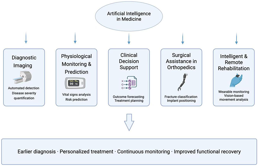 |
Figure 1 Overview of Artificial Intelligence Applications in Medicine. |
These advances have reshaped workflows in radiology,19 oncology,15 and surgical medicine, enabling earlier detection of disease, individualized treatment planning, and outcome forecasting. Notably, the core functions of AI—automatic classification, pattern recognition, and risk prediction—are also highly applicable to musculoskeletal disease management.
In orthopedics, AI has emerged as a valuable tool for automating fracture classification, evaluating implant positioning, and predicting surgical outcomes. More recently, its integration into postoperative rehabilitation has expanded through the development of intelligent recovery plans, wearable monitoring, and computer vision–based movement analysis. These innovations enable personalized rehabilitation while reducing reliance on in-person supervision.20
Given the chronic nature of many musculoskeletal conditions and the demand for long-term functional recovery, AI presents significant potential to bridge the gap between diagnosis, treatment, and rehabilitation. The following sections of this review will delve into how AI is transforming musculoskeletal care—particularly in surgery support, individualized rehabilitation strategies, and remote functional assessment.
Musculoskeletal Rehabilitation
Definition and Classification: Conservative vs Perioperative
Rehabilitation for musculoskeletal diseases encompasses both conservative (non-surgical) and postoperative interventions, each tailored to the patient’s clinical condition, functional goals, and stage of disease. Conservative rehabilitation focuses on delaying disease progression, relieving symptoms, and improving joint function through therapeutic exercise, physical modalities, and activity modification.21 This approach is commonly used in early-stage osteoarthritis, soft tissue injuries, or for patients who are not surgical candidates.22
In contrast, perioperative rehabilitation focuses on optimizing patient outcomes throughout the entire surgical continuum, encompassing preoperative, intraoperative, and postoperative phases. Preoperative interventions aim to enhance physical fitness, educate patients on surgical expectations, and reduce anxiety, while intraoperative strategies may include positioning and protective measures to minimize tissue stress. Postoperative rehabilitation transitions into protocols emphasizing early protection, progressive mobilization, muscle strengthening, and return-to-activity training.23 The choice of rehabilitation strategy is influenced by factors such as surgical procedure, joint involved, patient comorbidities, and individual recovery goals.24
Both conservative and postoperative rehabilitation share a common emphasis on individualized care and functional restoration. With the growing complexity of rehabilitation protocols and the need for personalized adjustment, integrating AI technologies—such as motion analysis, data-driven exercise progression, and real-time patient monitoring—presents a promising opportunity to optimize rehabilitation outcomes across the care continuum.
Role of Surgery and Rehabilitation Interplay
In the management of musculoskeletal diseases, surgical intervention and rehabilitation are deeply interconnected, forming a dynamic continuum rather than separate, sequential steps. Surgery aims to correct structural abnormalities, alleviate pain, and restore anatomical alignment, while rehabilitation ensures the functional translation of surgical outcomes into real-world mobility, strength, and quality of life. Without structured rehabilitation, surgical outcomes may be compromised by muscle atrophy, joint stiffness, or suboptimal biomechanical adaptations.
Among surgical approaches, arthroscopic surgery has become a preferred minimally invasive technique for managing intra-articular pathologies of the knee, shoulder, hip, and elbow. It enables real-time visualization and targeted intervention with minimal soft tissue disruption. Indications include meniscal tears, ligament injuries, cartilage lesions, and inflammatory joint conditions. Compared with open surgery, arthroscopy is associated with faster postoperative recovery, lower pain levels, and shorter hospitalization.25,26
Joint replacement surgery, most commonly involving the hip and knee, is the standard of care for advanced degenerative joint diseases such as osteoarthritis and rheumatoid arthritis. It involves excising diseased joint surfaces and implanting prosthetic components composed of metal alloys, polyethylene, or ceramics. Advancements in prosthetic design, surgical technique, and perioperative care have led to increased implant longevity, reduced complication rates, and enhanced postoperative functional recovery.27
Rehabilitation Advances in Joint Surgery
With the global increase in arthroscopic and joint replacement procedures, rehabilitation strategies for musculoskeletal diseases have progressed significantly. Arthroscopic surgery, utilizing minimally invasive techniques, offers advantages in reducing surgical trauma and accelerating early recovery. Meanwhile, joint replacement surgery—primarily involving the hip and knee—provides effective symptom relief and functional restoration for patients with advanced joint degeneration. This section summarizes representative rehabilitation principles across major joints and highlights where AI may support protocol individualization, remote monitoring, and progression assessment (Figure 2).
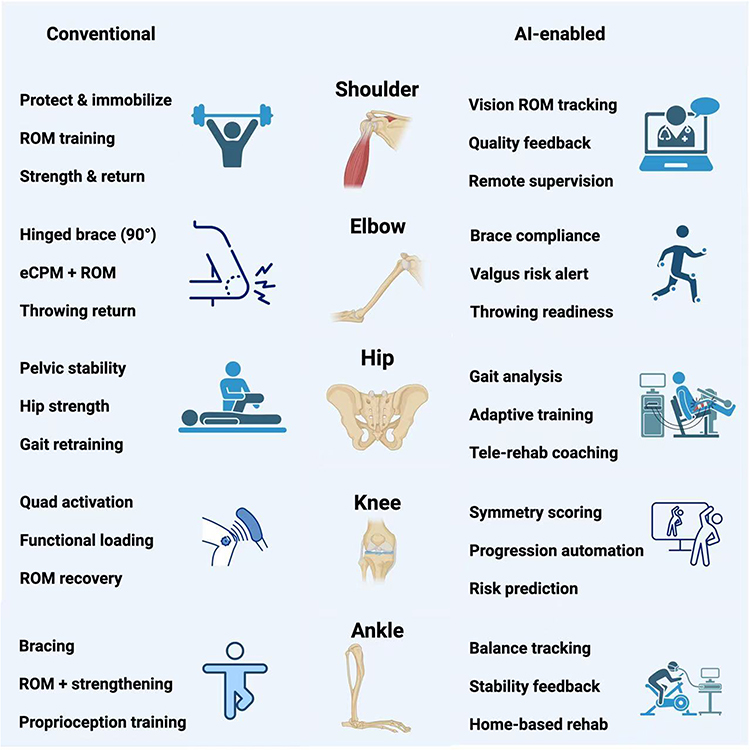 |
Figure 2 Conventional vs AI-Enabled Rehabilitation Pathways After Joint Surgery. Icons adapted from Flaticon:https://www.flaticon.com. |
Shoulder Joint
Postoperative rehabilitation following rotator cuff repair or total shoulder arthroplasty has been extensively studied, yet significant variations remain in immobilization duration, the initiation of active movement, and the progression of weight-bearing activities. The American Society of Shoulder and Elbow Therapists (ASSET) recommends strict shoulder immobilization for the first two weeks postoperatively, followed by phased passive range of motion (ROM) exercises. Active ROM is introduced around six weeks, with strengthening exercises beginning at twelve weeks.28 McBroom et al proposed an accelerated protocol that initiated passive and active-assisted ROM between weeks 2 and 4, and active ROM between weeks 4 and 6, with strengthening added between weeks 6 and 8. This approach, which discontinued sling use earlier than standard protocols, resulted in superior ROM at both 6 weeks and 3 months postoperatively.29 For SLAP lesions, rehabilitation typically includes a short protective phase followed by progressive ROM restoration and strengthening, with progression individualized according to surgical findings and tissue healing.30 In parallel, AI-based vision ROM tracking may help quantify shoulder motion and facilitate remote supervision with real-time exercise quality feedback.
Elbow Joint
Rehabilitation after medial collateral ligament (MCL) repair or reconstruction requires careful protection and professional supervision. In the early phase, valgus stress must be avoided to protect healing tissues. A hinged elbow brace is worn at 90° flexion during the first two weeks, followed by gradual passive and active ROM exercises until full flexion and extension are restored. Brace removal and return-to-throwing timelines differ between repair and reconstruction, with reconstruction generally requiring longer protection and a slower progression back to sport.31 Lihua et al demonstrated that combining early continuous passive motion (eCPM) with physical therapy (PT) significantly improves ROM and Mayo Elbow Performance Scores (MEPS) at multiple postoperative intervals compared to PT alone. A greater proportion of patients also achieved functional flexion (≥130°), supporting eCPM as a beneficial adjunct to conventional rehabilitation.32 In this context, wearable sensors and computer vision (CV) may help monitor brace compliance and detect potentially excessive valgus loading, supporting safer decisions on throwing readiness.
Hip Joint
For femoroacetabular impingement (FAI), conservative management and postoperative rehabilitation both emphasize hip strength, dynamic stability, and restoration of functional range of motion, with surgery generally reserved for patients who do not respond adequately to structured nonoperative treatment.33,34 Postoperative rehabilitation after hip arthroscopy typically focuses on pelvic control, hip strength, motor control, and restoration of functional ROM.35 In hip replacement surgery, Suetta et al showed that early supervised progressive resistance training improves hip strength and gait performance. Beginning exercises—like leg press and hip abductor training—within one week and continuing for four weeks led to sustained benefits over 12 months.36 Busato et al highlighted the effectiveness of combining fascial manipulation with standard PT, which significantly enhanced ROM and Harris Hip Scores within 10 days postoperatively.37 Di Monaco et al further emphasized the importance of ongoing rehabilitation beyond week six, including whole-body resistance training to improve gait speed, balance, and perceived independence.38 Emerging AI-based gait analysis may further support adaptive training strategies and telerehabilitation coaching after hip arthroscopy or total hip arthroplasty.
Knee Joint
Early rehabilitation after anterior cruciate ligament (ACL) reconstruction emphasizes pain control, recovery of range of motion, and quadriceps reactivation to support functional restoration and safe return to activity.39 After meniscal repair, rehabilitation generally follows a protected, milestone-based progression that balances tissue healing with gradual recovery of motion, strength, and function.40 Similarly, rehabilitation after major ligament repair or reconstruction, including medial collateral ligament injuries, requires staged progression based on healing status, knee stability, and recovery milestones.41 After total knee arthroplasty (TKA), early quadriceps weakness remains a major barrier to recovery, making progressive strengthening a central component of postoperative rehabilitation.42 Additional modalities, including cycling-based exercise and sensorimotor training,43 may further improve range of motion, balance, gait, and overall functional recovery. AI tools may assist in movement symmetry scoring, support progression decisions, and contribute to postoperative risk stratification after ACL reconstruction or total knee arthroplasty (TKA).
Ankle Joint
Grade I and II lateral ligament tears are typically managed non-surgically using the RICE protocol (Rest, Ice, Compression, Elevation) and short-term immobilization. Early ROM and neuromuscular training reduce stiffness and promote function. Proprioceptive training (eg, balance boards) begins around week 3–4 to improve joint stability.44 Previous studies have suggested that functional rehabilitation may provide favorable outcomes compared with prolonged immobilization or surgical treatment in selected patients with lateral ankle ligament injuries.45 Several meta-analyses also supported functional rehabilitation over casting, noting better outcomes and lower costs.46,47 AI-enabled balance tracking may provide stability feedback and support home-based rehabilitation, particularly during proprioceptive and neuromuscular retraining.
Future Integration with AI
While current rehabilitation protocols following arthroscopic and joint replacement surgeries are effective and evidence-based, they are often generalized and labor-intensive. These existing protocols provide a useful framework for introducing AI-assisted monitoring and individualized adjustment. Personalized training plans, real-time motion tracking, intelligent feedback, and adaptive progression systems powered by AI could elevate conventional rehabilitation to a more precise, efficient, and accessible level. Future work should focus on embedding AI technologies into validated clinical protocols to bridge the gap between evidence-based rehabilitation and intelligent, patient-specific care.
Integration of AI into Musculoskeletal Disease Management
As musculoskeletal diseases become increasingly prevalent and complex, there is a growing demand for scalable, precise, and patient-specific solutions in both diagnosis and rehabilitation. AI, with its capacity to analyze large volumes of multimodal data and derive actionable insights, offers a compelling opportunity to enhance the continuum of care in bone and joint disease management. This section explores the current and emerging roles of AI across key domains, including surgical decision support and intelligent rehabilitation.
AI in Orthopedic Surgery and Clinical Decision Support
AI has been increasingly explored in orthopedic surgery for preoperative planning, intraoperative guidance, and postoperative evaluation. Machine learning algorithms are increasingly used to classify fractures, detect joint degeneration, and assist in implant selection based on imaging modalities such as X-rays, CT, and MRI. Recent imaging-based studies have further reinforced this role; for example, deep learning models applied to knee radiographs have shown strong performance in osteoarthritis severity classification, supporting AI-assisted imaging assessment in clinically relevant decision-making. Intraoperatively, AI-assisted robotic platforms and 3D modeling technologies may support surgical guidance and anatomical reconstruction. AI has also played a role in prosthetic design by optimizing implant geometry and materials for improved biomechanical performance and durability. For instance, Bi et al demonstrated that AI-assisted 3D motion simulation in patients with elbow osteoarthritis helped identify osteophyte impingement and guide resection planning.48 Similarly, Jeung et al developed an augmented reality-based surgical guidance approach for wrist arthroscopy with bone-shift compensation, supporting more accurate arthroscopic navigation.49
Beyond image analysis and robotic assistance, large language models (LLMs) have introduced new possibilities for orthopedic decision support, patient education, and clinical information synthesis. These models can integrate patient histories, imaging findings, and guideline-based knowledge to generate preliminary recommendations, particularly in common musculoskeletal conditions involving the knee and shoulder. In a comparative evaluation of ChatGPT-3.5, ChatGPT-4, and Google Bard, ChatGPT-4 demonstrated the best performance in orthopedic clinical decision-making, achieving 76.3% accuracy within a 10% deviation margin and 86.6% within a 20% deviation margin.50 Although these results suggest that LLMs may be useful for standardized reasoning tasks, their current limitations in autonomy, clinical depth, and guideline concordance indicate that they should be regarded as adjunctive tools rather than independent decision-makers. Further development of domain-specific models and integration with validated medical knowledge bases may improve their reliability in orthopedic practice.
However, the performance of LLMs varies substantially across orthopedic tasks.50 While some reports have shown reasonable performance in MRI-based interpretation and preoperative patient education, others have identified limited concordance with guideline-based treatment recommendations and reduced reliability in complex or nuanced clinical scenarios.51–54 Taken together, the current evidence supports the use of LLMs as supplementary tools for communication, report interpretation, and preliminary information synthesis, but not as stand-alone systems for orthopedic decision-making.
AI in Musculoskeletal Rehabilitation
Traditional rehabilitation often suffers from limited personalization, delayed feedback, and heavy reliance on manual supervision—challenges that hinder efficient, adaptive recovery.7 The integration of AI into rehabilitation practice may help address these problems by enabling real-time data analysis, remote monitoring, and more individualized training programs.
AI-assisted systems may extend rehabilitation beyond hospital visits by supporting home-based monitoring, feedback, and follow-up. These technologies may improve rehabilitation accessibility and provide additional support for clinicians, particularly in home-based or remote settings. Key application areas include conversational agents for patient interaction, robotic exoskeletons for assisted mobility, gamified rehabilitation for engagement, and computer vision for movement assessment (Figure 3).
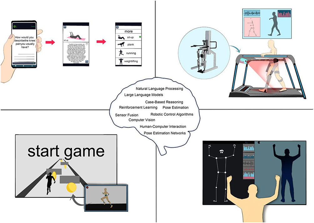 |
Figure 3 Applications and underlying technologies of artificial intelligence in musculoskeletal rehabilitation. |
AI-Enabled Mobile Applications and Conversational Support
AI-powered mobile applications are playing an increasingly important role in musculoskeletal rehabilitation by enabling real-time monitoring, personalized guidance, and adaptive intervention. Depending on the design, these platforms may incorporate wearable sensors, automated feedback, reminder systems, or conversational interfaces to support exercise guidance and patient adherence beyond clinical settings. These applications can be broadly grouped into three categories: self-management platforms for chronic musculoskeletal pain, osteoarthritis-focused exercise applications, and postoperative digital rehabilitation systems.
Among the first category, MyBehaviorCBP,55 SELFBACK,56 and Secaide,57 illustrate how AI-enabled mobile platforms can support chronic pain self-management through activity monitoring, adaptive exercise recommendation, and behavior-oriented prompting. These systems aim to improve adherence and extend rehabilitation beyond conventional clinical settings.
In osteoarthritis-focused rehabilitation, applications such as Rak Kao58 and Dr. Bart59 provide personalized exercise guidance, symptom monitoring, and behavior-based support for patients with knee or hip osteoarthritis. Collectively, these applications highlight the value of AI in improving exercise accuracy, patient engagement, and day-to-day self-management.
For postoperative rehabilitation, digital platforms such as SWORD Health60 combine wearable motion sensors with AI-driven biofeedback to support home-based recovery after orthopedic procedures such as total knee arthroplasty. This category is particularly relevant to remote rehabilitation because it links continuous monitoring with individualized exercise progression.
Overall, AI-enabled mobile applications demonstrate the potential of digital rehabilitation tools to improve precision, promote engagement, and expand access to care, particularly in chronic disease management and postoperative recovery.
AI-Powered Robotic Exoskeletons
Robotic exoskeletons represent a major advancement in physical rehabilitation, particularly when integrated with AI-enabled control systems. These wearable devices provide mechanical assistance and real-time sensor-driven feedback, enabling personalized support for patients with joint degeneration, post-surgical limitations, or motor impairments.61
Keeogo,62 for example, is a lower-limb exoskeleton equipped with AI-based gait analysis that adjusts support intensity dynamically based on knee biomechanics. In a randomized crossover study, users demonstrated improved functional performance in the 6-Minute Walk Test, TUG test, and stair climbing, as well as increased daily activity during home use. Similarly, single-joint exoskeletons targeting the hip joint, as studied by Mahla et al, have shown significant improvements in stride length, cadence, and oxygen efficiency, emphasizing their value in optimizing energy expenditure and gait symmetry.63
Following total knee arthroplasty (TKA), AI-powered robotic rehabilitation systems have been shown to accelerate recovery by restoring joint range of motion and muscle strength. A systematic review by Wu et al found that robotic-assisted rehabilitation (RAR) outperformed conventional therapy in improving ROM, functional scores, and hospital discharge readiness.64 Likewise, Japan’s Hybrid Assistive Limb (HAL-SJ) system uses bioelectrical signals (BESs) to synchronize robotic knee movement with patient intent.65 In controlled trials, HAL-SJ demonstrated superior gains in active ROM, muscle strength, and reduced postoperative pain when compared to standard care. The system has also been adapted for ACL reconstruction66 and total hip arthroplasty (THA),67 showing significant improvements in gait mechanics, neuromuscular control, and functional mobility across multiple clinical trials.
By continuously monitoring gait patterns, detecting deviations, and adjusting mechanical output in real time, AI-enhanced exoskeletons may provide targeted, adaptive therapy. In home rehabilitation, they provide quantifiable activity tracking and feedback, enabling safer, more autonomous recovery. As evidence grows, these systems may become valuable adjuncts for joint-specific rehabilitation, helping patients regain mobility and independence while reducing clinician workload and the risk of secondary injury.
Gamified Rehabilitation and Virtual Environments
Gamified rehabilitation leverages interactive video game platforms to enhance engagement, adherence, and functional outcomes. These systems offer a more enjoyable alternative to traditional exercise by transforming rehabilitation into an interactive experience—particularly valuable for home-based or elderly patient populations.68
One of the most widely studied platforms is the Nintendo Wii, originally popularized in neurological rehabilitation.69 In orthopedic care, Wii Fit and Wii Fit U have been adapted for post-operative and chronic musculoskeletal rehabilitation. Negus et al introduced TKR-POWER, a motion-controlled platform combining Wii Fit, balance boards, and motion sensors for total knee replacement (TKR) recovery.70 In a randomized trial, patients in the Wii group demonstrated superior improvements in muscle strength, joint flexibility, and gait performance compared to those undergoing standard rehabilitation.
Recent advances have integrated AI into these gamified systems to enhance motion tracking, postural assessment, and feedback accuracy. For example, an AI-assisted Wii Fit platform was evaluated in a randomized controlled trial for patients with ankle sprains.71 While all groups improved, the AI-enhanced Wii Fit group showed favorable trends in pain relief and functional performance, offering a cost-effective, engaging alternative to conventional therapy. Another study explored Wii Fit U for elderly individuals with chronic low back pain. Over eight weeks, participants experienced short-term improvements in pain and function, although long-term psychosocial outcomes remained unchanged—suggesting that game-based rehabilitation may benefit from complementary interventions for comprehensive care.72
These findings illustrate the potential of gamified rehabilitation—especially when enhanced by AI—to improve motivation, increase adherence, and support remote or underserved populations. While not a complete substitute for therapist-guided care, such platforms are increasingly valuable as adjunct tools within multimodal rehabilitation frameworks.
Computer Vision and Remote Movement Assessment
Computer vision (CV), a core domain of AI, has become an important approach for rehabilitation assessment by enabling non-contact, real-time motion analysis using standard cameras or depth sensors.73 Markerless CV systems are increasingly replacing traditional marker-based motion capture, offering scalable, accessible alternatives for clinical and home-based rehabilitation.74
Modern CV systems typically comprise four components: a camera (eg, RGB or depth), a body model, image processing modules, and AI algorithms. Deep learning networks—especially convolutional neural networks (CNNs)—allow precise joint localization, pose estimation, and action recognition. These systems can evaluate joint angles, gait mechanics, and compensatory movements in both simple (eg, knee flexion) and complex tasks (eg, squatting, walking), delivering real-time feedback to patients and clinicians.74
Clinical applications are rapidly expanding. Xu et al proposed a randomized controlled pilot trial to evaluate a computer vision-based exercise management system for patients with knee osteoarthritis.75 Wang et al designed a prospective randomized controlled trial to assess a digital rehabilitation program based on computer vision and augmented reality for isolated meniscus injury.76 Similarly, Mennella et al developed a deep learning–based CV system for elderly postoperative patients using a 2D pose estimation model (eg, MoveNet) to monitor ROM and detect improper movements. The system provided real-time corrective feedback, enhancing safety and self-management.77
In shoulder rehabilitation, Ramkumar et al validated an AI-enabled motion analysis SDK using consumer smartphones and wearables. The system accurately measured shoulder ROM compared to manual goniometers, demonstrating potential for remote monitoring and reducing clinic visits.78
Together, these CV-based systems may improve the accessibility and objectivity of rehabilitation assessment by supporting automated movement analysis, feedback, and remote monitoring. Particularly suited for home use and aging populations, they empower patients while optimizing clinician oversight and resource allocation.
Future Perspectives and Challenges
The Expanding Role of AI in Musculoskeletal Rehabilitation
AI may play an increasing role in musculoskeletal rehabilitation by helping clinicians monitor recovery more continuously and adjust rehabilitation programs more flexibly. Conventional rehabilitation mainly relies on scheduled clinical visits and therapist-based assessment, which may not fully capture day-to-day changes in pain, joint mobility, muscle strength, gait pattern, and exercise adherence. By integrating data from mobile applications, wearable sensors, robotic systems, and computer vision platforms, AI may enable more timely evaluation of functional recovery and individualized adjustment of rehabilitation programs.
Future AI-assisted rehabilitation may extend beyond simple exercise monitoring toward risk stratification, prediction of recovery trajectories, and early detection of patients who require additional intervention. Longitudinal data, including range of motion, movement symmetry, activity level, pain scores, and patient-reported outcomes, could be analyzed together to generate adaptive rehabilitation recommendations. This approach may be especially useful after arthroscopy, ligament reconstruction, joint replacement, and in chronic musculoskeletal diseases such as osteoarthritis.
Importantly, AI should be viewed as a supportive tool rather than a replacement for clinicians or therapists. By enabling closed-loop monitoring, real-time feedback, and remote supervision, AI may help extend professional rehabilitation guidance beyond the hospital and improve the precision, accessibility, and continuity of musculoskeletal rehabilitation.
Technical and Translational Challenges
Despite significant progress, the implementation of AI in musculoskeletal rehabilitation still faces critical technical and translational barriers that limit its scalability, clinical reliability, and user adoption—particularly among older adults and patients recovering from joint surgeries.
A primary limitation lies in system usability. Many AI-powered platforms lack intuitive interfaces, voice navigation, or conversational support, which can hinder engagement, especially in elderly populations with limited digital literacy. Overly complex app interactions, inconsistent feedback delivery, and visually dense dashboards often result in reduced user confidence and declining adherence over time.
Technical stability is another concern. AI-enhanced mobile applications and exoskeletons frequently depend on high-performance hardware or constant internet connectivity. Latency, motion capture inaccuracies, or power limitations can compromise real-time feedback and limit the safe execution of rehabilitation exercises—particularly in unsupervised home environments. Similarly, robotic systems, while promising, often suffer from excessive weight, limited battery life, and mechanical rigidity, restricting their prolonged use and clinical practicality.
On the algorithmic side, most current AI models are trained on relatively narrow datasets that fail to capture the diversity of functional impairments, body types, or movement compensation strategies seen in real-world orthopedic populations. This limits generalizability and increases the risk of inappropriate recommendations. Moreover, wearable sensors and motion capture tools often function as passive data collectors, providing visualizations without meaningful interpretation or adaptive decision support.
From a clinical perspective, many systems lack dynamic adaptability. Rehabilitation content is often standardized and static, unable to evolve with a patient’s changing capabilities. Multimodal data streams—such as joint ROM, pain scores, gait patterns, and muscle activation signals—are rarely integrated to provide holistic, context-aware feedback. These gaps prevent AI systems from delivering truly individualized, responsive rehabilitation care.
Addressing these challenges will require not only technological refinement but also stronger collaboration across clinicians, engineers, and interface designers. Without such integration, AI will remain a promising—but underutilized—tool in musculoskeletal rehabilitation.
Strategic Directions for Future Development
Realizing the full potential of AI in musculoskeletal rehabilitation will depend on the development of scalable, interoperable, and clinically validated systems that can adapt to individual needs while remaining feasible within real-world healthcare settings.
First, future development should focus on lightweight, edge-compatible AI models capable of running efficiently on consumer-grade devices. This would enable real-time, on-device processing of motion data, reducing dependence on constant internet access and cloud computing. Integration with embedded systems—such as wearables, smartphones, and exoskeleton controllers—will facilitate uninterrupted feedback and enhance safety during unsupervised training sessions.
Second, future systems should better integrate multiple sources of rehabilitation data. Combining motion tracking, surface electromyography (sEMG), pressure sensors, and pain self-report tools into a unified framework can allow for comprehensive monitoring of musculoskeletal function. AI algorithms should be designed to fuse these data streams, enabling predictive alerts, adaptive workload modulation, and early identification of maladaptive movement patterns.
Third, personalization should move beyond simple adjustment of exercise intensity or duration. Systems must learn from longitudinal recovery trajectories, user-specific biomechanics, and psychological engagement signals to continuously optimize rehabilitation intensity, progression, and content relevance. Behavioral reinforcement mechanisms—such as gamification and reward-based feedback—should be dynamically tuned to individual motivation patterns to promote long-term adherence.
On the clinical side, large-scale, multicenter randomized controlled trials (RCTs) are essential to validate AI-assisted rehabilitation protocols across diverse orthopedic populations and surgical contexts. These studies should include aging populations, comorbidity profiles, and post-arthroplasty cohorts, with outcomes that extend beyond function to include quality of life, healthcare cost reduction, and patient-reported satisfaction.
Finally, cross-disciplinary collaboration will be pivotal. Engineers, clinicians, data scientists, and rehabilitation specialists must co-develop systems that meet both technical feasibility and clinical relevance. Establishing standardized protocols for data interoperability, algorithm transparency, and regulatory compliance will be vital to promoting integration into mainstream musculoskeletal care.
Overall, continued advances in model design, multimodal data integration, and clinical validation may enable AI to support more predictive, personalized, and scalable musculoskeletal rehabilitation.
Declaration of AI and AI-Assisted Technologies in the Writing Process
During the preparation of this manuscript, the authors used AI-assisted language tools to improve grammar, clarity, and readability. The AI tools were not used to generate original scientific conclusions, perform data analysis, select references, or make clinical recommendations. All AI-assisted content was critically reviewed, revised, and approved by the authors, who take full responsibility for the accuracy and integrity of the final manuscript.
Acknowledgments
The authors would like to thank all colleagues and collaborators who provided insightful comments and technical assistance during the preparation of this review. All figures were prepared by the authors using icons obtained from Flaticon and were manually arranged and edited in Microsoft PowerPoint. No AI image-generation software was used for figure creation.
Author Contributions
All authors made a significant contribution to the work reported, whether that is in the conception, study design, execution, acquisition of data, analysis and interpretation, or in all these areas; took part in drafting, revising or critically reviewing the article; gave final approval of the version to be published; have agreed on the journal to which the article has been submitted; and agree to be accountable for all aspects of the work.
Funding
This study was funded by National Key R&D Program of China (2021YFA1102600), National Natural Science Foundation of China (82103781, 82472433), Science Fund for Distinguished Young Scholars of Liaoning Province (2024JH3/10200034), and Liaoning Revitalization Talents Program (XLYC2503157).
Disclosure
The authors declare that they have no competing interests in this work.
References
1. Steinmetz JD, Culbreth GT, Haile LM; Collaborators GBDO. Global, regional, and national burden of osteoarthritis, 1990-2020 and projections to 2050: a systematic analysis for the global burden of disease study 2021. Lancet Rheumatol. 2023;5(9):e508–15. doi:10.1016/S2665-9913(23)00163-7
2. Collaborators GBDOMD. Global, regional, and national burden of other musculoskeletal disorders, 1990-2020, and projections to 2050: a systematic analysis of the global burden of disease study 2021. Lancet Rheumatol. 2023;5(11):e670–e82. doi:10.1016/S2665-9913(23)00232-1
3. Cieza A, Causey K, Kamenov K, Hanson SW, Chatterji S, Vos T. Global estimates of the need for rehabilitation based on the global burden of disease study 2019: a systematic analysis for the global burden of disease study 2019. Lancet. 2021;396(10267):2006–2017. doi:10.1016/S0140-6736(20)32340-0
4. Molina-Garcia P, Mora-Traverso M, Prieto-Moreno R, Diaz-Vasquez A, Antony B, Ariza-Vega P. Effectiveness and cost-effectiveness of telerehabilitation for musculoskeletal disorders: a systematic review and meta-analysis. Ann Phys Rehabil Med. 2024;67(1):101791. doi:10.1016/j.rehab.2023.101791
5. Myers TG, Ramkumar PN, Ricciardi BF, Urish KL, Kipper J, Ketonis C. Artificial intelligence and orthopaedics: an introduction for clinicians. J Bone Joint Surg Am. 2020;102(9):830–840. doi:10.2106/JBJS.19.01128
6. Atik I, Polat O, Atik S, Gul E, Apalan D. Osteoarthritis severity classification in knee X-rays using optimized deep learning approaches. J Imaging Inform Med. 2026. doi:10.1007/s10278-025-01818-7
7. Sumner J, Lim HW, Chong LS, Bundele A, Mukhopadhyay A, Kayambu G. Artificial intelligence in physical rehabilitation: a systematic review. Artif Intell Med. 2023;146:102693. doi:10.1016/j.artmed.2023.102693
8. Luo Z, Wang Y, Zhang T, Wang J. Effectiveness of AI-assisted rehabilitation for musculoskeletal disorders: a network meta-analysis of pain, range of motion, and functional outcomes. Front Bioeng Biotechnol. 2025;13:1660524. doi:10.3389/fbioe.2025.1660524
9. Hamet P, Tremblay J. Artificial intelligence in medicine. Metabolism. 2017;69S:S36–S40. doi:10.1016/j.metabol.2017.01.011
10. Hashimoto DA, Witkowski E, Gao L, Meireles O, Rosman G. Artificial Intelligence in anesthesiology: current techniques, clinical applications, and limitations. Anesthesiology. 2020;132(2):379–394. doi:10.1097/ALN.0000000000002960
11. Hashimoto DA, Rosman G, Rus D, Meireles OR. Artificial Intelligence in Surgery: promises and Perils. Ann Surg. 2018;268(1):70–76. doi:10.1097/SLA.0000000000002693
12. Gui Y, Zhang J. Research progress of artificial intelligence in the grading and classification of meningiomas. Acad Radiol. 2024;31(8):3346–3354. doi:10.1016/j.acra.2024.02.003
13. Sogandi F. Identifying diseases symptoms and general rules using supervised and unsupervised machine learning. Sci Rep. 2024;14(1):17956. doi:10.1038/s41598-024-69029-8
14. Frank SM, Qi A, Ravasio D, Sasaki Y, Rosen EL, Watanabe T. Supervised learning occurs in visual perceptual learning of complex natural images. Curr Biol. 2020;30(15):2995–3000e3. doi:10.1016/j.cub.2020.05.050
15. Bhinder B, Gilvary C, Madhukar NS, Elemento O. Artificial intelligence in cancer research and precision medicine. Cancer Discov. 2021;11(4):900–915. doi:10.1158/2159-8290.CD-21-0090
16. Nagpal K, Foote D, Liu Y, et al. Development and validation of a deep learning algorithm for improving Gleason scoring of prostate cancer. NPJ Digit Med. 2019;2:48. doi:10.1038/s41746-019-0112-2
17. Mirsadeghi M, Behnam H, Shalbaf R, Jelveh Moghadam H. Characterizing awake and anesthetized states using a dimensionality reduction method. J Med Syst. 2016;40(1):13. doi:10.1007/s10916-015-0382-4
18. Shalbaf A, Saffar M, Sleigh JW, Shalbaf R. Monitoring the depth of anesthesia using a new adaptive neurofuzzy system. IEEE J Biomed Health Inform. 2018;22(3):671–677. doi:10.1109/JBHI.2017.2709841
19. Driver CN, Bowles BS, Bartholmai BJ, Greenberg-Worisek AJ. Artificial intelligence in radiology: a call for thoughtful application. Clin Transl Sci. 2020;13(2):216–218. doi:10.1111/cts.12704
20. Tjardes T, Heller RA, Pforringer D, Lohmann R, Back DA; DGOU AGDd. Artificial intelligence in orthopedics and trauma surgery. Chirurg. 2020;91(3):201–205. doi:10.1007/s00104-019-01091-9
21. O’Brian DA, Mattock J, Gibson J, Pappas E. Rehabilitation after arthroscopic shoulder stabilisation: are we letting patients down by being too conservative? Br J Sports Med. 2025;59(9):627–629. doi:10.1136/bjsports-2024-109290
22. Muaidi QI. Rehabilitation of patellar tendinopathy. J Musculoskelet Neuronal Interact. 2020;20(4):535–540.
23. Cavanaugh JT. Rehabilitation of meniscal injury and surgery. J Knee Surg. 2014;27(6):459–478. doi:10.1055/s-0034-1394299
24. Berman A, Studenski S. Musculoskeletal rehabilitation. Clin Geriatr Med. 1998;14(3):641–659. doi:10.1016/S0749-0690(18)30110-1
25. O’Connor D, Johnston RV, Brignardello-Petersen R, et al. Arthroscopic surgery for degenerative knee disease (osteoarthritis including degenerative meniscal tears). Cochrane Database Syst Rev. 2022;3(3):CD014328. doi:10.1002/14651858.CD014328
26. Mancini MR, Horinek JL, Phillips CJ, Denard PJ. Arthroscopic rotator cuff repair: a review of surgical techniques and outcomes. Clin Sports Med. 2023;42(1):81–94. doi:10.1016/j.csm.2022.08.004
27. Cadossi M, Sambri A, Tedesco G, Mazzotti A, Terrando S, Faldini C. Anterior approach in total hip replacement. Orthopedics. 2017;40(3):e553–e6. doi:10.3928/01477447-20161202-06
28. Kennedy JS, Garrigues GE, Pozzi F, et al. The American society of shoulder and elbow therapists’ consensus statement on rehabilitation for anatomic total shoulder arthroplasty. J Shoulder Elbow Surg. 2020;29(10):2149–2162. doi:10.1016/j.jse.2020.05.019
29. McBroom TJ, Abraham PF, Varady NH, et al. Accelerated versus standard physical therapy in patients with transtendinous rotator cuff repair: a propensity-matched cohort study. J Shoulder Elbow Surg. 2022;31(6S):S123–S30. doi:10.1016/j.jse.2021.10.039
30. Manske R, Prohaska D. Superior labrum anterior to posterior (SLAP) rehabilitation in the overhead athlete. Phys Ther Sport. 2010;11(4):110–121. doi:10.1016/j.ptsp.2010.06.004
31. Wilk KE, Arrigo CA, Bagwell MS, Rothermich MA, Dugas JR. Repair of the ulnar collateral ligament of the elbow: rehabilitation following internal brace surgery. J Orthop Sports Phys Ther. 2019;49(4):253–261. doi:10.2519/jospt.2019.8215
32. Lihua Huang MM, Wang Y, Ma Y, Ding J, Lu S. Does early continuous passive motion improves effectiveness of physical therapy after arthroscopic release of elbow contracture: a prospective randomized trial. Am J Phys Med Rehabil. 2024;2024:1.
33. Emara K, Samir W, Motasem El H, Ghafar KA. Conservative treatment for mild femoroacetabular impingement. J Orthop Surg. 2011;19(1):41–45. doi:10.1177/230949901101900109
34. Kekatpure AL, Ahn T, Kim CH, Lee SJ, Yoon KS, Yoon PW. Clinical outcomes of an initial 3-month trial of conservative treatment for femoroacetabular impingement. Indian J Orthop. 2017;51(6):681–686. doi:10.4103/ortho.IJOrtho_212_16
35. Terrell SL, Olson GE, Lynch J. Therapeutic exercise approaches to nonoperative and postoperative management of femoroacetabular impingement syndrome. J Athl Train. 2021;56(1):31–45. doi:10.4085/1062-6050-0488.19
36. Suetta C, Magnusson SP, Rosted A, et al. Resistance training in the early postoperative phase reduces hospitalization and leads to muscle hypertrophy in elderly hip surgery patients--a controlled, randomized study. J Am Geriatr Soc. 2004;52(12):2016–2022. doi:10.1111/j.1532-5415.2004.52557.x
37. Busato M, Quagliati C, Magri L, et al. Fascial manipulation associated with standard care compared to only standard postsurgical care for total hip arthroplasty: a randomized controlled trial. PM R. 2016;8(12):1142–1150. doi:10.1016/j.pmrj.2016.04.007
38. Di Monaco M, Vallero F, Tappero R, Cavanna A. Rehabilitation after total hip arthroplasty: a systematic review of controlled trials on physical exercise programs. Eur J Phys Rehabil Med. 2009;45(3):303–317.
39. Buckthorpe M, Gokeler A, Herrington L, et al. Optimising the early-stage rehabilitation process post-ACL reconstruction. Sports Med. 2024;54(1):49–72. doi:10.1007/s40279-023-01934-w
40. Wedge C, Crowell M, Mason J, Pitt W. Rehabilitation and return to play following meniscus repair. Sports Med Arthrosc Rev. 2021;29(3):173–179. doi:10.1097/JSA.0000000000000303
41. Acevedo J, Boden AL, Greif DN, et al. Distal MCL grade III injuries in collegiate football players: operative management, rehabilitation, and return to play. J Athl Train. 2020. doi:10.4085/465-19
42. Petterson SC, Mizner RL, Stevens JE, et al. Improved function from progressive strengthening interventions after total knee arthroplasty: a randomized clinical trial with an imbedded prospective cohort. Arthritis Rheum. 2009;61(2):174–183. doi:10.1002/art.24167
43. Sattler LN, Hing WA, Vertullo CJ. Pedaling-based protocol superior to a 10-exercise, non-pedaling protocol for postoperative rehabilitation after total knee replacement: a randomized controlled trial. J Bone Joint Surg Am. 2019;101(8):688–695. doi:10.2106/JBJS.18.00898
44. Lynch SA, Renstrom PA. Treatment of acute lateral ankle ligament rupture in the athlete. Conservative versus surgical treatment. Sports Med. 1999;27(1):61–71. doi:10.2165/00007256-199927010-00005
45. Eiff MP, Smith AT, Smith GE. Early mobilization versus immobilization in the treatment of lateral ankle sprains. Am J Sports Med. 1994;22(1):83–88. doi:10.1177/036354659402200115
46. Chen P, Wang L, Zhan J, Wei Y, Wang L. Effects of physical therapy on proprioception in individuals with chronic ankle instability: a systematic review with pairwise and network meta-analyses. Am J Phys Med Rehabil. 2026;105(7):571–578. doi:10.1097/PHM.0000000000002905
47. Cheng W, Tang Q, Chen J, Yang J, Sun P, Liao Y. Differences in neuromuscular function of the ankle joint following lateral ankle sprain between copers and cai individuals: a systematic review and meta-analysis. BMC Sports Sci Med Rehabil. 2026;18(1). doi:10.1186/s13102-026-01629-5
48. Bi W, Guo B, Xu J, et al. Effectiveness analysis of computer-aided technology in the treatment of primary elbow osteoarthritis combined with stiffness under arthroscopy. Zhongguo Xiu Fu Chong Jian Wai Ke Za Zhi. 2022;36(3):268–273. doi:10.7507/1002-1892.202109034
49. Jeung D, Jung K, Lee HJ, Hong J. Augmented reality-based surgical guidance for wrist arthroscopy with bone-shift compensation. Comput Methods Programs Biomed. 2023;230:107323. doi:10.1016/j.cmpb.2022.107323
50. Agharia S, Szatkowski J, Fraval A, Stevens J, Zhou Y. The ability of artificial intelligence tools to formulate orthopaedic clinical decisions in comparison to human clinicians: an analysis of ChatGPT 3.5, ChatGPT 4, and Bard. J Orthop. 2024;50:1–7. doi:10.1016/j.jor.2023.11.063
51. Nietsch KS, Shrestha N, Mazudie Ndjonko LC, et al. Can large language models (LLMs) predict the appropriate treatment of acute hip fractures in older adults? Comparing appropriate use criteria with recommendations from ChatGPT. J Am Acad Orthop Surg Glob Res Rev. 2024;8(8). doi:10.5435/JAAOSGlobal-D-24-00206
52. Truhn D, Weber CD, Braun BJ, et al. Author Correction: a pilot study on the efficacy of GPT-4 in providing orthopedic treatment recommendations from MRI reports. Sci Rep. 2024;14(1):5431. doi:10.1038/s41598-024-56029-x
53. Kienzle A, Niemann M, Meller S, Gwinner C. ChatGPT may offer an adequate substitute for informed consent to patients prior to total knee arthroplasty-yet caution is needed. J Pers Med. 2024;14(1). doi:10.3390/jpm14010069
54. Li LT, Sinkler MA, Adelstein JM, Voos JE, Calcei JG. ChatGPT responses to common questions about anterior cruciate ligament reconstruction are frequently satisfactory. Arthroscopy. 2024;40(7):2058–2066. doi:10.1016/j.arthro.2023.12.009
55. Rabbi M, Aung MS, Gay G, Reid MC, Choudhury T. Feasibility and acceptability of mobile phone-based auto-personalized physical activity recommendations for chronic pain self-management: pilot study on adults. J Med Internet Res. 2018;20(10):e10147. doi:10.2196/10147
56. Marcuzzi A, Nordstoga AL, Bach K, et al. Effect of an artificial intelligence-based self-management app on musculoskeletal health in patients with neck and/or low back pain referred to specialist care: a randomized clinical trial. JAMA Network Open. 2023;6(6):e2320400. doi:10.1001/jamanetworkopen.2023.20400
57. Itoh N, Mishima H, Yoshida Y, Yoshida M, Oka H, Matsudaira K. Evaluation of the effect of patient education and strengthening exercise therapy using a mobile messaging app on work productivity in japanese patients with chronic low back pain: open-label, randomized, parallel-group trial. JMIR Mhealth Uhealth. 2022;10(5):e35867. doi:10.2196/35867
58. Thiengwittayaporn S, Wattanapreechanon P, Sakon P, et al. Development of a mobile application to improve exercise accuracy and quality of life in knee osteoarthritis patients: a randomized controlled trial. Arch Orthop Trauma Surg. 2023;143(2):729–738. doi:10.1007/s00402-021-04149-8
59. Pelle T, Bevers K, van der Palen J, van den Hoogen FHJ, van den Ende CHM. Effect of the dr. Bart application on healthcare use and clinical outcomes in people with osteoarthritis of the knee and/or hip in the Netherlands; a randomized controlled trial. Osteoarthritis Cartilage. 2020;28(4):418–427. doi:10.1016/j.joca.2020.02.831
60. Correia FD, Nogueira A, Magalhaes I, et al. Medium-term outcomes of digital versus conventional home-based rehabilitation after total knee arthroplasty: prospective, parallel-group feasibility study. JMIR Rehabil Assist Technol. 2019;6(1):e13111. doi:10.2196/13111
61. Velez-Guerrero MA, Callejas-Cuervo M, Mazzoleni S. Artificial intelligence-based wearable robotic exoskeletons for upper limb rehabilitation: a review. Sensors. 2021;21(6):1.
62. McGibbon C, Sexton A, Jayaraman A, et al. Evaluation of a lower-extremity robotic exoskeleton for people with knee osteoarthritis. Assist Technol. 2022;34(5):543–556. doi:10.1080/10400435.2021.1887400
63. Daliri M, Ghorbani M, Akbarzadeh A, et al. Powered single hip joint exoskeletons for gait rehabilitation: a systematic review and meta-analysis. BMC Musculoskelet Disord. 2024;25(1):80. doi:10.1186/s12891-024-07189-4
64. Wu K, Pan HH, Lin CH. Robotic exoskeletons and total knee arthroplasty: the future of knee rehabilitation and replacement - A meta-analysis. Medicine. 2024;103(17):e37876. doi:10.1097/MD.0000000000037876
65. Kotani N, Morishita T, Saita K, et al. Feasibility of supplemental robot-assisted knee flexion exercise following total knee arthroplasty. J Back Musculoskelet Rehabil. 2020;33(3):413–421. doi:10.3233/BMR-181482
66. Soma Y, Mutsuzaki H, Yoshioka T, et al. Single-joint hybrid assistive limb in knee rehabilitation after ACL reconstruction: an open-label feasibility and safety trial. Prog Rehabil Med. 2022;7:20220036. doi:10.2490/prm.20220036
67. Setoguchi D, Kinoshita K, Kamada S, et al. Hybrid assistive limb improves restricted hip extension after total hip arthroplasty. Assist Technol. 2022;34(1):112–120. doi:10.1080/10400435.2020.1712498
68. Graves L, Stratton G, Ridgers ND, Cable NT. Energy expenditure in adolescents playing new generation computer games. Br J Sports Med. 2008;42(7):592–594.
69. Holmes JD, Jenkins ME, Johnson AM, Hunt MA, Clark RA. Validity of the Nintendo Wii(R) balance board for the assessment of standing balance in Parkinson’s disease. Clin Rehabil. 2013;27(4):361–366. doi:10.1177/0269215512458684
70. Negus JJ, Cawthorne DP, Chen JS, Scholes CJ, Parker DA, March LM. Patient outcomes using Wii-enhanced rehabilitation after total knee replacement - the TKR-POWER study. Contemp Clin Trials. 2015;40:47–53. doi:10.1016/j.cct.2014.11.007
71. Punt IM, Armand S, Ziltener JL, Allet L. Effect of Wii Fit exercise therapy on gait parameters in ankle sprain patients: a randomized controlled trial. Gait Posture. 2017;58:52–58. doi:10.1016/j.gaitpost.2017.06.284
72. Zadro JR, Shirley D, Simic M, et al. Video-game-based exercises for older people with chronic low back pain: a randomized controlledtable trial (GAMEBACK). Phys Ther. 2019;99(1):14–27. doi:10.1093/ptj/pzy112
73. Gao Z, Yu Y, Zhou Y, Du S. Leveraging two kinect sensors for accurate full-body motion capture. Sensors. 2015;15(9):24297–24317. doi:10.3390/s150924297
74. Hellsten T, Karlsson J, Shamsuzzaman M, Pulkkis G. The potential of computer vision-based marker-less human motion analysis for rehabilitation. Rehabil Process Outcome. 2021;10:11795727211022330. doi:10.1177/11795727211022330
75. Xu Y, Chen X, Wang L, et al. Efficacy of a computer vision-based system for exercise management in patients with knee osteoarthritis: a study protocol for a randomised controlled pilot trial. BMJ Open. 2024;14(11):e077455. doi:10.1136/bmjopen-2023-077455
76. Wang L, Chen X, Deng Q, et al. Effectiveness of a digital rehabilitation program based on computer vision and augmented reality for isolated meniscus injury: protocol for a prospective randomized controlled trial. J Orthop Surg Res. 2023;18(1):936. doi:10.1186/s13018-023-04367-3
77. Mennella C, Maniscalco U, Pietro G, Esposito M. A deep learning system to monitor and assess rehabilitation exercises in home-based remote and unsupervised conditions. Comput Biol Med. 2023;166:107485. doi:10.1016/j.compbiomed.2023.107485
78. Ramkumar PN, Haeberle HS, Navarro SM, et al. Mobile technology and telemedicine for shoulder range of motion: validation of a motion-based machine-learning software development kit. J Shoulder Elbow Surg. 2018;27(7):1198–1204. doi:10.1016/j.jse.2018.01.013
 © 2026 The Author(s). This work is published and licensed by Dove Medical Press Limited. The
full terms of this license are available at https://www.dovepress.com/terms
and incorporate the Creative Commons Attribution
- Non Commercial (unported, 4.0) License.
By accessing the work you hereby accept the Terms. Non-commercial uses of the work are permitted
without any further permission from Dove Medical Press Limited, provided the work is properly
attributed. For permission for commercial use of this work, please see paragraphs 4.2 and 5 of our Terms.
© 2026 The Author(s). This work is published and licensed by Dove Medical Press Limited. The
full terms of this license are available at https://www.dovepress.com/terms
and incorporate the Creative Commons Attribution
- Non Commercial (unported, 4.0) License.
By accessing the work you hereby accept the Terms. Non-commercial uses of the work are permitted
without any further permission from Dove Medical Press Limited, provided the work is properly
attributed. For permission for commercial use of this work, please see paragraphs 4.2 and 5 of our Terms.