Back to Journals » Clinical Ophthalmology » Volume 20
Influence of Anti-VEGF Injections on Longitudinal Changes in Vascular Metrics Measured by OCTA in Age Related Macular Degeneration: A Retrospective Real-World Study
Authors Zhu Y, Gumustop SS ![]() , Wang L, Tong A, Ding X, Ploumi I
, Wang L, Tong A, Ding X, Ploumi I ![]() , Romano F, Chen C
, Romano F, Chen C ![]() , Nodecker KN, Shah SH, Wagner SL, Vavvas DG, Husain D
, Nodecker KN, Shah SH, Wagner SL, Vavvas DG, Husain D ![]() , Miller JW, Patel NA, Kim LA
, Miller JW, Patel NA, Kim LA ![]() , Wu DM, Miller JB
, Wu DM, Miller JB ![]()
Received 21 August 2025
Accepted for publication 11 November 2025
Published 23 May 2026 Volume 2026:20 561121
DOI https://doi.org/10.2147/OPTH.S561121
Checked for plagiarism Yes
Review by Single anonymous peer review
Peer reviewer comments 2
Editor who approved publication: Dr Yousef Fouad
Ying Zhu,1,* Selin S Gumustop,1,* Leiyu Wang,1 Aurora Tong,1 Xinyi Ding,1 Ioanna Ploumi,1 Francesco Romano,1 Chong Chen,1 Kayla N Nodecker,1 Shivesh H Shah,1 Sarah L Wagner,1 Demetrios G Vavvas,2 Deeba Husain,2 Joan W Miller,2 Nimesh A Patel,2 Leo A Kim,2 David M Wu,2 John B Miller1,2
1Harvard Retinal Imaging Lab, Massachusetts Eye and Ear Infirmary, Harvard Medical School, Boston, MA, USA; 2Retina Service, Massachusetts Eye and Ear Infirmary, Harvard Medical School, Boston, MA, USA
*These authors contributed equally to this work
Correspondence: John B Miller, Retina Service, Massachusetts Eye and Ear Infirmary Harvard Medical School, 243 Charles Street, Boston, MA, 02114, USA, Email [email protected]
Purpose: To investigate the possible influence of repeated anti-VEGF injections on vascular metrics measured by optical coherence tomography angiography (OCTA) in patients with age-related macular degeneration (AMD).
Methods: This retrospective longitudinal study included AMD patients with a follow-up time of at least 18 months from 2019 to 2024. Swept-source OCTA was performed on all eyes. Based on whether an eye received injections or not during follow-up, all eyes were divided into two groups (non-injection group only included non-exudative AMD). Vessel density, Vessel skeleton density in the superficial, deep, and retina slab, as well as foveal avascular zone (FAZ) size, circularity and perimetry of Angio 6mm× 6mm were calculated. Change in vascular metrics between baseline and last follow-up were compared between the two groups using t-test or Mann–Whitney U-test. Correlation between change in vascular metrics and visual acuity was investigated by Spearman’s Rank Correlation test.
Results: A total of 164 eyes from 107 patients were included. The average follow-up time was 34 months. No statistically significant difference in baseline vascular metrics was detected between the injection group (57 eyes) and non-injection (107 eyes) group. The injection group received 12.56 injections during follow-up. Among all the parameters, only change in FAZ size during follow-up showed a statistically significant difference between the two groups (0.03 vs. 0.02 mm2, P=0.043). No correlation was found between change in vascular metrics and change in visual acuity (P> 0.05).
Conclusion: In this retrospective longitudinal study of 164 eyes, repeated intravitreal anti-VEGF injections were associated with no relevant significant changes in OCTA vascular metrics over time.
Keywords: anti-VEGF, age-related macular degeneration, OCTA
Introduction
Age-related macular degeneration (AMD) is a leading cause of vision loss and constitutes 6% to 9% of global legal blindness.1 Approximately 20 million individuals in the US were living with AMD in 2019, and 1.49 million with late-stage AMD.2 The social economic burden of late-stage AMD was estimated at $49.4 billion annually in the US.3
Intravitreal anti-vascular endothelial growth factor (anti-VEGF) injections are the first line treatment for exudative AMD and long-term repetitive anti-VEGF injections are required to stabilize the disease.4,5 VEGF, a key regulator of angiogenesis, plays a pivotal role in normal development, wound healing, reproductive processes and maintenance of adult organ homeostasis.6,7 It remains unclear whether the long-term suppression of VEGF may alter the normal retinal vasculature, which may raise concerns about possible side effects of repeated anti-VEGF injections over a long time.
As a powerful tool in visualizing retinal and choroidal vessels, optical coherence tomography angiography (OCTA) has been widely used in ophthalmology research.8–10 In a range of retinal disease, OCTA metrics served as potential biomarkers for both qualitative11–13 and quantitative analysis.14–16 The non-invasive nature of OCTA makes it a vital tool to follow up retinal vessel changes overtime, and changes in OCTA metrics serve as useful indicators of disease progression.17 The present study aimed to investigate the possible influence of repeated anti-VEGF injections on vascular metrics measured by swept-source OCTA in AMD patients. It may provide real-world evidence on possible vascular side effects of the long-term anti-VEGF injections.
Materials and Methods
Participants
A retrospective, longitudinal study was conducted at Massachusetts Eye and Ear from 2019 to 2024. AMD patients with a follow-up time of at least 18 months were included. Based on whether an eye received anti-VEGF injections or not during follow-up, all eyes were divided into two groups (injection and non-injection group). Stable exudative AMD eyes receiving no injections during follow-up were excluded, so the non-injection group only included non-exudative AMD eyes. Other exclusion criteria included eyes with concomitant retinal diseases, severe media opacities, and low image quality preventing accurate image processing. This study was approved by the institutional review board of Massachusetts General Brigham (2019P001863), and informed consent was obtained from all subjects. All procedures adhered to the tenets of the Declaration of Helsinki and Health Insurance Portability and Accountability Act regulations.
Image Acquisition
For each visit, all participants underwent ophthalmic examination including Snellen best-corrected visual acuity (BCVA), slit-lamp examination, intraocular pressure and dilated fundus examination. All subjects were imaged with a 100 kHz swept source OCTA instrument (Plex® Elite 9000, Carl Zeiss Meditec Inc., Dublin, CA). Multiple scan protocols were performed for each patient: Angio (3 mm × 3 mm), Angio (6 mm × 6 mm) and Angio (12 mm × 12 mm) centered on fovea. Vascular layers were segmented automatically using the built-in custom segmentation of the device.
Image Analysis
All images were screened for possible segmentation error and all segmentation error were manually corrected before image analysis. Vessel density (VD), Vessel skeleton density (VSD) in the superficial, deep, and retina slab, as well as foveal avascular zone (FAZ) size, circularity and perimetry of Angio 6 mm×6 mm were calculated with algorithm (Macular Density v0.7.3.3) provided by Advanced Research and Innovation (ARI) Network. VD is defined as the total area of perfused vasculature per unit area in a region of measurement (range: 0~1). VSD is defined as the total length of perfused vasculature per unit area in a region of measurement to treat all vessels equally (unit: 1/mm). Representative original images and processed images are shown in Figure 1.
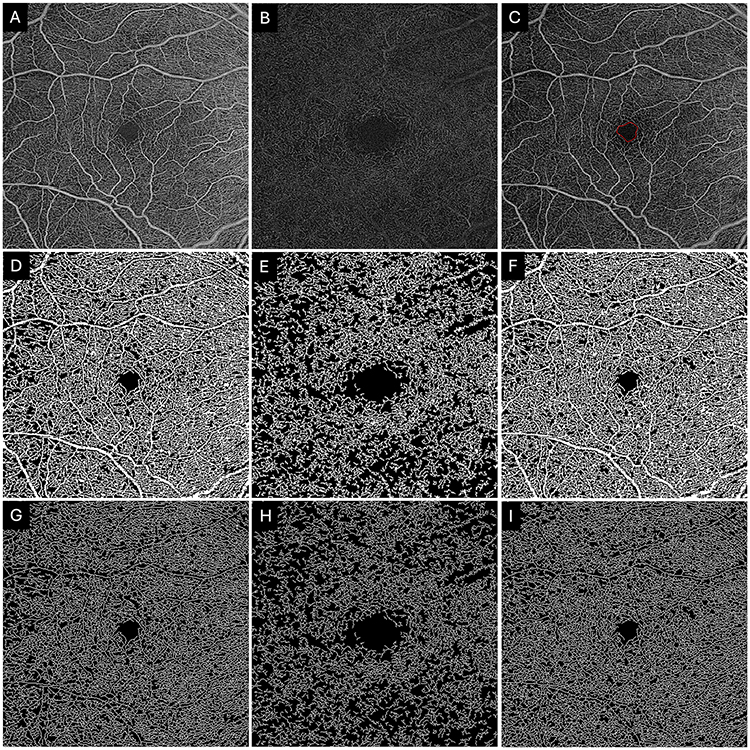 |
Figure 1 Representative original OCTA images (A–C) and processes images by Advanced Research and Innovation (ARI) Network for vascular metrics calculations (D–I) were shown. (A-C) Original Angio 6mm×6mm OCTA en face images showing the Superficial (A), Deep (B), and Retina (C) slabs, respectively. The foveal avascular zone (FAZ) was delineated in red in the Retina slab (C). (D–F) Binarized perfusion maps generated by the Advanced Research and Innovation (ARI) Network, where white pixels represent perfused vasculature, utilized for calculating vessel density (VD). (G–I) Corresponding skeletonized vessel maps (vessel trace) used to calculate vessel skeleton density (VSD) by reducing vascular segments to 1-pixel width, thereby minimizing the influence of vessel diameter on density metrics. |
Statistical Analysis
Statistical analyses were performed using R version 4.4.2. Change in vascular metrics between baseline and last follow-up were compared between the two groups using t-test (for normally distributed data) or Mann–Whitney U-test (for non-normally distributed data). Correlation between change in vascular metrics and visual acuity was investigated by Spearman’s Rank Correlation test. A 2-tailed p value of less than 0.05 was considered statistically significant for all analysis.
Results
A total of 164 eyes from 107 patients were included. The average follow-up time was 34 months with a range of 18 to 69 months. Patients in the non-injection group were slightly younger than those in the injection group (71.36±6.38 vs. 74.26±5.71, p=0.005). No other statistically significant difference in demographics or baseline vascular metrics was detected between the injection group (33 participants, 57 eyes) and non-injection (74 participants,107 eyes) group (Table 1). For the non-injection group, 25 were early AMD, 74 were intermediate AMD and 8 were late-stage non-exudative AMD at baseline. Majority of the injection group were exudative AMD at baseline and a total of eight eyes turned from intermediate AMD to exudative AMD during follow-up were included in the injection group. The injection group received 12.56 (95% CI: 10.31 to 14.81) injections during follow-up on average.
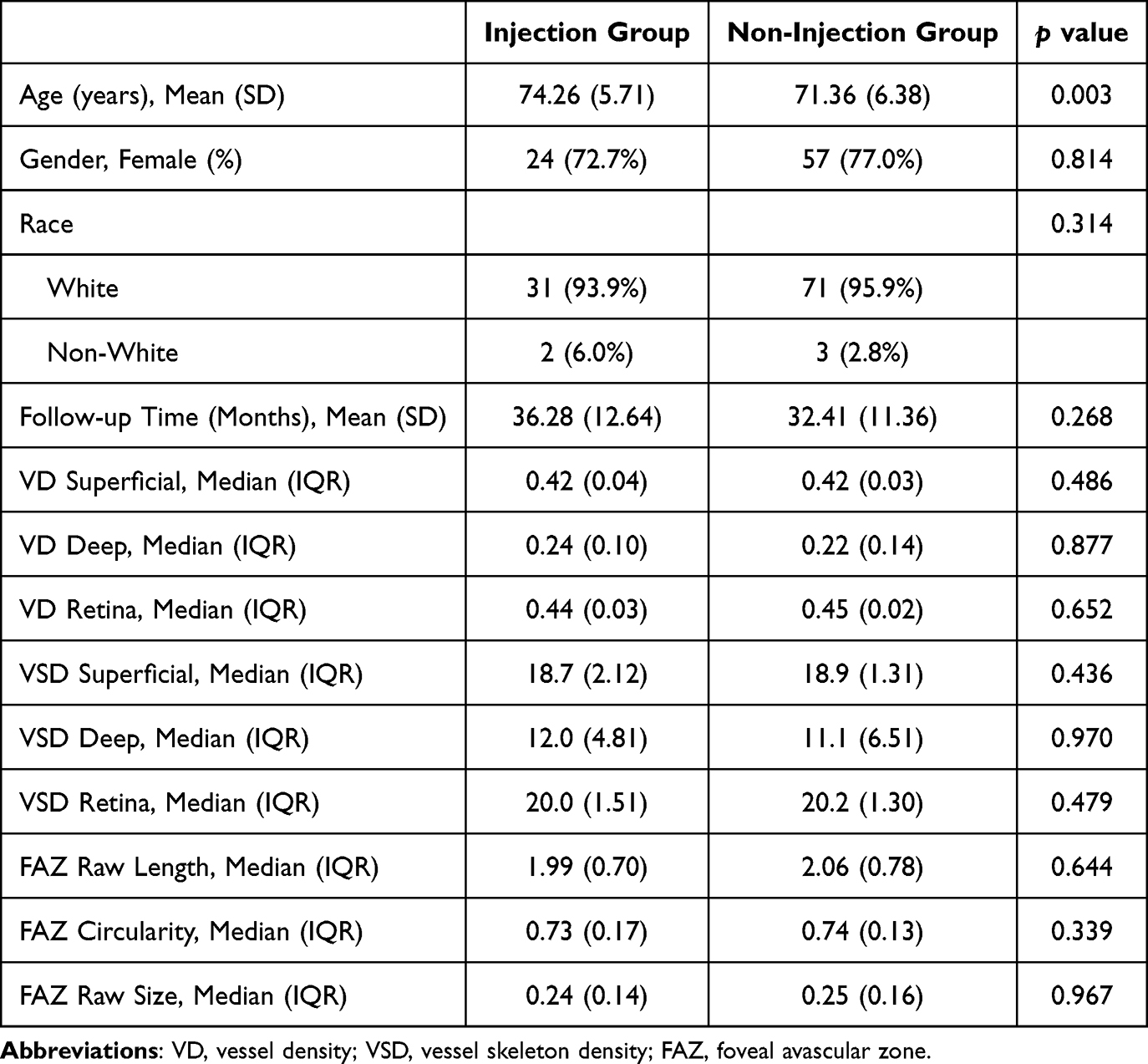 |
Table 1 Demographics and Baseline Vascular Metrics Between Injection and Non-Injection Group |
The differences between follow-up and baseline vascular metrics of Angio 6 mm×6 mm were compared between the two groups using Mann–Whitney U-test (Table 2). Among all the parameters, only FAZ size showed a statistically significant difference between the two groups (0.03 (0.01,0.09) vs. 0.02 (−0.02,0.06) mm2, p=0.043). The injection group showed a slightly larger change in FAZ area during follow-up. Slight deterioration in logarithm of the minimum angle of resolution (LogMAR) visual acuity from 0.156 (0.203) to 0.206 (0.291) was observed but not statistically significant (95% CI: −0.004 to 0.105, p=0.07). In addition, no correlation was found between change in vascular metrics and change in visual acuity (p>0.05) (Table 3).
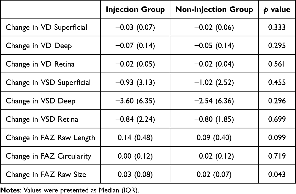 |
Table 2 Comparison of Change in Vascular Metrics from Baseline to Follow-Up Between Two Groups |
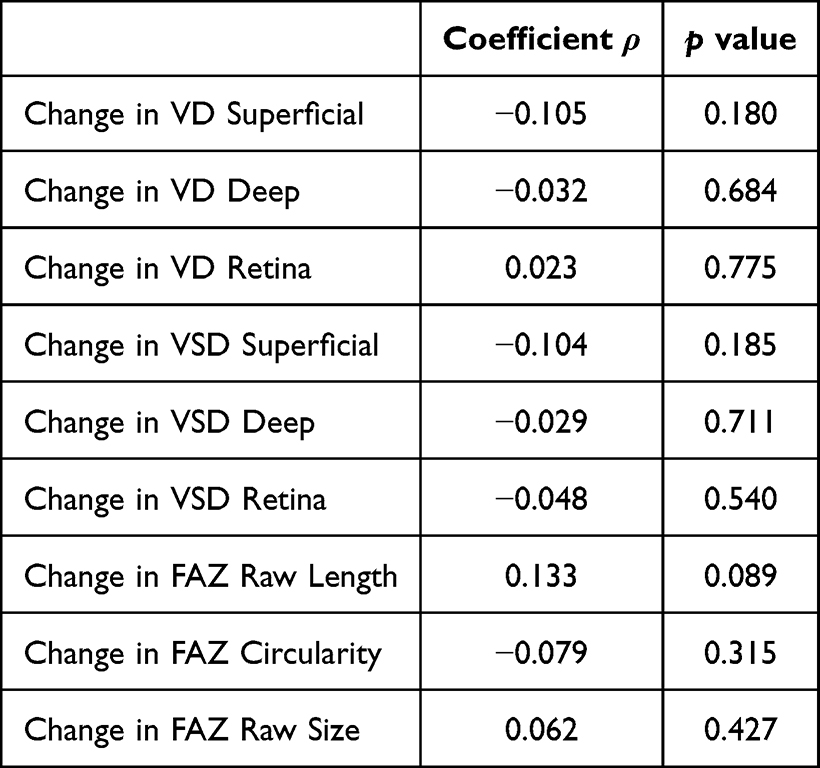 |
Table 3 Correlation Analysis Between Change in Vascular Metrics and Change in Visual Acuity |
Discussion
The present retrospective longitudinal study included 164 AMD eyes and investigated the possible influence of repeated anti-VEGF injections on OCTA vascular metrics. Only FAZ size change over time showed a statistical difference between the injection group and the non-injection group, yet the difference was small to likely not be clinically important. The results provided more evidence for the safety of long-term, repeated anti-VEGF injections in the real-world setting.
Considering the important role of VEGF in physiological angiogenesis,18 the concern over potential side-effects of long-term anti-VEGF treatments persists for both patients and clinicians. Among the variety retinal diseases receiving anti-VEGF injections, exudative AMD generally requires high frequency and long-term treatment regimen.4,19–21 Not being a retinal vascular disease itself, the pathophysiology of AMD does not affect most OCTA vascular metrics directly. These advantages make AMD a good candidate to investigate the possible influence of anti-VEGF injections on retinal perfusion.
Several cross-sectional studies reported decreased vessel density in macular or peripapillary area in anti-VEGF treated AMD eyes compared to control.22,23 Immediate decrease of retinal and choroidal perfusion after injections were observed with OCTA and laser speckle flowgraphy.24–26 However, in Lee’s cross-sectional study, a total of 9 injections on average did not impact retinal vascular measurements.27 Short-term longitudinal studies revealed mixed results of vessel density change in superficial, deep and choriocapillaris slabs.28–31 The conflicting results might be attributed to different follow-up time, scan protocol, slab chosen for analysis, control group design (including but not limited to fellow eye, healthy control eye, nonexudative AMD control). The present study provided results from a relatively large cohort with long follow-up duration. Among all the vascular metrics from superficial, deep and retina slab of Angio 6 mm × 6 mm, no vessel density or skeleton vessel density showed statistically significant differences between the injection and non-injection group. The results supported the theory that decreased perfusion due to anti-VEGF injections appears to be transient and no long-term effects were observed in a real-world clinical setting.
No differences in FAZ size, perimeter or circularity were detected between exudative and non-exudative AMD in a cross-sectional study.27 In a retrospective cohort study, FAZ size and perimetry increased over two years and no difference was found between exudative and non-exudative eyes.32 Our study echoed their findings with comparable baseline vascular metrics between the two groups. The injection group showed a statistically larger FAZ size change during follow-up compared to the non-injection group; however, the difference was only 0.01 mm2, which we do not think is clinically meaningful. Larger prospective studies are needed to further validate the differences.
No relationship between change in visual acuity and change in vascular metrics was observed during the follow-up. In the case of exudative AMD, the presence of macular neovascularization and its response to treatment were the dominant determining factor for vision, which might outweigh the vascular metrics. However, the subgroup analysis of non-exudative AMD showed no correlation either (data not shown). In Shin’s cross-sectional study for dry AMD, BCVA was correlated with vessel density in univariate linear regression, but the correlation was no longer valid in multi-variate analysis.33 Due to the retrospective nature of the current study, visual acuity was BCVA measured by Snellen chart instead of ETDRS chart, which makes it less sensitive to detect early vision change in intermediate AMD patients that constituted the majority of our non-injection group. Lee et al found BCVA could affect the repeatability of vessel density measurement in OCTA,34 which may contribute as a confounding factor in our study too.
There were several limitations of the study: 1) retrospective nature; 2) limited sample size (esp. the injection group); 3) potential selective bias due to OCTA image quality control; 4) BCVA was measured by Snellen chart instead of ETDRS chart; 5) majority of eyes in the injection group received aflibercept and not suitable for comparison between different anti-vegf agents; 6) axial length data were not acquired for calculation correction; however, the influence was limited since the key outcome measures focused on longitudinal change of the same eye.
Conclusion
In summary, this retrospective longitudinal study showed repeated intravitreal anti-VEGF injections do not impact OCTA vascular metrics significantly through the treatment course. Only FAZ size change over time showed a statistical difference between the injection group and the non-injection group, yet the difference was too small to be considered clinically relevant. Future prospective, randomized controlled clinical trials would help confirm our findings.
Acknowledgments
This paper has been uploaded to [IOVS] as a preprint: [https://iovs.arvojournals.org/article.aspx?articleid=2807724].
Author Contributions
All authors made a significant contribution to the work reported, whether that is in the conception, study design, execution, acquisition of data, analysis and interpretation, or in all these areas; took part in drafting, revising or critically reviewing the article; gave final approval of the version to be published; have agreed on the journal to which the article has been submitted; and agree to be accountable for all aspects of the work.
Funding
This work was supported by The Hamilton Company Charitable Foundation.
Disclosure
J.W.M. is a consultant for Sumitomo Pharma America, Inc. and ONL Therapeutics, LLC. J.W.M. has a patent US 7,811,832 licensed to ONL Therapeutics, various patents licensed to QLT/Bausch and Lomb (US 5,798,349; US 6,225,303; US 6,610,679; CA 2,185,644; CA 2,536,069) with royalties paid to Mass Eye and Ear and distributed per institutional policy; PDT for AMD largely supplanted by anti-VEGF therapy. She also reports grants from Lowy Medical Research Institute, Ltd., NEI R01 EY030088-01A1; Consulting fees from ONL Therapeutics, LLC, Sumitomo Pharma America, Inc.; Honoraria from Atlantic Coast Retina Conference/Macula 2026, UCLA, University of North Carolina Chapel Hill, NYU/Langone, Atlantic Coast Retina Conference/Macula 2022, Connecticut Society of Eye Physicians; Meeting/travel support from Doheny Eye Institute, NYU Grossman School of Medicine, Wills Eye Hospital, Nova Scotia Health Authority, Portuguese Society of Ophthalmology Annual National Congress; Leadership or fiduciary roles from Club Jules Gonin, Harvard Health Publishing, Drusolv Therapeutics, Ophthalmology Retina, Ophthalmology, Macula Society, Heed Ophthalmic Foundation, Association of University Professors in Ophthalmology (AUPO), Aptinyx, Inc., Massachusetts Eye and Ear Associates, Inc., Foundation of the Massachusetts Eye and Ear Infirmary; Stock or stock options from Ciendias Bio, ONL Therapeutics, LLC, and Aptinynx, Inc., outside the submitted work. N.A.P. is a consultant for Apellis, Alcon, Allergan, Alimera, Atheneum, Biogen, Dorc, Eyepoint, Genentech, Lifesciences, Guidepoint, Regeneron, and Gerson Lehrman Group, Inc. L.A.K. is part of the Scientific Advisory Board for and owns stock options from Ingenia Therapeutics and Pykus Therapeutics; he also has a patent “compositions and methods for the treatment of aberrant angiogenesis” licensed to X1 Biotech. J.B.M. is a consultant for Alcon, Allergan, Carl Zeiss, Sunovion, Topcon, and Genentech. The authors report no other conflicts of interest in this work.
References
1. Fleckenstein M, Schmitz-Valckenberg S, Chakravarthy U. Age-related macular degeneration: a review. JAMA. 2024;331:147–8. doi:10.1001/jama.2023.26074
2. Rein DB, Wittenborn JS, Burke-Conte Z, et al. Prevalence of age-related macular degeneration in the US in 2019. JAMA Ophthalmol. 2022;140:1202–1208. doi:10.1001/jamaophthalmol.2022.4401
3. Paudel N, Brady L, Stratieva P, et al. Economic burden of late-stage age-related macular degeneration in Bulgaria, Germany, and the US. JAMA Ophthalmol. 2024;142:1123–1130. doi:10.1001/jamaophthalmol.2024.4401
4. Sunaga T, Maeda M, Saulle R, et al. Anti-vascular endothelial growth factor biosimilars for neovascular age-related macular degeneration. Cochrane Database Syst Rev. 2024;6:Cd015804. doi:10.1002/14651858.CD015804.pub2
5. Arpa C, Khalid H, Chandra S, et al. Ten-year survival trends of neovascular age-related macular degeneration at first presentation. Br J Ophthalmol. 2021;105:1688–1695. doi:10.1136/bjophthalmol-2020-317161
6. Perez-Gutierrez L, Ferrara N. Biology and therapeutic targeting of vascular endothelial growth factor A. Nat Rev Mol Cell Biol. 2023;24:816–834. doi:10.1038/s41580-023-00631-w
7. Apte RS, Chen DS, Ferrara N. VEGF in signaling and disease: beyond discovery and development. Cell. 2019;176:1248–1264. doi:10.1016/j.cell.2019.01.021
8. Laíns I, Wang JC, Cui Y, et al. Retinal applications of swept source optical coherence tomography (OCT) and optical coherence tomography angiography (OCTA). Prog Retin Eye Res. 2021;84:100951. doi:10.1016/j.preteyeres.2021.100951
9. Wang JC, Miller JB. Optical coherence tomography angiography: review of current technical aspects and applications in chorioretinal disease. Semin Ophthalmol. 2019;34:211–217. doi:10.1080/08820538.2019.1620797
10. Spaide RF, Fujimoto JG, Waheed NK, Sadda SR, Staurenghi G. Optical coherence tomography angiography. Prog Retin Eye Res. 2018;64:1–55. doi:10.1016/j.preteyeres.2017.11.003
11. Cui Y, Zhu Y, Lu ES, et al. Widefield swept-source oct angiography metrics associated with the development of diabetic vitreous hemorrhage: a prospective study. Ophthalmology. 2021;128:1312–1324. doi:10.1016/j.ophtha.2021.02.020
12. Ding X, Romano F, Garg I, et al. Longitudinal assessment of intraretinal microvascular abnormalities in diabetic retinopathy using swept-source optical coherence tomography angiography. Invest Ophthalmol Vis Sci. 2024;65:29. doi:10.1167/iovs.65.8.29
13. Rudnick ND, Vingopoulos F, Wang JC, et al. Characterising collateral vessels in eyes with branch retinal vein occlusions using widefield swept-source optical coherence tomography angiography. Br J Ophthalmol. 2023;107:1887–1891. doi:10.1136/bjo-2021-320356
14. Moon JY, Garg I, Cui Y, et al. Wide-field swept-source optical coherence tomography angiography in the assessment of retinal microvasculature and choroidal thickness in patients with myopia. Br J Ophthalmol. 2023;107:102–108. doi:10.1136/bjophthalmol-2021-319540
15. Romano F, Ding X, Yuan M, et al. Progressive choriocapillaris changes on optical coherence tomography angiography correlate with stage progression in AMD. Invest Ophthalmol Vis Sci. 2024;65:21. doi:10.1167/iovs.65.8.21
16. Garg I, Uwakwe C, Le R, et al. Nonperfusion area and other vascular metrics by wider field swept-source oct angiography as biomarkers of diabetic retinopathy severity. Ophthalmol Sci. 2022;2. 10.1016/j.xops.2022.100144
17. Marques IP, Ribeiro ML, Santos T, et al. Patterns of progression of nonproliferative diabetic retinopathy using non-invasive imaging. Transl Vis Sci Technol. 2024;13:22. doi:10.1167/tvst.13.5.22
18. Ferrara N. Role of vascular endothelial growth factor in regulation of physiological angiogenesis. Am J Physiol Cell Physiol. 2001;280:C1358–66.
19. Virgili G, Curran K, Lucenteforte E, Peto T, Parravano M. Anti-vascular endothelial growth factor for diabetic macular oedema: a network meta-analysis. Cochrane Database Syst Rev. 2023;2023:Cd007419. doi:10.1002/14651858.CD007419.pub7
20. Shalchi Z, Mahroo O, Bunce C, Mitry D. Anti-vascular endothelial growth factor for macular oedema secondary to branch retinal vein occlusion. Cochrane Database Syst Rev. 2020;7:Cd009510. doi:10.1002/14651858.CD009510.pub3
21. Ohno-Matsui K, Ikuno Y, Lai TYY, Gemmy Cheung CM. Diagnosis and treatment guideline for myopic choroidal neovascularization due to pathologic myopia. Prog Retin Eye Res. 2018;63:92–106. doi:10.1016/j.preteyeres.2017.10.005
22. Türksever C, Hoffmann L, Hatz K. Peripapillary and macular microvasculature in neovascular age-related macular degeneration in long-term and recently started anti-VEGF therapy versus healthy controls. Front Med. 2022;9:1080052. doi:10.3389/fmed.2022.1080052
23. Resch MD, Balogh A, Kurth T, Nagy ZZ, DeBuc DC, Papp A. Atrophy of retinal vessels in neovascular age-related macular degeneration following long-term treatment with 20 intravitreal anti-VEGF injections. BMC Ophthalmol. 2022;22:469. doi:10.1186/s12886-022-02700-8
24. Pilotto E, Frizziero L, Daniele AR, et al. Early OCT angiography changes of type 1 CNV in exudative AMD treated with anti-VEGF. Br J Ophthalmol. 2019;103:67–71. doi:10.1136/bjophthalmol-2017-311752
25. Mursch-Edlmayr AS, Luft N, Podkowinski D, Ring M, Schmetterer L, Bolz M. Effects of three intravitreal injections of aflibercept on the ocular circulation in eyes with age-related maculopathy. Br J Ophthalmol. 2020;104:53–57. doi:10.1136/bjophthalmol-2019-313919
26. Kato N, Haruta M, Furushima K, Arai R, Matsuo Y, Yoshida S. Decrease in ocular blood flow thirty minutes after intravitreal injections of brolucizumab and aflibercept for neovascular age-related macular degeneration. Clin Ophthalmol. 2023;17:1187–1192. doi:10.2147/OPTH.S407249
27. Lee SC, Tran S, Amin A, et al. Retinal vessel density in exudative and nonexudative age-related macular degeneration on optical coherence tomography angiography. Am J Ophthalmol. 2020;212:7–16. doi:10.1016/j.ajo.2019.11.031
28. Cennamo G, Montorio D, D’Alessandro A, Napolitano P, D’Andrea L, Tranfa F. Prospective study of vessel density by optical coherence tomography angiography after intravitreal bevacizumab in exudative age-related macular degeneration. Ophthalmol Ther. 2020;9:77–85. doi:10.1007/s40123-019-00221-0
29. Hikichi T, Agarie M. Reduced vessel density of the choriocapillaris during anti-vascular endothelial growth factor therapy for neovascular age-related macular degeneration. Invest Ophthalmol Vis Sci. 2019;60:1088–1095. doi:10.1167/iovs.18-24522
30. Park YS, Moon H, Woo JM, Min JK. Changes in the foveal avascular zone area and retinal vessel density after anti-vegf therapy for neovascular age-related macular degeneration. Semin Ophthalmol. 2021;36:110–114. doi:10.1080/08820538.2021.1889612
31. Resch MD, Balogh A, Deák GG, Nagy ZZ, Papp A. Vascular density in age-related macular degeneration after one year of antiVEGF treatment with treat-and-extend and fixed regimens. PLoS One. 2020;15:e0229388. doi:10.1371/journal.pone.0229388
32. Lee SC, Rusakevich AM, Amin A, et al. Long-term retinal vascular changes in age-related macular degeneration measured using optical coherence tomography angiography. Ophthalmic Surg Lasers Imaging Retina. 2022;53:529–536. doi:10.3928/23258160-20220919-01
33. Shin YI, Kim JM, Lee MW, Jo YJ, Kim JY. Characteristics of the foveal microvasculature in asian patients with dry age-related macular degeneration: an optical coherence tomography angiography study. Ophthalmologica. 2020;243:145–153. doi:10.1159/000503295
34. Lee MW, Kim KM, Lim HB, Jo YJ, Kim JY. Repeatability of vessel density measurements using optical coherence tomography angiography in retinal diseases. Br J Ophthalmol. 2018. doi:10.1136/bjophthalmol-2018-312516
 © 2026 The Author(s). This work is published and licensed by Dove Medical Press Limited. The
full terms of this license are available at https://www.dovepress.com/terms
and incorporate the Creative Commons Attribution
- Non Commercial (unported, 4.0) License.
By accessing the work you hereby accept the Terms. Non-commercial uses of the work are permitted
without any further permission from Dove Medical Press Limited, provided the work is properly
attributed. For permission for commercial use of this work, please see paragraphs 4.2 and 5 of our Terms.
© 2026 The Author(s). This work is published and licensed by Dove Medical Press Limited. The
full terms of this license are available at https://www.dovepress.com/terms
and incorporate the Creative Commons Attribution
- Non Commercial (unported, 4.0) License.
By accessing the work you hereby accept the Terms. Non-commercial uses of the work are permitted
without any further permission from Dove Medical Press Limited, provided the work is properly
attributed. For permission for commercial use of this work, please see paragraphs 4.2 and 5 of our Terms.