Back to Journals » International Medical Case Reports Journal » Volume 18
Hypertriglyceridemia-Induced Acute Pancreatitis with Lipemic Samples in a Type 2 Diabetic Patient: A Case Report in a Resource-Limited Setting
Authors Ahmed HM, Osman MH, Hassan SA ![]() , Dirie HM, Mohamed MA
, Dirie HM, Mohamed MA
Received 23 January 2025
Accepted for publication 28 May 2025
Published 30 May 2025 Volume 2025:18 Pages 645—649
DOI https://doi.org/10.2147/IMCRJ.S516349
Checked for plagiarism Yes
Review by Single anonymous peer review
Peer reviewer comments 2
Editor who approved publication: Dr Gates Colbert
Hussein Mahdi Ahmed,1 Mohamed Hassan Osman,1,2 Shafie Abdulkadir Hassan,2 Hassan Mohamud Dirie,1 Mowlid Abdikarin Mohamed2
1Department of Internal Medicine, Royal Hospital, Mogadishu, Somalia; 2Faculty of Medicine and Health Sciences, Jamhuriya University of Science and Technology, Mogadishu, Somalia
Correspondence: Shafie Abdulkadir Hassan, Faculty of Medicine and Health Sciences, Jamhuriya University of Science and Technology, Hodon District, Mogadishu, Somalia, Tel +252615206907, Email [email protected]
Background: Hypertriglyceridemia (HTG) is a known but relatively uncommon cause of acute pancreatitis (AP), accounting for approximately 1– 7% of cases. Hypertriglyceridemia-induced acute pancreatitis (HTG-AP) can lead to significant morbidity if not promptly identified and managed. This case report describes a patient with poorly controlled type 2 diabetes mellitus (T2DM) who presented with HTG-AP, characterized by a lipemic blood sample, in a resource-limited setting.
Case Presentation: A 45-year-old male with a history of poorly controlled T2DM and hyperlipidemia presented with a 24-hour history of severe epigastric abdominal pain, fatigue, and vomiting. Clinical examination revealed diffuse abdominal tenderness, tachypnea, tachycardia, and a habitus consistent with central obesity. His BMI was 33.2 kg/m². Initial laboratory findings included seriously elevated triglycerides (1509 mg/dL), lipase (83 U/L), and amylase (161 U/L), along with hyperglycemia (465mg/dL). Abdominal computed tomography (CT) scan showed peripancreatic fatty stranding, consistent with early acute pancreatitis, as well as a fatty liver and a focal hypodense lesion in the right lobe. Treatment included intravenous insulin, dextrose, and potassium infusions to reduce triglyceride levels, analgesics, intravenous fluids for electrolyte imbalances, and thromboprophylaxis with enoxaparin.
Conclusion: This case highlights the importance of early recognition of HTG-AP in patients with poorly controlled diabetes and hyperlipidemia. Prompt triglyceride-lowering therapy, primarily with insulin in resource-limited settings, is crucial for improving patient outcomes and preventing complications.
Keywords: hypertriglyceridemia, acute pancreatitis, diabetes mellitus, triglyceride-lowering therapy, lipemia, case report
Introduction
Hypertriglyceridemia-induced acute pancreatitis (HTG-AP) is a relatively uncommon cause of acute pancreatitis, typically occurring when serum triglyceride levels exceed 1000 mg/dL (11.3 mmol/L).1,2 The pathophysiology of HTG-AP involves the hydrolysis of excess triglycerides within pancreatic capillaries by pancreatic lipase, releasing high concentrations of toxic free fatty acids (FFAs).3 These FFAs subsequently induce inflammation, ischemia, and tissue damage.1,4 While it accounts for a small percentage of all acute pancreatitis cases, HTG-AP can lead to substantial morbidity and mortality if not promptly managed.5,6
Common risk factors associated with HTG-AP include poorly controlled T2DM and underlying hyperlipidemia.7,8 It is also well-established that patients with diabetes have a higher risk of acquiring infections.9 This case report describes an instance involving a 45-year-old man with poorly controlled T2DM and a history of hyperlipidemia who developed HTG-AP. A key diagnostic indicator during his presentation was the lipemic blood sample; indeed, only the types of HTG that lead to a lipemic sample (secondary to markedly increased chylomicrons) are typically implicated in acute pancreatitis.10,11 This case underscores the importance of early recognition and rapid treatment initiation, especially considering challenges such as managing multiple comorbidities and potential resource limitations. Furthermore, this report contributes to the literature by demonstrating a successful outcome using insulin-based therapy as a primary modality when plasmapheresis is unavailable, offering practical insights for clinical practice in similar settings.
Case Presentation
A 45-year-old male with a history of type 2 diabetes mellitus presented with a 24-hour history of vomiting, fatigue, and severe epigastric abdominal pain. He had a habitus of central obesity, and his body mass index (BMI) was 33.2 kg/m². He exhibited tachypnea, tachycardia, and diffuse abdominal tenderness upon clinical examination. Initial laboratory analysis showed seriously elevated triglycerides (1509 mg/dL), lipase (83 U/L), and amylase (161 U/L), and these findings were accompanied by hyperglycemia (465mg/dL). Follow-up evaluations were performed at intervals of 2 days, 4 weeks (28 days), and approximately 3.5 months (110 days) after the initial assessment, as detailed in Table 1. An abdominal computed tomography (CT) scan revealed peripancreatic fatty stranding, which was consistent with early acute pancreatitis, as shown in Figure 1, alongside the identification of a fatty liver and a focal hypodense lesion in the right lobe. Furthermore, the pronounced lipemic appearance of the patient’s blood samples, immediately indicative of hypertriglyceridemia with chylomicronemia, is presented in Figure 2. The patient’s treatment involved an intravenous infusion of adjustable short-acting insulin (Actrapid) accompanied by dextrose and potassium replacement (80 mEq TDS for the first 3 days) aimed at lowering triglyceride levels. Pain control was initiated with paracetamol (1g TDS on day 1), and aggressive intravenous fluid therapy was administered, starting with 6 liters of crystalloids (Normal Saline and Ringer’s Lactate) on the first day, followed by maintenance fluids (500 mL every 6 hours for the next 3 days) to correct dehydration and electrolyte imbalances. Furthermore, he received standard thromboprophylaxis with enoxaparin to prevent venous thromboembolism (VTE). His total hospital admission lasted 9 days. After stabilization, the patient recovered and was discharged with a comprehensive follow-up plan.
 |
Table 1 Key Laboratory Findings in a Patient with Hypertriglyceridemia-Induced Acute Pancreatitis |
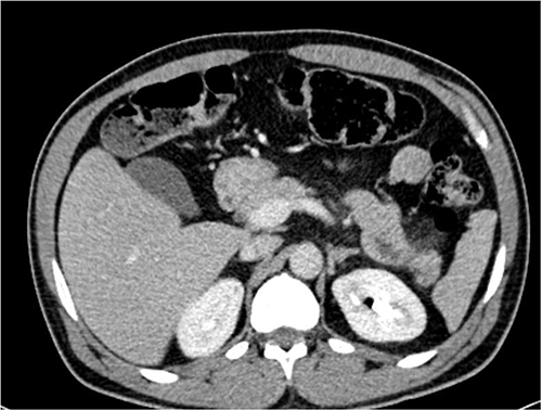 |
Figure 1 A non-contrast abdominal CT scan revealed peri-pancreatitis fatty stranding of the pancreas. |
 |
Figure 2 Lipemic blood samples were drawn from the cephalic vein. |
Discussion
This case demonstrates a typical presentation of HTG-AP in a patient with poorly controlled T2DM and hyperlipidemia. The patient had a history of vomiting, fatigue, and epigastric abdominal pain. Clinical examination revealed tachypnea, tachycardia, and diffuse abdominal tenderness; notably, the patient lacked xanthomas, which are a predictive sign of familial and secondary hypertriglyceridemia (HTG) complications such as pancreatitis. The absence of xanthomas can contribute to a delay in the diagnosis of HTG-AP.10
Diagnosing hypertriglyceridemia-induced acute pancreatitis (HTG-AP) can be challenging due to the potential interference of elevated triglyceride (TG) levels (above 500 mg/dL) with both lipase and amylase activity.7 Specifically, excessive TG levels, causing lipemic serum, can interfere with the colorimetric testing of amylase, potentially leading to deceptively normal results.12 Despite this potential for misleading enzyme levels, the presence of lipemic serum itself is a critical finding that should focus the clinician on the likely presence of chylomicronemia characteristic of severe HTG and hyperlipidemia.
In this case, the patient’s initial amylase values were within the normal range, which is due to the interference of elevated triglyceride (TG) levels that were severely elevated at 1509 mg/dL, with a moderately elevated lipase of 83 mg/dL. These levels, while substantial, were lower than those reported in previous case reports.7,13
The CT imaging further confirmed the presence of acute pancreatitis and associated fatty changes. The pathophysiology of HTG-AP involves high triglyceride levels leading to the production of excessive FFAs, which induce inflammation and pancreatic damage.14,15 This process is further exacerbated by conditions such as T2DM and obesity, which promote insulin resistance and hyperglycemia, leading to increased lipogenesis.16–18
In this case, the patient’s presentation aligns with literature data showing a strong association between HTG, metabolic disorders, and AP.2,7,19 This patient lacked the xanthomas, typical signs of HTG complications, leading to potential diagnostic delay.20 Severe hypertriglyceridemia causing hyperlipemic serum presents a diagnostic challenge, as it can interfere with key laboratory assays used for the diagnosis of acute pancreatitis; this may lead clinicians to overlook HTG-associated acute pancreatitis despite relatively low or normal serum amylase values.
In resource-limited settings, plasmapheresis, a standard therapy for severe HTG-AP, may not be available. In this case, an insulin infusion with dextrose and potassium proved to be a practical and effective alternative for the rapid reduction of triglycerides.
This case demonstrates the successful management of hypertriglyceridemia-induced acute pancreatitis (HTG-AP) through the timely administration of insulin therapy within a resource-limited setting. The case underscores the crucial importance of early recognition of HTG-AP, particularly in patients with type 2 diabetes mellitus (T2DM) presenting with abdominal pain. Furthermore, the presence of lipemic samples serves as a readily available and immediate visual diagnostic clue, prompting consideration of HTG as a potential cause of acute pancreatitis (AP).
Clinicians should also remain mindful of potential diagnostic challenges, as high triglyceride levels may interfere with enzyme activity assays. In situations where plasmapheresis is not accessible, insulin can be a highly effective and cost-efficient intervention for reducing triglyceride levels in HTG-AP. Additionally, close monitoring of laboratory values during the treatment phase is paramount to ensure optimal patient outcomes. Finally, this case aligns with existing literature, further emphasizing the pressing need for standardized protocols to guide the management of HTG-AP.
Conclusion
This case underscores the clinical significance of HTG-AP in patients with poorly controlled T2DM and hyperlipidemia. Early suspicion of HTG-AP in patients presenting with acute T2DM, hyperlipidemia, and lipemic serum. In settings with limited resources, intravenous insulin infusion with dextrose and potassium can effectively manage HTG-AP. This report emphasizes the need for early diagnosis, vigilant lipid monitoring, and good glycemic control to prevent such complications. Future research should focus on the development of standardized, resource-adapted protocols for managing HTG-AP, ensuring that all patients, especially those in resource-limited environments, have access to effective care.
Ethical Considerations
This case report was conducted with ethical approval obtained from the Ethics Committee of Jamhuriya University of Science and Technology.
Consent for Publication
Informed consent was obtained from the patient for the publication of this case.
Author Contributions
All authors made a significant contribution to the work reported, whether that is in the conception, study design, execution, acquisition of data, analysis and interpretation, or all these areas; took part in drafting, revising or critically reviewing the article; gave final approval of the version to be published; have agreed on the journal to which the article has been submitted; and agree to be accountable for all aspects of the work.
Disclosure
The authors report no conflicts of interest in this work.
References
1. Garg R, Rustagi T. Management of hypertriglyceridemia induced acute pancreatitis. Biomed Res Int. 2018;2018(1):4721357. doi:10.1155/2018/4721357
2. Melnick S, Nazir S, Gish D, Aryal MR. Hypertriglyceridemic pancreatitis associated with confounding laboratory abnormalities. J Community Hosp Intern Med Perspect. 2016;6(3):31808. doi:10.3402/jchimp.v6.31808
3. Kiss L, Fűr G, Pisipati S, et al. Mechanisms linking hypertriglyceridemia to acute pancreatitis. Acta Physiol. 2023;237(3):1–21. doi:10.1111/apha.13916
4. Qiu M, Zhou X, Zippi M, et al. Comprehensive review on the pathogenesis of hypertriglyceridaemia-associated acute pancreatitis. Ann Med. 2023;55(2):2265939. doi:10.1080/07853890.2023.2265939
5. Kumar BGV, Prasad K, Singh D, Sethy PC. Hypertriglyceridemia induced acute pancreatitis: 4 years’ experience from a tertiary care institute and quick literature review. J Fam Med Prim care. 2022;11(6):3360–3367. doi:10.4103/jfmpc.jfmpc_1426_21
6. Aldhaleei WA, Alnuaimi A, Bhagavathula AS. Hypertriglyceridemia-induced acute pancreatitis in a patient with type 2 diabetes mellitus. Cureus. 2020;12(7):e9414. doi:10.7759/cureus.9414
7. Aldhaleei WA, Alnuaimi A, Bhagavathula AS. Hypertriglyceridemia–induced acute pancreatitis in a patient with type 2 diabetes mellitus. Cureus. 2020;12(7):10–13. doi:10.7759/cureus.9414
8. Valdivielso P, Ramírez-Bueno A, Ewald N. Current knowledge of hypertriglyceridemic pancreatitis. Eur J Intern Med. 2014;25(8):689–694. doi:10.1016/j.ejim.2014.08.008
9. Hassan SA, Ahmed YMA, Hassan GD. Antimicrobial susceptibility of Escherichia coli isolated from diabetic patients in Mogadishu, Somalia. Front Microbiol. 2023;14:1–6. doi:10.3389/fmicb.2023.1204052
10. Van Elslande J, Hijjit S, De Vusser K, et al. Delayed diagnosis and treatment of extreme hypertriglyceridemia due to rejection of a lipemic sample. Biochem Medica. 2021;31(2):21002. doi:10.11613/BM.2021.021002
11. Joury A, Alshehri M, Mahendra A, Anteet M, Yousef MA, Khan AM. Therapeutic approaches in hypertriglyceridemia-induced acute pancreatitis: a literature review of available therapies and case series. J Clin Apher. 2020;35(2):131–137. doi:10.1002/jca.21763
12. Aryal MR, Mainali NR, Gupta S, Singla M. Acute pancreatitis owing to very high triglyceride levels treated with insulin and heparin infusion. BMJ Case Rep. 2013;2013:bcr2013008550. doi:10.1136/bcr-2013-008550
13. Chen WW, Yang Q, Li XY, et al. Identification of a novel and heterozygous LMF1 nonsense mutation in an acute pancreatitis patient with severe hypertriglyceridemia, severe obesity and heavy smoking. Lipids Health Dis. 2019;18(1):68. doi:10.1186/s12944-019-1012-9
14. Hidalgo NJ, Pando E, Alberti P, et al. The role of high serum triglyceride levels on pancreatic necrosis development and related complications. BMC Gastroenterol. 2023;23(1):51. doi:10.1186/s12876-023-02684-9
15. Song K, Wu Z, Meng J, et al. Hypertriglyceridemia as a risk factor for complications of acute pancreatitis and the development of a severity prediction model. HPB. 2023;25(9):1065–1073. doi:10.1016/j.hpb.2023.05.006
16. Ma M, Liu H, Yu J, et al. Triglyceride is independently correlated with insulin resistance and islet beta cell function: a study in population with different glucose and lipid metabolism states. Lipids Health Dis. 2020;19(1):121. doi:10.1186/s12944-020-01303-w
17. Subramanian S, Chait A. Hypertriglyceridemia secondary to obesity and diabetes. Biochim Biophys Acta Mol Cell Biol Lipids. 2012;1821(5):819–825. doi:10.1016/j.bbalip.2011.10.003
18. Mathuram Thiyagarajan U, Ponnuswamy A, Chung A. An enigmatic triad of acute pancreatitis, diabetic ketoacidosis and hypertriglyceridaemia: who is the culprit? BMJ Case Rep. 2019;12(7):10–12. doi:10.1136/bcr-2016-217272
19. Yang AL, McNabb-Baltar J. Hypertriglyceridemia and acute pancreatitis. Pancreatology. 2020;20(5):795–800. doi:10.1016/j.pan.2020.06.005
20. Lee SY, Sheth CA. Eruptive xanthoma associated with severe hypertriglyceridemia and poorly controlled type 1 diabetes mellitus. J Community Hosp Intern Med Perspect. 2019;9(4):344–346. doi:10.1080/20009666.2019.1650591
 © 2025 The Author(s). This work is published and licensed by Dove Medical Press Limited. The
full terms of this license are available at https://www.dovepress.com/terms
and incorporate the Creative Commons Attribution
- Non Commercial (unported, 4.0) License.
By accessing the work you hereby accept the Terms. Non-commercial uses of the work are permitted
without any further permission from Dove Medical Press Limited, provided the work is properly
attributed. For permission for commercial use of this work, please see paragraphs 4.2 and 5 of our Terms.
© 2025 The Author(s). This work is published and licensed by Dove Medical Press Limited. The
full terms of this license are available at https://www.dovepress.com/terms
and incorporate the Creative Commons Attribution
- Non Commercial (unported, 4.0) License.
By accessing the work you hereby accept the Terms. Non-commercial uses of the work are permitted
without any further permission from Dove Medical Press Limited, provided the work is properly
attributed. For permission for commercial use of this work, please see paragraphs 4.2 and 5 of our Terms.