Back to Journals » International Medical Case Reports Journal » Volume 19
Hyperbaric Oxygen Therapy in Postoperative Intracranial Pneumocephalus After Lumbar Decompression: A Case Study
Authors Chen Z, Xiang Z, Wang P, Zhang M, Yan M, Wang X, Hu B ![]()
Received 11 January 2026
Accepted for publication 10 March 2026
Published 12 March 2026 Volume 2026:19 595460
DOI https://doi.org/10.2147/IMCRJ.S595460
Checked for plagiarism Yes
Review by Single anonymous peer review
Peer reviewer comments 2
Editor who approved publication: Dr Tanvi Dhere
ZhiWu Chen,* Zhong Xiang,* Pan Wang, Meng Zhang, MengYao Yan, XingYi Wang, Bin Hu
Department of Orthopaedics, The Fourth Hospital of Changsha, Chang Sha Hospital of Hunan Normal University, Changsha, People’s Republic of China
*These authors contributed equally to this work
Correspondence: Bin Hu, Department of Orthopaedics, The Fourth Hospital of Changsha, Changsha Hospital of Hunan Normal University, Changsha, People’s Republic of China, Email [email protected]
Abstract: Intracranial pneumocephalus is an extremely rare complication following posterior lumbar surgery. We present a case of a 67-year-old female who underwent posterior lumbar interbody fusion (PLIF) at L4/5 for lumbar disc herniation. During the procedure, severe adhesions between the ligamentum flavum and dura mater were encountered, resulting in an inadvertent dural tear with cerebrospinal fluid (CSF) leakage. Six hours postoperatively, the patient abruptly developed dysarthria, expressive aphasia, and right-sided hemiparesis. Emergency cranial computed tomography revealed pneumocephalus predominantly in the bilateral frontotemporal sulci and suprasellar cistern, with greater involvement on the right side. Conservative management, including strict bed rest, intravenous hydration, analgesia, and hyperbaric oxygen therapy (HBOT), was initiated, leading to gradual neurological recovery. This case highlights the clinical presentation, management, and outcomes of this rare complication, with emphasis on the potential role of HBOT in treatment.
Keywords: posterior lumbar surgery, intracranial pneumocephalus, dural injury, cerebrospinal fluid leakage, hyperbaric oxygen therapy
Introduction
Intracranial pneumocephalus, a known complication of craniofacial trauma and cranial surgery, is exceptionally rare following spinal procedures. It typically results from an incidental dural tear, with air entering the intracranial space via mechanisms such as a one-way valve. Although reported sporadically after cervical, thoracic, or lumbar surgery, it remains under-recognized in spinal surgery, and management is not standardized. Hyperbaric oxygen therapy (HBOT) has been utilized in other etiologies of pneumocephalus but is rarely documented in post-spinal surgery cases. We present a rare case of symptomatic tension pneumocephalus after posterior lumbar interbody fusion (PLIF) due to a small dural tear. The patient developed acute neurological decline that resolved completely after emergent HBOT. This report highlights pneumocephalus as a critical differential in acute postoperative neurological deterioration and discusses the potential role of HBOT in its management.
Case Report
A 67-year-old woman presented with a 3-month history of low back pain and right lower extremity radiculopathy. Her medical history included a right L5/S1 discectomy and hemilaminectomy performed 20 years earlier. Physical examination revealed limited lumbar range of motion, tenderness at the L4/5 interspinous space with radiation to the right lower extremity, and a positive right straight leg raise test at 40° with reinforcement. CT and MRI confirmed a right posterolateral L4/5 disc herniation with thecal sac compression and right nerve root impingement (Figure 1). Preoperative evaluations were unremarkable. After unsuccessful conservative treatment, she underwent posterior lumbar interbody fusion (PLIF) at L4/5. Intraoperatively, severe adhesions between the ligamentum flavum and dura mater were encountered, and a small incidental dural tear (approximately 2 mm) occurred during adhesiolysis. Following discectomy and interbody cage placement, hemostasis was achieved. A closed suction drain was placed prior to wound closure.
 |
Figure 1 Preoperative MRI demonstrates right posterocentral disc herniation at L4/5 level. Arrows indicate the herniated disc. |
The patient was initially stable and asymptomatic following anesthesia. At 6 hours postoperatively, she acutely developed dysarthria, expressive aphasia, and right hemiparesis. Neurological examination demonstrated right hemiparesis with Medical Research Council scores of 1/5 in the upper extremity and 2/5 in the lower extremity, alongside preserved tone and intact sensation. Emergency cranial CT (Figure 2) revealed pneumocephalus, predominantly in the bilateral frontotemporal sulci and suprasellar cistern (right > left). At 10 hours postoperation, emergent hyperbaric oxygen therapy (HBOT) was initiated (2.5 ATA for 90 minutes), combined with strict supine positioning, intravenous dexamethasone (10 mg q12h), mannitol (125 mL q8h) for cerebral edema, volume replacement, and analgesia. By 18 hours, dysarthria and mental status deterioration persisted, though right upper extremity strength had improved to 4+/5. A second HBOT session was repeated at 26 hours using identical parameters. At 28 hours postoperation, speech disturbances had completely resolved. Follow-up cranial CT (Figure 3) showed complete resolution of the pneumocephalus.
 |
Figure 2 Postoperative cranial CT following PLIF demonstrates pneumocephalus predominantly involving bilateral frontotemporal sulci and suprasellar cistern (right > left). Arrows indicate the presence of intracranial pneumocephalus. |
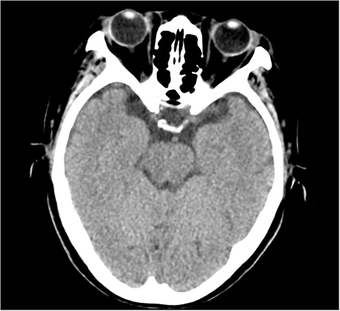 |
Figure 3 Follow-up cranial CT after two HBOT sessions demonstrates complete resolution of previously noted pneumocephalus in bilateral frontotemporal sulci and suprasellar cistern. |
Discussion
Intracranial pneumocephalus refers to the presence of air within the ventricular system, brain parenchyma, or epidural/subdural/subarachnoid spaces (which may be intra-axial or extra-axial).1 Its etiology is associated with traumatic brain injury, infections, neoplasms, iatrogenic procedures, spontaneous/ congenital causes, and barotrauma.2–4 Notably, 74% of pneumocephalus cases are trauma-related.5 A 2021 literature review identified only 25 cases associated with spinal surgery,6 underscoring its rarity as a surgical complication.
The clinical manifestations of pneumocephalus are highly variable, depending on air distribution and volume. Most patients remain asymptomatic, with headache being the most common symptom. Associated neurological presentations may include:7 Gastrointestinal symptoms (nausea/vomiting); Motor symptoms (tonic-clonic seizures); Higher cortical dysfunction (encephalopathy); Focal deficits (hemiparesis/visual disturbances). Spinal surgery-related pneumocephalus typically correlates with dural tears and CSF leakage. Sasaki et al8 reported a 76-year-old patient developing pneumocephalus post-cervical surgery due to dural injury. Notably, Zhao et al9 (lumbar surgery), Zhao et al10 (thoracic surgery), and Li et al11 (UBE technique) systematically demonstrated its occurrence across spinal regions and surgical approaches.
The pathophysiological cascade involves four synergistic mechanisms:12 (1) Inverted soda-bottle model: Postural changes induce gas migration through CSF flow dynamics; (2) One-way valve mechanism: Tissue flap at dural defect facilitates unidirectional air entry during Valsalva; (3) Negative-pressure model: CSF loss creates intrathecal negative pressure (−15 to −20 cmH2O), driving air influx per Boyle’s law; (4) Elastic compartment model: CSF system compliance generates siphon effects promoting intracranial gas migration.
These mechanisms explain why minor dural tears (>2 mm) may cause tension pneumocephalus. Nitrogen (79% of intracranial air) is poorly soluble, permitting prolonged tissue retention and neural compression.13 Hyperbaric oxygen therapy (HBOT) accelerates gas resorption through two mechanisms:14,15 Mechanical compression: Boyle’s law-mediated volume reduction; Nitrogen washout: 100% oxygen breathing enhances nitrogen diffusion gradients. Although not standard for pneumocephalus, we adapted HBOT protocols from epidural-related cases.15 Remarkably, one session improved motor deficits, and two sessions achieved near-complete radiologic resolution with rapid neurological recovery, demonstrating therapeutic efficacy for this rare complication.
Conclusion
This case report highlights the critical need for spine surgeons to maintain a high index of suspicion for intracranial pneumocephalus when evaluating postoperative neurological deficits (including but not limited to dysarthria, expressive aphasia, and hemiparesis). This rare yet potentially life-threatening complication should be incorporated into the differential diagnosis of acute postoperative neurological deterioration. Furthermore, our experience demonstrates that hyperbaric oxygen therapy (HBOT) represents a viable therapeutic option for managing post-spinal surgery pneumocephalus, warranting consideration in similar clinical scenarios.
Data Sharing Statement
The data supporting the findings of this case report are available within the article. Further details can be requested from the corresponding author upon reasonable request.
Ethics Approval
This case report was conducted in accordance with the ethical standards of the institutional and/or national research committee and with the 1964 Helsinki Declaration and its later amendments or comparable ethical standards. The Fourth Hospital of Changsha, Changsha Hospital of Hunan Normal University has approved publication of the case details.
Consent for Publication
Written informed consent was obtained from the patient for publication of this case report and any accompanying images.
Acknowledgments
The authors acknowledge the Department of Radiology for providing the essential clinical data and thank the nursing and hyperbaric medicine staff for their dedicated care. We are also grateful to the patient’s family for their kind cooperation.
Disclosure
The authors report no conflicts of interest in this work.
References
1. Schirmer CM, Heilman CB, Bhardwaj A. Pneumocephalus: case illustrations and review. Neurocrit Care. 2010;13(1):152–5. doi:10.1007/s12028-010-9363-0
2. Candan S, Katelioğlu M, Ceylan S, Köksal I. Otogenic brain abscess with pneumocephalus. Infection. 1990;18(3):191–192. doi:10.1007/BF01642114
3. Reasoner DK, Todd MM, Scamman FL, Warner DS. The incidence of pneumocephalus after supratentorial craniotomy. Observations on the disappearance of intracranial air. Anesthesiology. 1994;80(5):1008–1012. doi:10.1097/00000542-199405000-00009
4. Finch MD, Morgan GA. Traumatic pneumocephalus following head injury. A complication of general anaesthesia. Anaesthesia. 1991;46(5):385–387. doi:10.1111/j.1365-2044.1991.tb09552.x
5. Markham JW. The clinical features of pneumocephalus based upon a survey of 284 cases with report of 11 additional cases. Acta Neurochir. 1967;16(1):1–78. doi:10.1007/BF01401900
6. Abu-Hamdiyah OJ, Al Sharie S, Awadi S, Khamees A, Athamneh MJ. Pneumocephalus secondary to a spinal surgery: a literature review and a case report. Int J Surg Case Rep. 2021;86(C):106342. doi:10.1016/j.ijscr.2021.106342
7. Jones JM, Gouveia JP, Rodrigues NM. Pneumocephalus and seizures following combined spinal-epidural for labor. J Clin Anesth. 2018;44:123–124. doi:10.1016/j.jclinane.2017.11.021
8. Sasaki K, Matsumoto T, Mizuno T, et al. Pneumocephalus associated with cerebrospinal fluid fistula as a complication of spinal surgery: a case report. Case Rep Med. 2010;2010:328103. doi:10.1155/2010/328103
9. Zhao JR, Chen W, Deng Q, et al. Intracranial pneumocephalus following lumbar spondylolisthesis surgery: a case report. Chin J Spine Spinal Cord. 2018;28(8):757–760.
10. Zhao GS, Quan ZX, Zhong WY, et al. Intracranial pneumocephalus after spinal surgery: a case report. Chin J Spine Spinal Cord. 2015;25(12):1134–1136.
11. Li JZ, Wang YY, Zheng XY, et al. Intracranial pneumocephalus following unilateral biportal endoscopic spinal surgery: a case report. J Pract Orthopaedics. 2025;31(2):179–181.
12. Walker FO, Vern BA. The mechanism of pneumocephalus formation in patients with CSF fistulas. J Neurol Neurosurg Psychiatry. 1986;49(2):203–205. doi:10.1136/jnnp.49.2.203
13. Shih CC, Tsai SH, Liao WI, Wang JC, Hsu CW. Successful treatment of epidural anesthesia-induced severe pneumocephalus by hyperbaric oxygen therapy. Am J Emerg Med. 2015;33(8):
14. Paiva WS, de Andrade AF, Figueiredo EG, Amorim RL, Prudente M, Teixeira MJ. Effects of hyperbaric oxygenation therapy on symptomatic pneumocephalus. Ther Clin Risk Manag. 2014;10:769–773. doi:10.2147/TCRM.S45220
15. Castedo J, Ferreira AP, Camacho Ó. Hyperbaric oxygen therapy in the treatment of pneumocephalus associated with epidural block: case report. Braz J Anesthesiol. 2021;71(3):295–298. doi:10.1016/j.bjane.2021.02.058
 © 2026 The Author(s). This work is published and licensed by Dove Medical Press Limited. The
full terms of this license are available at https://www.dovepress.com/terms
and incorporate the Creative Commons Attribution
- Non Commercial (unported, 4.0) License.
By accessing the work you hereby accept the Terms. Non-commercial uses of the work are permitted
without any further permission from Dove Medical Press Limited, provided the work is properly
attributed. For permission for commercial use of this work, please see paragraphs 4.2 and 5 of our Terms.
© 2026 The Author(s). This work is published and licensed by Dove Medical Press Limited. The
full terms of this license are available at https://www.dovepress.com/terms
and incorporate the Creative Commons Attribution
- Non Commercial (unported, 4.0) License.
By accessing the work you hereby accept the Terms. Non-commercial uses of the work are permitted
without any further permission from Dove Medical Press Limited, provided the work is properly
attributed. For permission for commercial use of this work, please see paragraphs 4.2 and 5 of our Terms.