
Back to Journals » International Medical Case Reports Journal » Volume 19
Hydroa Vacciniforme–Like Lymphoproliferative Disorder in the Elderly: A Case Report and Diagnostic Challenges
Authors Long X, Lai L  , Lin Q, Duan D, Huang D, Yu M
Received 4 April 2026
Accepted for publication 4 July 2026
Published 20 July 2026 Volume 2026:19 611417
DOI https://doi.org/10.2147/IMCRJ.S611417
Checked for plagiarism Yes
Review by Single anonymous peer review
Peer reviewer comments 2
Editor who approved publication: Professor Thomas E Hutson
Xihan Long,1 Liyan Lai,1 Qiaomin Lin,1 Dejian Duan,2 Dan Huang,3 Muyuan Yu2
1The Seventh Clinical College of Guangzhou University of Chinese Medicine, Shenzhen, Guangdong, People’s Republic of China; 2Department of Dermatology, Shenzhen Bao’an Chinese Medicine Hospital, Shenzhen, Guangdong, People’s Republic of China; 3Department of Dermatology, Shenzhen Traditional Chinese Medicine Hospital, Shenzhen, Guangdong, People’s Republic of China
Correspondence: Dejian Duan, Department of Dermatology, Shenzhen Bao’an Chinese Medicine Hospital, 25 Yu’an 2nd Road, 30 District, Baoan District, Shenzhen, Guangdong, People’s Republic of China, Email [email protected]
Background: Hydroa vacciniforme–like lymphoproliferative disorder is a rare Epstein–Barr virus -associated T/NK cell proliferation predominantly.
Case Presentation: A 72-year-old female presented with persistent skin lesions and high fever. The atypical distribution of lesions, along with the spontaneous regression of some of them, and the positive Staphylococcus aureus culture, initially made the diagnostic process complicated. However, skin biopsies revealed CD3+CD8+ T-cell infiltration, partial loss of CD5 expression, a high proliferative index, and positive Epstein-Barr virus-encoded RNA in situ hybridization. Reactive lymph node hyperplasia ruled out systemic lymphoma, thus supporting the diagnosis of HVLPD.
Conclusion: HVLPD can present in the elderly with lesions in unexposed areas. Diagnostic challenges require accounting for infectious confounders.
Keywords: hydroa vacciniforme–like lymphoproliferative disorder, elderly, Epstein–Barr virus, diagnosis
Introduction
In the 2022 WHO classification, HVLPD exists on a spectrum from indolent cutaneous lesions to aggressive systemic lymphoma.1 Over the past three decades, slightly more than 160 cases have been documented in the literature.2 The disorder predominantly affects children and adolescents.1,2 Studies suggest that adult patients tend to follow a more protracted disease course and have a poorer prognosis than pediatric patients.3
Case Presentation
A 72-year-old female presented with a two-month history of recurrent papules, vesicles, and ulcers on the trunk and extremities, accompanied by four days of high fever. Lesions began on the upper extremities without an identifiable trigger, progressing to involve the trunk and lower limbs, with vesiculation, necrosis, ulceration, and resultant pockmark-like depressed scars. Prior treatment for “allergy” was ineffective. Her medical history included hypertension, type 2 diabetes, coronary artery disease, and hypothyroidism, managed with long-term medications.
On admission, the physical examination revealed a temperature of 38.3°C and scattered erythematous papules, vesicles, and ulcers, some crusted and markedly tender (Figure 1). Soft, mobile lymphadenopathy (0.5–1.5 cm) was palpable in cervical, axillary, and inguinal regions. Laboratory studies showed hemoglobin 87 g/L, C-reactive protein 28.1 mg/L, and erythrocyte sedimentation rate 72 mm/h. Lesional exudate culture grew Staphylococcus aureus (3+). Despite antibiotic therapy, fever persisted and skin lesions progressed.
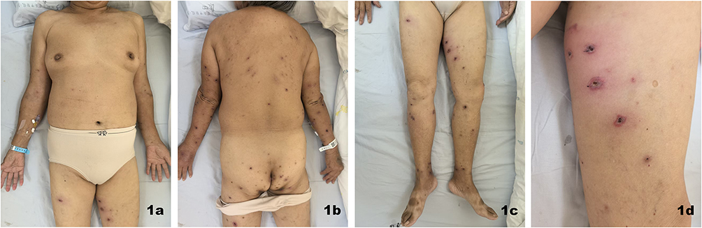 |
Figure 1 Cutaneous manifestations at presentation. (a–c) Scattered erythematous patches, papules, vesicles, and superficial ulcers on the trunk and limbs, some of which have formed scabs; (d) There are some red patches visible on the inner side of the left thigh, with a depressed ulcer in the center of each patch. |
Skin biopsy of the left thigh revealed dense perivascular lymphocytic infiltration extending into subcutaneous adipose tissue (Figure 2). The computed tomography of the chest and abdomen revealed multiple enlarged superficial lymph nodes but showed no hepatosplenomegaly, visceral lesions, or evidence of systemic lymphoma. Since the patient continued to experience recurrent high fever and progressive skin lesions despite antimicrobial therapy, and neoplastic diseases had been excluded, a therapeutic trial of intravenous methylprednisolone (40 mg/day) was initiated while awaiting definitive pathological results. Based on the recurrent papulonecrotic lesions, coexistence of lesions at different stages of evolution, varioliform scarring, and a history of partial spontaneous regression before treatment, the differential diagnoses initially included lymphomatoid papulosis (LyP), pityriasis lichenoides et varioliformis acuta (PLEVA), and other inflammatory or lymphoproliferative disorders. LyP, a primary cutaneous CD30⁺ lymphoproliferative disorder, presents clinically as a chronic, recurrent, and self-limiting condition, which closely aligns with the patient’s presentation.4 PLEVA is an uncommon inflammatory dermatosis characterized by recurrent papulonecrotic lesions that may heal with varioliform scarring.5
 |
Figure 2 Skin biopsy of the left thigh. (a) The pathological findings for the left thigh reveal diffuse oedema in the superficial dermis. A substantial population of lymphocytes, accompanied by sporadic eosinophils and focal neutrophil infiltration, is evident throughout the dermis, spanning from the superficial to the deep layers, with the predominance in proximity to blood vessels (HE×40); (b) In the subcutaneous fat layer, there is also evidence of lymphocytic infiltration, primarily around blood vessels, as well as scattered eosinophils and focal neutrophil infiltration. (HE×40); (c) Lymphocytic invasion of blood vessels is also observed. (HE×200). |
Immunohistochemical analysis demonstrated that the atypical infiltrating lymphocytes expressed CD3, CD4, CD8, Granzyme B (GrB), and βF1, with partial loss of CD5 expression. The cells were negative for CD30, CD56, TCRδ, and PD-1. Ki-67 staining showed a proliferative index of approximately 80%. CD20 highlighted scattered background B lymphocytes. Epstein–Barr virus-encoded RNA (EBER) in situ hybridization demonstrated strong positivity in the infiltrating lymphocytes (Figure 3). Direct immunofluorescence showed granular C3 deposition in dermal vessel walls. An excisional biopsy of the axillary lymph node revealed reactive lymphoid hyperplasia without evidence of systemic lymphoma (Figure 4). Integrating these findings, a final diagnosis of HVLPD was established.
 |
Figure 3 Details of the immunopathological phenotype of the left thigh (×40). (a) CD3-positive infiltrating T lymphocytes; (b) CD8-positive cytotoxic T cells; (c) Strong EBER positivity; (d) Ki-67 proliferation index approximately 80%; (e) Partial loss of CD5 expression. |
 |
Figure 4 Histopathology and Immunohistochemistry of Lymph Nodes in the Right Axilla. (a) Lymph nodes as observed under low magnification (HE×40); (b) KI67 approximately 95% (×40); (c) Positive for the Bcl-2 expression (×40). |
The patient was continued on a tapering methylprednisolone regimen combined with tripterygium glycosides. Tripterygium glycosides, an immunomodulatory agent widely used in China for inflammatory and autoimmune diseases, were added as a steroid-sparing therapy after the diagnosis had been established.6 At discharge, skin lesions had largely resolved (Figure 5), and during follow-up, only occasional isolated new lesions appeared with overall stable disease. The patient’s clinical course is summarized in Figure 6.
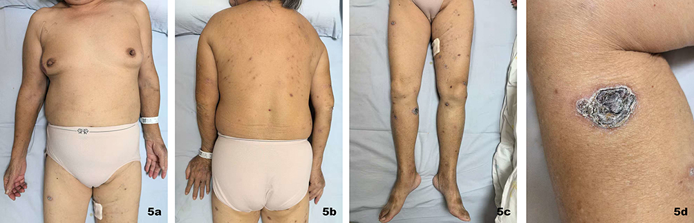 |
Figure 5 Skin lesions at discharge. (a–c) The scattered erythematous lesions across the body have faded and the ulcers have dried up and healed; (d) The image herein portrays aclose-up view of adesiccated ulcer located on the inner aspect of the right lower leg. |
 |
Figure 6 Clinical timeline. |
Discussion
This case underscores the diagnostic complexity of HVLPD in elderly patients, where atypical lesion distribution and secondary infection can confound clinical assessment. The hallmark histopathological features—angiocentric lymphocytic infiltration and subcutaneous extension—were present but required careful interpretation due to inflammatory admixture.7 The infiltrating lymphocytes exhibited a cytotoxic T-cell immunophenotype, characterized by expression of CD3, CD8, Granzyme B, and βF1. Loss of CD5 expression has been reported in EBV-associated T-cell lymphoproliferative disorders and may reflect aberrant T-cell immunophenotypes.8 Immunophenotypic analysis revealed a cytotoxic T-cell profile with CD8 and Granzyme B expression, partial loss of CD5 expression, and strong EBER positivity. The absence of CD30 expression effectively ruled out lymphomatoid papulosis, and the elevated Ki-67 index reflected brisk proliferative activity. PLEVA was also considered because of the papulonecrotic lesions, crust formation, and varioliform scarring observed in the present case. However, several findings were inconsistent with PLEVA. Histopathologically, the skin biopsy demonstrated angiocentric lymphoid infiltration extending into the subcutaneous tissue. These features are not characteristic of PLEVA and instead support an Epstein–Barr virus-associated lymphoproliferative disorder. Therefore, PLEVA was excluded. Despite palpable lymphadenopathy and a germinal center Ki-67 approaching 95%, preserved nodal architecture and lack of clonal T-cell expansion supported reactive hyperplasia rather than systemic lymphoma.1,9 Furthermore, no evidence of hepatosplenic involvement or extranodal visceral disease was identified, supporting the conclusion that the disease was primarily confined to the skin at the time of diagnosis. A strong positive EBER-ISH result remains the defining feature of HVLPD. In the present case, the favorable response to corticosteroid therapy prompted reconsideration of the differential diagnosis and supported the possibility of an underlying inflammatory or immune-mediated disorder. Nevertheless, the diagnosis of HVLPD was ultimately established through comprehensive clinicopathological correlation rather than therapeutic response. Close clinicopathological correlation remains essential for establishing an accurate diagnosis of HVLPD.
In diagnosis, HVLPD must be distinguished from several conditions, including hydroa vacciniforme, chronic active Epstein–Barr virus infection, extranodal NK/T-cell lymphoma, nasal type, and pityriasis lichenoides et varioliformis acuta (PLEVA) (Table 1).
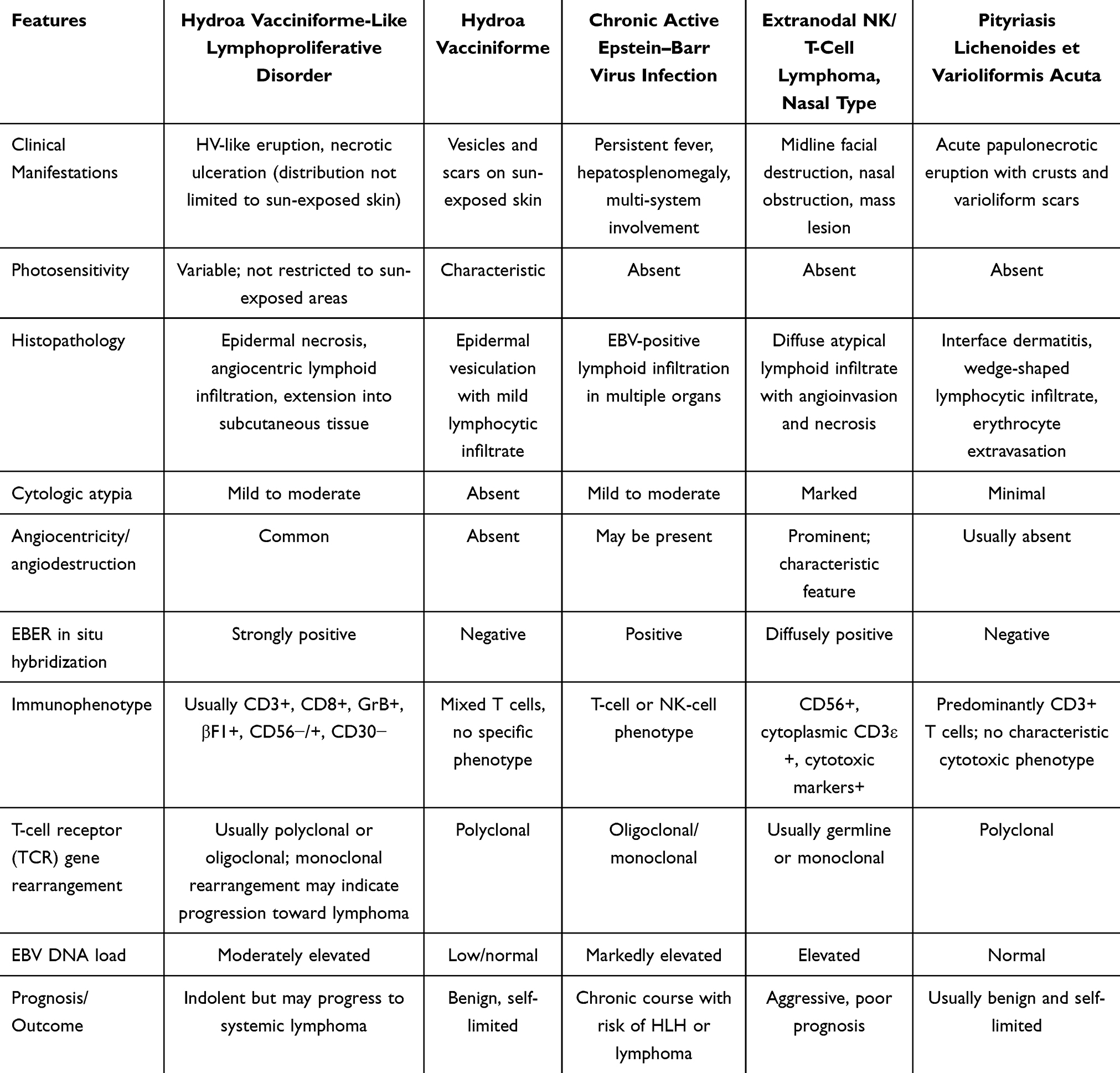 |
Table 1 Differential Diagnosis Table |
Limitations include the absence of T-cell receptor gene rearrangement analysis, EBV-DNA load quantification and bone marrow, as the patient declined further investigations. However, the integration of characteristic EBER positivity, reactive lymph node pathology, and comprehensive immunophenotypic findings provides robust evidence supporting HVLPD.10 Because bone marrow examination and quantitative EBV-DNA testing were not performed, occult systemic involvement cannot be completely excluded. However, the absence of hepatosplenomegaly, lack of visceral organ involvement, and reactive lymph node pathology favored a diagnosis of cutaneous HVLPD rather than CAEBV.
Given the disease’s rarity, no standardized treatment protocol exists. Current strategies advocate stratified management based on disease activity and systemic involvement.1 Allogeneic hematopoietic stem cell transplantation is currently the only potentially curative approach for HVLPD and associated lymphoma.2
Conclusions
This case highlights that HVLPD may occur in elderly patients and present with atypical lesions involving non–sun-exposed areas. Secondary infection and spontaneous lesion regression may obscure the diagnosis. Comprehensive clinicopathological correlation, including EBV studies and immunophenotypic analysis, is essential for accurate diagnosis and timely management.
Generative AI Statement
The authors declared that generative AI was not used in the creation of this manuscript. All scientific content, data interpretation, clinical decisions, and conclusions were developed, verified, and approved by the authors. The authors take full responsibility for the accuracy and integrity of the manuscript.
Data Sharing Statement
The original contributions presented in this study are included in this article, further inquiries can be directed to the corresponding author.
Ethics Statement
Ethical approval is not required to publish the case details in accordance with local or national guidelines. The patient provided written informed consent for the publication of this case report and accompanying images.
Author Contributions
XL: Conceptualization, data curation, investigation, formal analysis, visualization, and writing—original draft. LL: Investigation, data curation, and writing—review and editing. QL: Data curation, literature review, and writing—review and editing. DH: data curation, investigation, formal analysis. MY: Histopathological interpretation, supervision, and writing—review and editing. DD: Conceptualization, supervision, project administration, and writing—review and editing. All authors made a significant contribution to the work reported, whether that is in the conception, study design, execution, acquisition of data, analysis and interpretation, or in all these areas; took part in drafting, revising or critically reviewing the article; gave final approval of the version to be published; have agreed on the journal to which the article has been submitted; and agree to be accountable for all aspects of the work.
Funding
The authors declared that financial support was not received for this work and/or its publication.
Disclosure
The authors declare that the research was conducted in the absence of any commercial or financial relationships that could be construed as a potential conflict of interest.
References
1. Khoury JD, Solary E, Abla O, et al. The 5th edition of the World Health Organization classification of haematolymphoid tumours: myeloid and histiocytic/dendritic neoplasms. Leukemia. 2022;36(7):1703–8. doi:10.1038/s41375-022-01613-1
2. Liquidano-Perez E, Maza-Ramos G, Yamazaki-Nakashimada M, et al. Case report: hydroa vacciniforme-like lymphoproliferative disorder, an EBV-associated disease, successfully treated with hematopoietic stem cell transplantation. Front Immunol. 2025;16:1511385. doi:10.3389/fimmu.2025.1511385
3. Wang X, Wang P, Wang A, et al. Hydroa vacciniforme-like lymphoproliferative disorder in an adult invades the liver and bone marrow with clear pathological evidence: a case report and literature review. BMC Infect Dis. 2021;21(1):17. doi:10.1186/s12879-020-05697-x
4. Nowicka D, Mertowska P, Mertowski S, et al. Etiopathogenesis, diagnosis, and treatment strategies for lymphomatoid papulosis with particular emphasis on the role of the immune system. Cells. 2022;11(22):3697. doi:10.3390/cells11223697
5. Arias-Rodriguez C, Hoyos-Gaviria JG, Muñoz-Monsalve AM, et al. Dermoscopy of pityriasis lichenoides et varioliformis acuta (PLEVA). An Bras Dermatol. 2024;99(1):120–123. doi:10.1016/j.abd.2022.04.017
6. Shan Y, Zhao J, Wei K, et al. A comprehensive review of Tripterygium wilfordii hook. f. in the treatment of rheumatic and autoimmune diseases: bioactive compounds, mechanisms of action, and future directions. Front Pharmacol. 2023;14:1282610. doi:10.3389/fphar.2023.1282610
7. Chang L, Zhang C, Lu J, et al. Clinical and pathological features of hydroa vacciniforme-like lymphoproliferative disorder along with risk factors indicating poor prognosis. Infect Drug Resist. 2023;16:1545–1559. doi:10.2147/IDR.S402040
8. Luo Y, Zhang H, Tang K, et al. Single-cell dissection reveals immune dysregulation after CD5 or CD7-directed chimeric antigen receptor T-cell therapy. Advanced Science. 2026;13(8):e09259. doi:10.1002/advs.202509259
9. Cai Q, Chen K, Young KH. Epstein-Barr virus-positive T/NK-cell lymphoproliferative disorders. Exp Mol Med. 2015;47(1):e133. doi:10.1038/emm.2014.105
10. Chen Z, Huang H, Fang X, et al. Dynamic monitoring of circulating cell-free EBV-DNA for risk assessment in early-stage natural killer/T-cell lymphoma. Blood Adv. 2026;10(10):3457–3466. doi:10.1182/bloodadvances.2025018855
 © 2026 The Author(s). This work is published and licensed by Dove Medical Press Limited. The
full terms of this license are available at https://www.dovepress.com/terms
and incorporate the Creative Commons Attribution
- Non Commercial (unported, 4.0) License.
By accessing the work you hereby accept the Terms. Non-commercial uses of the work are permitted
without any further permission from Dove Medical Press Limited, provided the work is properly
attributed. For permission for commercial use of this work, please see paragraphs 4.2 and 5 of our Terms.
© 2026 The Author(s). This work is published and licensed by Dove Medical Press Limited. The
full terms of this license are available at https://www.dovepress.com/terms
and incorporate the Creative Commons Attribution
- Non Commercial (unported, 4.0) License.
By accessing the work you hereby accept the Terms. Non-commercial uses of the work are permitted
without any further permission from Dove Medical Press Limited, provided the work is properly
attributed. For permission for commercial use of this work, please see paragraphs 4.2 and 5 of our Terms.