Back to Journals » International Journal of Chronic Obstructive Pulmonary Disease » Volume 12
Heterogeneity of asthma–COPD overlap syndrome
Authors Joo H ![]() , Han D
, Han D ![]() , Lee JH
, Lee JH ![]() , Rhee CK
, Rhee CK
Received 22 December 2016
Accepted for publication 30 January 2017
Published 22 February 2017 Volume 2017:12 Pages 697—703
DOI https://doi.org/10.2147/COPD.S130943
Checked for plagiarism Yes
Review by Single anonymous peer review
Peer reviewer comments 2
Editor who approved publication: Prof. Dr. Richard Russell
Hyonsoo Joo,1 Deokjae Han,1 Jae Ha Lee,2 Chin Kook Rhee1
1Division of Pulmonary, Allergy and Critical Care Medicine, Department of Internal Medicine, Seoul St Mary’s Hospital, College of Medicine, The Catholic University of Korea, Seoul, 2Division of Pulmonology, Department of Internal Medicine, Inje University College of Medicine, Haeundae Paik Hospital, Busan, Republic of Korea
Abstract: Many patients suffering from asthma or COPD have overlapping features of both diseases. However, a phenotypical approach for evaluating asthma–COPD overlap syndrome (ACOS) has not been established. In this report, we examined the phenotypes in patients with ACOS. Patients diagnosed with ACOS between 2011 and 2015 were identified and classified into four phenotype groups. Group A was composed of patients who smoked <10 pack years and had blood eosinophil counts ≥300. Group B was composed of patients who smoked <10 pack years and had blood eosinophil counts <300. Group C was composed of patients who smoked ≥10 pack years and had blood eosinophil counts ≥300. Group D was composed of patients who smoked ≥10 pack years and had blood eosinophil counts <300. Clinical characteristics were analyzed and compared among groups. Comparisons were made among 103 ACOS patients. Patients in group D were oldest, while patients in group A were youngest. There were relatively more female patients in groups A and B; the majority of patients in groups C and D were male. The degree of airflow obstruction was most severe in group C. The rate of being free of severe exacerbation was significantly lower in group C than in the other groups. In this study, each ACOS phenotype showed different characteristics. The proportion of patients free of severe exacerbation differed significantly among groups. At this time, further studies on the phenotypes of ACOS are required.
Keywords: phenotype, smoking, eosinophil, exacerbation
Introduction
Asthma and COPD are common diseases characterized by the presence of airway obstruction and have traditionally been considered different diseases. However, many patients with asthma or COPD have overlapping features of both diseases. Recently, the Global Initiative for Asthma (GINA) and the Global Initiative for Chronic Obstructive Lung Disease (GOLD) committees accepted the concept of asthma–COPD overlap syndrome (ACOS), described as follows:
ACOS is characterized by persistent airflow limitation with several features usually associated with asthma and several features usually associated with COPD. ACOS is therefore identified by the features that it shares with both asthma and COPD.1,2
However, a clear definition remains unavailable.
Asthma and COPD are heterogeneous disorders with many phenotypes. Thus, ACOS may show a spectrum of phenotypes, which is particularly true when the diagnostic criteria of ACOS are broad. Different diagnostic criteria (broad and narrow) for ACOS have been proposed by various groups.1–6 Depending on the various definitions, the prevalence of ACOS also varies.7 One broad definition of ACOS has been proposed by Gibson and Simpson.3 Patients with fixed airflow obstruction with bronchodilator reversibility (BDR) or bronchial hyperresponsiveness (BHR) can be considered to have ACOS.
Studies on the classification and clinical features of ACOS have been steadily increasing. However, a phenotypical approach for the classification of ACOS is not yet available. Some experts discussed the heterogeneity of ACOS in previous review articles.5,8,9 However, few studies have explored the phenotype of ACOS. Rhee5 previously proposed four phenotypes of ACOS classified mainly based on eosinophilic inflammation and smoking history. Each phenotype has a different underlying pathophysiology and requires different medications. In this study, we applied this ACOS phenotype in clinical practice, examined the prevalence of each phenotype, and determined whether there are different clinical characteristics between these disorders.
Methods
Study population
We identified all outpatients diagnosed with COPD at Seoul St Mary’s Hospital (1,356 bed tertiary referral hospital) between May 2011 and May 2015. Among them, we selected patients who were managed by a pulmonary specialist. We retrospectively reviewed the medical records during the study period and applied the definition of ACOS from a previous study by Gibson and Simpson.3 We utilized this definition because it is much simpler and clearer than GINA/GOLD ACOS definition. We enrolled patients with symptoms of increased variability of airflow and incompletely reversible airflow obstruction. Patients with ACOS were eligible if their post-bronchodilator forced expiratory volume in 1 second (FEV1)/forced vital capacity (FVC) ratio was <0.7 with a positive BDR (>200 mL and >12% increase in FEV1) and/or positive BHR (positive methacholine or mannitol provocation test). Patients with lung cancer or cystic fibrosis were excluded in this study.
This study was approved by the Institutional Review Board of Seoul St Mary’s Hospital. The requirement for written informed consent from each patient was waived due to the retrospective nature of the study.
Clinical evaluation
Clinical data, including demographic data, smoking history, and laboratory tests such as eosinophil counts, COPD assessment test (CAT), pulmonary function test (PFT), frequency of severe exacerbation, and use of medication, were collected by retrospective review of medical records. For data regarding exacerbation, only severe exacerbation was recorded due to the potential for underdetection of mild or moderate exacerbation based on chart review. Severe exacerbation was defined as a worsening of any respiratory symptoms or increased dyspnea, which required an emergency room (ER) visit or hospitalization with prescription of systemic corticosteroid and/or antibiotics. History of medication use included inhaled corticosteroid (ICS) with long-acting beta agonist (LABA), long-acting muscarinic antagonist (LAMA), LABA, phosphodiesterase 4 inhibitor (PDE4I), and leukotriene receptor antagonist (LTRA).
Classification of the ACOS phenotype
The enrolled patients were divided into four phenotype groups according to blood eosinophil counts and smoking history. The classification was based on Rhee’s previous article5 and modified in a simple way. ACOS phenotype A group was composed of patients who smoked <10 pack years and had blood eosinophil counts ≥300. Group B was composed of patients who smoked <10 pack years and had blood eosinophil counts <300. Group C was composed of patients who smoked ≥10 pack years and had blood eosinophil counts ≥300. Group D was composed of patients who smoked ≥10 pack years and had blood eosinophil counts <300 (Figure 1). Patients were classified as high blood eosinophil group if blood eosinophil count was ≥300 even once among multiple measurements. We analyzed the characteristics and compared differences among the four groups.
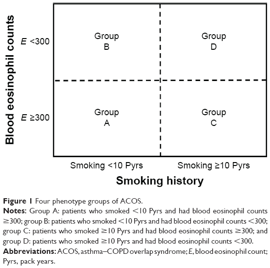 | Figure 1 Four phenotype groups of ACOS. |
PFT
PFTs were performed for all patients by experienced technicians. PFT was performed following the American Thoracic Society (ATS)/European Respiratory Society (ERS) guidelines in a licensed laboratory. Bronchodilator test was performed 15 minutes after the administration of 400 μg of salbutamol. Positive test for methacholine provocation was defined as provocative concentrations of methacholine required to decrease FEV1 (PC20) by 20% (≤16 mg/mL). Positive test for mannitol provocation was defined as PD15 (≤635 mg). Medications that can affect BDR or BHR were stopped before the test according to ATS guideline. Baseline PFT data were used in this study.
Statistical analysis
Categorical data were analyzed using χ2 or Fisher’s exact test. For normally distributed data, Student’s t-test or one-way analysis of variance was utilized for between-group comparisons. For nonnormally distributed data, we used the Mann–Whitney U-test or the Kruskal–Wallis test for between-group comparisons. The proportion of patients free of severe exacerbation was compared using the log-rank test for the four groups. The proportion of patients free of severe exacerbation was also compared between ICS (± LABA) users and nonusers. All statistical analyses were performed using the SPSS statistical package (version 18.0; SPSS Inc, Chicago, IL, USA); P-values <0.05 were considered statistically significant.
Results
Baseline characteristics
A total of 1,017 COPD patients were identified during the study period. Of these, 120 (11.8%) patients showed fixed airflow obstruction with BDR or BHR. Due to a lack of laboratory tests or smoking history, 17 patients were excluded. In total, 103 patients were included in the study (Figure 2). Figure 3 shows the number of patients classified into each group. The mean age (range: 35–94 years) in each group differed significantly (Table 1). Patients in group D were oldest, while group A patients were youngest. The percentage of males also differed significantly (Figure 4, P<0.001). There were relatively more female patients in groups A and B; the majority of patients in groups C and D were male. There were no significant differences in body mass index (BMI) among these groups. Post-bronchodilator FVC (%) or FEV1 (%) did not differ significantly among the four groups. However, FEV1/FVC (%) differed significantly (P=0.003) and was highest in group A and lowest in group C. There were no significant differences in CAT scores among the four groups.
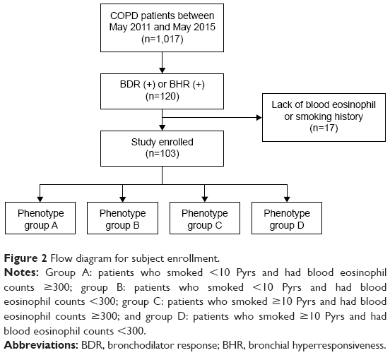 | Figure 2 Flow diagram for subject enrollment. |
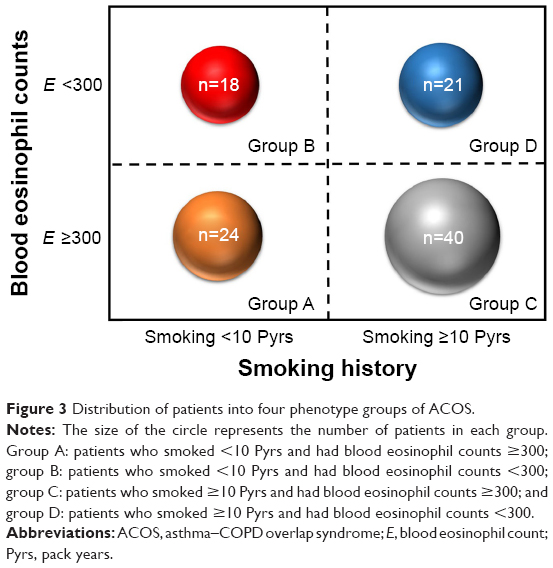 | Figure 3 Distribution of patients into four phenotype groups of ACOS. |
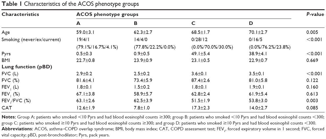 | Table 1 Characteristics of the ACOS phenotype groups |
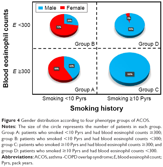 | Figure 4 Gender distribution according to four phenotype groups of ACOS. |
Frequency of medication use
There was a different pattern of medication prescription among the four groups (Figure 5). Overall, ICS + LABA was most frequently prescribed. For LAMA, the prescription rate was highest in group D followed by groups C, B, and A. In contrast, the LTRA prescription rate was highest in group A followed by groups B, D, and C. LABA and PDE4I were more frequently prescribed in groups C/D than in groups A/B.
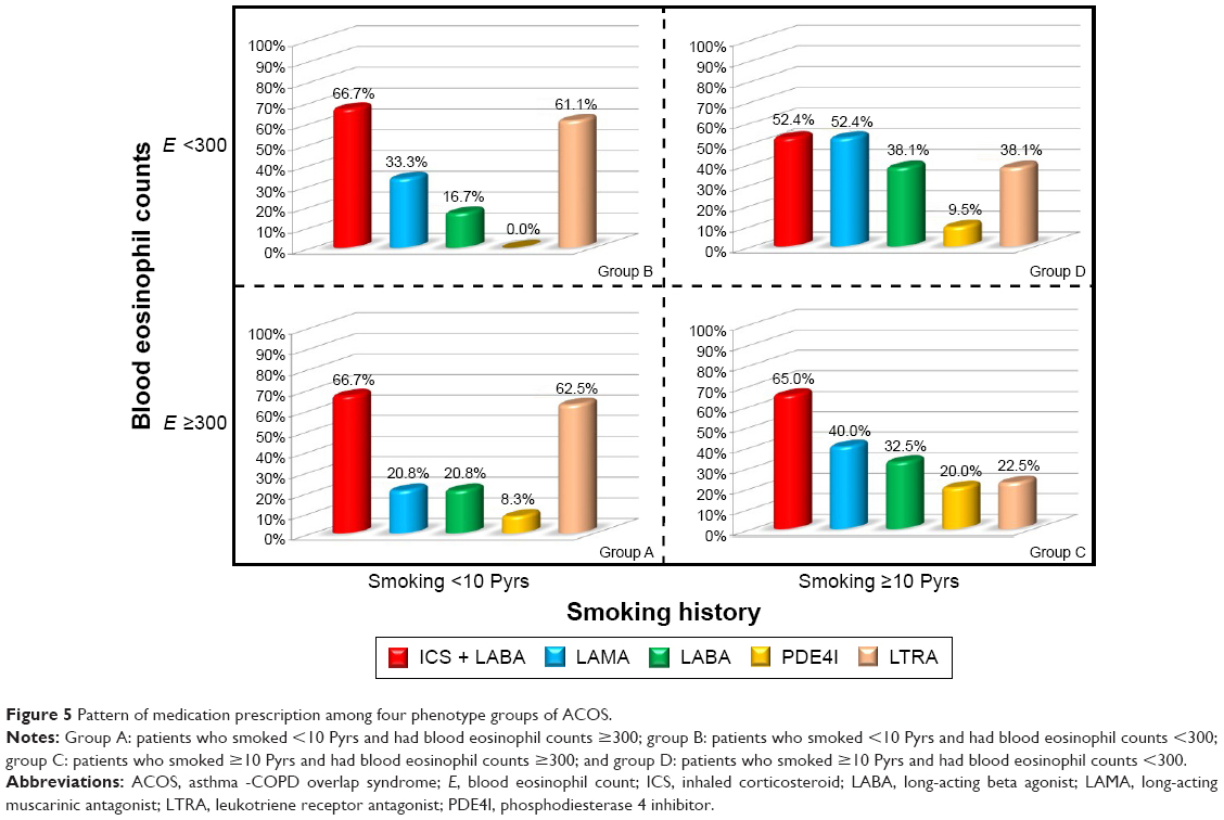 | Figure 5 Pattern of medication prescription among four phenotype groups of ACOS. |
Severe exacerbation
The mean follow-up period of enrolled patients was 1,229.8±152.2 days (mean ± standard error of the mean). The proportion of patients free of severe exacerbation was analyzed using log-rank tests among the four groups (Figure 6). There were significant differences in the proportion of patients free of severe exacerbation (P=0.016). The survival rate for patients free of exacerbation was lower in group C than in other groups.
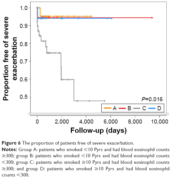 | Figure 6 The proportion of patients free of severe exacerbation. |
Among 103 patients, ICS was prescribed to two patients and ICS + LABA was prescribed to 65 patients. The proportion of patients free of severe exacerbation was compared between ICS group (patients with ICS or ICS + LABA) and non-ICS group. There was no significant difference between the two groups (P=0.663). Among 40 group C patients, ICS was prescribed to zero patient and ICS + LABA was prescribed to 14 patients. The proportion of patients free of severe exacerbation was compared between ICS and non-ICS groups among group C patients. There was also no significant difference between the two groups (P=0.383).
Discussion
After the GINA and GOLD committees accepted and published the concept of ACOS,1,2 several articles have been published regarding ACOS.5,6,8–13 However, the definition of ACOS remains unclear. The GINA and GOLD committees provided a table to help clinicians diagnose ACOS. However, this table is not clinically practical because it requires extensive time to complete. Moreover, some questions are vague and the answer can be very subjective. Even for the same patient, the answer can be changed according to the clinician who checks the table. Thus, few ACOS studies14 have utilized this table for the definition of ACOS. In contrast, Gibson and Simpson3 proposed a definition of ACOS based on spirometry results. Compared to the GINA/GOLD definition, this definition is much simpler and very clear. It is also simple to apply in clinical practice. For these reasons, there have been many ACOS studies11,15,16 using this definition. However, compared to the definition by GINA and GOLD, this definition is considered broad. Thus, a relatively large number of patients can be classified as ACOS according to this definition. It is expected that patients with predominant features of asthma or COPD are likely to be considered as ACOS according to this definition.
The purpose of this study was to examine how many patients are enrolled by this definition and determine whether patients can be classified into four different phenotypes. Interestingly, among 1,017 COPD patients, a relatively small percentage (11.8%) of patients showed positive BDR or BHR. In addition, COPD patients with BDR or BHR could be classified into four different phenotypes. Although phenotype C was dominant (38.8%), several patients were classified into other phenotypes (A: 23.3%, B: 17.4%, and D: 20.4%). This result suggests that heterogeneity exists even in patients with ACOS.
This is the first study to show the heterogeneity of ACOS. Several previous studies have explored the heterogeneity of asthma17–19 or COPD.20–29 However, few studies have explored the heterogeneity of ACOS. The concept of heterogeneity in ACOS has been suggested in previous review articles by experts.5,8,9 However, few studies have supported the concept of heterogeneity in ACOS. In this study, we used COPD patients managed by a pulmonary specialist in a tertiary referral hospital. We then classified patients into four categories according to relatively simple criteria – blood eosinophils and smoking history. There are different characteristics among the four groups. Moreover, the prognosis (exacerbation) differed significantly among the four groups. These results are valuable in that we demonstrated heterogeneity in ACOS patients.
Phenotypic classification based on the underlying disease mechanism is important for the appropriate management of individual patients. Eosinophilic inflammation and a history of smoking are important components in the classification of ACOS. According to these factors, Rhee5 proposed four phenotypes of ACOS and specific treatment options for each phenotype. Phenotype A is an allergic asthma-predominant phenotype. Phenotype B is characterized by features of severe noneosinophilic asthma. Phenotype C is very compatible with ACOS in that these patients have features of both asthma and COPD. Phenotype D is relatively pure COPD with reversibility. In this study, we investigated baseline characteristics and clinical features of each phenotype in real-world patients with ACOS. There are a relatively large number of patients in group C; however, the number of patients was relatively evenly distributed among groups. This study validated that the previously proposed phenotypes of ACOS by Rhee are clinically applicable and meaningful.
One interesting result of this study is the different pattern of medication use in each group. All enrolled patients were managed by a pulmonary specialist in tertiary referral hospitals. Because these patients were considered as ACOS, ICS + LABA was prescribed most commonly across the four phenotypes. However, LAMA, LABA, and PDE4I, which are mainly COPD medications, were more frequently prescribed in COPD-predominant phenotypes (C and D). In contrast, LTRA, which is mainly an asthma medication, was more frequently prescribed in asthma-predominant phenotypes (A and B). This phenomenon also supports the usefulness of ACOS classification by Rhee. The medication prescription in this study was determined by an experienced pulmonary specialist. They can classify phenotypes of ACOS patients and prescribe appropriate medication in a personalized manner. However, for general practitioners (GPs), it can be difficult to classify ACOS patients into subtypes. Thus, simple criteria to classify ACOS will be helpful for GPs. Based on our results, patients are well-characterized by two simple clinically relevant criteria – blood eosinophil count and smoking history. Thus, GPs can easily diagnose ACOS patients (fixed airflow obstruction with BDR or BHR) and subsequently classify these patients into four phenotypes based on blood eosinophil count and smoking history. Finally, GPs can choose a specific medication according to the phenotype. This allows GPs to treat ACOS patients in a personalized manner.
Preventing exacerbations in patients with ACOS is one of the unmet needs in the pharmacological treatment, similar to asthma and COPD. In this study, the differences that exist in severe exacerbation among the four phenotype groups is one of the important outcomes. When classified into the four phenotypes, severe exacerbation is most frequent in phenotype C. Based on these results, pulmonary function is not the reason for the differences in the frequency of acute exacerbation. This is because the frequency of acute exacerbation in phenotype C was worst while its pulmonary function was not. This suggests that different phenotypes exist in ACOS, and each phenotype has a different prognosis. Actually, it is well known that the prognosis of ACOS was poorer than that of COPD alone.10,30 They have more symptoms25 and exacerbate more frequently.31 The poor prognosis of phenotype C in this study is in agreement with previous reports because phenotype C is very compatible with ACOS. The ACOS definition based only on BDR or BHR is a broad definition. In contrast, including other criteria such as blood eosinophil count and history of smoking results in a narrow definition. Rhee5 proposed a broad and narrow definition of ACOS in a previous review article. This narrow definition, which is compatible with phenotype C, is similar to other definitions proposed by Spanish colleagues.4 Thus, it is understandable that patients with phenotype C show more exacerbations.
There are some limitations in this study. 1) The sample size may not be sufficiently large to clarify the clinical features of each phenotype group. In addition, this was a single-center study. However, there has been no prior classification of ACOS phenotypes or studies on clinical patients. The results of this study should be validated in a large-scale cohort study. Whether two simple criteria – blood eosinophil count 300 and 10 pack years smoking history – used in this study will be useful should be validated in future study. Moreover, whether different prognoses of four groups will be replicated or not is also important. 2) It is likely that there was bias in this study due to its retrospective nature. The history of exacerbation may not be correct. Due to this limitation, only severe exacerbation was analyzed in this study; mild or moderate exacerbation was not considered. By reviewing the charts, it was difficult to detect mild or moderate exacerbation.
Conclusion
In this study, we classified ACOS patients into four phenotypes. Each phenotype showed different characteristics. The proportion of patients free of severe exacerbation differed significantly among groups. However, further studies on the phenotype of ACOS are required.
Disclosure
CKR received consulting/lecture fees from MSD, AstraZeneca, Novartis, GSK, Takeda, Mundipharma, Sandoz, Boehringer-Ingelheim, and Teva-Handok. The other authors report no conflicts of interest in this work.
References
Global Strategy for the Diagnosis, Management, and Prevention of Chronic Obstructive Pulmonary Disease. 2016. Available from: http://goldcopd.org/global-strategy-diagnosis-management-prevention-copd-2016/. Accessed October 15, 2016. | ||
Global Initiative for Asthma. Global Strategy for Asthma Management and Prevention. 2015. Available from: http://ginasthma.org/wp-content/uploads/2016/01/GINA_Report_2015_Aug11-1.pdf. Accessed October 15, 2016. | ||
Gibson PG, Simpson JL. The overlap syndrome of asthma and COPD: what are its features and how important is it? Thorax. 2009;64(8):728–735. | ||
Soler-Cataluna JJ, Cosio B, Izquierdo JL, et al. Consensus document on the overlap phenotype COPD-asthma in COPD. Arch Bronconeumol. 2012;48(9):331–337. | ||
Rhee CK. Phenotype of asthma–chronic obstructive pulmonary disease overlap syndrome. Korean J Intern Med. 2015;30(4):443–449. | ||
Sin DD, Miravitlles M, Mannino DM, et al. What is asthma–COPD overlap syndrome? Towards a consensus definition from a round table discussion. Eur Respir J. 2016;48(3):664–673. | ||
Wurst KE, Kelly-Reif K, Bushnell GA, Pascoe S, Barnes N. Understanding asthma–chronic obstructive pulmonary disease overlap syndrome. Respir Med. 2016;110:1–11. | ||
Bateman ED, Reddel HK, van Zyl-Smit RN, Agusti A. The asthma–COPD overlap syndrome: towards a revised taxonomy of chronic airways diseases? Lancet Respir Med. 2015;3(9):719–728. | ||
Reddel HK. Treatment of overlapping asthma–chronic obstructive pulmonary disease: can guidelines contribute in an evidence-free zone? J Allergy Clin Immunol. 2015;136(3):546–552. | ||
Rhee CK, Yoon HK, Yoo KH, et al. Medical utilization and cost in patients with overlap syndrome of chronic obstructive pulmonary disease and asthma. COPD. 2014;11(2):163–170. | ||
Lee HY, Kang JY, Yoon HK, et al. Clinical characteristics of asthma combined with COPD feature. Yonsei Med J. 2014;55(4):980–986. | ||
Hardin M, Cho M, McDonald ML, et al. The clinical and genetic features of COPD-asthma overlap syndrome. Eur Respir J. 2014;44(2):341–350. | ||
Barrecheguren M, Roman-Rodriguez M, Miravitlles M. Is a previous diagnosis of asthma a reliable criterion for asthma–COPD overlap syndrome in a patient with COPD? Int J Chron Obstruct Pulmon Dis. 2015;10:1745–1752. | ||
Gao Y, Zhai X, Li K, et al. Asthma COPD overlap syndrome on CT densitometry: a distinct phenotype from COPD. COPD. 2016;13(4):471–476. | ||
Lee SY, Park HY, Kim EK, et al. Combination therapy of inhaled steroids and long-acting beta2-agonists in asthma–COPD overlap syndrome. Int J Chron Obstruct Pulmon Dis. 2016;11:2797–2803. | ||
Menezes AM, Montes de Oca M, Perez-Padilla R, et al. Increased risk of exacerbation and hospitalization in subjects with an overlap phenotype: COPD-asthma. Chest. 2014;145(2):297–304. | ||
Wenzel SE. Asthma phenotypes: the evolution from clinical to molecular approaches. Nat Med. 2012;18(5):716–725. | ||
Skloot GS. Asthma phenotypes and endotypes: a personalized approach to treatment. Curr Opin Pulm Med. 2016;22(1):3–9. | ||
Lotvall J, Akdis CA, Bacharier LB, et al. Asthma endotypes: a new approach to classification of disease entities within the asthma syndrome. J Allergy Clin Immunol. 2011;127(2):355–360. | ||
Agusti A, Bel E, Thomas M, et al. Treatable traits: toward precision medicine of chronic airway diseases. Eur Respir J. 2016;47(2):410–419. | ||
Agusti A. The path to personalised medicine in COPD. Thorax. 2014;69(9):857–864. | ||
Agusti A. Phenotypes and disease characterization in chronic obstructive pulmonary disease. Toward the extinction of phenotypes? Ann Am Thorac Soc. 2013;10(suppl):S125–S130. | ||
Agusti A, Edwards LD, Rennard SI, et al. Persistent systemic inflammation is associated with poor clinical outcomes in COPD: a novel phenotype. PLoS One. 2012;7(5):e37483. | ||
de Oca MM, Halbert RJ, Lopez MV, et al. The chronic bronchitis phenotype in subjects with and without COPD: the PLATINO study. Eur Respir J. 2012;40(1):28–36. | ||
Izquierdo-Alonso JL, Rodriguez-Gonzalezmoro JM, de Lucas-Ramos P, et al. Prevalence and characteristics of three clinical phenotypes of chronic obstructive pulmonary disease (COPD). Respir Med. 2013;107(5):724–731. | ||
Jamieson DB, Matsui EC, Belli A, et al. Effects of allergic phenotype on respiratory symptoms and exacerbations in patients with chronic obstructive pulmonary disease. Am J Respir Crit Care Med. 2013;188(2):187–192. | ||
Kim V, Han MK, Vance GB, et al. The chronic bronchitic phenotype of COPD: an analysis of the COPDGene Study. Chest. 2011;140(3):626–633. | ||
Miravitlles M, Soler-Cataluna JJ, Calle M, Soriano JB. Treatment of COPD by clinical phenotypes: putting old evidence into clinical practice. Eur Respir J. 2013;41(6):1252–1256. | ||
Hurst JR, Vestbo J, Anzueto A, et al. Susceptibility to exacerbation in chronic obstructive pulmonary disease. N Engl J Med. 2010;363(12):1128–1138. | ||
Nielsen M, Barnes CB, Ulrik CS. Clinical characteristics of the asthma–COPD overlap syndrome – a systematic review. Int J Chron Obstruct Pulmon Dis. 2015;10:1443–1454. | ||
de Marco R, Pesce G, Marcon A, et al. The coexistence of asthma and chronic obstructive pulmonary disease (COPD): prevalence and risk factors in young, middle-aged and elderly people from the general population. PLoS One. 2013;8(5):e62985. |
 © 2017 The Author(s). This work is published and licensed by Dove Medical Press Limited. The
full terms of this license are available at https://www.dovepress.com/terms
and incorporate the Creative Commons Attribution
- Non Commercial (unported, 3.0) License.
By accessing the work you hereby accept the Terms. Non-commercial uses of the work are permitted
without any further permission from Dove Medical Press Limited, provided the work is properly
attributed. For permission for commercial use of this work, please see paragraphs 4.2 and 5 of our Terms.
© 2017 The Author(s). This work is published and licensed by Dove Medical Press Limited. The
full terms of this license are available at https://www.dovepress.com/terms
and incorporate the Creative Commons Attribution
- Non Commercial (unported, 3.0) License.
By accessing the work you hereby accept the Terms. Non-commercial uses of the work are permitted
without any further permission from Dove Medical Press Limited, provided the work is properly
attributed. For permission for commercial use of this work, please see paragraphs 4.2 and 5 of our Terms.