Back to Journals » International Medical Case Reports Journal » Volume 17
Guillain-Barre Syndrome with Acute Lymphoblastic Leukemia: Case Report
Authors Almatar ERE, Alsharidah S, Bourusly M, Ahmed RL, Alabdulhadi MA
Received 12 September 2024
Accepted for publication 23 November 2024
Published 10 December 2024 Volume 2024:17 Pages 1013—1020
DOI https://doi.org/10.2147/IMCRJ.S496062
Checked for plagiarism Yes
Review by Single anonymous peer review
Peer reviewer comments 2
Editor who approved publication: Professor Thomas E Hutson
Eman RE Almatar,1,* Sondus Alsharidah,1,* Maha Bourusly,1 Rania Lotfi Ahmed,1,* Mariam Ahmed Alabdulhadi2,*
1Department of Pediatrics, NBK Children Specialized Hospital, Sabah, Kuwait; 2College of Medicine, Arabian Gulf University, Manama, Kuwait
*These authors contributed equally to this work
Correspondence: Eman RE Almatar, Department of Pediatrics, NBK Children Specialized Hospital, Sabah, Kuwait, Email [email protected]
Abstract: Guillain-Barré syndrome (GBS) is a rare complication in children with acute lymphoblastic leukemia (ALL). This case report explores the presentation and management of GBS in a 16-year-old male with a history of ALL, who developed GBS during maintenance therapy. The patient exhibited progressive symmetrical weakness, sensory loss, and autonomic dysfunction. Diagnostic workup, including nerve conduction studies and lumbar puncture, confirmed the diagnosis of GBS. Differentiating GBS from vincristine-induced neuropathy, a common challenge in this population, was crucial for appropriate management. The patient responded well to intravenous immunoglobulin and supportive care. This case highlights the importance of considering GBS in the differential diagnosis of neurological complications in children with ALL and emphasizes the need for prompt diagnosis and treatment.
Keywords: acute lymphoblastic leukemia, pediatric oncology, neuropathy, immune-mediated disease
Introduction
Guillain-Barre syndrome (GBS) is an immune-mediated disease affecting the peripheral nervous system that can affect both children and adults. The body’s immune system mistakenly attacks peripheral nerves. The classic presentation of GBS is characterized by progressive symmetrical, ascending muscle weakness.1 GBS is rarely reported in children with acute lymphoblastic leukemia (ALL) and may be difficult to differentiate from vincristine-induced neuropathy.2 In children undergoing maintenance therapy for B-cell precursor acute lymphoblastic leukemia (BCP-ALL), GBS can be particularly concerning. Maintenance therapy for BCP-ALL typically includes chemotherapy agents such as methotrexate and mercaptopurine, which suppress the immune system to prevent leukemia relapse. This immunosuppression increases the risk of infections, autoimmune reactions, and other complications, which may contribute to the onset of GBS.
In addition, ALL could theoretically trigger GBS through paraneoplastic mechanisms, where aberrant immune responses target the nervous system. In addition to that, ALL patients are susceptible to infections due to neutropenia, which might trigger GBS.
This case report explores the occurrence of Guillain-Barre syndrome in a 16-year-old male with a history of B-cell lymphoblastic leukemia.
Case Report
A 16-year-old male with a history of ALL in maintenance therapy presented to the hospital with increasing numbness in the left cheek, left arm, and lower limbs. He also reported dizziness and could walk only with support against the wall. His symptoms worsened, resulting in numbness throughout his body, including his face, along with dysarthria, severe headache, and neck pain.
The patient’s history indicated a diagnosis of B-cell lymphoblastic leukemia since 2022. His cerebrospinal fluid (CSF) was clear of blasts, and his cytogenetics were normal. He completed an induction chemotherapy course as per the UK-ALL Regimen B protocol and subsequently shifted to maintenance therapy under the more intensive UK-ALL Regimen C protocol due to poor bone marrow biopsy (BMB) result and no minimal residual disease (MRD) marker. Chemotherapy was temporarily halted for 4 days because he had experienced neutropenia and developed septicemia Staphylococcus Aureus (S. aureus). He was treated intravenously with Tazocin, Clindamycin, and Vancomycin. While GBS is typically linked to pathogens like Campylobacter jejuni or viruses (eg, cytomegalovirus), cases related to S. aureus are rare.
Additionally, he had detected atrial thrombus twice and was on a daily dose of 20 mg oral anticoagulant (Rivaroxaban).
On general physical examination, the patient was conscious with a Glasgow Coma Scale (GCS) score of 15/15, mild left upper motor neuron facial palsy, intact facial sensation, normal deep tendon reflexes in the lower limb, and diminished reflexes in upper limbs. No orthopedic or organic findings were noted. Blood tests, coagulation profile, CT scan (head), MRI, MRA, and MRV results were all normal (Figures 1 and 2).
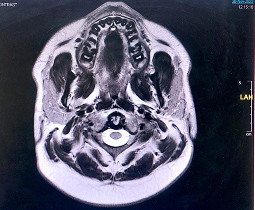 |
Figure 1 MRI Report. Normal brain parenchymal MR signal intensity with no focal lesion. No abnormal brain parenchymal or mennigeal enhancement. Normal signal, thickness and enhancement of both facial nerves. Normal size and configuration of the cerebral ventricles with no middle line shift or deformities. Normal gray- white matter differentiation. Normal cerebellum, brain stem and cervico- medullary junction. Both cerebellopontine angles are free. Normal cellar region. Normal extra axial spaces. Apart from variable degrees of para-nasal sinuses inflammatory changes with bubbly appearance in the sphenoid sinus and to a less extent the maxillary sinuses suggesting acute exacerbation. |
 |
Figure 2 MRV Report. Normal MRV appearance of the major venous sinus and superficial veins with no evidence of thrombosis, obstruction, filling defects or infiltration. |
 |
Figure 3 EMG – NCV Study Report for Motor Nerve Results. |
 |
Figure 4 NCV Study Report for Sensory Nerve Results: the findings of demyelinating and axonal peripheral neuropathy. There is significant proximal conduction block, very prolonged F waves, moderately slow motor conduction velocities, low motor responses and sural sparing pattern. The findings support the clinical diagnosis of Guillain-Barre syndrome. A follow-up after 3-4 weeks is recommended. |
The following day, the patient’s condition worsened, with severe loss of sensation in the lower limbs and anal area, dysphagia, and choking. An immediate tests and examinations were done resulting in normal blood tests, coagulation profile, essential unremarkable non-enhanced CT study (brain) and normal MRI, MRA and MRV examination of the brain with para-nasal sinuses chronic inflammatory changes with features suggestive of acute exacerbation.
An urgent nerve conduction velocity (NCV) and electromyography (EMG) study was conducted to rule out Guillain-Barre Syndrome. The findings indicated demyelinating and axonal peripheral neuropathy with a significant proximal conduction block and very prolonged F waves. Motor conduction velocities were moderately slow, with low motor responses and a sural sparing pattern Figures 3 and 4. The patient also underwent MRI examination of the dorsal and lumbo-sacral spine for diagnostic purposes, and the analysis showed abnormal smooth thickening and enhancement of the intrathecal nerve roots, which are consistent with Guillain-Barre and very high protein levels and the presence of monoclonal antibodies in the CSF, confirming the diagnosis of Guillain-Barre syndrome.
A diagnosis of Guillain-Barre syndrome was made based on the criteria and findings that strongly supported the diagnosis.
Features required to rule out diagnoses other than GBS include no history of hexacarbon abuse, no evidence of porphyria, no history or culture evidence of diphtheria, no history or evidence of lead intoxication, symptoms not purely sensory, and no evidence of poliomyelitis or toxic neuropathy.
Immediate therapeutic management included intravenous immunoglobulin (IVIG) at 50 g daily for five days a week and plasma transfusion. The patient chemotherapeutic plan was resumed under the UK- ALL Regimen C guidelines without vincristine. The patient developed tachypnea without respiratory distress (RD) and hypoxia, necessitating transfer to the ICU. The plasma exchange transfusion was started in the ICU after right femoral hemodialysis catheter was inserted.
After two days of therapeutic treatment, the patient showed gradual improvement in reverse chronological manner with the appearance of symptoms, including better swallowing of solid food, sensation and movement in the anal area and lower limbs, resolution of headaches and neck pain, and gradually regressing numbness. After four plasma exchange sessions and three weeks with physiotherapy, the patient reached near-complete recovery. His treatment plan includes the continuation of IVIG for long-term GBS management and physiotherapy to enhance quality of life.
Discussion
Guillain-Barre syndrome (GBS) is a critically acquired condition characterized by acute evolution, immune mediation, and inflammatory disorder of the peripheral nervous system, leading to demyelination and axonal loss. Clinical hallmarks include symmetrical flaccid muscle paresis, areflexia, increased cerebrospinal fluid protein content, and electrophysiologic evidence of evolving demyelination.3
GBS in children with ALL is rare, with few reported cases.4–6 Out of the five cases reported, three were from a single center.5 ALL is a hematologic malignancy, characterized by various genetic abnormalities, including chromosomal translocations, fusion genes, and other mutations, that lead to uncontrolled proliferation of lymphoid precursor cells. These genetic abnormalities often align with specific subtypes of ALL and can influence disease prognosis, treatment response, and clinical trajectory.
Most patients develop weakness starting in the lower extremities, progressing due to peripheral nerve demyelination, resulting in ascending paralysis and loss of cranial nerve function.7 Manifestations may be acute or chronic and temporary or permanent, depending on the degree of neuronal destruction.8 Muscle stretch reflexes are typically depressed, and sensory loss is variable. Weakness is usually symmetric but can involve the upper extremities.9–11 Elevated CSF protein in patients with ascending paresis is indicative of GBS.11
An important consideration in children with ALL developing neuropathy during chemotherapy is vincristine-induced neuropathy. However, the clinical and electrodiagnostic findings for vincristine-induced neuropathy are distinct.12 Timely differentiation is important to initiate immunomodulatory therapies for GBS and avoid unnecessary withdrawal of vincristine, which could worsen ALL symptoms.
Treatment for GBS varies based on symptom severity. Common complications include ventilatory failure and cardiovascular instability, necessitating intensive care support. Ventilatory failure results from involvement of the airway and respiratory muscles, particularly the diaphragm.13 Corticosteroids have shown no benefit.14 Plasmapheresis is a well-investigated, efficacious immunomodulatory therapy, shown to decrease ventilator dependence in severe GBS cases.3
Conclusion
This case highlights the rare but serious complication of Guillain-Barré syndrome (GBS) in children with acute lymphoblastic leukemia (ALL), particularly during maintenance therapy. In children undergoing maintenance therapy for B-cell precursor acute lymphoblastic leukemia (BCP-ALL), GBS can be particularly concerning. The overlap in clinical presentation between GBS and vincristine-induced neuropathy underscores the importance of thorough diagnostic evaluation to ensure accurate diagnosis and prompt treatment. In this case, the patient responded positively to intravenous immunoglobulin and supportive care, demonstrating the efficacy of early intervention in managing GBS. This report emphasizes the need for healthcare providers to maintain a high index of suspicion for GBS in similar clinical settings, as timely differentiation and management can significantly impact patient outcomes. Further research is needed to better understand the pathophysiology of GBS in the pediatric ALL population and to develop targeted therapeutic strategies.
Human and Animal Guidelines
“Not applicable” as this patient was presented at the hospital as a regular patient.
Medical Writing Support
Medical Writing support was provided by Al Essa Medical and Scientific Group.
Abbreviations
GBS, Guillain-Barré syndrome; ALL, acute lymphoblastic leukemia; GCS, Glasgow Coma Scale; NCV, nerve conduction velocity; EMG, electromyography; CSF, cerebrospinal fluid; IVIG, intravenous immunoglobulin; RD, respiratory distress.
Ethical Approval
Ethical approval is not recommended in this case report because the patient was referred to the hospital for normal treatment. Patient was treated and discharged according to the hospital normal procedures. However, since such cases are rare in the medical literature, we decided to publish this case and got a written consent from the guardians of the pediatric patient.
Informed Consent
Written informed consent was obtained from legal guardians for the publication of any potentially identifiable images or data included in this article.
Funding
The publication of this article was supported by NBK Children Specialized Hospital, Kuwait.
Disclosure
The authors declare no conflict of interest in this work.
References
1. Kozyreva AA, Druzhinina ES, Zavadenko NN, et al. Guillain-Barre syndrome in children. Zh N evrol Psikhiatr Im S S Korsakova. 2023;123(9. Vyp.2):20–32. doi:10.17116/jnevro202312309220
2. Rajeswari B, Krishnan S, Sarada C, Kusumakumary P. Guillain-Barre syndrome with acute lymphoblastic leukemia. Indian Pediatr. 2013;50(8):791–792. doi:10.1007/s13312-013-0201-2
3. Hund EF, Borel CO, Hanley DF, et al. Intensive management and treatment of severe Guillain-Barre syndrome. Crit Care Med. 1993;21(3):433–446. doi:10.1097/00003246-199303000-00023
4. Aral YZ, Gursel T, Ozturk G, Serdaroglu A. Guillain- Barre syndrome in a child with acute lymphoblastic leukemia. Pediatr Hematol Oncol. 2001;18(5):343–346. doi:10.1080/088800101300312618
5. Brigo F, Balter R, Marradi P, et al. Vincristine related neuropathy versus acute inflammatory demyelinating polyradiculoneuropathy in children with acute lymphoblastic leukemia. J Child Neurol. 2012;27(7):867–874. doi:10.1177/0883073811428379
6. Norman M, Elinder G, Finkel Y. vincristine neuropathy and a Guillain-Barre syndrome: a case with acute lymphatic leukemia and quadriparesis. Eur J Haematol. 1987;39(1):75–76. doi:10.1111/j.1600-0609.1987.tb00168.x
7. Nadkarni N, Lisak RP. Guillain-Barre syndrome(GBS) with bilateral optic neuritis and central white matter disease. Neurology. 1993;43(4):842–843. doi:10.1212/WNL.43.4.842
8. Murray DP. Impaired mobility: guillain-Barre syndrome. J Neurosci Nursing. 1993;25(2):100–104. doi:10.1097/01376517-199304000-00006
9. Mendell JR. Chronic inflammatory demyelinating polyradiculopathy. Ann Rev Med. 1993;44(1):211–219. doi:10.1146/annurev.me.44.020193.001235
10. John RP. Guillain-Barre syndrome: a case report. J CCA. 1995;39(2):80.
11. Hegen H, Ladstätter F, Bsteh G. Cerebrospinal fluid protein in Guillain- Barre syndrome: need for age dependent interpretation. Eur J Neurol. 2021;28(3):965–973.
12. Pal PK. Clinical and electrophysiological studies in vincristine induced neuropathy. Electromyogr Clin Neurophysiol. 1999;39(6):323–330.
13. Leonhard SE, Mandarakas MR, Gondim FAA, et al. Diagnosis and management of Guillain-Barre syndrome in ten steps. Consensus Statement Neurol. 2019;15:671–683.
14. Hughes RA, Newsom-Davis JM, Perkin GD, et al. Controlled trial of prednisolone in acute polyneuropathy. Lancet. 1978;2(8093):750–753. doi:10.1016/S0140-6736(78)92644-2
 © 2024 The Author(s). This work is published and licensed by Dove Medical Press Limited. The
full terms of this license are available at https://www.dovepress.com/terms
and incorporate the Creative Commons Attribution
- Non Commercial (unported, 3.0) License.
By accessing the work you hereby accept the Terms. Non-commercial uses of the work are permitted
without any further permission from Dove Medical Press Limited, provided the work is properly
attributed. For permission for commercial use of this work, please see paragraphs 4.2 and 5 of our Terms.
© 2024 The Author(s). This work is published and licensed by Dove Medical Press Limited. The
full terms of this license are available at https://www.dovepress.com/terms
and incorporate the Creative Commons Attribution
- Non Commercial (unported, 3.0) License.
By accessing the work you hereby accept the Terms. Non-commercial uses of the work are permitted
without any further permission from Dove Medical Press Limited, provided the work is properly
attributed. For permission for commercial use of this work, please see paragraphs 4.2 and 5 of our Terms.