
Back to Journals » International Journal of Women's Health » Volume 18
Giant Vulvar Vascular Lesion in Pregnancy: A Case Report
Authors Jin Y, Gao J, Luo L, Wu Y
Received 17 December 2025
Accepted for publication 12 March 2026
Published 9 April 2026 Volume 2026:18 589720
DOI https://doi.org/10.2147/IJWH.S589720
Checked for plagiarism Yes
Review by Single anonymous peer review
Peer reviewer comments 2
Editor who approved publication: Dr Matteo Frigerio
Yanrong Jin,1 Jie Gao,1 Li Luo,2 Yingfeng Wu3
1Department of Obstetrics and Gynecology, Beijing Luhe Hospital, Capital Medical University, Beijing, 101100, People’s Republic of China; 2Department of Pathology, Beijing Luhe Hospital, Capital Medical University, Beijing, 101100, People’s Republic of China; 3Department of Vascular Surgery, Beijing Luhe Hospital, Capital Medical University, Beijing, 101100, People’s Republic of China
Correspondence: Yanrong Jin, Department of Obstetrics and Gynecology, Beijing Luhe Hospital, Capital Medical University, No. 82, Xinhua South Road, Tongzhou District, Beijing, 101100, People’s Republic of China, Email [email protected] Yingfeng Wu, Department of Vascular surgery, Beijing Luhe Hospital, Capital Medical University, No. 82, Xinhua South Road, Tongzhou District, Beijing, 101100, People’s Republic of China, Email [email protected]
Abstract: Vulvar vascular lesions are rare entities that include both vascular tumors and vascular malformations. However, vulvar involvement is extremely rare. We report a case of a giant vulvar vascular lesion that developed during pregnancy and demonstrated rapid progression in the puerperal period. No vulvar mass was identified during the first and second trimesters. A soft mass was first detected in the right labium majus at 36 weeks of gestation, with a positive postural test. Superficial ultrasonography revealed a subcutaneous, partially cystic mass with associated vulvar varicosities, suggestive of a low-flow venous-type lesion. Between 36 and 39 weeks of gestation, no significant enlargement was observed. At 39 weeks, a cesarean section was performed due to the presence of the mass and concurrent vulvar varicosities. During the puerperal period, the lesion enlarged progressively and was accompanied by pain, numbness and compressive symptoms involving the right lower limb. Further vascular imaging, including ovarian, iliac, and lower-extremity venography, was performed to exclude pelvic vascular malformations or vascular tumors. Surgical excision was subsequently performed. Histopathological examination showed an elastic layer within the cystic wall, composed of myofibrous tissue with granulation tissue formation, consistent with venous thrombosis and thrombus organization. No pathological features suggestive of hemangioblastoma were identified.
Keywords: pregnancy, vulva, vascular lesion, venous malformation, thrombosis
Introduction
Hemangiomas are benign vascular tumors characterized by abnormal proliferation of endothelial cells and the formation of disorganized vascular channels. Although these mesoderm-derived tumors can arise in virtually any anatomical site, approximately 60% occur in the cervicofacial region. In contrast, vulvar hemangiomas are exceedingly rare and are most commonly located in the labia majora, with occasional involvement of the mons veneris. Due to their low incidence, only a limited number of cases have been reported in the literature.
Treatment strategies for vulvar vascular lesions depend on lesion size, symptom severity, growth dynamics, and patient-specific factors. Reported modalities include conservative observation, pharmacological therapy (such as propranolol or corticosteroids), sclerotherapy, laser ablation, transarterial embolization, and surgical excision.1 Small, asymptomatic lesions may undergo spontaneous regression or remain stable with conservative management. Interventional approaches, including sclerotherapy and laser therapy, have demonstrated satisfactory cosmetic and symptomatic improvement in selected cases, although multiple treatment sessions may be required. Surgical excision is generally reserved for large, symptomatic, rapidly enlarging, or complicated lesions and has been reported to achieve high success rates with low recurrence when complete resection is feasible. However, the optimal management during pregnancy remains challenging due to hormonal influences, increased vascularity, and concerns regarding maternal-fetal safety.
Here, we describe a case of a massive vulvar vascular lesion that developed during pregnancy and progressed in the puerperal period, ultimately requiring surgical excision after delivery.
Case Report
A 33-year-old multiparous woman (G2P1) presented with a vulvar mass discovered late in pregnancy. Her first pregnancy and delivery had been uneventful, and no vulvar mass or vascular abnormality had been noted previously. No symptoms or abnormal findings were noted during the first and second trimesters, and no vulvar lesions were detected during routine gynecological examinations. At 36 weeks of gestation, a painless mass measuring 5.1×3.7 cm was identified beneath the right labium majus. The patient reported that the mass had not been noticed prior to this time. The postural test was positive, with the lesion prominent in the standing position but nearly invisible when supine.
Superficial perineal ultrasonography performed at 36 and 38 weeks showed a subcutaneous lesion with heterogeneous echogenicity, partial cystic components, and compressible low-velocity venous flow signals on Doppler imaging. A 0.35-cm deep opening was identified. When examined in the supine position with applied pressure, the lesion collapsed as its contents regressed through the opening, suggesting a subcutaneous lesion associated with vulvar varicosities and consistent with a low-flow venous-type lesion (Figure 1A and B). Routine obstetric ultrasound examinations during pregnancy revealed no additional pelvic masses or vascular abnormalities. Between 36 and 39 weeks, serial clinical examinations did not demonstrate rapid enlargement of the lesion.
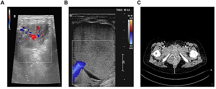 |
Figure 1 Ultrasound images at 36 weeks (A) and 38 weeks (B) of gestationshowing a heterogeneous cystic mass with associated vulvar varicosities, suggestive of a low-flow venous-type lesion. (C) CTV shows that the boundary of vulvar masses is clear. |
At 39 weeks of gestation, a lower-segment cesarean section was performed due to the presence of the mass and concurrent vulvar varicosities. The procedure was successful and the patient was discharged in stable condition.
On postpartum day 25, the vulvar lesion enlarged progressively and was accompanied by right lower-limb pain and swelling. Given the rapid enlargement during the puerperal period, differential diagnoses including vascular malformations and hemangioblastoma were considered. Ovarian venography, iliac venography, and lower-extremity venography revealed no evidence of pelvic vascular malformations or direct vascular communication with the lesion (Figure 1C). These imaging modalities were used for preoperative evaluation and surgical planning to assess vascular supply and minimize intraoperative bleeding risk.
Surgical excision of the vulvar vascular lesion was performed (Figure 2A). The tumor was large and contained tortuous venous plexuses with small arterial components at the base (Figure 2B and C). Complete resection was achieved (Figure 2D). Adequate preoperative blood preparation was undertaken, and the estimated blood loss was about 35 mL.
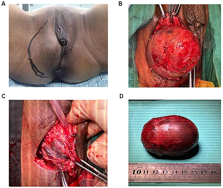 |
Figure 2 (A) Examination of the vulva with a lesion between the vaginal vestibule and the perineal region. (B) Surgical resection process. (C) After stripping, the basal tortuous vein and artery mass. (D) Complete resection of the hemangioma tumor body. |
Histopathological examination revealed a well-formed elastic lamina within the vascular wall, composed of the myofibrous tissue with granulation tissue formation, consistent with venous thrombosis and thrombus organization. Organized thrombus formation within the lesion was observed, which may explain the rapid postpartum enlargement. Immunohistochemistry demonstrated CD31 (vascular +), CD34 (vascular +), SMA (++), Desmin (++), WT-1 (-), D2–40 (++), Vimentin (++), and EMA (-). No histological features of hemangioblastoma, such as stromal cells with vacuolated cytoplasm or characteristic capillary networks, were identified. Special staining with EVG and HE confirmed the presence of elastic fibers (Figure 3).
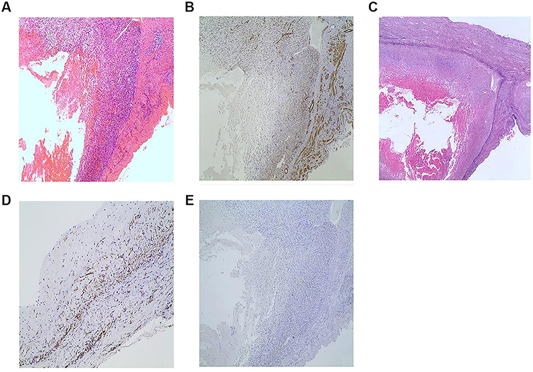 |
Figure 3 The histology, immunohistochemical staining histochemical staining of the lesion. (A) The vascular lumen was dilated, the vascular wall was locally thickened and locally thinned, and there was thrombosis in the vascular lumen (Hematoxylin and eosin staining, 40x magnification). Immunohistochemical staining showed that vascular wall was positive for smooth muscle actin. (B) Immunohistochemical staining, 40x magnification) and CD31. Elastic fiber staining outlined the complete vessel shape (C, histochemical staining, 20x magnification). (D) Immunohistochemical staining, 20x magnification), but was negative for WT1. (E) Immunohistochemical staining, 20x magnification). |
There was no clinical evidence suggesting a congenital hemangioma, as the lesion had not been present since birth or during the patient’s first pregnancy. The findings are more consistent with a venous malformation that became clinically apparent and enlarged under pregnancy-related hemodynamic changes, rather than the coexistence of both hemangioma and vascular malformation.
Written informed consent was obtained from the patient. During two years of follow-up, no recurrence has been observed.
Discussion
According to the ISSVA classification, vascular anomalies are broadly divided into vascular tumors (such as hemangiomas) and vascular malformations. From a biological standpoint, vascular anomalies are categorized based on clinical manifestations and the histological behavior of vascular endothelial cells into two major categories: true hemangiomas and vascular malformations.2 True hemangiomas are proliferative lesions characterized by active endothelial turnover and potential spontaneous involution, whereas vascular malformations are congenital structural abnormalities composed of mature, quiescent endothelial cells that typically persist without regression.3
Vulvar vascular tumors and malformations are rare. While hemangiomas most commonly affect the head and neck region, vulva involvement has been sporadically reported.4 These lesions typically arise in the labia majora and may remain asymptomatic for prolonged periods. However, large or deep lesions can cause swelling, pain, dyspareunia, cosmetic concerns, and, in rare cases, life-threatening hemorrhage.2,5 Hormonal and hemodynamic changes during pregnancy may promote lesion enlargement, likely due to increased pelvic blood flow, progesterone-mediated venous relaxation, and inferior vena cava compression by the gravid uterus.6,7
Given the rarity of vulvar vascular lesions, we reviewed representative previously published case reports to better characterize their clinical presentation, management strategies, and outcomes. Most reported cases involved cavernous hemangiomas or venous malformations of the labia majora. Management approaches varied according to lesion size, vascularity, and symptom severity, including observation, surgical excision, transarterial embolization, or combined strategies. Overall, complete surgical excision has been associated with favorable outcomes and low recurrence rates when feasible, whereas embolization appears particularly effective in cases complicated by hemorrhage or high-flow vascular lesions. A summary of representative cases is provided in Table 1 to assist clinicians in therapeutic decision-making.
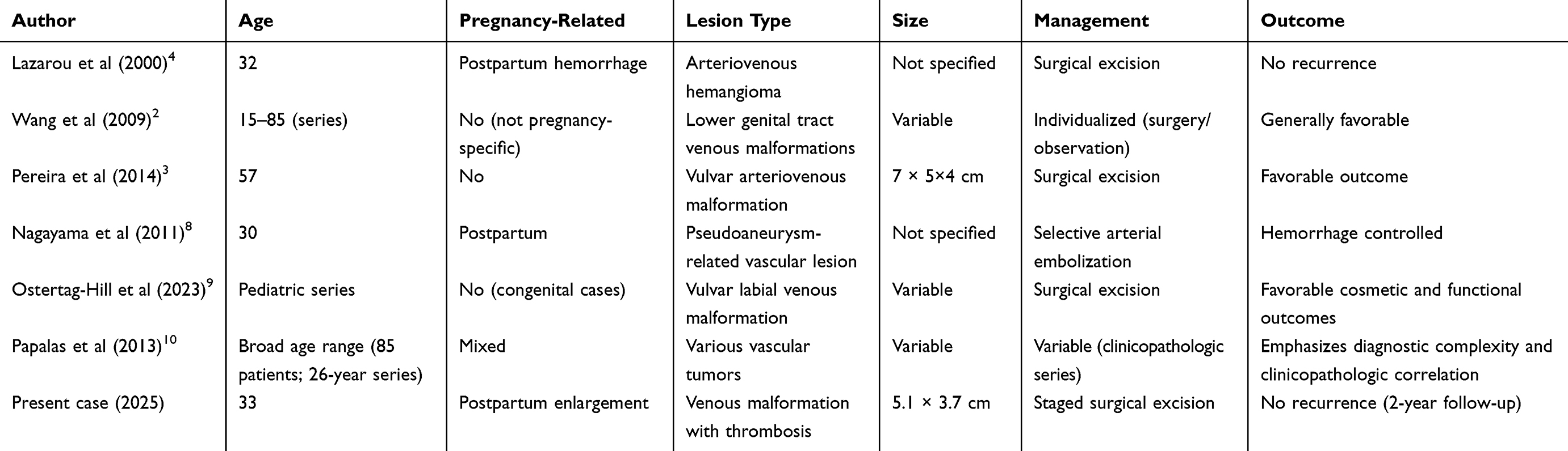 |
Table 1 Representative Previously Reported Cases and Series of Vulvar Hemangiomas and Vascular Malformations |
Therapeutic options for vulvar vascular lesions should be individualized according to lesion size, vascularity, symptom severity, and bleeding risk. Small, asymptomatic lesions may be managed conservatively with observation, whereas sclerotherapy, laser therapy, or transarterial embolization may serve as alternative or adjunctive treatments in selected cases, particularly when surgical bleeding risk is high. Surgical excision remains the most definitive approach for large, symptomatic, or complicated lesions, as it enables complete removal and provides a definitive pathological diagnosis. Careful preoperative planning is essential, as some lesions extend deeply or communicate with adjacent vessels. Magnetic resonance imaging and vascular studies can help delineate lesion boundaries and feeding vessels, and selective embolization may reduce intraoperative bleeding in high-risk cases.8,9
In selected postpartum cases without significant symptoms or progressive enlargement, minimally invasive approaches such as sclerotherapy or selective embolization may potentially reduce lesion size and alleviate symptoms, thereby avoiding or delaying surgical intervention. However, in the present case, the lesion demonstrated rapid postpartum enlargement accompanied by thrombosis-related compression symptoms and failure of spontaneous regression. Moreover, imaging studies did not identify a discrete feeding vessel suitable for targeted embolization. Therefore, surgical excision was considered the most appropriate and definitive management strategy.
With regard to surgical timing in this case, excision was not performed during the cesarean section. At the time of delivery, the lesion exhibited prominent vascular components and indistinct margins, and its relationship with surrounding structures had not been fully delineated, raising concern for uncontrolled hemorrhage if simultaneous excision were attempted. Furthermore, concurrent vulvar surgery during cesarean delivery would have prolonged operative time and increased peripartum risks. A staged surgical approach was therefore adopted, allowing improved vascular assessment and more controlled operative conditions after hemodynamic stabilization.
The rapid enlargement during the puerperal period was likely multifactorial. Postpartum hypercoagulability, venous stasis following cesarean section, and persistent pelvic venous congestion may have contributed to thrombosis within the lesion, resulting in secondary expansion and compression symptoms. Histopathological examination confirmed organized venous thrombosis, supporting this mechanism.
Based on intraoperative findings of tortuous venous plexuses and arterial channels at the base of the lesion, together with the presence of an elastic layer on histopathology and negative WT1 staining, the lesion most likely originated from a vulvar venous vascular pedicle rather than representing a true proliferative hemangioma. Although no direct communication with major pelvic vessels was identified on venography, these findings suggest a local venous origin with secondary thrombosis rather than a high-flow arteriovenous malformation.
According to the ISSVA classification, lesions historically termed “hemangiomas” may represent either proliferative vascular tumors or vascular malformations characterized by structural abnormalities with slow endothelial turnover.10 Immunohistochemical analysis assists in differentiation. WT1 expression has been associated with proliferative vascular tumors, whereas vascular malformations are typically WT1-negative.11 In this case, negative WT1 staining supported the diagnosis of a venous malformation with secondary thrombosis rather than a true proliferative hemangioma.
The importance of this case lies in its demonstration of rapid postpartum progression with secondary thrombosis, highlighting the dynamic behavior of vulvar venous malformations under pregnancy-related hemodynamic changes. It also underscores the need for careful differential diagnosis and individualized treatment planning, particularly when considering whether minimally invasive alternatives may be feasible before proceeding to definitive surgical management.
Conclusion
In conclusion, this case highlights the importance of considering vulvar venous vascular lesions in the differential diagnosis of postpartum vulvar masses. Clinicians should be aware that pregnancy- and puerperium-related hemodynamic changes may promote lesion enlargement and secondary thrombosis, leading to symptomatic compression. Early recognition, appropriate imaging evaluation, and individualized treatment planning are essential to avoid misdiagnosis, prevent severe complications such as hemorrhage, and achieve favorable clinical outcomes. This report underscores the need for careful assessment of postpartum vulvar masses and consideration of both minimally invasive and surgical management options based on lesion behavior, vascular characteristics, and patient-specific factors.
Data Sharing Statement
The analyzed data sets generated during the present study are available from the two corresponding author on reasonable request.
Ethics Approval and Consent to Participate
Institutional approval for the publication of this case report was obtained from the Ethics Committee of Beijing Luhe Hospital, Capital Medical University. All methods were carried out in accordance with Declaration of Helsinki.
Consent for Publication
The author obtained permission from the patient to publish this case report. The patient explicitly agreed to the anonymized reporting of their case details, in accordance with the Declaration of Helsinki and the Ethics Committee of Beijing Luhe Hospital, Capital Medical University ethical guidelines.
Author Contributions
All authors made a significant contribution to the work reported, whether that is in the conception, study design, execution, acquisition of data, analysis and interpretation, or in all these areas; took part in drafting, revising or critically reviewing the article; gave final approval of the version to be published; have agreed on the journal to which the article has been submitted; and agree to be accountable for all aspects of the work.
Funding
This study was not funded.
Disclosure
The authors declared that they have no conflicts of interest regarding this work.
References
1. Siu WYS, Chen YC, Ding DC. Vulvar hemangioma: a review. Diagnostics. 2025;15(10):1270. doi:10.3390/diagnostics15101270
2. Wang S, Lang JH, Zhou HM. Venous malformations of the female lower genital tract. Eur J Obstet Gynecol Reprod Biol. 2009;145(2):205–7. doi:10.1016/j.ejogrb.2009.05.017
3. Pereira N, Dormosh M, Mirmanesh M, Guilfoil DS. Arteriovenous malformation of the vulva: a case report. J Lower Genit Tract Dis. 2014;18(1):E12–E15. doi:10.1097/LGT.0b013e318286b770
4. Lazarou G, Goldberg MI. Vulvar arteriovenous hemangioma. A case report. J Reprod Med. 2000;45(5):439–441.
5. Hong HR, Hwang KR, Kim SA, et al. A case of vulvar hematoma with rupture of pseudoaneurysm of pudendal artery. Obstetr Gynecol Sci. 2014;57(2):168–171. doi:10.5468/ogs.2014.57.2.168
6. Gavrilov SG. Vulvar varicosities: diagnosis, treatment, and prevention. Int J Women’s Health. 2017;9:463–475. doi:10.2147/IJWH.S126165
7. Ryo E, Unno N, Nagasaka T, et al. Changes in the size of maternal inferior vena cava during pregnancy. J Perinat Med. 2004;32(4):327–331. doi:10.1515/JPM.2004.061
8. Nagayama C, Gibo M, Nitta H, et al. Rupture of pseudoaneurysm after vaginal delivery successfully treated by selective arterial embolization. Arch Gynecol Obstetrics. 2011;283(1):37–40. doi:10.1007/s00404-009-1271-1
9. Ostertag-Hill CA, Mulliken JB, Dickie BH, et al. Surgical resection of vulvar labial venous malformations: a single center experience. J Pediatr Surg. 2023;58(10):2038–2042. doi:10.1016/j.jpedsurg.2023.03.002
10. Papalas JA, Sangueza OP, Puri PK, et al. Vulvar vascular tumors: a clinicopathologic study of 85 patients. Am J Dermatopathol. 2013;35(1):
11. Karpathiou G, Chauleur C, Da Cruz V, Forest F, Peoc’h M. Vascular lesions of the female genital tract: clinicopathologic findings and application of the ISSVA classification. Pathophysiology. 2017;24(3):161–167. doi:10.1016/j.pathophys.2017.04.002
 © 2026 The Author(s). This work is published and licensed by Dove Medical Press Limited. The
full terms of this license are available at https://www.dovepress.com/terms
and incorporate the Creative Commons Attribution
- Non Commercial (unported, 4.0) License.
By accessing the work you hereby accept the Terms. Non-commercial uses of the work are permitted
without any further permission from Dove Medical Press Limited, provided the work is properly
attributed. For permission for commercial use of this work, please see paragraphs 4.2 and 5 of our Terms.
© 2026 The Author(s). This work is published and licensed by Dove Medical Press Limited. The
full terms of this license are available at https://www.dovepress.com/terms
and incorporate the Creative Commons Attribution
- Non Commercial (unported, 4.0) License.
By accessing the work you hereby accept the Terms. Non-commercial uses of the work are permitted
without any further permission from Dove Medical Press Limited, provided the work is properly
attributed. For permission for commercial use of this work, please see paragraphs 4.2 and 5 of our Terms.