
Back to Journals » International Journal of Nanomedicine » Volume 21
From Antibody Drugs to RNA Interference and Nanotherapy: The Evolution and Integration of Targeted Strategies in Gastric Cancer
Authors Wang S, Dong Y, Ma L  , Wu J, Long D, Mei Z , Jiang C, Lyu X
Received 22 April 2026
Accepted for publication 26 June 2026
Published 22 July 2026 Volume 2026:21 618409
DOI https://doi.org/10.2147/IJN.S618409
Checked for plagiarism Yes
Review by Single anonymous peer review
Peer reviewer comments 2
Editor who approved publication: Dr Yan Shen
Songlin Wang,1,* Yinhong Dong,1,* Lan Ma,2– 4,* Jiangfeng Wu,2– 4 Daoguo Long,1 Zhao Mei,5,6 Chuanlin Jiang,1 Xiaoguang Lyu1,7
1Zhongxiang People’s Hospital, Zhongxiang, People’s Republic of China; 2Hubei Key Laboratory of Tumor Microenvironment and Immunotherapy, China Three Gorges University, Yichang, People’s Republic of China; 3College of Basic Medical Science, China Three Gorges University, Yichang, People’s Republic of China; 4Institute of Organ Fibrosis and Targeted Drug Delivery, China Three Gorges University, Yichang, People’s Republic of China; 5Department of Pharmacy, The First College of Clinical Medical Science, China Three Gorges University, Yichang, People’s Republic of China; 6Department of Pharmacy, Yichang Central People’s Hospital, Yichang, People’s Republic of China; 7Department of Gastroenterology, Renmin Hospital of Wuhan University, Wuhan, People’s Republic of China
*These authors contributed equally to this work
Correspondence: Chuanlin Jiang, Email [email protected] Xiaoguang Lyu, Email [email protected]
Abstract: Gastric cancer is a leading cause of cancer death worldwide. Targeted therapy is shifting from blocking signaling pathways to precise gene regulation. This review covers three key areas: antibody drugs, RNAi, and nanocarriers. Anti-HER2 agents, VEGFR2 inhibitors, Claudin 18.2-directed therapies and PD-1/PD-L1 inhibitors improve survival in biomarker-selected patients—but resistance and low response rates (ORR < 20% in unselected groups) limit their use. RNAi silences key oncogenes to reverse chemoresistance and reprogram the immunosuppressive tumor microenvironment, but it suffers from rapid degradation, poor endosomal escape, off-target effects, and weak tumor penetration. Nanocarrier systems—including lipid nanoparticles (LNPs), stimuli-responsive micelles, and mesoporous silica—combined with aptamers enable targeted delivery of therapeutic antibodies and siRNA, penetration across stromal barriers, and controlled, tumor microenvironment–triggered cargo release. Altogether, the future of gastric cancer therapy is moving beyond monotherapy to a five-part strategy: antibody targeting, RNA silencing, nanocarrier delivery, AI-guided decisions, and immune remodeling.
Keywords: gastric cancer, target therapy, RNA interference, siRNA, shRNA, nanoparticles
Gastric cancer remains a significant global health challenge—it is the fifth most commonly diagnosed cancer and the fourth leading cause of cancer-related death worldwide, with 684,000 lives lost in 2022 (GLOBOCAN).1 In China, gastric cancer is the third in incidence and the second in mortality—accounting for 478,000 new cases and 373,000 deaths annually.2,3 Due to its asymptomatic nature in the early stage, 70% of patients are diagnosed at advanced stage, resulting in a five-year overall survival rate of less than 10%.4–7 These figures underscore an urgent need for more effective therapies to delay tumor progression and extend survival time.
Antibody-based drugs launched targeted therapy for gastric cancer. Trastuzumab validated HER2, ramucirumab enabled anti-angiogenesis, and immune checkpoint inhibitors achieved durable responses in biomarker-selected patients.8–11 But resistance—due to target loss, pathway redundancy, or immune evasion—and low response rates in unselected patients remain key challenges. 10 RNA interference (RNAi) offers the capacity to silence pathogenic genes at the mRNA level.12 In gastric cancer, siRNAs directed against HER2, c-MET, or KRAS mutants have shown promising results in preclinical studies. 13 The translational barrier, however, is delivery: naked nucleic acids are rapidly degraded, poorly internalized, and prone to off-target effects.12,13 It is here that nanotechnology becomes indispensable. Nanocarriers—such as lipid nanoparticles and polymeric micelles—protect siRNA from degradation, enhance cellular uptake, and enable controlled release.14,15 They can also penetrate the dense gastric tumor stroma and respond to microenvironmental cues like low pH or elevated enzyme levels.14,16 Most promising is delivering antibodies and siRNA in the nanocarrier to combine extracellular targeting with intracellular gene silencing.13,17–19
This review traces the evolution from antibody drugs to RNAi and nanotherapeutics in gastric cancer. We argue that monotherapy is inherently limited and that the future lies in synergistic integration: antibodies for targeting, RNAi for gene silencing, and nanotechnology for precision delivery.14,16
Clinically Validated Molecular Targets in Gastric Cancer for Targeted Therapy and Immunotherapy (Table 1)
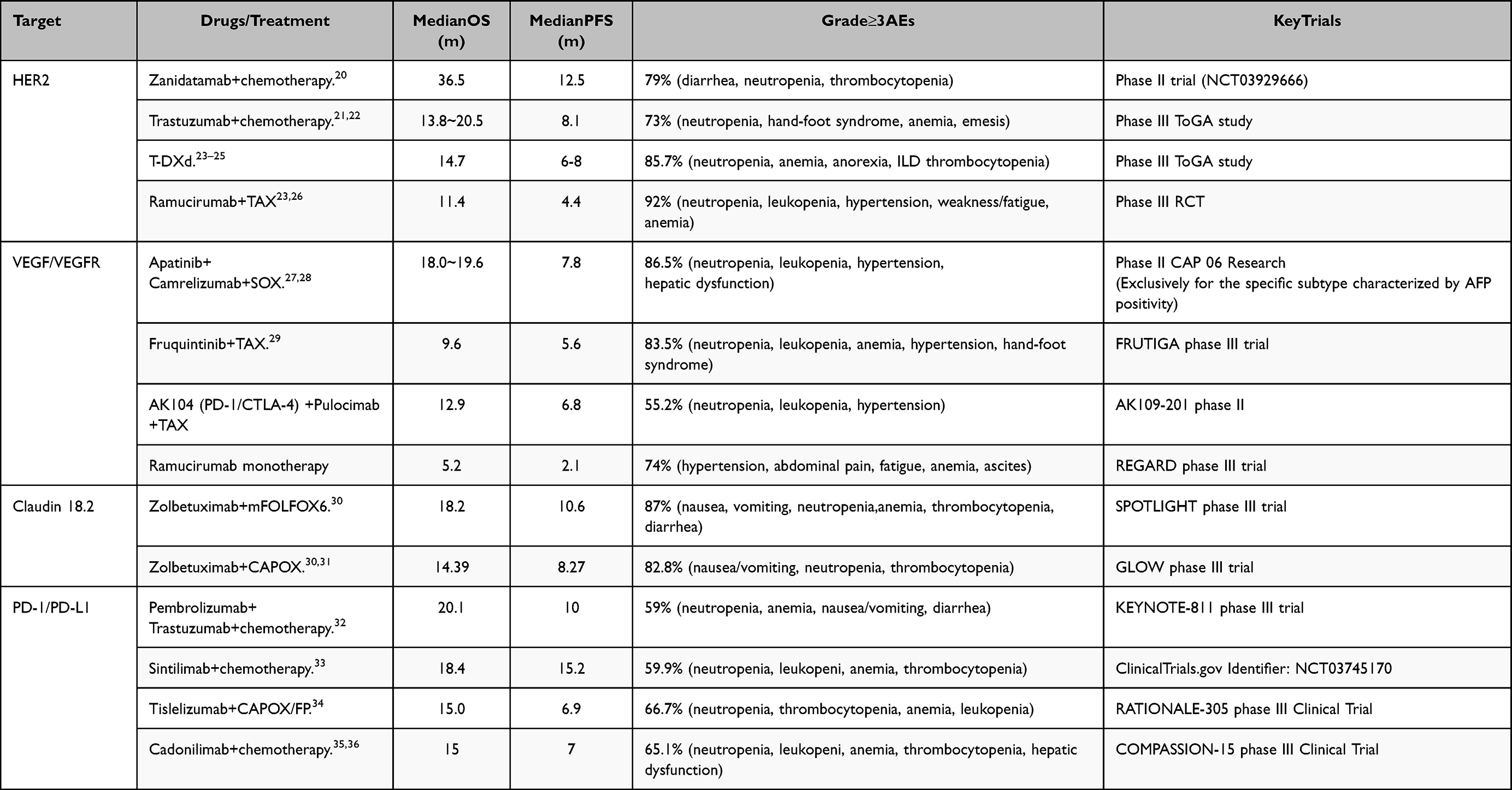 |
Table 1 Gastric Cancer Targeted Drugs Currently Under Clinical Investigation |
The Following are the Crucial Molecular Targets
Targeting the HER2 Receptor
HER2 (ErbB2), a ligand-independent receptor tyrosine kinase, preferentially heterodimerizes with HER3 or EGFR, triggering constitutive activation of the PI3K/AKT/mTOR and RAS/RAF/MEK/ERK pathways in the absence of ligand binding.37–41 These cascades promote cell survival, proliferation, and immune evasion through effectors including mTORC1, Elk-1, c-Myc, and STAT3.42
PI3K/AKT/mTOR Pathway
When HER2 heterodimerizes with HER3, it recruits and phosphorylates the PI3K regulatory subunit, thereby activating the enzyme. Activated PI3K converts PIP2 to PIP3, which recruits and activates AKT (protein kinase B). AKT then promotes cell survival and suppresses apoptosis by phosphorylating key targets—including BAD and caspase-9—and enhancing pro-survival signaling.43–45 AKT also activates mTORC1, driving protein synthesis, cell growth, proliferation, and metabolic adaptation.46–48
RAS/RAF/MEK/ERK Pathway
HER2 activation recruits Grb2-SOS complex, triggering RAS activation. Active RAS activates RAF (MAPKKK), which phosphorylates and activates MEK (a MAPKK); MEK then phosphorylates and activates ERK (a MAPK). Nuclear translocation of activated ERK leads to phosphorylation of transcription factors—including Elk-1, c-Fos, and c-Myc—driving G1-to-S-phase cell cycle progression, proliferation, differentiation, and motility.46,49 Both pathways also phosphorylate STAT3, thereby modulating JAK/STAT signaling to promote immune evasion and reshape the tumor microenvironment.50
Both pathways phosphorylate STAT3, thus modulating JAK/STAT signaling to promote immune evasion and reshape the tumor microenvironment.46,50
Targeting the VEGF Receptor (VEGFR)
The VEGF family includes VEGF-A, VEGF-C, and VEGF-D. VEGF-A drives angiogenesis via VEGFR-1/VEGFR-2 (KDR); VEGF-C/VEGF-D drive lymphangiogenesis via VEGFR-3.10 In gastric cancer, VEGF-A and VEGFR-2 are overexpressed relative to adjacent normal tissue and correlate with microvessel density (MVD). This promotes leaky vasculature, fueling hypoxia, acidosis, invasion, and metastasis: high MVD supports tumor growth and spread—VEGF enhances permeability to facilitate intravasation; VEGF-C/VEGFR-3 drives lymphangiogenesis and lymph node metastasis. VEGF also induces an immunosuppressive microenvironment by impairing dendritic cell maturation and T-cell infiltration, expanding Tregs, and upregulating PD-L1—contributing to “cold tumor” resistance to checkpoint inhibitors.10,51,52 Consequently, targeting VEGF/VEGFR (eg, fruquintinib [FRUTIGA trial, HR 0.67] and surufatinib) improves survival in heavily pretreated Chinese patients.53
Targeting the Claudin 18.2
CLDN18.2 is a tight junction protein normally confined to the apical surface of gastric epithelial cells, maintaining epithelial barrier integrity and polarity.30,54 In gastric cancer, loss of polarity exposes it across the entire tumor cell membrane—converting it from an immunologically “hidden” to a “conspicuous” target.5,55 Epigenetic derepression drives its overexpression: promoter hypomethylation lifts transcriptional silencing, enabling stable, high-level expression. This fuels oncogenesis via three key mechanisms: (1) PI3K/AKT activation → suppressed apoptosis and enhanced survival/proliferation; (2) ERK/MAPK-driven AP-1 activation → upregulation of cell-cycle genes; and (3) CLDN18-ARHGAP26 fusion (in ~15% of diffuse-type cases) → constitutive RhoA activation, cytoskeletal remodeling, increased motility, EMT, and invasion. CLDN18.2+ tumors also create an immunosuppressive microenvironment—recruiting Tregs, M2 macrophages, and MDSCs; impairing dendritic cell maturation and T-cell infiltration; and disrupting antigen presentation—enabling immune escape.56,57 Critically, its stable epigenetic expression—and selective pressure in metastatic niches—elevates CLDN18.2 in metastases, reinforcing its value as a metastatic target.54,58 Clinically, zolbetuximab + chemotherapy improved PFS (7.5 vs 5.3 mo) and OS (14.4 vs 12.2 mo) in SPOTLIGHT and GLOW trials, leading to FDA/NMPA approval in 2024.30,31 Claudin 18.2–directed CAR-T (CT053) achieved a 57.1% ORR in heavily pretreated patients.
Target of Immune Checkpoint Inhibitor Therapy
PD-1/PD-L1
In gastric adenocarcinoma, PD-L1 is upregulated by inflammatory cytokines—including IFN-γand IL-6—via JAK/STAT and NF-κB signaling, enabling immune evasion. Approximately 10% of cases are EBV-positive; these tumors consistently overexpress PD-L1 and show enhanced response to PD-1 blockade. PD-1 is expressed on T cells, B cells, and NK cells, whereas its ligand PD-L1 is upregulated on tumor cells and antigen-presenting cells (eg, macrophages, dendritic cells). PD-1/PD-L1 binding recruits SHP-1 and SHP-2 phosphatases, inhibiting TCR signaling—thereby suppressing T-cell proliferation, cytokine production (IFN-γ, IL-2), and effector function—and facilitating tumor immune escape.59
Analysis of the Current Limitations in Targeted Therapy for Gastric Cancer
Dependence on Specific Target Expression
Targeted therapy requires uniform, high-level expression of tumor-specific markers—but many gastric cancers show low or heterogeneous expression. For example, only 7.3–20.2% of patients overexpress HER2, typically in focal, intensely stained regions.60 Claudin 18.2 positivity varies widely (30–70%) depending on the IHC assay and cutoff: early studies using the 43–14A antibody reported >60% positivity, whereas the VENTANA SP97 assay (≥2+in ≥75%) yields 35–50% in both Western and Asian cohorts.61 This variability underscores the urgent need for standardized, companion diagnostic—approved testing.
Prominent Drug Resistance Challenges
Targeted therapies face primary and secondary resistance. Primary resistance arises from compensatory pathway activation (eg, MAPK, PI3K/AKT) or baseline mutations; secondary resistance stems from acquired target-gene mutations (eg, HER2 L755S/V777L), phenotypic shifts, epigenetic changes, or co-amplifications (MYC, CCNE1, CDK12).62–65 For example, trastuzumab resistance often involves HER2 alterations, IGF-1R upregulation, or bypass signaling via MET/FGFR. Apatinib may promote immune evasion by activating MAPK or inhibiting STAT3/PKM2.64–66 VEGF further contributes to a “cold tumor” microenvironment by impairing dendritic cell maturation and T-cell infiltration while expanding Tregs.67
Abnormal Tumor Vasculature
Tumor blood vessels are chaotic, tortuous, leaky, and dysfunctional—causing poor drug delivery and an immunosuppressive microenvironment. Hypoxia from abnormal perfusion recruits immunosuppressive cells and factors, impairing immune cell infiltration and function. Anti-VEGF agents (eg, ramucirumab, apatinib) were initially intended to “starve” tumors. However, short-term, moderate VEGF inhibition could induce transient “vascular normalization”—a temporary restoration of vessel structure and function. Yet this window is narrow, dynamic, and highly variable across patients, making it hard to identify and easy to miss clinically. Moreover, tumors rapidly activate bypass pathways (eg, FGF, Angiopoietin) upon VEGF blockade, driving vascular regrowth and undermining combination therapy. This adaptation not only reduces efficacy but may also promote a more invasive phenotype.
Safety Concerns
Some targeted agents cause significant toxicities. Topoisomerase inhibitors like exatecan induce grade ≥3 neutropenia in 20–30% of patients.68 CCR8×CLDN18.2 bispecific antibodies (eg, LM-108) are still in early development; their long-term safety and efficacy remain undefined. Bemarituzumab causes vision impairment, dry eye, or corneal damage in >25% of patients—mostly reversible but potentially affecting adherence.69 Long-term data on Claudin 18.2-targeted therapies are scarce, so the risks of chronic toxicities (eg, interstitial lung disease) are unclear.
Economic and Accessibility Barriers
High treatment costs limit the broad use of targeted therapies. Trastuzumab costs over 300,000 yuan annually; CAR-T therapy—typically priced above 1 million yuan per treatment—presents a substantially higher financial barrier, critically constraining patient access. So far, only one Claudin 18.2-targeted agent, zolbetuximab (not trastuzumab), has received global marketing approval; ADCs and CAR-T therapies remain under clinical investigation. First-line guidelines increasingly recommend “targeted therapy + immunotherapy + chemotherapy”—a regimen far costlier than chemotherapy alone. Biomarker testing (eg, for Claudin 18.2) adds further financial strain.
RNA Interference (RNAi) Technology Demonstrates Significant Potential in the Field of Gastric Cancer Therapy (Table 2); However, It Also Encounters Various Challenges
Key Advantages of RNAi-Based Therapeutic Approaches in Gastric Cancer Management
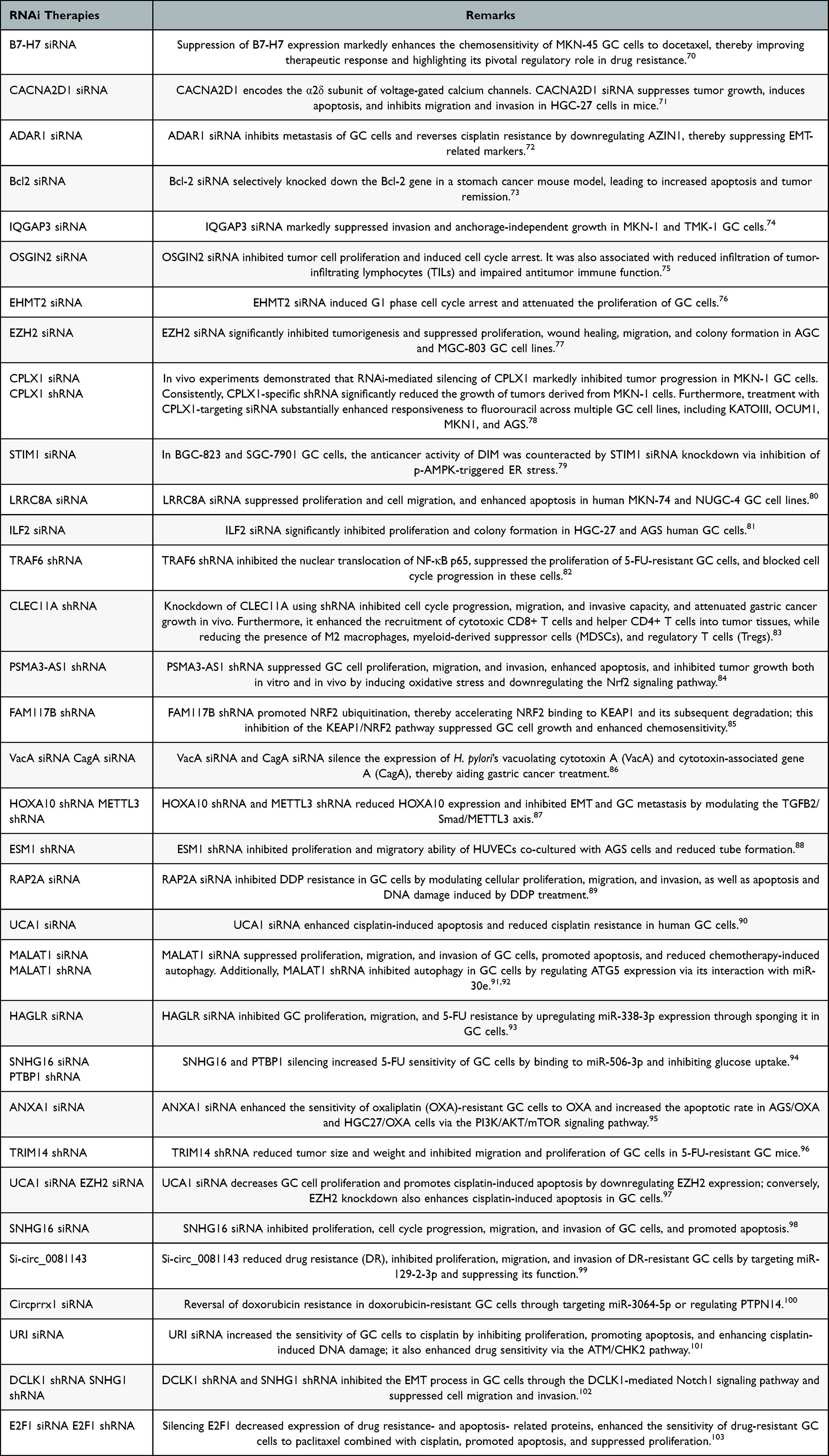 |
Table 2 Recent Advances in Experimental Research on RNAi Therapy for Gastric Cancer |
2) This technology effectively addresses drug resistance commonly encountered in conventional therapies. For example, knockdown of the lncRNA NCK-AS1 reverses cisplatin resistance and improves chemotherapeutic sensitivity.106 Targeting the E3 ubiquitin ligase SYVN1 has also been shown to significantly inhibit gastric cancer cell (GC cell) proliferation.107
3) RNAi technology demonstrates the capacity to modulate the tumor microenvironment (TME). Silencing of the NAT10 gene interrupts the CXCL2/STAT3 signaling axis, thereby suppressing M2 macrophage polarization—with the proportion of M2 macrophages in liver metastases declining from 42% to baseline levels—resulting in a 78% reduction in liver metastasis.108 Furthermore, the novel delivery system TMAB3, which carries a RIG-I agonist RNA, enhances CD8⁺ T cell infiltration and effectively reverses the “cold tumor” phenotype, thereby augmenting anti-tumor immune responses.109
4) The technology is associated with low toxicity and minimal side effects. For example, under physiological conditions, AE1 is exclusively expressed in red blood cells, and siRNA delivery vehicles are unable to cross the bone marrow-blood barrier (MBB).105 Consequently, no hemolytic or hepatorenal toxicities were observed in preclinical animal studies.
Current Challenges and Limitations of RNAi Therapy
1) Instability of RNA molecules: Unprotected RNA is highly susceptible to degradation by endogenous nucleases in vivo, leading to a short half-life in blood and tissues. This instability prevents the maintenance of sustained therapeutic concentrations, thereby compromising treatment efficacy.110–112
2) Suboptimal delivery efficiency: Conventional delivery systems—such as liposomes—exhibit limited targeting specificity. The lack of highly efficient and selective delivery platforms hinders the precise accumulation of RNAi molecules at tumor sites, thereby constraining their broad clinical applicability.100 Consequently, numerous studies are currently focused on developing highly specific, tumor-targeted delivery systems—including aptamers and nanoparticles—to enhance the efficient delivery of siRNA and shRNA to tumor sites (Figure 1 and Table 3).111,113–115 Tumor-targeted lipid nanoparticles (such as Onpattro-like platforms) and GalNAc-conjugated siRNAs are currently undergoing phase I/II trials for solid tumors. Aptamer-siRNA chimeras specific to gastric cancer and pH-responsive nanoparticles have achieved over 50-fold tumor enrichment in orthotopic models.
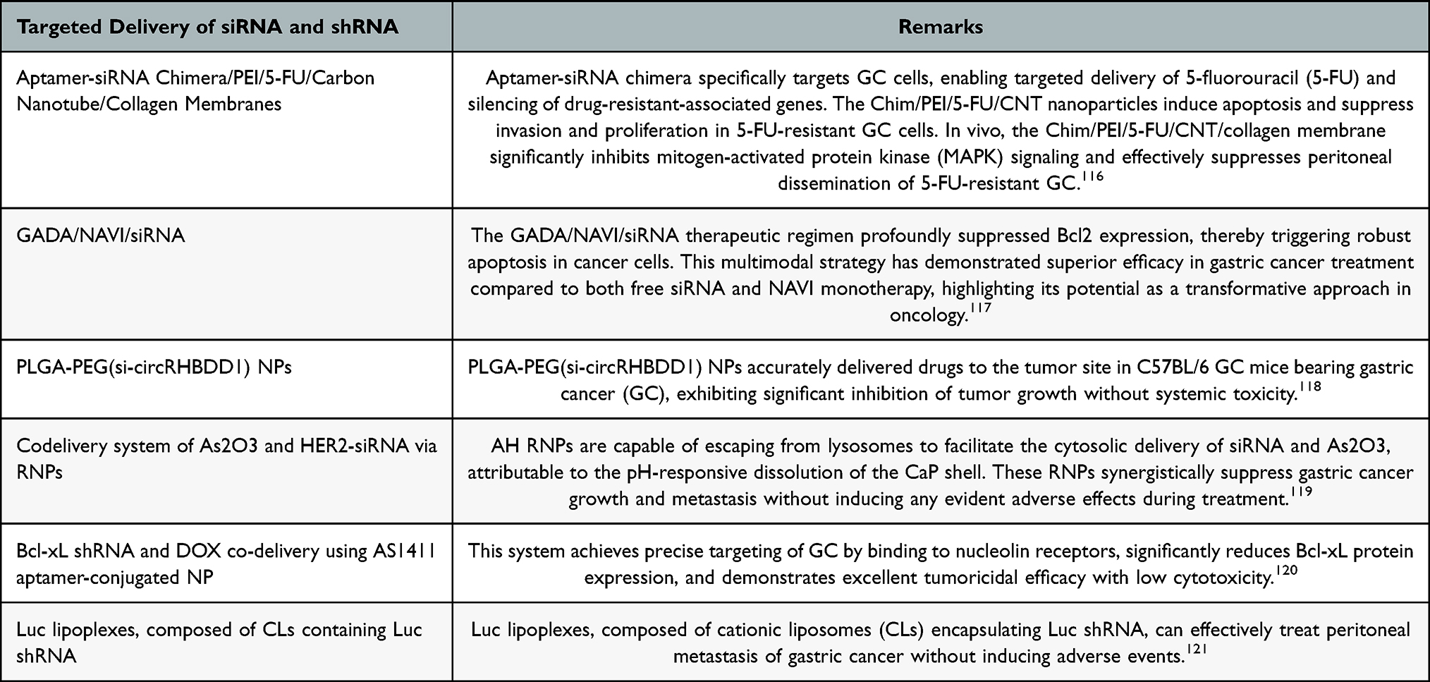 |
Table 3 Recent Advances in Experimental Research on Targeted Delivery of siRNA and shRNA for RNAi Therapy in Gastric Cancer |
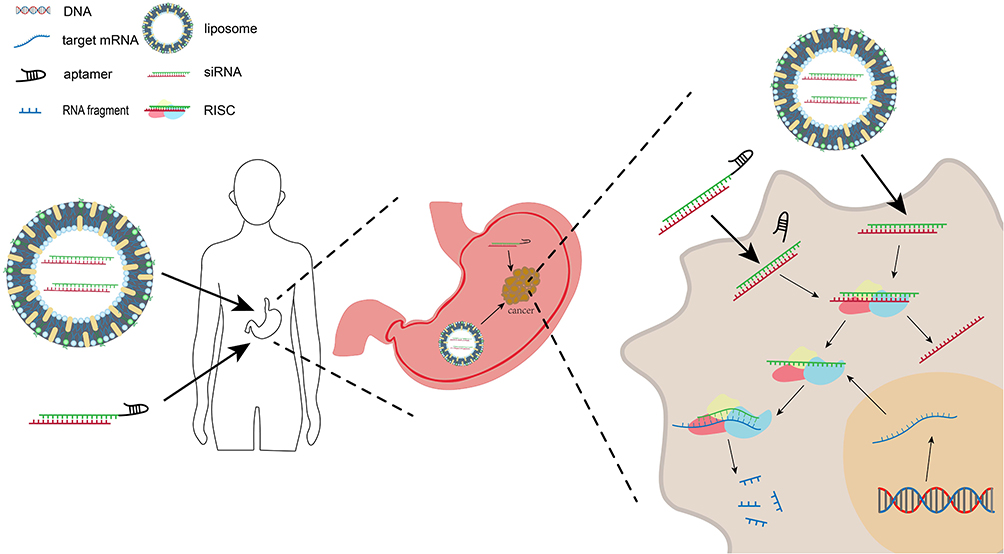 |
Figure 1 Targeted delivery systems, such as aptamers and nanoparticles, improve the efficient and precise delivery of siRNA to tumor sites, thereby enhancing therapeutic efficacy and enabling potent antitumor effects. |
3) Potential off-target effects: RNAi molecules may hybridize with non-target mRNA sequences, leading to unintended gene silencing and posing potential risks of adverse physiological effects.115,122
4) Immune activation potential: Exogenous RNA can activate the innate immune system, inducing the production of inflammatory mediators that may compromise both the safety and tolerability of RNAi-based therapies.122
5) Production complexity and cost constraints: The synthesis, purification, and quality control of RNAi therapeutics involve technically demanding processes, contributing to high manufacturing costs. These challenges hinder large-scale production and widespread clinical adoption. Moreover, complementary genetic testing (eg, assessment of NAT10 and KLF5 expression levels) imposes additional economic and therapeutic burdens on patients.
6) Inadequate long-term safety data: The majority of current evidence is derived from animal models, and comprehensive long-term safety evaluations in humans—particularly regarding immunogenicity and organ-specific accumulation—remain lacking.122 Furthermore, epigenetic targets such as NAT10 may disrupt normal RNA modification pathways in healthy cells, raising concerns about potential yet undefined biological risks.
Nanoparticle-Based Therapeutics for Gastric Cancer: Advancing from Passive Targeting to Intelligent, Multimodal Synergy
Evolution of Targeting Strategies: From Active Targeting to Microenvironment-Responsive Delivery
Conventional passive targeting relies on the enhanced permeability and retention (EPR) effect. However, EPR effect is highly heterogeneous across and within gastric tumors, limiting its reliability. To improve targeting, nanoparticles are surface-modified with ligands—such as folate, transferrin, antibodies, or aptamers—that bind receptors overexpressed on GC cells. For example, folate receptor alpha (FR ɑ) is overexpressed in >60% of gastric cancers; folate-conjugated nanoparticles increase tumor accumulation 3-5-fold compared with non-targeted controls in mouse models.123 Microenvironment-responsive nanoparticles offer greater precision: they release drug only in response to gastric tumor-specific cues—such as low pH (6.0–6.8), high GSH concentrations (2–10 mM), elevated ROS, or overexpression of matrix metalloproteinases MMP-2 and MMP-9—using cleavable linkers like hydrazone, disulfide, or thioacetal bonds. A pH/ROS dual-responsive docetaxel nanomicelle achieved 79.6% tumor growth inhibition in subcutaneous gastric cancer xenografts—significantly higher than that of free docetaxel.124
Overcoming Chemotherapy Resistance: From Gene Silencing to Pathway Intervention
Chemotherapy resistance is a major cause of treatment failure in gastric cancer. Nanoparticles can deliver both chemotherapeutic drugs and gene therapeutics—such as siRNA, miRNA, or CRISPR-Cas systems—to reverse resistance at its source. Two key strategies are employed: (1) Gene silencing—target ABC transporters (eg, P-gp) or anti-apoptotic proteins (eg, Survivin, Bcl-2); and (2) Pathway inhibition—block pro-survival signals such as Notch, Wnt/β-catenin, or NF-κB. For example, liposomal nanoparticles co-delivering P-gp siRNA and paclitaxel reduced the IC50 by ~80% in resistant SGC7901/VCR cells.125 Similarly, SPIONs functionalized with the WSGC peptide inhibited Notch signaling, suppressing tumor growth by >60% and enhancing apoptosis in xenografts.123
Emerging Physical Therapies for Cancer Treatment: Integrating Photothermal Therapy, Ferroptosis Induction, and Immunomodulation
Nanoparticles serve as programmable theranostic platforms—functioning either as photonic energy transducers or as precision inducers of regulated cell death.
Photothermal Therapy (PTT)
Carbon quantum dots, MXene, and polydopamine nanoparticles absorb near-infrared light (808 nm or 1064 nm) to generate heat, raising tumor temperature above 50 °C and killing cancer cells. Limitation: poor tissue penetration limits their use in deep gastric tumors or peritoneal metastases.126,127
Ferroptosis Therapy
Nanoparticles deliver Fe2⁺/Fe3⁺ or inhibit GPX4, thereby inducing lipid peroxidation and ferroptosis. The biomimetic nanocrystal TRNC@P+L co-delivers chemotherapeutic drugs and ferroptosis inducers, showing synergistic efficacy in preclinical models of gastric cancer peritoneal metastasis.128
Immunomodulatory Nanoparticles
In a gastric carcinoma mouse model, M2pep-Cs NPs/Plerixafor nanoparticles suppress tumor progression by concurrently blocking the CXCL12-CXCR4 axis and reprogramming tumor-associated macrophages (TAMs)—specifically shifting them from immunosuppressive M2 phenotype to immunostimulatory M1 phenotypes—which enhances T-cell infiltration, activation, and tumor antigen recognition.129
Nanometer-Scale Chemotherapeutic Agents: Advancing Therapeutic Efficacy While Minimizing Systemic Toxicity
The real-world study of NPMP (paclitaxel nanomicelles) combined with immunotherapy and chemotherapy—though non-randomized and uncontrolled—achieved an objective response rate (ORR) of 42.4%, exceeding the typical ORR of 25–35% observed with conventional chemotherapy. Neutropenia rates did not increase significantly compared with historical controls, suggesting that the nanomicellar formulation improves the therapeutic index through tumor-selective drug accumulation.130 SN BioScience has received FDA Orphan Drug Designation for SNB-101 in gastric cancer—the third such designation for this novel nanoparticle formulation of SN-38. As the world’s first polymer-based nanoparticle formulation of the highly water-insoluble anticancer agent SN-38, SNB-101 is designed to enhance efficacy and reduce toxicity. The designation addresses a critical unmet medical need in gastric cancer, where advanced disease has limited treatment options and a 5-year survival rate of only 36%.
Future Development of Gastric Cancer Treatment (Figure 2)
Future directions in gastric cancer treatment focus on four key areas: 1) Precision oncology: Multi-omics profiling (genomics, proteomics, metabolomics) identifies targetable molecular drivers; CRISPR/Cas9 and animal models validate function and therapeutic potential; resulting agents—small molecules, mAbs, or bispecific antibodies—are matched to patients’ molecular profiles (eg, mutations, protein expression, immune contexture) for stratified therapy. 2) Next-generation ADCs: Optimized DAR, cleavable/conditionally active linkers, and novel payloads improve tumor selectivity and safety; rational combinations—eg, with chemo, immunomodulators, or dual-target inhibitors—overcome resistance and broaden indications. 3) CLDN18.2-directed CAR-T: Now in randomized clinical trials (eg, CT041-ST-01), this “living drug” bypasses vascular dependence, enabling direct tumor cell killing. It shows durable survival benefit after standard therapy failure, with manageable CRS and no neurotoxicity in trials. Limitations remain: strict CLDN18.2+ selection (≥40% tumor cells), complex manufacturing/logistics, high cost, and persistent challenges of solid tumors—including immunosuppressive microenvironments and target heterogeneity.
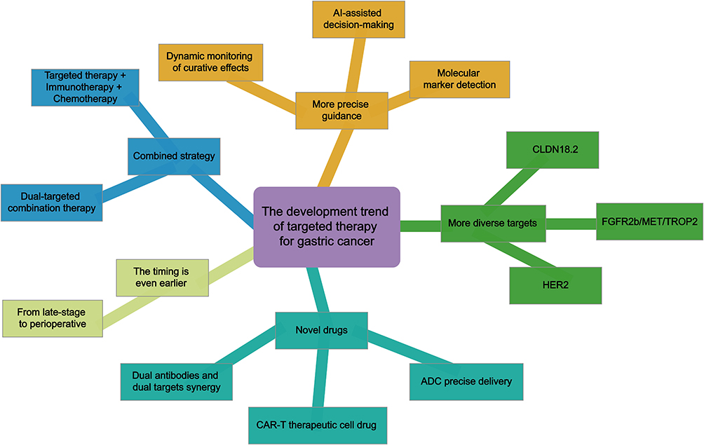 |
Figure 2 Trends in the Development of Targeted Therapy for Gastric Cancer. |
Systemic therapy for gastric cancer has evolved from sequential monotherapy to rational combinations of immune checkpoint inhibitors, targeted agents, and chemotherapy. Aptamer- and nanocarrier-based delivery platforms represent a paradigm shift—enabling spatiotemporally controlled treatment, not merely incremental improvement. Current standard therapies (eg, anti-HER2, CLDN18.2-targeted, and dual PD-1/CTLA-4 blockade) provide clinical benefit but suffer from narrow therapeutic indices due to on-target/off-tumor toxicity. Aptamers—synthetic, high-affinity, low-immunogenicity nucleic acid ligands—precisely target gastric cancer drivers. Nanocarriers exploit both passive (enhanced permeability and retention, EPR) and active (ligand-mediated) tumor accumulation and release their payloads selectively in response to the tumor microenvironment—within the tumor parenchyma or the immunosuppressive stroma. Integrating these modalities enables intelligent nanosystems: either for localized intraperitoneal delivery or as multi-epitope tandem aptamer–drug conjugates (ADCs) that address gastric cancer’s core challenges—inter- and intra-tumoral heterogeneity and its strong propensity for peritoneal dissemination. This dual-strategy approach tackles three key clinical challenges in peritoneal metastases: (1) poor drug accumulation due to low peritoneal–plasma barrier permeability; (2) emergence of therapy-resistant clones under single-target treatment; and (3) systemic toxicities that limit dose intensity and duration. Clinical translation requires solving three hurdles: aptamer degradation by nucleases in vivo, RES-mediated nanoparticle clearance, and scalable GMP-compliant manufacturing with strict quality control. A phaseItrial of this aptamer-nanocarrier platform in gastric cancer patients with peritoneal metastases is expected within five years. Primary endpoints include safety plus quantitative pharmacodynamic biomarkers—tumor-to-plasma drug ratio, target saturation kinetics, and spatially resolved intratumoral payload distribution. Critically, these mechanistic metrics—not just response rates—will determine whether the platform evolves from innovation to standard therapy.
The aptamer-nanodrug system’s key advantage in gastric cancer treatment is not antibody replacement, but programmable, precise targeting. Unlike antibodies, aptamers can both bind targets and execute logic operations—eg, releasing drugs in response to tumor microenvironment cues—via rational structural design. Yet current research focuses excessively on aptamer loading onto nanoparticles, overlooking a fundamental question: What target profiles do distinct gastric cancer molecular subtypes actually require? Future progress depends less on generating more aptamers and more on establishing a clinical-translational paradigm: begin with molecular subtyping, reverse-define target requirements, then custom-design the aptamer–nanoparticle system. Critically, this system must integrate into a closed loop with liquid biopsy–based early detection and post-surgical minimal residual disease monitoring—to translate precise targeting into real survival benefits.
Acknowledgments
This study was supported by the National Natural Science Foundation of China (Grant number [81670555]); State Key Laboratory of Medical Molecular Biology (Grant number [State Key Laboratory Special Fund 206024]); 2025 Yichang Medical and Health Research Project (No. A25-2-178); Chen Xiao-Ping Foundation for the Development of Science and Technology of Hubei Province (No. CXPJJH125001-2509).
Since English is not our native language, the article was refined with the assistance of Youdao Translate (Version: 11.0.0).
Author Contributions
All authors made a significant contribution to the work reported, whether that is in the conception, study design, execution, acquisition of data, analysis and interpretation, or in all these areas; took part in drafting, revising or critically reviewing the article; gave final approval of the version to be published; have agreed on the journal to which the article has been submitted; and agree to be accountable for all aspects of the work.
Disclosure
The authors declare no conflicts of interest.
References
1. Röcken C. Predictive biomarkers in gastric cancer. J Cancer Res Clin Oncol. 2022;149(1):467–15. doi:10.1007/s00432-022-04408-0
2. Högner A, Moehler M. Immunotherapy in gastric cancer. Curr Oncol. 2022;29(3):1559–1574. doi:10.3390/curroncol29030131
3. Huang RJ, Laszkowska M, In H, Hwang JH, Epplein M. Controlling gastric cancer in a world of heterogeneous risk. Gastroenterology. 2023;164(5):736–751. doi:10.1053/j.gastro.2023.01.018
4. Guan W-L, He Y, Rui-Hua X. Gastric cancer treatment: recent progress and future perspectives. J Hematol Oncol. 2023;16(1):57. doi:10.1186/s13045-023-01451-3
5. Mathias-Machado MC, de Jesus VHF, Jácome A. Claudin 18.2 as a new biomarker in gastric cancer-what should we know? Cancers. 2024;16(3):679. doi:10.3390/cancers16030679
6. Smyth EC, Nilsson M, Grabsch HI, van Grieken NC, Lordick F. Gastric cancer. Lancet. 2020;396:635–648. doi:10.1016/S0140-6736(20)31288-5
7. Siegel RL, Giaquinto AN, Jemal A. Cancer statistics, 2024. Cancer J Clin. 2024;74:12–49. doi:10.3322/caac.21820
8. Wu Z, Ni J, Xu Q. Clinical advances and challenges of anti-angiogenic targeted therapy in gastric cancer. Front Oncol. 2025;15. doi:10.3389/fonc.2025.1654300
9. Wang Y, Chong G. Immunotherapy in gastric adenocarcinoma – a rapidly evolving treatment landscape. Crit Rev Oncol Hematol. 2025;216:104941. doi:10.1016/j.critrevonc.2025.104941
10. Luo D, Liu Y, Zhengmao L, et al. Targeted therapy and immunotherapy for gastric cancer: rational strategies, novel advancements, challenges, and future perspectives. Mol Med. 2025;31(1):52. doi:10.1186/s10020-025-01075-y
11. Scheck MK, Hofheinz RD, Lorenzen S. HER2-positive gastric cancer and antibody treatment: state of the art and future developments. Cancers. 2024;16(7):1336. doi:10.3390/cancers16071336
12. Liu R, Zhou J, Chen X, et al. Diagnostic and therapeutic advances of RNAs in precision medicine of gastrointestinal tumors. Biomedicines. 2024;13(1):47. doi:10.3390/biomedicines13010047
13. Hashemi M, Aparviz R, Beickzade M, et al. Advances in RNAi therapies for gastric cancer: targeting drug resistance and nanoscale delivery. Biomed Pharmacother. 2023;169:115927. doi:10.1016/j.biopha.2023.115927
14. Wang S, Li J, Zhang Z, et al. Advances in nanomedicine and delivery systems for gastric cancer research. Front Bioeng Biotechnol. 2025;13:1565999
15. Mingzhu L, Ningxin L, Piao H, et al. Nanomaterials for targeted drug delivery for immunotherapy of digestive tract tumors. Front Immunol. 2025;16:1562766. doi:10.3389/fimmu.2025.1562766
16. Basirinia G, Ali M, Comelli A, et al. Theranostic approaches for gastric cancer: an overview of in vitro and in vivo investigations. Cancers. 2024;16(19):3323. doi:10.3390/cancers16193323
17. Rodríguez-Criado J, Quiñonero F, Prados J, et al. Nanoparticles combining gene therapy and chemotherapy as a treatment for gastrointestinal tumors: a systematic review. Appl Sci. 2024;14(17):7872. doi:10.3390/app14177872
18. Chen C, Li J, Hua X, et al. Nano-enabled strategies for targeted immunotherapy in gastrointestinal cancers. Front Immunol. 2025;16:1653829.
19. Zhang M, An Z, Jiang Y, et al. Self-assembled redox-responsive BRD4 siRNA nanoparticles: formulation and its in vitro delivery in gastric cancer cells. J Chemother. 2024.
20. Elimova E, Ajani J, Burris H, et al. Zanidatamab plus chemotherapy as first-line treatment for patients with HER2-positive advanced gastro-oesophageal adenocarcinoma: primary results of a multicentre, single-arm, Phase 2 study. Lancet Oncol. 2025;26(7):847–859. doi:10.1016/S1470-2045(25)00287-6
21. Tabernero J, Shen L, Elimova E, et al. HERIZON-GEA-01: zanidatamab + chemo ± tislelizumab for 1L treatment of HER2-positive gastroesophageal adenocarcinoma. Future Oncol. 2022;18(29):3255–3266. doi:10.2217/fon-2022-0595
22. Shiying W, Pinglong X, Zhang F. Advances in targeted therapy for gastric cancer based on tumor driver genes. Zhejiang Da Xue Xue Bao Yi Xue Ban. 2023;53(1):73–83. doi:10.3724/zdxbyxb-2023-0522
23. Shitara K, Van Cutsem E, Gümüş M, et al. Trastuzumab Deruxtecan or ramucirumab plus paclitaxel in gastric cancer. N Engl J Med. 2025;393(4):336–348. doi:10.1056/NEJMoa2503119
24. Jubashi A, Nakayama I, Koganemaru S. Prognostic and predictive factors for the efficacy and safety of trastuzumab deruxtecan in HER2-positive gastric or gastroesophageal junction cancer. Gastric Cancer. 2024;28(1):63–73. doi:10.1007/s10120-024-01560-z
25. Van Cutsem E, Di Bartolomeo M, Smyth E, et al. Trastuzumab deruxtecan in Western patients with HER2-positive advanced gastric or gastroesophageal junction cancer with disease progression on or after a trastuzumab-containing regimen: results from a phase 2, single-arm study. Lancet Oncol. 2023;24(7):744–756. doi:10.1016/S1470-2045(23)00215-2
26. ElHalawani H, Abdel-Rahman O. Critical evaluation of ramucirumab in the treatment of advanced gastric and gastroesophageal cancers. Ther Clin Risk Manag. 2015;11:1123–1132. doi:10.2147/TCRM.S71045
27. Chen X, Xu H, Chen X, et al. First-line camrelizumab (a PD-1 inhibitor) plus apatinib (an VEGFR-2 inhibitor) and chemotherapy for advanced gastric cancer (SPACE): a Phase 1 study. Signal Transduct Target Ther. 2024;9(1):73. doi:10.1038/s41392-024-01773-9
28. Wang Y, Jialin L, Chong X, et al. PD-1 antibody camrelizumab plus apatinib and SOX as first-line treatment in patients with AFP-producing gastric or gastro-esophageal junction adenocarcinoma (CAP 06): a multi-center, single-arm, phase 2 trial. Signal Transduct Target Ther. 2025;10(1):100. doi:10.1038/s41392-025-02193-z
29. Wang F, Shen L, Guo W, et al. Fruquintinib plus paclitaxel versus placebo plus paclitaxel for gastric or gastroesophageal junction adenocarcinoma: the randomized Phase 3 FRUTIGA trial. Nat Med. 2024;30(8):2189–2198. doi:10.1038/s41591-024-02989-6
30. Kubota Y, Shitara K. Zolbetuximab for Claudin18.2-positive gastric or gastroesophageal junction cancer. Ther Adv Med Oncol. 2024;16:1758835923121.
31. Shah MA, Shitara K, Ajani JA, et al. Zolbetuximab plus CAPOX in CLDN18.2-positive gastric or gastroesophageal junction adenocarcinoma: the randomized, phase 3 GLOW trial. Nat Med. 2023;29(8):2133–2141. doi:10.1038/s41591-023-02465-7
32. Janjigian YY, Rha SY, Bai Y. Pembrolizumab in HER2+ Gastric Cancer. N Engl J Med. 2024;391(14):1360–1362. doi:10.1056/NEJMc2408121
33. Jianming X, Jiang H, Pan Y, et al. Sintilimab Plus chemotherapy for unresectable gastric or gastroesophageal junction cancer: the orient-16 randomized clinical trial. JAMA. 2023;330(21):2064–2074. doi:10.1001/jama.2023.19918
34. Qiu M-Z, Do-Youn O, Kato K, et al. Tislelizumab plus chemotherapy versus placebo plus chemotherapy as first line treatment for advanced gastric or gastro-oesophageal junction adenocarcinoma: RATIONALE-305 randomised, double blind, phase 3 trial. BMJ. 2024;385:e078876. doi:10.1136/bmj-2023-078876
35. Gao X, Ji K, Jia Y, et al. Cadonilimab with chemotherapy in HER2-negative gastric or gastroesophageal junction adenocarcinoma: the phase 1b/2 COMPASSION-04 trial. Nat Med. 2024;30(7):1943–1951. doi:10.1038/s41591-024-03007-5
36. Shen L, Zhang Y, Ziyu L, et al. Cadonilimab with chemotherapy in HER2-negative gastric or gastroesophageal junction adenocarcinoma: the phase 1b/2 COMPASSION-04 trial. Nat Med. 2024;30(7):1943–1951.
37. Zhu K, Yang X, Tai H, et al. HER2-targeted therapies in cancer: a systematic review. Biomark Res. 2024;12(1):16. doi:10.1186/s40364-024-00565-1
38. Robinson HR, Messersmith WA, Lentz RW. HER2-positive metastatic colorectal cancer. Curr Treat Options Oncol. 2024;25(5):585–604. doi:10.1007/s11864-024-01183-7
39. Xia X, Gong C, Zhang Y, et al. The history and development of her2 inhibitors. Pharmaceuticals. 2023;16(10):1450. doi:10.3390/ph16101450
40. Friedlaender A, Subbiah V, Russo A, et al. EGFR and HER2 exon 20 insertions in solid tumours: from biology to treatment. Nat Rev Clin Oncol. 2022;19(1):51–69. doi:10.1038/s41571-021-00558-1
41. Bhagyalalitha M, Shankaranarayana AH, Kumar SA, et al. Advances in HER2-Targeted Therapies: from monoclonal antibodies to dual inhibitors developments in cancer treatment. Bioorg Chem. 2024;151:107695. doi:10.1016/j.bioorg.2024.107695
42. Ying Y, Yang Y, Hui L, et al. Targeting HER2 alterations in non-small cell lung cancer. Therapeutic Breakthrough and Challenges Cancer Treat Rev. 2023;,14:102520.
43. Zhang X-N, Gao Y, Zhang X-Y, et al. Detailed curriculum vitae of HER2-targeted therapy. Pharmacol Ther. 2023;245:108417. doi:10.1016/j.pharmthera.2023.108417
44. Astore S, Oneda E, Zaniboni A. A therapeutic algorithm guiding subsequent therapy selection after CDK4/6 inhibitors’ failure: a review of current and investigational treatment for HR+/Her2- breast cancer. Crit Rev Oncol Hematol. 2024;204:104535. doi:10.1016/j.critrevonc.2024.104535
45. da Silva JL, de Carvalho Oliveira LJ, de de Resende CAA, et al. Understanding and overcoming CDK4/6 inhibitor resistance in HR+/HER2- metastatic breast cancer: clinical and molecular perspectives. Ther Adv Med Oncol. 2025;17:17588359251353623. doi:10.1177/17588359251353623
46. Morgos D-T, Stefani C, Miricescu D, et al. Targeting PI3K/AKT/mTOR and MAPK signaling pathways in gastric cancer. Int J Mol Sci. 2024;25(3):1848. doi:10.3390/ijms25031848
47. Sun S, Wenqian Y, Zhang G, et al. Potential mechanism of traditional Chinese medicine intervention in gastric cancer: targeted regulation of autophagy. Front Pharmacol. 2025;16:1548672. doi:10.3389/fphar.2025.1548672
48. Kang BW, Chau I. Molecular target: pan-AKT in gastric cancer. ESMO Open. 2020;5(5):e000728. doi:10.1136/esmoopen-2020-000728
49. Patrad E, Khalighfard S, Amiriani T, et al. Molecular mechanisms underlying the action of carcinogens in gastric cancer with a glimpse into targeted therapy. Cell Oncol. 2022;45(6):1073–1117.
50. Kipkeeva F, Muzaffarova T, Korotaeva A, et al. MicroRNA in Gastric Cancer Development. Mechanisms and Biomarkers Diagnostics. 2020;10(11):891.
51. Fathi D, Elballal MS, Elesawy AE, et al. An emphasis on the interaction of signaling pathways highlights the role of miRNAs in the etiology and treatment resistance of gastric cancer. Life Sci. 2023;322:121667. doi:10.1016/j.lfs.2023.121667
52. Patel SA, Nilsson MB, Xiuning L, et al. Molecular mechanisms and future implications of VEGF/VEGFR in cancer therapy. Clin Cancer Res. 2023;29(1):30–39. doi:10.1158/1078-0432.CCR-22-1366
53. Shaw P, Dwivedi SKD, Bhattacharya R, et al. VEGF signaling: role in angiogenesis and beyond. Biochim Biophys Acta Rev Cancer. 2024;1879(2):189079. doi:10.1016/j.bbcan.2024.189079
54. Ooki A, Yamaguchi K. The Dawn of precision medicine in diffuse-type gastric cancer. Ther Adv Med Oncol. 2022;14:17588359221083049. doi:10.1177/17588359221083049
55. Fassan M, Kuwata T, Matkowskyj KA, et al. Claudin-18.2 immunohistochemical evaluation in gastric and gastroesophageal junction adenocarcinomas to direct targeted therapy: a practical approach. Mod Pathol. 2024;37(11):100589. doi:10.1016/j.modpat.2024.100589
56. Kage H, Flodby P, Zhou B, et al. Dichotomous roles of claudins as tumor promoters or suppressors: lessons from knockout mice. Cell Mol Life Sci. 2019;76(23):4663–4672. doi:10.1007/s00018-019-03238-7
57. Poniewierska-Baran A, Plewa P, Żabicka Z, et al. Claudin18.2 as a promising therapeutic target in gastric cancer. Cells. 2025;14(16):1285. doi:10.3390/cells14161285
58. Wu LW, Jang SJ, Shapiro C, et al. Diffuse gastric cancer: a comprehensive review of molecular features and emerging therapeutics. Target Oncol. 2024;19(6):845–865. doi:10.1007/s11523-024-01097-2
59. Chen Y, Bai B. Anti-PD-1 combined with targeted therapy: theory and practice in gastric and colorectal cancer. Biochim Biophys Acta Rev Cancer. 2022;1877(5):188775.
60. Zhu Y, Zhu X, Wei X, et al. HER2-targeted therapies in gastric cancer. Biochim Biophys Acta Rev Cancer. 2021;1876(1):188549. doi:10.1016/j.bbcan.2021.188549
61. Kubota Y, Kawazoe A, Mishima S, et al. Comprehensive clinical and molecular characterization of claudin 18.2 expression in advanced gastric or gastroesophageal junction cancer. ESMO Open. 2023;8(1):100762. doi:10.1016/j.esmoop.2022.100762
62. Pectasides E, Stachler MD, Derks S, et al. Genomic heterogeneity as a barrier to precision medicine in gastroesophageal adenocarcinoma. Cancer Discov. 2018;8:37–48. doi:10.1158/2159-8290.CD-17-0395
63. Zhifei L, Zhao H, Hu H. Mechanisms of resistance to trastuzumab in HER2-positive gastric cancer. Chin J Cancer Res. 2024;36(3):306–321. doi:10.21147/j.issn.1000-9604.2024.03.07
64. Teng F, Zhiyuan X, Chen J, et al. DUSP1 induces apatinib resistance by activating the MAPK pathway in gastric cancer. Oncol Rep. 2018;40(3):1203–1222. doi:10.3892/or.2018.6520
65. Zhang R-S, Zhi-Ke L. WZB117 enhanced the anti-tumor effect of apatinib against melanoma via blocking STAT3/PKM2 axis. Front Pharmacol. 2022;16(13):976117.
66. Van Herpe F, Van Cutsem E. The role of cMET in gastric cancer—a review of the literature. Cancers. 2023;15(7):1976. doi:10.3390/cancers15071976
67. Tamura R, Tanaka T, Akasaki Y, et al. The role of vascular endothelial growth factor in the hypoxic and immunosuppressive tumor microenvironment: perspectives for therapeutic implications. Med Oncol. 2019;37(1):2. doi:10.1007/s12032-019-1329-2
68. Liu J, Yang J, Sun Y. CLDN18.2-targeting antibody-drug conjugate IBI343 in advanced gastric or gastroesophageal junction adenocarcinoma: a phase 1 trial. Nat Med. 2025;31(9):3028–3036. doi:10.1038/s41591-025-03783-8
69. Sidaway P. Bemarituzumab enters the FIGHT. Nat Rev Clin Oncol. 2023;20(1):2.
70. Bolandi N, Ansari MHK, Rasmi Y, et al. Cooperative treatment of gastric cancer using b7-h7 sirna and docetaxel; how could they modify their effectiveness? Adv Pharm Bull. 2023;13(3):573–582. doi:10.34172/apb.2023.055
71. Inoue H, Shiozaki A, Kosuga T, et al. Functions and clinical significance of cacna2d1 in gastric cancer. Ann Surg Oncol. 2022;20(29):4522–4535. doi:10.1245/s10434-022-11752-5
72. Wang H, Yang L, Liu R, et al. ADAR1 affects gastric cancer cell metastasis and reverses cisplatin resistance through AZIN1. Anticancer Drugs. 2023;34(10):1132–1145.
73. Afrin H, Esquivel SV, Kumar R. β-glucan-mediated oral codelivery of 5FU and Bcl2 siRNA attenuates stomach cancer. ACS Appl Mater Interfaces. 2023;15(27):32188–32200. doi:10.1021/acsami.3c03528
74. Jinawath N, Shiao M-S, Chanpanitkitchote P, Svasti J, Furukawa Y, Nakamura Y. Enhancement of migration and invasion of gastric cancer cells by IQGAP3. Biomolecules. 2020;10(8):1194. doi:10.3390/biom10081194
75. Wang P, Zhu Y, Jia X, et al. Clinical prognostic value of OSGIN2 in gastric cancer and its proliferative effect in vitro. Sci Rep. 2023;13(1):5775. doi:10.1038/s41598-023-32934-5
76. Ryu TY, Tae IH, Han T-S, et al. Epigenetic alterations of TP53INP1 by EHMT2 regulate the cell cycle in gastric cancer. Exp Hematol Oncol. 2024;13(1):86. doi:10.1186/s40164-024-00554-y
77. Huang B, Mu P, Yu Y. Inhibition of EZH2 and activation of ERRγ synergistically suppresses gastric cancer by inhibiting FOXM1 signaling pathway. Gastric Cancer. 2021;24(1):72–84. doi:10.1007/s10120-020-01097-x
78. Tanaka H, Kanda M, Shimizu D, et al. Transcriptomic profiling on localized gastric cancer identified CPLX1 as a gene promoting malignant phenotype of gastric cancer and a predictor of recurrence after surgery and subsequent chemotherapy. J Gastroenterol. 2022;57(9):640–653. doi:10.1007/s00535-022-01884-6
79. Ye Y, Li X, Wang Z. Diindolylmethane induces gastric cancer cells death via STIM1 mediated store-operated calcium entry. Int J Biol Sci. 2021;17(5):1217–1233. doi:10.7150/ijbs.56833
80. Kurashima K, Shiozaki A, Kudou M. LRRC8A influences the growth of gastric cancer cells via the p53 signaling pathway. Gastric Cancer. 2021;24(5):1063–1075. doi:10.1007/s10120-021-01187-4
81. Liu S-S, Wan Q-S, Cong L, et al. Integrating trans-omics, cellular experiments and clinical validation to identify ILF2 as a diagnostic serum biomarker and therapeutic target in gastric cancer. BMC Cancer. 2024;24(1):465. doi:10.1186/s12885-024-12175-z
82. Chen S, Zhang D, Du Y, et al. Targeting TRAF6/IRF3 axis to inhibit NF-κB-p65 nuclear translocation enhances the chemosensitivity of 5-FU and reverses the proliferation of gastric cancer. Cell Death Dis. 2024;15(12):924. doi:10.1038/s41419-024-07290-5
83. Zheng Q, Gong Z, Li B. Identification and characterization of CLEC11A and its derived immune signature in gastric cancer. Front Immunol. 2024;15:1324959. doi:10.3389/fimmu.2024.1324959
84. Kan L, Yang M, Zhang H. Long noncoding RNA PSMA3-AS1 functions as a competing endogenous RNA to promote gastric cancer progression by regulating the miR-329-3p/ALDOA axis. Biol Direct. 2023;18(1):36. doi:10.1186/s13062-023-00392-8
85. Zhou Y, Chen Y, Shi Y, et al. FAM117B promotes gastric cancer growth and drug resistance by targeting the KEAP1/NRF2 signaling pathway. J Clin Invest. 2023;133(3):e158705. doi:10.1172/JCI158705
86. Reza MN, Mahmud S, Ferdous N, et al. Gene silencing of Helicobacter pylori through newly designed siRNA convenes the treatment of gastric cancer. Cancer Med. 2023;12(24):22407–22419. doi:10.1002/cam4.6772
87. Song C, Zhou C. HOXA10 mediates epithelial-mesenchymal transition to promote gastric cancer metastasis partly via modulation of TGFB2/Smad/METTL3 signaling axis. J Exp Clin Cancer Res. 2021;40(1):62. doi:10.1186/s13046-021-01859-0
88. Yang J, Shu G, Chen T. ESM1 interacts with c-met to promote gastric cancer peritoneal metastasis by inducing angiogenesis. Cancers. 2023;16(1):194. doi:10.3390/cancers16010194
89. Zhang J, Wei Y, Min J, et al. Knockdown of RAP2A gene expression suppresses cisplatin resistance in gastric cancer cells. Oncol Lett. 2019;19(1):350–358. doi:10.3892/ol.2019.11086
90. Cheng H, Sharen G, Wang Z, Zhou J. LncRNA UCA1 enhances cisplatin resistance by regulating CYP1B1-mediated apoptosis via miR-513a-3p in human gastric cancer. Cancer Manag Res. 2021;13:367–377. doi:10.2147/CMAR.S277399
91. Dai Q, Zhang T, Chen L. LncRNA MALAT1 Regulates the Cell Proliferation and Cisplatin Resistance in Gastric Cancer via PI3K/AKT Pathway. Cancer Manag Res. 2020;12:1929–1939. doi:10.2147/CMAR.S243796
92. Zhang Y-F, Chang-Sheng L, Zhou Y, et al. Propofol facilitates cisplatin sensitivity via lncRNA MALAT1/miR-30e/ATG5 axis through suppressing autophagy in gastric cancerLife Sci. Life Sciences. 2020;244:117280. doi:10.1016/j.lfs.2020.117280
93. Jing H, Huang L, Ding Q, et al. Long noncoding RNA HAGLR sponges miR-338-3p to promote 5-Fu resistance in gastric cancer through targeting the LDHA-glycolysis pathway. Cell Biol Int. 2022;46(2):173–184. doi:10.1002/cbin.11714
94. Ding Y, Gao S, Zheng J, et al. Blocking lncRNA-SNHG16 sensitizes gastric cancer cells to 5-Fu through targeting the miR-506-3p-PTBP1-mediated glucose metabolism. Cancer Metab. 2022;10:20. doi:10.1186/s40170-022-00293-w
95. Ren J, Zhiqing H, Niu G, et al. Annexin A1 induces oxaliplatin resistance of gastric cancer through autophagy by targeting PI3K/AKT/mTOR. FASEB J. 2023;37(3):e22790. doi:10.1096/fj.202200400RR
96. Xiao F, Ouyang B, Zou J, et al. Trim14 promotes autophagy and chemotherapy resistance of gastric cancer cells by regulating AMPK/mTOR pathway. Drug Dev Res. 2020;81:544–550. doi:10.1002/ddr.21650
97. Dai Q, Zhang T, Pan J, et al. LncRNA UCA1 promotes cisplatin resistance in gastric cancer via recruiting EZH2 and activating PI3K/AKT pathway. J Cancer. 2020;11(13):3882–3892. doi:10.7150/jca.43446
98. Zhao -J-J, Liu -J-J, Zhang Y-Y, et al. SNHG16 lncRNAs are overexpressed and may be oncogenic in human gastric cancer by regulating cell cycle progression. Neoplasma. 2022;69(1):49–58. doi:10.4149/neo_2021_210518N680
99. Wenting O, Lin L, Chen R, et al. Circ_0081143 contributes to gastric cancer malignant development and doxorubicin resistance by elevating the expression of yes1 by targeting mziR-129-2-3p. Gut Liver. 2022;16(6):861–874. doi:10.5009/gnl210354
100. Wang S, Ping M, Song B, et al. Exosomal CircPRRX1 Enhances Doxorubicin Resistance in Gastric Cancer by Regulating MiR-3064-5p/PTPN14 Signaling. Yonsei Med J. 2020;61(9):750–761. doi:10.3349/ymj.2020.61.9.750
101. Bian H, Gu Y, Chen C, et al. Silence of URI in gastric cancer cells promotes cisplatin-induced DNA damage and apoptosis. Am J Cancer Res. 2023;13(3):936–949.
102. Liu Z-Q, Wei-Feng H, Yang-Jie W, et al. LncRNA SNHG1 promotes EMT process in gastric cancer cells through regulation of the miR-15b/DCLK1/Notch1 axis. BMC Gastroenterol. 2020;20:156. doi:10.1186/s12876-020-01272-5
103. Zheng H, Wang -J-J, Yang X-R, et al. Upregulation of miR-34c after silencing E2F transcription factor 1 inhibits paclitaxel combined with cisplatin resistance in gastric cancer cells. World J Gastroenterol. 2020;26(5):499–513. doi:10.3748/wjg.v26.i5.499
104. Kara G, Calin GA, Ozpolat B. RNAi-based therapeutics and tumor targeted delivery in cancer. Adv Drug Deliv Rev. 2022;182:114113. doi:10.1016/j.addr.2022.114113
105. Suo W-H, Zhang N, Wu -P-P. Anti-tumour effects of small interfering RNA targeting anion exchanger 1 in experimental gastric cancer. Br J Pharmacol. 2012;165(1):135–147. doi:10.1111/j.1476-5381.2011.01521.x
106. Cheng Y, Shen X, Zheng M, et al. Knockdown Of lncRNA NCK-AS1 regulates cisplatin resistance through modulating miR-137 in osteosarcoma cells. Onco Targets Ther. 2019;12:11057–11068. doi:10.2147/OTT.S228199
107. Lijiao H, Zhang X, Zhang S, et al. Pylori-facilitated tert/wnt/β-catenin triggers spasmolytic polypeptide-expressing metaplasia and oxyntic atrophy. Adv Sci. 2025;12(3):e2401227.
108. Chen C, Wang Z, Lin Q, et al. NAT10 promotes gastric CANCER liver metastasis by modulation of m2 macrophage polarization and metastatic tumor cell hepatic adhesion. Adv Sci. 2025;12(15):e2410263. doi:10.1002/advs.202410263
109. Quijano E, Martinez-Saucedo D, Ianniello Z, et al. Systemic administration of an RNA binding and cell-penetrating antibody targets therapeutic RNA to multiple mouse models of cancer. Sci Transl Med. 2025;17(807):eadk1868. doi:10.1126/scitranslmed.adk1868
110. Hu B, Zhong L, Weng Y, et al. Therapeutic siRNA: state of the art. Signal Transduct Target Ther. 2020;5(1):101. doi:10.1038/s41392-020-0207-x
111. Dong Y, Siegwart DJ, Anderson DG, et al. Strategies, design, and chemistry in siRNA delivery systems. Adv Drug Deliv Rev. 2019;144:133–147. doi:10.1016/j.addr.2019.05.004
112. Zaidi SSA, Fatima F, Zaidi SAA, et al. Engineering siRNA therapeutics: challenges and strategies. J Nanobiotechnology. 2023;21(1):381. doi:10.1186/s12951-023-02147-z
113. Moazzam M, Zhang M, Hussain A, et al. The landscape of nanoparticle-based siRNA delivery and therapeutic development. Mol Ther. 2024;32(2):284–312. doi:10.1016/j.ymthe.2024.01.005
114. Friedrich M, Aigner A. Achim Aigner. Therapeutic siRNA: state-of-the-Art and Future Perspectives. BioDrugs. 2022;36(5):549–571. doi:10.1007/s40259-022-00549-3
115. Zhang J, Chen B, Gan C, et al. A comprehensive review of small interfering RNAs (siRNAs): mechanism, therapeutic targets, and delivery strategies for cancer therapy. Int J Nanomed. 2023;Volume 18:7605–7635. doi:10.2147/IJN.S436038
116. Chen W, Yang S, Wei X, et al. Construction of aptamer-sirna chimera/pei/5-fu/carbon nanotube/collagen membranes for the treatment of peritoneal dissemination of drug-resistant gastric cancer. Adv Healthc Mater. 2020;9(21):e2001153. doi:10.1002/adhm.202001153
117. Kumar R, Afrin H, Himanshu N, et al. Mucoadhesive carrier-mediated oral co-delivery of bcl2 inhibitors improves gastric cancer treatment. ACS Appl Mater Interfaces. 2024;16(1):305–317. doi:10.1021/acsami.3c15226
118. Yanna L, Wang Z, Gao P, et al. CircRHBDD1 promotes immune escape via IGF2BP2/PD-L1 signaling and acts as a nanotherapeutic target in gastric cancer. J Transl Med. 2024;22(1):704. doi:10.1186/s12967-024-05498-9
119. Wang Q, Tian Y, Liu L, et al. Precise targeting therapy of orthotopic gastric carcinoma by siRNA and chemotherapeutic drug codelivered in ph-sensitive nano platform. Adv Healthc Mater. 2021;10(20):e2100966. doi:10.1002/adhm.202100966
120. Taghavi S, Nia AH, Abnous K, et al. Polyethylenimine- functionalized carbon nanotubes tagged with AS1411 aptamer for combination gene and drug delivery into human gastric cancer cells. Int J Pharm. 2017;516(1–2):301–312. doi:10.1016/j.ijpharm.2016.11.027
121. Ando-Matsuoka R, Ando H, Lila ASA, et al. I.p.-injected cationic liposomes are retained and accumulate in peritoneally disseminated tumors. J Control Release. 2022;341:524–532. doi:10.1016/j.jconrel.2021.12.004
122. Saw PE, Song E-W. siRNA therapeutics: a clinical reality. Sci China Life Sci. 2020;63(4):485–500. doi:10.1007/s11427-018-9438-y
123. Song D, Liu Q, Yan Z, et al. WSGC@FA@PEG/PEI-SPIONs mitigate chemoresistance in gastric adenocarcinoma by modulating the notch signaling pathway and mitophagy. Adv Sci. 2025;12(36):e15840. doi:10.1002/advs.202415840
124. Junjia W, Kun D. Yan Shi, et al. Docetaxel-loaded pH/ROS dual-responsive nanoparticles for the targeted treatment of gastric cancer. Cancer Nanotechnol. 2025;1–18.
125. Peng C, Zhengrong Z. Wang Yanling, et al. Targeted inhibition of gastric cancer progression via a chitosan-RNU11 siRNA nanoparticle delivery system: mechanistic insights and therapeutic potential. Cancer Nanotechnol. 2025;16:1868–6958.
126. Guo R, Wang J, Duan J, et al. Fabrication of NIR and ROS-responsive β-cyclodextrin/hyaluronic acid-based hybrid hydrogel for the treatment of HER2-positive gastric cancer cells. Int J Biol Macromol. 2026;355:151472. doi:10.1016/j.ijbiomac.2026.151472
127. Doryani F, Ahmadi A, Nikmanesh H, et al. A dual- functional cobalt ferrite nanocomplex for targeted cisplatin prodrug delivery and MALAT1 gene silencing in gastric cancer cells. Nature Sci Rep. 2026;16:2676.
128. Zhao Q, Ting W, Tang C, et al. Biomimetic nanocrystals co-deliver paclitaxel and small-molecule LF3 for ferroptosis-combined chemotherapy for gastric cancer. Colloids Surf B. 2025;251:114586. doi:10.1016/j.colsurfb.2025.114586
129. Cao Q, Cheng X, Rongbin L, et al. Nanoparticle-mediated CXCL12–CXCR4 inhibition reprograms macrophages and suppresses gastric carcinoma. Adv Sci. 2025;12(30):e00225. doi:10.1002/advs.202500225
130. Cao Y-Q, Song M, Huang X-M, et al. Sintilimab plus nanoparticle polymeric micellar paclitaxel and s-1 as first-line treatment in her2-negative advanced gastric cancer: a real-world study. J Gastrointest Cancer. 2026;57(1):9. doi:10.1007/s12029-025-01390-x
 © 2026 The Author(s). This work is published and licensed by Dove Medical Press Limited. The
full terms of this license are available at https://www.dovepress.com/terms
and incorporate the Creative Commons Attribution
- Non Commercial (unported, 4.0) License.
By accessing the work you hereby accept the Terms. Non-commercial uses of the work are permitted
without any further permission from Dove Medical Press Limited, provided the work is properly
attributed. For permission for commercial use of this work, please see paragraphs 4.2 and 5 of our Terms.
© 2026 The Author(s). This work is published and licensed by Dove Medical Press Limited. The
full terms of this license are available at https://www.dovepress.com/terms
and incorporate the Creative Commons Attribution
- Non Commercial (unported, 4.0) License.
By accessing the work you hereby accept the Terms. Non-commercial uses of the work are permitted
without any further permission from Dove Medical Press Limited, provided the work is properly
attributed. For permission for commercial use of this work, please see paragraphs 4.2 and 5 of our Terms.