
Back to Journals » International Journal of Women's Health » Volume 18

Follicle-Stimulating Hormone and Its Emerging Role in Coronary Atherosclerosis Among Postmenopausal Women: A Comprehensive Review

Received 1 October 2025
Accepted for publication 25 February 2026
Published 13 March 2026 Volume 2026:18 571490
DOI https://doi.org/10.2147/IJWH.S571490
Checked for plagiarism Yes
Review by Single anonymous peer review
Peer reviewer comments 2
Editor who approved publication: Dr Everett Magann
Zijing Wang,1 Hongmei Yao2
1The First Clinical Medical College, Shanxi Medical University, Taiyuan, Shanxi, 030000, People’s Republic of China; 2Department of Cardiology, First Hospital of Shanxi Medical University, Taiyuan, Shanxi, 030000, People’s Republic of China
Correspondence: Hongmei Yao, Department of Cardiology, First Hospital of Shanxi Medical University, Taiyuan, Shanxi, 030000, People’s Republic of China, Tel +86 13653436566, Fax +86 351 3985328, Email [email protected]
Abstract: After menopause, the incidence of cardiovascular disease in women rises sharply, especially coronary artery disease (CAD), which has become a major health concern for women worldwide. Previous perspectives have held that decreased estrogen levels are the main cause of the elevated risk among postmenopausal women. In recent years, increasing evidence has suggested that follicle-stimulating hormone (FSH), whose levels surge postmenopausally, may play a pivotal role in this process. This review aims to systematically summarize current evidence from clinical and animal studies to evaluate the relationship between FSH and CAD risk in postmenopausal women, exploring the conflicting evidence and examining its potential effects in both the pathogenesis of atherosclerosis and the regulation of cardiometabolic risk factors. It has been confirmed by multiple studies that elevated FSH, by engaging follicle-stimulating hormone receptor (FSHR) in the liver and inflammatory cells, disrupts lipid metabolism and exacerbates vascular inflammation, thereby promoting the progression of CAD. Epidemiological evidence also supports this point. However, some clinical studies show contrasting evidence, indicating that FSH might exert a protective effect in certain contexts, possibly by influencing various metabolic pathways. In conclusion, there is increasing evidence suggesting that FSH levels play a role in the development of CAD in postmenopausal women, yet the current body of evidence is characterized by predominantly observational study designs and inconsistent findings. Therefore, it is particularly important for future research to further verify this association and clarify the causal relationship. By synthesizing the proposed mechanistic pathways, this review highlights implications for future research, which can explore the potential of FSH and FSHR as biomarkers and therapeutic targets for coronary atherosclerosis risk stratification, assessment, and intervention in this postmenopausal population.
Plain Language Summary: The risk of coronary artery disease increases significantly in women after menopause. Traditionally, doctors have attributed this to the loss of the hormone estrogen. However, another hormone called follicle-stimulating hormone(FSH), also rises dramatically, and a growing body of research suggests it may be an independent and important player in heart health. To clarify this, we conducted a comprehensive review of all existing clinical and laboratory studies. By analyzing the full scope of existing research, we find that elevated FSH appears to harm cardiovascular health through a network of interconnected effects. It can directly impair blood vessel function, trigger inflammation, and disrupt the body’s management of fats and sugars. These disturbances often lead to problematic conditions like high blood pressure, unfavorable cholesterol levels, and increased abdominal fat, all of which work together to accelerate the hardening and narrowing of arteries. The scientific picture is nuanced, with some studies suggesting potential protective aspects of FSH, indicating its role is complex and not entirely one-sided. While current evidence is compelling, it comes largely from observational studies with some inconsistencies. Therefore, we need robust, long-term studies to confirm cause and effect. Establishing FSH as a key player could transform how we assess heart disease risk in postmenopausal women, potentially making it a valuable biomarker for earlier detection and a target for entirely new prevention strategies.
Keywords: follicle-stimulating hormone, cardiovascular disease, coronary artery disease, atherosclerosis, postmenopausal women
Introduction
Coronary artery disease (CAD) primarily refers to heart disease caused by coronary artery atherosclerotic lesions, which lead to the narrowing or obstruction of the vascular lumen, resulting in myocardial ischemia, hypoxia, or necrosis.1 According to the “Overview of the Report on Cardiovascular Health and Disease in China”, the current number of individuals with CAD in China is 11.39 million, and the prevalence continues to rise. Recognized risk factors for CAD include hypertension, hyperlipidemia, diabetes, chronic kidney disease, and psychological factors related to sleep.2 Previous studies have shown that the incidence of CAD in premenopausal women is significantly lower than that in age-matched men. However, after menopause, the incidence of CAD rises sharply, becoming similar to that in men.3 Menopause-induced hormonal changes, particularly the decline in estrogen (E2), have long been considered a significant factor contributing to the increased risk of cardiovascular diseases.4 However, recent studies have found that elevated levels of follicle-stimulating hormone (FSH) may play an even more important role in the development of cardiovascular diseases in postmenopausal women.5
FSH is a glycoprotein hormone secreted by the anterior pituitary, primarily regulating ovarian follicular development and reproductive function in women.6 After menopause, with the decline in ovarian function, E2 levels decrease significantly, leading to a marked increase in FSH levels. Although the classical functions of FSH are centered on the reproductive system, research has revealed its broader physiological reach. Functional FSHR are expressed in key extragonadal tissues implicated in the pathophysiology of CAD, including inflammatory cells,7 hepatocytes,8 and vascular endothelial cells.9 By engaging these receptors, FSH directly modulates lipid metabolism, inflammatory responses, and endothelial function.10,11 Furthermore, the persistently elevated FSH levels postmenopause may contribute to hypertension by stimulating catecholamine synthesis in the adrenal medulla12 and to glucose metabolism disorders by suppressing the cAMP/PKA signaling pathway in pancreatic β-cells,13 leading to impaired insulin secretion. These multifaceted pathways regulated by FSH underpin its significant role in the development and progression of CAD. This article reviews the relationship between FSH and the risk of coronary heart disease in postmenopausal women, exploring the potential mechanisms of FSH in metabolism, inflammatory response, and vascular function regulation. Current conclusions regarding the association between FSH and postmenopausal CAD remain divided. Major studies, including a large-scale longitudinal cohort from the United States14 and recent analyses from China,15 associate elevated FSH with increased CAD risk and adverse metabolic profiles. Conversely, other studies present a different perspective, suggesting that FSH may exert protective influences on certain metabolic parameters. A multicenter randomized controlled trial conducted in Korea reported that higher FSH levels correlated with a lower prevalence of metabolic syndrome and more favorable lipid parameters.16 Another research has also observed an inverse correlation between FSH levels and carotid intima-media thickness.17 While existing studies have separately revealed the association between FSH and increased coronary heart disease risk in postmenopausal women, as well as its potential protective metabolic effects, certain limitations remain. Currently, comprehensive reviews that systematically reconcile the inconsistent clinical correlations of FSH with disease progression, while integrating them into a coherent pathophysiological framework, are still limited. Moreover, the translational potential of these findings is not yet clear. For instance, whether FSH and its receptor could function as biomarkers for risk stratification or as therapeutic targets requires further investigation.
Therefore, elucidating the complex role of FSH in CAD among postmenopausal women holds considerable clinical significance. Postmenopausal women are a high-risk group for CAD, yet current risk assessment and prevention strategies fail to fully address their unique pathophysiological features. A more in-depth exploration of FSH may not only uncover novel disease mechanisms, but serum FSH levels could also act as a new biomarker for risk stratification. Additionally, interventions targeting the FSH signaling pathway may pave new ways for the prevention and treatment of cardiovascular diseases in postmenopausal women. This review seeks to systematically synthesize and integrate existing clinical and basic research evidence, clarify the pathophysiological mechanisms by which FSH impacts CAD, and lay a research foundation for its translational potential as a biomarker and therapeutic target.
Materials and Methods
This systematic review was conducted in accordance with the Preferred Reporting Items for Systematic Reviews and Meta-Analyses (PRISMA) guidelines.18 The literature search was performed in two databases: PubMed and Web of Science. Separate search terms combined with Boolean operators were adopted for each database, as follows: PubMed: “FSH” AND (“cardiovascular” OR “metabolic syndrome”) AND (“postmenopausal” OR “menopause” OR “postmenopause”); Web of Science: “FSH” AND (“cardiovascular”) AND (“postmenopausal” OR “menopause” OR “postmenopause”). The search was restricted to articles published between 2015 and 2025 (past 10 years). Inclusion criteria: Study subjects must be postmenopausal women; english literature related to FSH and cardiovascular diseases; literature with novel topics and viewpoints. Exclusion criteria consisted of male participants, non-English, review articles, conference papers, books, and book chapters, and no full text available. The literature search was conducted by Zijing Wang and Hongmei Yao. Zijing Wang independently screened titles and abstracts against the eligibility criteria and subsequently assessed the full texts of studies meeting the inclusion standards. To ensure consistency and methodological rigor, the screening process and inclusion decisions were discussed and verified with Hongmei Yao Data extraction was performed by the primary reviewer Zijing Wang and cross-checked by Hongmei Yao to ensure accuracy and reliability. The study selection process is summarized in the PRISMA flow diagram (Figure 1).
 |
Figure 1 PRISMA flow diagram. PRISMA flow diagram illustrating the identification, screening, eligibility assessment, and inclusion of studies in the systematic review. |
The Relationship Between FSH and the Risk of CAD
The following outlines current clinical and animal studies on the relationship between FSH and CAD risk.
FSH Regulation and Postmenopausal Changes
FSH plays a crucial role in gonadal development, maturation, and function. In females, it promotes follicular growth and maturation, as well as E2 production, while in males, it supports spermatogenesis.19 FSH exerts its effects by binding to specific receptors and activating various downstream signaling pathways. FSHR was once considered gonad-specific;20 however, recent studies have shown that it is also expressed in a range of extragonadal sites, including human osteoclasts21 and monocytes,7 tumor vasculature and metastasis,22 liver,8 and endothelial cells.9
In women of reproductive age, FSH production is controlled by gonadotropin-releasing hormone (GnRH) from the hypothalamus and is negatively regulated by ovarian estrogen. When estrogen levels are high, FSH secretion is inhibited, whereas when estrogen levels are low, FSH secretion is promoted. However, after menopause, with the decline in ovarian function and a sharp decrease in estrogen levels, the inhibition of FSH secretion is lost, leading to a significant increase in serum FSH levels.23 After menopause, FSH levels in women are typically several times, or even tens of times, higher than those before menopause. In recent years, there has been widespread attention on whether this change is related to changes in cardiovascular function.24 Some studies suggest that the significant increase in FSH levels may affect the cardiovascular system through direct or indirect pathways, thereby increasing the risk of coronary heart disease.12,25
Research on the Association Between FSH and CAD
Current epidemiological evidence, though limited in scope, reveals a multifaceted interplay between FSH and CAD. A large-scale longitudinal study from the United States found women with elevated FSH levels exhibited a significantly elevated incidence of coronary heart disease,14 with analyses showing higher FSH correlated with dyslipidemia, insulin resistance, and abdominal obesity—critical atherogenic factors. Stefanka et al conducted a study exploring the relationship between FSH and cardiovascular risk factors in postmenopausal women. They found that FSH levels were significantly associated with the likelihood of developing impaired fasting glucose (IFG), diabetes, and homeostatic model assessment of insulin resistance (HOMA-IR) > 2.0 within five years, whereas the correlation between E2 levels and these risk factors was weaker.26 Additionally, FSH levels were significantly lower in postmenopausal women with metabolic syndrome (MetS).27 A prospective cohort study in Canada revealed that patients treated with GnRH agonists had a higher incidence of CAD compared to those treated with GnRH antagonists. Additionally, case-control studies have also indicated that males with cardiovascular diseases have higher FSH/T levels.28 This difference can be attributed to the distinct mechanisms through which these treatments affect FSH level. While both GnRH agonists and antagonists initially stimulate FSH secretion, previous studies have shown that GnRH antagonists are more effective at suppressing FSH levels than GnRH agonists.29 A recent cross-sectional study in China found that the risk of MetS is higher in perimenopausal women compared to premenopausal women. In perimenopausal women, blood pressure and lipid levels are positively correlated with FSH, independent of age and estrogen levels. Specifically, FSH was positively associated with serum total cholesterol (TC) and low-density lipoprotein cholesterol (LDL-C) levels. This suggests that elevated FSH levels may serve as a risk factor for MetS in perimenopausal women, potentially lead to CAD.12 Previous studies have shown that MetS and its elements are significantly associated with a higher risk of CAD.30 Liu et al conducted separate studies on premenopausal and postmenopausal women and found a positive correlation between FSH and fat mass index (FMI), with this correlation being more pronounced in postmenopausal women.31 FMI is a more effective indicator of obesity, and recent studies have also shown that it can be used to predict cardiovascular risk in young individuals.32
However, a multicenter randomized controlled trial conducted in Korea arrived at a contrary conclusion. It found that FSH levels were negatively correlated with cardiac metabolic risk factors, such as body mass index (BMI), body weight, fasting blood glucose, and the prevalence of MetS, while positively correlated with high-density lipoprotein cholesterol (HDL-C). This association remained significant even after adjusting for age, body weight, and E2, indicating that lower FSH levels independently increase the risk of coronary artery disease (CAD) and MetS in postmenopausal women.16 Bertone-Johnson et al also observed that, after adjusting for age, E2, and other factors, higher FSH levels in postmenopausal women were negatively correlated with the prevalence and incidence of type 2 diabetes (T2DM).33 Additionally, the mean carotid artery intima-media thickness (IMT) decreased as FSH levels increased.17 Lee et al conducted a study on the relationship between FSH and MetS in postmenopausal women. They found that serum FSH levels were significantly lower in the MetS group compared to the non-MetS group. After adjusting for age, years since menopause, BMI, and E2, a negative correlation was observed between FSH and body fat mass (BFM) as well as HOMA-IR.34 A study conducted in China by Xu et al found that, after adjusting for confounding factors such as BMI, age, and E2, FSH levels in postmenopausal women remained negatively correlated with LDL-C. They hypothesized that lower FSH levels may increase the likelihood of lipid abnormalities, particularly the risk of elevated LDL-C, which is an important factor in the increased risk of CAD in postmenopausal women.15 Huang et al found in their study of postmenopausal women that, in the absence of significant differences in E2 levels, adiponectin was positively correlated with FSH levels in healthy women. When GnRH agonists blocked FSH release in breast cancer patients, adiponectin levels were significantly lower compared to those who received only tamoxifen treatment.35 Adiponectin is a cytokine secreted by adipose tissue that has anti-atherosclerotic properties.36 Therefore, FSH may influence the risk of atherosclerosis through its effect on adiponectin. A cross-sectional study from East China also revealed that, in subgroups with or without central obesity, diabetes, hypertension, hyperlipidemia, and MetS, the 10-year CAD risk in postmenopausal women decreased as FSH quartiles increased. Concurrently, the prevalence of overweight, obesity, hyperlipidemia, hypertension, diabetes, and MetS also declined. However, since the predictive algorithm is based on several cardiovascular metabolic risk factors, including waist circumference, blood pressure, blood lipids, and diabetes, the causal relationship remains to be clarified.37 Moreover, studies have found that lower FSH levels are associated with lower HDL-C and higher levels of triglycerides (TG), TC, and LDL-C.38 These findings suggest that FSH levels may be an independent risk factor for cardiovascular disease, independent of E2, age, and other factors (Table 1).
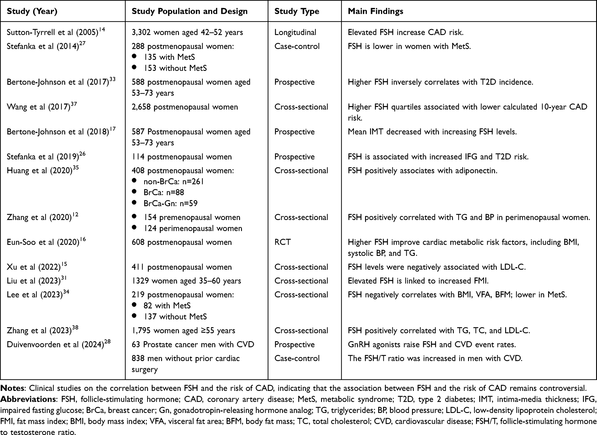 |
Table 1 Clinical Studies on the Association Between FSH and CAD |
Recent animal studies have also focused on the role of FSH and its relationship with coronary atherosclerosis and associated risk factors. Piao et al demonstrated in animal experiments that FSH plays a role in exacerbating atherosclerosis.25 Similarly, another animal study showed that elevated FSH levels are responsible for the accelerated atherosclerosis observed in mice following castration. Mechanistically, FSH increases the expression of vascular cell adhesion molecule 1 (VCAM-1) through signal transduction, promoting endothelial inflammation and subsequently increasing the risk of coronary artery disease.11 Recent animal experiments in China have demonstrated that FSH induces endothelial-mesenchymal transition (EndMT) and atherosclerotic plaque formation in ApoE-/- mice.39 The aberrant occurrence of EndMT has been identified as a key driver of endothelial dysfunction and atherosclerosis.40 Guo et al conducted a series of animal experiments and found that when the E2 levels were comparable across the experimental groups, mice receiving exogenous FSH exhibited elevated FSH levels, along with significant increases in serum TC, LDL-C, and free cholesterol (FC) concentrations. In contrast, mice treated with FSHβ antibody (FSHAb) showed a significant reduction in serum TC levels. These findings suggest that FSH can independently affect lipid levels in the body by binding to the FSHR, and this effect can be reversed by blocking FSH.41 Yu et al conducted animal experiments and found that in ovariectomized (OVX) mice receiving physiological doses of estrogen to simulate normal estrogen levels, exogenous FSH administration fully reversed the decline in renin, angiotensin II, and blood pressure. This effect may be due to FSH stimulating the FSHR on juxtaglomerular cells, promoting renin secretion, which leads to vasoconstriction and increased aldosterone secretion, ultimately elevating blood pressure and triggering a series of physiological responses.42 Wu et al found that in OVX mice receiving estrogen supplementation, elevated levels of FSH significantly increased cortisol and blood pressure. Furthermore, administration of a gonadotropin-releasing hormone analogue (GnRHa) to stimulate FSH production resulted in a further rise in both cortisol and blood pressure.43 These findings provide strong evidence for the potential role of FSH in the development of CAD and further demonstrate that FSH can exert its effects independently of E2 (Table 2).
 |
Table 2 Animal Studies on the Association Between FSH and CAD |
Sources of Heterogeneity Across Studies
The current evidence regarding FSH and the risk of coronary heart disease in postmenopausal women presents notable contradictions. This heterogeneity is not coincidental but stems from multi-level differences in study design, population characteristics, methodology, and analytical models. A systematic analysis of these differences is crucial for understanding the complex biological role of FSH and for integrating conflicting findings. Research supporting the role of FSH in exacerbating CAD in postmenopausal women is not limited to cross-sectional studies but also includes high-quality prospective designs. For instance, longitudinal studies conducted in diverse populations in the United States14 and a prospective cohort study in Poland26 have identified a positive correlation between FSH levels and coronary artery disease, as well as cardiovascular metabolic risk factors, through long-term follow-up, large sample sizes, and clear timelines. These studies, along with findings from animal experiments demonstrating the role of FSH in promoting atherosclerosis, provide a mutually validating framework that connects clinical observations with mechanistic explanations. However, the studies supporting the potential protective role of FSH on cardiometabolic factors exhibit significant variability in evidence quality. While one of these studies is a rigorously designed multicenter randomized controlled trial from Korea,16 its conclusions have limited generalizability due to the constraints of the research context. This study involved a strictly selected cohort of postmenopausal female volunteers from Korea, whose health status and metabolic characteristics do not accurately represent those of the general community population. The analysis was essentially a secondary exploration based on existing clinical trial data, rather than a prospective study specifically designed to examine the relationship between FSH and CAD risk, thus imposing limitations on causal inference. Most other studies supporting a protective role for FSH adopt a cross-sectional design, which fails to establish the temporal sequence between FSH and metabolic indices and is susceptible to selection bias. These factors collectively represent an important source of inconsistency in the current research conclusions.
Studies show notable heterogeneity in the racial and geographic composition of their populations. Prospective cohorts from the United States, such as SWAN and NHANES, include racially diverse populations. Their findings consistently show that higher FSH levels are associated with an increased risk of CAD, and these results are more readily generalizable to broader populations. In contrast, reports of an inverse relationship between FSH and metabolic parameters often come from racially and geographically homogeneous samples. These include healthy, stringently screened clinical trial volunteers in South Korea and the Eastern Finnish community cohort study,17 where population specific characteristics may limit external validity. Methodological variation in the definition of menopausal status and in the handling of other hormonal influences further complicates comparisons across studies. The work by Stefanska and colleagues26 used clear and consistent criteria and therefore provides more reliable inferences. In many cross sectional studies from China,15,35 menopause is classified only by the duration of amenorrhea and no uniform FSH threshold is applied for confirmation. The East China study37 even further combined naturally and surgically menopausal women in its analysis. Control of key covariates, including baseline estrogen levels and prior hormone therapy, is also inconsistent, leaving considerable scope for residual confounding. Differences in baseline health status add another layer of complexity. The Korean randomized controlled trial, for example, enrolled strictly healthy volunteers, so the associations observed may not be directly applicable to community based populations of women who already have metabolic disturbances.
Regarding FSH assay methodology, studies generally employed chemiluminescent immunoassays for measurement, ensuring data reliability. However, the use of different detection platforms, with their distinct antibody characteristics and calibration standards, makes direct comparison of absolute values challenging. Furthermore, FSH exhibits a pulsatile secretion pattern. The vast majority of studies rely on a single fasting FSH measurement to represent an individual’s long-term exposure level, while only a very limited number of prospective designs incorporate repeated measurements. This single-point approach may introduce random measurement error, thereby attenuating true association strength or contributing to inconsistent findings. Existing research predominantly relies on relative classification of FSH levels within their respective study cohorts for analysis, rather than employing a universally applicable, pathologically meaningful absolute concentration threshold. This approach substantially limits the ability to compare conclusions uniformly across studies.
The substantial heterogeneity across studies likely explains the discrepancies in current findings. Future research should prioritize prospective, multi-center, multi-ethnic cohort studies with refined stratification for menopausal status and hormone therapy history. Study designs should account for FSH’s pulsatile secretion by using repeated measurements to derive average values. Standardizing FSH assay protocols across research centers is also necessary. In statistical analyses, variables like BMI and estrogen should be examined not just as confounders, but also as potential mediators to more accurately delineate how FSH exerts its effects. Furthermore, future investigations should identify potential pathological FSH thresholds related to CAD, rather than relying solely on relative rankings within study populations.
In conclusion, current research mainly focuses on the association between FSH and CAD in postmenopausal women, along with the potential risk factors involved. Most studies, after adjusting for confounders such as age, menopausal duration, and E2 levels, still find significant positive or negative correlations between FSH and CAD, atherosclerosis, and related risk factors. Animal studies suggest that changes in FSH levels may influence the progression of atherosclerosis and are closely related to lipid metabolism and vascular function. However, most existing studies have been conducted in China, which limits the global generalizability of the findings, and these studies generally share several common limitations. Most studies are cross-sectional or retrospective, making it difficult to assess the temporal relationship between changes in FSH levels and corresponding outcomes, thus preventing the establishment of causality; the sample sizes in existing studies are small, with homogeneous study populations that were not randomly selected and a lack of multi-racial and multi-center comparisons, making the findings potentially not fully representative of the general postmenopausal female population; existing studies generally evaluate FSH levels at a single time point, but since FSH is released in pulses with a frequency of every 1 to 4 hours, this may affect measurement accuracy, limiting the ability to assess the impact of FSH fluctuations on the outcomes; the age range of study populations is narrow; and certain special groups, such as women with premature ovarian failure, have not been included, which limits the generalizability of the results. The threshold of FSH and its underlying mechanisms remain insufficiently explained, and the potential influence of other sex hormones, such as luteinizing hormone and androgens, on FSH levels has not been considered. Therefore, the relationship between FSH and the risk of CAD in postmenopausal women still requires further research for confirmation. Future research should focus on large-scale, multi-center, randomized controlled trials across different geographical regions and ethnic groups, while exploring the mechanisms by which FSH affects CAD and its related risk factors.
The Potential Role of FSH in the Pathogenesis of Coronary Heart Disease
The following explores the potential pathological mechanisms through which FSH may influence the development and progression of cardiovascular diseases.
Structural and Signaling Basis of FSHR
As a member of the glycoprotein hormone family, FSH exerts its physiological effects through its specific receptor, FSHR. FSHR is a classic class A G protein-coupled receptor (GPCR). Its structural characteristic includes a large extracellular domain (ECD) composed of leucine-rich repeats (LRR), which is connected to a seven-transmembrane domain via a hinge region and is responsible for high affinity and specificity in recognizing FSH.45 Cryo-electron microscopy studies have revealed that in the activated state, the extracellular domain of FSHR adopts an almost vertical conformation. Upon FSH binding, key sequences in the hinge region become ordered and form specific interactions with the hormone, driving a rigid rotation of the entire extracellular domain. This movement translates the chemical signal into conformational changes in the transmembrane region, ultimately opening the G protein binding pocket and initiating downstream signaling cascades such as cAMP.46 This model has been supported by independent research. An integrative study combining chemical cross-linking and computational modeling indicated that due to the stabilizing effect of disulfide bonds, the conformation of the hinge region is relatively rigid, making large-scale rearrangements unlikely. This confirms that the key step in transmembrane signaling is the overall rotation of the ECD, rather than local distortion of the hinge region.47 This precise structural activation mechanism not only serves as the structural basis for the activation of the classic Gαs/cAMP/PKA pathway but also likely provides the physical premise for achieving signal bias and producing tissue-specific effects.
For a long time, the action of FSH has been considered mainly mediated by this classic pathway.48 However, it has been discovered that the FSHR gene can produce various receptor subtypes through alternative splicing, among which the full-length FSHR1 and the truncated FSHR3 are representative. FSHR1 is a typical GPCR, primarily mediating the Gαs/cAMP/PKA pathway to regulate reproductive functions, while FSHR3 exhibits a structure more akin to growth factor receptors, with a signaling bias towards activating proliferation pathways such as MAPK.49 This fundamental difference in signaling output provides a crucial explanation for how FSH mediates different or even opposing biological effects in gonadal and extragonadal tissues. In gonadal tissues, it mainly regulates metabolism and differentiation through FSHR1; whereas in extragonadal sites such as vascular endothelium, adipose tissue, and liver, FSHR3 and other subtypes may dominate, directing FSH stimulation towards promoting inflammation, cell proliferation, and lipid synthesis, potentially leading to atherosclerosis.
Furthermore, studies indicate that activated FSHR can connect to a complex signaling network that includes Gαi, Gαq, and β-arrestin. Different ligands or variations within the receptor itself can selectively stabilize specific active conformations of FSHR, favoring the activation of certain downstream pathways rather than uniformly activating all signals.48 This theory has received direct pharmacological evidence support, as research has found that different small-molecule allosteric ligands acting on the same FSHR can stabilize distinct receptor conformations, thereby selectively activating specific pathways. For example, compound B3 exhibits super agonist activity in recruiting β-arrestin 2, while compound B1 significantly favors the Gαs/cAMP pathway. In contrast, endogenous FSH acts as a balanced agonist, relatively uniformly activating multiple pathways. Such bias is also reflected in the dynamics of signaling and directly influences downstream functional outputs, such as transcriptional activity dependent on cAMP response elements.50 Therefore, the signaling output of FSHR is not solely determined by ligand concentration but rather depends on the inherent biased characteristics of the ligand and the complex interactions with the local microenvironment of the tissue. This provides a critical molecular and pharmacological framework for understanding the contradictory roles of FSH as a risk factor for atherosclerosis in postmenopausal women, with some studies showing its association with protective metabolic parameters.
Future research should move beyond a singular focus on circulating FSH levels and delve deeper into the expression profiles of dominant FSHR subtypes in target tissues and the specific signaling networks they mediate, to more accurately elucidate the complex role of FSH in cardiovascular diseases.
Impact on Lipid Metabolism
A pivotal factor in the progression of atherosclerosis is lipid metabolism dysregulation. Under the influence of various risk factors, metabolic disturbances occur, impairing the body’s ability to effectively clear or suppress excessive cholesterol production, which ultimately leads to pathological accumulation within the vascular walls and subsequent atherosclerotic development.51 LDL-C, as the primary carrier of cholesterol, is one of the risk factors for atherosclerosis.52 In contrast, HDL-C exerts a protective effect on the cardiovascular system by facilitating reverse cholesterol transport.53 Studies have shown that elevated FSH levels are closely associated with lipid metabolism disturbances in postmenopausal women. An animal experiments further demonstrate that FSH reduces the degradation of LDL-C by inhibiting the expression of LDL receptor (LDLR) in liver tissue.54 Furthermore, another animal experiment revealed the presence of FSHR in the liver, where FSH binds to FSHR on hepatocytes, activating the sterol regulatory element-binding protein (SREBP), which subsequently upregulates the expression of HMG-CoA reductase.41 HMG-CoA reductase is a key enzyme in hepatic cholesterol biosynthesis, and its overexpression is linked to excessive cholesterol production.55 Additionally, a study suggest that FSH activates Gαi proteins through binding to FSHR, thereby regulating intracellular Ca2⁺ concentrations. This signaling pathway ultimately activates the cAMP response element-binding protein (CREB) transcription factor, which regulates fat accumulation and its redistribution in the abdominal and visceral areas.56 Liu et al confirmed the presence of FSHR protein in both white and brown adipose tissues of mice through immunostaining. Their study demonstrated that FSHR inhibits cAMP production and stimulates the expression of adipogenesis-related genes fatty acid synthase (Fas) and lipoprotein lipase (Lpl), resulting in an increase in fat accumulation in mice.57 Cui et al reported that FSH treatment enhanced the expression of genes involved in fatty acid metabolism, retinol metabolism, and peroxisome proliferator-activated receptor (PPAR) signaling pathways, leading to an increase in abdominal fat weight and lipid accumulation in adipocytes in chickens.58 Therefore, women with elevated FSH levels often exhibit higher cholesterol and LDL-C levels, along with lower HDL-C levels. This may contribute to the development of atherosclerosis, subsequently increasing the risk of coronary heart disease.
Regulation of Inflammatory Response
Another key factor in the development of atherosclerosis is the dysregulation of the inflammatory response. Recent studies suggest that FSH may influence cardiovascular health by regulating inflammatory responses. This could potentially contribute to the progression of atherosclerosis through mechanisms that involve immune cell activation, cytokine release, and endothelial function. Mouse studies have shown that the FSHR is expressed in various inflammatory cells, including macrophages, monocytes, and neutrophils. FSH can promote the expression of VCAM-1 through the FSHR/GαS/cAMP/PKA pathway. This, in turn, contributes to the recruitment, migration, and differentiation of foam cells, as well as the promotion of monocyte release of the inflammatory cytokine IL-6. Together, these processes contribute to the progression of atherosclerosis.11 FSH can also promote the expression of IL-1β in macrophages, increasing the macrophage’s chemotactic migration ability toward MCP-1. This process contributes to the development of atherosclerosis by facilitating the recruitment and activation of inflammatory cells at the site of vascular injury, thus exacerbating the inflammatory response in the arterial wall.44 In addition, FSH upregulates the expression of genes related to inflammation, such as Ms4a4a, CD68, and Ms4a7, leading to excessive activation of the sympathetic nervous system in mice. This promotes an increase in extramedullary monocytic cells, resulting in the enhanced infiltration of monocyte-derived macrophages into vascular and myocardial tissues. Such processes further aggravate endothelial injury and are considered early events in the development of coronary heart disease.59
Direct Vascular Effects
Beyond lipid metabolism abnormalities and inflammatory responses, vascular endothelial dysfunction or impairment predisposes arteries to a hyperconstricted state. This pathophysiological condition blood vessels hypersensitive, where even minor stimuli can trigger exaggerated vasoconstrictive responses or vasospasm, leading to cardiac ischemia,60 which may precipitate clinical events including angina pectoris and myocardial infarction. Recent in vitro cell studies and animal experiments have shown that FSH receptors are not only expressed in the reproductive system but are also widely distributed in various extragonadal tissues. For instance, FSH can stimulate the FSHRs on zona fasciculata cells of mouse adrenal glands and ATC7 cells, inducing cortisol secretion in vitro;43 FSH binds to FSHRs on osteoclasts in mice, promoting osteoclast energy metabolism through the CREB-MDH2-NAD+ axis, thereby exacerbating bone loss.61 FSHRs are also expressed in the human endometrium, where they are functionally active and may regulate endometrial cell function.62 FSHRs are also expressed in atherosclerotic plaques in the carotid, coronary, and femoral arteries.63 These receptors have also been detected in muscle tissue, although their precise role remains unclear.64 In mouse chondrocytes, these receptors promote cell proliferation and maturation.65 In the mouse cortex and hippocampus, FSHR expression is linked to the onset of Alzheimer’s disease (AD) and cognitive impairments.66 In human breast cancer cells, FSHRs enhance their survival and proliferation.67 In a prospective study, Chen et al found that FSH promotes atrial fibrosis in postmenopausal women with atrial fibrillation through oxidative stress. Furthermore, the inhibition of FSHR in the atria of mice significantly reduced the cumulative incidence of atrial fibrillation.68 However, the majority of the aforementioned studies were conducted on subjects other than postmenopausal women, making it essential to further investigate the role of FSHRs in this population. FSHR has been identified in endothelial cells and cardiomyocytes.69 Elevated levels of FSH may affect endothelial function by binding to these receptors, leading to impaired vasoconstrictive function and subsequently increasing the risk of coronary artery disease. Previous studies have detected FSHR transcripts and FSHR protein in human umbilical vein endothelial cells (HUVECs).70,71 Consequently, Rocca et al performed further cellular experiments using HUVECs to validate the effects of FSH on endothelial cells.10 FSH exerts dual mechanisms on vascular endothelial cells: (1) facilitating calcium ion release via opening IP3-sensitive calcium channels, and (2) stimulating nitric oxide (NO) production. The concerted actions of calcium signaling and NO on human umbilical vein endothelial cells (HUVECs) modulate the organization of vascular endothelial cadherin (VE-cadherin) in murine vasculature. Functionally intact VE-cadherin is essential for promoting intercellular adhesion, thereby safeguarding endothelial barrier integrity.72 Therefore, altered expression or mislocalization of this protein may impair endothelial integrity. In experimental settings, rhFSH stimulation induced disrupted spatial organization of VE-cadherin, resulting in diminished intercellular adhesive junction integrity within the vascular endothelium. These findings demonstrate that elevated FSH levels may compromise endothelial barrier function, potentially contributing to coronary artery disease pathogenesis (Figure 2).
 |
Figure 2 Pro-Atherogenic and Potentially Protective Mechanisms of FSH in CAD. Abbreviations: FSH, follicle-stimulating hormone; CAD, coronary artery disease; FSHR, Follicle-Stimulating Hormone Receptor; LDL-C, low-density lipoprotein cholesterol; GRCPs, G protein-coupled receptors; IP3, inositol triphosphate; CaM, calmodulin; NOS, nitric oxide synthase; TG, triglycerides; HDL-C, high-density lipoprotein cholesterol. Notes: Dual mechanisms of FSH in the development of CAD. (A–C) Pro-atherogenic mechanisms: (A) FSH increases hepatic cholesterol synthesis and inhibits LDL-C clearance; (B) FSH promotes inflammation via cytokine release and adhesion molecule expression; (C) FSH compromises endothelial integrity. (D–F) Potentially protective mechanisms: (D) Physiological FSH levels enhance pancreatic β-cell insulin secretion in response to glucose; (E) FSH upregulates adiponectin secretion from adipocytes; (F) FSH correlates with favorable metabolic profiles, including increased HDL-C and reduced triglycerides. The figure was created by Figdraw and is original. |
The Impact of Cardiac Metabolic Risk Factors
Unhealthy lifestyle habits can lead to multiple cardiac metabolic risk factors, which interact with each other and further increase the risk of CAD. Therefore, in addition to the previously mentioned lipid metabolism, there are numerous studies focusing on other cardiac metabolic risk factors. Regarding blood pressure, a 10-year prospective study found that with the progression of menopause in women, FSH levels increased from 7.7 IU/L at baseline to 65.3 IU/L after 10 years, and both systolic and diastolic blood pressures tended to rise. In addition, adverse changes were also observed in lipid levels and glycated hemoglobin.73 Animal studies have also concluded that the increase in FSH is associated with elevated blood pressure in postmenopausal women. This study further proposed the intracellular mechanism by which FSH promotes catecholamine synthesis in PC12 cells and chromaffin cells of the adrenal medulla in rats.74
Numerous clinical studies have demonstrated a significant increase in the prevalence of T2DM among postmenopausal women.75–77The decline in E2 levels partially accounts for the development of diabetes after menopause.78 In clinical practice, we have observed that E2 replacement therapy does not completely eliminate this risk.79 Therefore, Cheng et al studied the relationship between FSH levels and T2DM in postmenopausal women and confirmed that elevated serum FSH levels are positively correlated with the diabetes risk in this population. The pathogenic mechanism involves dual signaling regulation. Through experiments on human pancreatic β-cells, the FSHR was identified as exhibiting a dose-dependent bidirectional regulation in response to FSH stimulation. Under physiological conditions, FSH activates the cAMP/PKA and calcium signaling pathways via Gαs protein coupling, promoting glucose-stimulated insulin secretion. However, under conditions of FSH overload (greater than 40 IU/L), the receptor switches to Gαi protein coupling, significantly inhibiting the cAMP/PKA signaling pathway, thereby leading to defects in insulin secretion.13 Therefore, prolonged exposure to high FSH levels can lead to insulin deficiency, ultimately progressing to T2DM.
An increase in FSH levels has been shown to be a major risk factor for the progression of obesity in postmenopausal women.80 The metabolic reasons behind FSH-induced obesity are multifaceted, including the lipid metabolism abnormalities and impaired insulin secretion leading to glucose metabolism dysfunction, as mentioned earlier. However, the primary factor contributing to the progression of obesity is the accumulation of fat. FSH has been confirmed to promote fat generation, particularly visceral fat, leading to central obesity characterized by abdominal fat accumulation.57 In white adipose tissue, activation of the downstream FSHR elevates intracellular cAMP levels, thereby activating protein kinase A (PKA). PKA phosphorylates a cascade of transcription factors, which upregulate the expression of adipogenic genes—including LPL, FAS, and peroxisome proliferator-activated receptor gamma (PPARγ)—in white adipose tissue. This molecular cascade promotes the maturation of preadipocytes into functional adipocytes, enabling their lipid synthesis and storage capabilities.58 In beige adipose tissue, FSHR couples with the Gαi protein. Gαi signaling reduces intracellular cAMP levels, thereby suppressing the activation of mitochondrial uncoupling protein-1 (UCP1). UCP1, a master regulator of beige adipose tissue, normally enhances ATP production and oxidative processes in mitochondria when activated. This thermogenic mechanism enables beige adipocytes to catabolize stored lipids and elevate metabolic rates. FSH reduces fat tissue expenditure by blocking the activation of UCP1, leading to fat accumulation. This process can ultimately result in the development of obesity.81
Integration of Mechanistic and Clinical Evidence
Numerous clinical studies have observed that elevated levels of FSH in postmenopausal women are associated with an increased risk of CAD and adverse cardiac metabolic characteristics. This mainstream finding aligns closely with basic research revealing the mechanisms by which FSH promotes atherosclerosis. In terms of lipid metabolism, clinical investigations consistently report that high FSH levels correlate with elevated TC and LDL-C. This directly corresponds to mechanistic studies indicating that FSH binds to FSHR in hepatocytes, activating the SREBP-2/HMGCR pathway,41 thereby enhancing cholesterol synthesis in the liver while inhibiting the expression of LDLR, resulting in reduced clearance of LDL-C. Furthermore, the association between FSH and increased visceral fat along with abdominal obesity can be explained by its action in adipose tissue. FSH interacts with FSHR in white adipose tissue, inhibiting cAMP production and upregulating genes related to fat synthesis, thus directly promoting fat accumulation; in beige adipose tissue, FSH decreases energy expenditure by inhibiting UCP1-mediated thermogenesis.
FSH can activate inflammatory cells such as monocytes and macrophages, leading to the expression and release of inflammatory factors like VCAM-1, IL-6,11 and IL-1β,44 which exacerbate the inflammatory response within the vascular wall and further worsen atherosclerosis, consequently increasing CAD risk. The mechanisms linking elevated blood pressure and insulin resistance to CAD risk have also been supported: FSH stimulates the adrenal medulla to synthesize catecholamines and induces juxtaglomerular cells to secrete renin, resulting in hypertension; in pancreatic beta cells, high levels of FSH switch to a Gαi signaling pathway, inhibiting the cAMP/PKA pathway and leading to impaired insulin secretion. These multi-level evidences, ranging from cellular models to population studies, collectively form the pathophysiological basis for FSH’s role in driving atherosclerosis development.
Some studies have reported protective effects of FSH on cardiac metabolic markers; however, this does not negate the aforementioned mechanisms promoting atherosclerosis, but can be understood through the complexity, bias, and tissue specificity of the FSHR signaling pathway.48 Under certain metabolic contexts, FSH may produce seemingly beneficial signals in specific tissues. For instance, FSH may transiently activate the Gαs/cAMP pathway in pancreatic beta cells, momentarily enhancing insulin secretion, which might be reflected as a protective effect in cross-sectional studies. In summary, the inconsistencies observed in clinical findings highlight the multifaceted and context-dependent nature of FSH’s actions. Chronic exposure to elevated levels of FSH can promote the development of atherosclerosis through pathways involving enhanced lipid synthesis, inflammation, endothelial damage, and metabolic disturbance. Those seemingly contradictory clinical associations may reflect the temporary activation of different signaling pathways under specific subpopulations or conditions. Therefore, this integrative understanding strongly suggests that future research should extend beyond merely measuring circulating FSH levels to investigate the expression profiles of dominant FSHR isoforms in target tissues and their specific signaling networks, combined with genetic backgrounds and metabolic phenotypes, to more accurately assess individualized CAD risk, elucidate the complex role of FSH in cardiovascular disease, and explore intervention strategies targeting specific FSHR signaling pathways (Table 3).
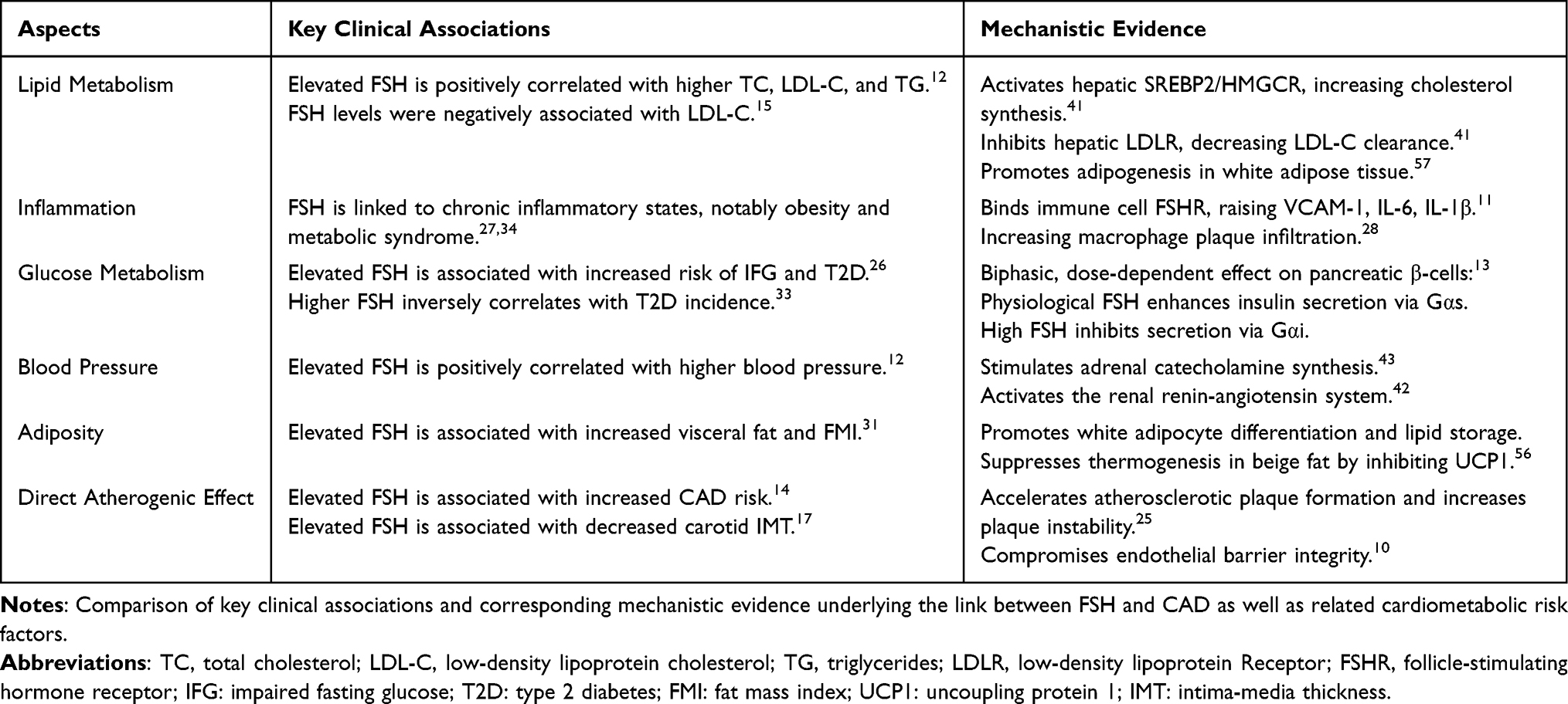 |
Table 3 Comparison of Clinical and Mechanistic Evidence |
Unresolved Questions and Future Directions
Elevated FSH levels influence cardiovascular health through several mechanisms, including disruption of liver cholesterol metabolism, activation of inflammatory responses, and impairment of endothelial function, all of which contribute to atherosclerosis. Furthermore, the contradictory conclusions observed in clinical studies may be related to the biological characteristics of FSH receptors, including signaling bias where different FSHR receptor subtypes activate distinct intracellular pathways that lead to varying biological outcomes and tissue specificity which refers to the distribution of FSHR receptors in different tissues affecting how FSH exerts its effects. Additionally, these conclusions may be influenced by confounding factors such as the baseline cardiac metabolic health status and ethnic characteristics of the studied population. This review contributes to gradually revealing the complex role of FSH in cardiovascular diseases.
Current research largely shares several key limitations. First, most studies are observational or cross-sectional in design, making it impossible to clearly establish causal relationships between FSH levels and CAD. Second, there is considerable heterogeneity in trial design, definitions of menopausal status, FSH detection protocols, and control of confounding factors across studies. Third, known risk factors for CAD, such as smoking and chronic kidney disease, have not been adequately considered, necessitating further investigation into their impact on the relationship between FSH levels and CAD risk. Finally, the geographical concentration of existing studies limits the generalizability of findings. A significant portion of clinical research comes from single populations, particularly in China. While these results provide important insights for this specific group, factors such as FSHR genetic polymorphisms and lifestyle differences may influence the relationship between FSH and CAD across ethnicities. Validating these associations in diverse multi-ethnic cohorts is essential for future research.
Although no FSH/FSHR-targeted therapies currently exist for cardiovascular diseases, compelling preclinical evidence from related fields supports their therapeutic potential. For instance, monoclonal antibodies that block FSH action, such as HF257 and Hu6,82 activate thermogenesis in adipose tissue, reduce fat accumulation, improve lipid profiles, and increase energy expenditure in obese mouse models. Mechanistic studies indicate that FSH promotes liver cholesterol synthesis through the FSHR/Gi2α/β-arrestin-2/Akt/SREBP-2 pathway,41 suggesting that blocking this pathway could prevent hypercholesterolemia. The humanized FSH-blocking antibody MS-Hu6 has demonstrated efficacy in osteoporosis models by inhibiting bone resorption83 and alleviating neuroinflammation and amyloid pathology in Alzheimer’s disease models.84 These findings support the clinical significance of interventions targeting the FSH pathway, providing a foundation for its application in cardiovascular diseases.
This potential new approach contrasts with the primary intervention for managing cardiovascular risk in postmenopausal women, hormone replacement therapy (HRT). Hormone replacement therapy (HRT) remains the primary intervention for managing cardiovascular risk in postmenopausal women, several previous studies have indicated that initiating HRT during the perimenopausal or early postmenopausal period can alleviate vasomotor and urogenital symptoms,85 and significantly reduce the risk of osteoporotic fractures.86 Some evidence suggests that HRT may also offer protective effects on cardiovascular health by improving lipid profiles and lowering blood pressure.87 Certain studies have shown that HRT does not significantly affect lipid levels in some women and may lead to lipid metabolism disturbances.88 Findings by Gu and others suggest that while HRT may improve arterial dilation, it does not significantly reduce all-cause mortality or the incidence of cardiovascular events.89 These uncertainties highlight the value of exploring non-estrogen-dependent targets like FSH. Unlike HRT, which aims to supplement what is lacking, targeting the FSH pathway focuses on mitigating abnormally activated signals, potentially providing a more precise approach that is not limited by traditional estrogen-related risks. Future research should focus on multi-ethnic cohort studies to validate the association between FSH levels and CAD risk, as well as explore targeted therapies that modulate FSH pathways, offering new options for clinical practice.
Conclusions
In summary, this review highlights that elevated FSH levels after menopause are a significant independent risk factor for CAD in postmenopausal women, challenging the traditional focus on estrogen decline. FSH contributes to atherosclerosis through dyslipidemia, chronic inflammation, and endothelial dysfunction, with evidence supporting a positive correlation between FSH levels and CAD risk. Furthermore, seemingly contradictory clinical data indicate that FSH’s role in the cardiovascular system reflects its pleiotropic functions, influenced by receptor biology and individual physiological contexts.
Measuring FSH as a stable biomarker for cardiovascular risk assessment in postmenopausal women is clinically valuable. Targeting the FSH/FSHR pathway also presents a promising therapeutic approach for gender-specific strategies against cardiovascular diseases, which is significant for improving cardiovascular health in postmenopausal women. Future research should prioritize large-scale, multi-ethnic studies to clarify the causal relationship between FSH and CAD, explore FSH signaling mechanisms in cardiovascular tissues, and evaluate the impact of modulating the FSH pathway on cardiovascular outcomes.
Acknowledgments
This work was supported by the Shanxi Provincial Health and Wellness Commission Scientific Research Project under Grant No. 2024053. The authors would like to express their gratitude for the financial support provided, which made this research possible.
Disclosure
The authors report no conflicts of interest in this work.
References
1. Savo MT, De Amicis M, Cozac DA, et al. Comparative Prognostic Value of Coronary Calcium Score and Perivascular Fat Attenuation Index in Coronary Artery Disease. J Clin Med. 2024;13(17):5205. doi:10.3390/jcm13175205
2. Hu SS. Report on cardiovascular health and diseases in China 2021: an updated summary. J Geriatr Cardiol. 2023;20(6):399–18. doi:10.26599/1671-5411.2023.06.001
3. Nudy M, Buerger J, Dreibelbis S, et al. Menopausal hormone therapy and coronary heart disease: the roller-coaster history. Climacteric J Int Menopause Soc. 2024;27(1):81–88. doi:10.1080/13697137.2023.2282690
4. El Khoudary SR. Gaps, limitations and new insights on endogenous estrogen and follicle stimulating hormone as related to risk of cardiovascular disease in women traversing the menopause: A narrative review. Maturitas. 2017;104:44–53. doi:10.1016/j.maturitas.2017.08.003
5. Lizneva D, Rahimova A, Kim SM, et al. FSH beyond fertility. Front Endocrinol. 2019;10:136. doi:10.3389/fendo.2019.00136
6. Recchia K, Jorge AS, de Pessôa LVF, et al. Actions and roles of FSH in germinative cells. Int J Mol Sci. 2021;22(18):10110. doi:10.3390/ijms221810110
7. Pawlikowski M. Direct actions of gonadotropins beyond the reproductive system and their role in human aging and neoplasia [Bezpośrednie działanie gonadotropin poza układem rozrodczym i ich rola w starzeniu się i nowotworzeniu u człowieka]. Endokrynol Pol. 2019;70(5):437–444. doi:10.5603/EP.a2019.0034
8. Sun D, Bai M, Jiang Y, et al. Roles of follicle stimulating hormone and its receptor in human metabolic diseases and cancer. Am J Transl Res. 2020;12(7):3116–3132.
9. Garrido MP, Bruneau N, Vega M, Selman A, Tapia JC, Romero C. Follicle-stimulating hormone promotes nerve growth factor and vascular endothelial growth factor expression in epithelial ovarian cells. Histol Histopathol. 2020;35(9):961–971. doi:10.14670/HH-18-226
10. Rocca MS, Pannella M, Bayraktar E, et al. Extragonadal function of follicle-stimulating hormone: evidence for a role in endothelial physiology and dysfunction. Mol Cell Endocrinol. 2024;594:112378. doi:10.1016/j.mce.2024.112378
11. Wang Q, Han J, Liang Z, et al. FSH is responsible for androgen deprivation therapy-associated atherosclerosis in mice by exaggerating endothelial inflammation and monocyte adhesion. Arterioscler Thromb Vasc Biol. 2024;44(3):698–719. doi:10.1161/ATVBAHA.123.319426
12. Zhang C, Zhao M, Li Z, Song Y. Follicle-Stimulating hormone positively associates with metabolic factors in perimenopausal women. Int J Endocrinol. 2020;2020:7024321. doi:10.1155/2020/7024321
13. Cheng Y, Zhu H, Ren J, et al. Follicle-stimulating hormone orchestrates glucose-stimulated insulin secretion of pancreatic islets. Nat Commun. 2023;14(1):6991. doi:10.1038/s41467-023-42801-6
14. Sutton-Tyrrell K, Wildman RP, Matthews KA, et al. Sex-hormone-binding globulin and the free androgen index are related to cardiovascular risk factors in multiethnic premenopausal and perimenopausal women enrolled in the Study of Women Across the Nation (SWAN). Circulation. 2005;111(10):1242–1249. doi:10.1161/01.CIR.0000157697.54255.CE
15. Xu Z, Gu S, Wu X, Zhou Y, Li H, Tang X. Association of follicle stimulating hormone and serum lipid profiles in postmenopausal women. Medicine. 2022;101(39):e30920. doi:10.1097/MD.0000000000030920
16. Jung ES, Choi EK, Park BH, Chae SW. Serum follicle-stimulating hormone levels are associated with cardiometabolic risk factors in post-menopausal Korean women. J Clin Med. 2020;9(4):1161. doi:10.3390/jcm9041161
17. Bertone-Johnson ER, Virtanen JK, Nurmi T, et al. Follicle-Stimulating hormone levels and subclinical atherosclerosis in older postmenopausal women. Am J Epidemiol. 2018;187(1):16–26. doi:10.1093/aje/kwx174
18. Page MJ, McKenzie JE, Bossuyt PM, et al. The PRISMA 2020 statement: an updated guideline for reporting systematic reviews. BMJ. 2021:
19. Santi D, Crépieux P, Reiter E, et al. Follicle-stimulating Hormone (FSH) action on spermatogenesis: a focus on physiological and therapeutic roles. J Clin Med. 2020;9(4):1014. doi:10.3390/jcm9041014
20. Ongaro L, Zhou X, Wang Y, et al. Muscle-derived myostatin is a major endocrine driver of follicle-stimulating hormone synthesis. Science. 2025;387(6731):329–336. doi:10.1126/science.adi4736
21. Qian W, Yaping L. Research progress on the role of follicle-stimulating hormone in bone metabolism. J Int Endocrinol Metabol. 2023;43(04):305–308,316. doi:10.3760/cma.j.cn121383-20220307-03016
22. Radu A, Pichon C, Camparo P, et al. Expression of follicle-stimulating hormone receptor in tumor blood vessels. N Engl J Med. 2010;363(17):1621–1630. doi:10.1056/NEJMoa1001283
23. Peacock K, Carlson K, Ketvertis KM. Menopause. In: StatPearls. Treasure Island (FL): StatPearls Publishing; 2025. PMID: 29939603.
24. Zhang J, Liu ML. Menopause hormone therapy and prevention of cardiovascular disease. Zhonghua Xin Xue Guan Bing Za Zhi. 2020;48(3):259–262. doi:10.3760/cma.j.cn112148-20190815-00495
25. Piao J, Yin Y, Zhao Y, et al. Follicle-Stimulating hormone accelerates atherosclerosis by activating PI3K/Akt/NF-κB pathway in mice with androgen deprivation. J Vasc Res. 2022;59(6):358–368. doi:10.1159/000527239
26. Stefanska A, Cembrowska P, Kubacka J, Kuligowska-Prusinska M, Sypniewska G. Gonadotropins and their association with the risk of prediabetes and type 2 diabetes in middle-aged postmenopausal women. Dis Markers. 2019;2019:2384069. doi:10.1155/2019/2384069
27. Stefanska A, Ponikowska I, Cwiklinska-Jurkowska M, Sypniewska G. Association of FSH with metabolic syndrome in postmenopausal women: a comparison with CRP, adiponectin and leptin. Biomarker Med. 2014;8(7):921–930. doi:10.2217/bmm.14.49
28. Duivenvoorden WCM, Margel D, Subramony Gayathri V, et al. Follicle-Stimulating hormone exacerbates cardiovascular disease in the presence of low or castrate testosterone levels. JACC Basic Transl Sci. 2024;9(3):364–379. doi:10.1016/j.jacbts.2023.10.010
29. Davey P, Kirby MG. Cardiovascular risk profiles of GnRH agonists and antagonists: real-world analysis from UK general practice. World J Urol. 2021;39(2):307–315. doi:10.1007/s00345-020-03433-3
30. Alshammary AF, Alharbi KK, Alshehri NJ, Vennu V, Ali Khan I. Metabolic syndrome and coronary artery disease risk: a meta-analysis of observational studies. Int J Environ Res Public Health. 2021;18(4):1773. doi:10.3390/ijerph18041773
31. Liu X, Xu J, Wei D, Chen Y. Associations of serum follicle-stimulating hormone and luteinizing hormone levels with fat and lean mass during menopausal transition. Obes Facts. 2023;16(2):184–193. doi:10.1159/000528317
32. Rg SM, RC R, Lm S, et al. A new approach using BMI and FMI as predictors of cardio-vascular risk factors among Mexican young adults. Eur J Investig Health Psychol Educ. 2023;13(10). doi:10.3390/ejihpe13100146
33. Bertone-Johnson ER, Virtanen JK, Niskanen L, et al. Association of follicle-stimulating hormone levels and risk of type 2 diabetes in older postmenopausal women. Menopause N Y N. 2017;24(7):796–802. doi:10.1097/GME.0000000000000834
34. Lee SW, Hwang IS, Jung G, Kang HJ, Chung YH. Relationship between metabolic syndrome and follicle-stimulating hormone in postmenopausal women. Medicine. 2022;101(18):e29216. doi:10.1097/MD.0000000000029216
35. Huang WY, Chen DR, Kor CT, et al. Relationships between follicle-stimulating hormone and adiponectin in postmenopausal women. Metabolites. 2020;10(10):420. doi:10.3390/metabo10100420
36. Gianopoulos I. Adiponectin and adiponectin receptors in atherosclerosis. Endocr Rev. 2025;46(1).
37. Wang N, Shao H, Chen Y, et al. Follicle‐Stimulating hormone, its association with cardiometabolic risk factors, and 10‐year risk of cardiovascular disease in postmenopausal women. J Am Heart Assoc. 2017;6(9):e005918. doi:10.1161/JAHA.117.005918
38. Zhang W, Chen Y, Zhang X, et al. Association of follicle-stimulating hormone with lipid profiles in older women: a cross-sectional SPECT-China study. BMJ Open. 2023;13(7):e072796. doi:10.1136/bmjopen-2023-072796
39. Li P, Xiang Y, Wei J, et al. Follicle-stimulating hormone promotes EndMT in endothelial cells by upregulating ALKBH5 expression. Cell Mol Biol Lett. 2025;30(1):41. doi:10.1186/s11658-025-00720-y
40. D M, Z Y, C M, et al. ASF1A-dependent P300-mediated histone H3 lysine 18 lactylation promotes atherosclerosis by regulating EndMT. Acta Pharm Sin B. 2024;14(7). doi:10.1016/j.apsb.2024.03.008
41. Guo Y, Zhao M, Bo T, et al. Blocking FSH inhibits hepatic cholesterol biosynthesis and reduces serum cholesterol. Cell Res. 2019;29(2):151–166. doi:10.1038/s41422-018-0123-6
42. Yu Z, Yang J, Huang WJ, et al. Follicle stimulating hormone promotes production of renin through its receptor in juxtaglomerular cells of kidney. Diabetol Metab Syndr. 2022;14(1):65. doi:10.1186/s13098-022-00816-x
43. Wu JG, Zhao P, Yang J, et al. Activation of follicle-stimulating hormone receptor in adrenal zona fasciculata cells promotes cortisol secretion: implications for the development of menopause-associated diseases. Exp Clin Endocrinol Diabetes. 2025;133(1):8–19. doi:10.1055/a-2376-5952
44. Han JL, Song YX, Yao WJ, Zhou J, Du Y, Xu T. Follicle-Stimulating hormone provokes macrophages to secrete IL-1β contributing to atherosclerosis progression. J Immunol. 2022;ji2200475. doi:10.4049/jimmunol.2200475
45. UA A, Z T, JV E, GS R, Ja D. Structure-Function relationships of the follicle-stimulating hormone receptor. Front Endocrinol. 2018;9. doi:10.3389/fendo.2018.00707
46. Hendrickson WA, Gong Z. Structural and evolutionary insights into the functioning of glycoprotein hormones and their receptors. Andrology. 2025. doi:10.1111/andr.70001
47. Nagel M, Moretti R, Paschke R, von Bergen M, Meiler J, Kalkhof S. Integrative model of the FSH receptor reveals the structural role of the flexible hinge region. Structure. 2022;30(10):1424–1431.e3. doi:10.1016/j.str.2022.07.007
48. Landomiel F, Gallay N, Jégot G, et al. Biased signalling in follicle stimulating hormone action. Mol Cell Endocrinol. 2014;382(1):452–459. doi:10.1016/j.mce.2013.09.035
49. B D, P H. An overview of FSH-FSHR biology and explaining the existing conundrums. J Ovarian Res. 2021;14(1). doi:10.1186/s13048-021-00880-3
50. De Pascali F, Ayoub MA, Benevelli R, et al. Pharmacological characterization of low molecular weight biased agonists at the follicle stimulating hormone receptor. Int J Mol Sci. 2021;22(18):9850. doi:10.3390/ijms22189850
51. Libby P. The changing landscape of atherosclerosis. Nature. 2021;592(7855):524–533. doi:10.1038/s41586-021-03392-8
52. Abdullah SM, Defina LF, Leonard D, et al. Long-Term association of low-density lipoprotein cholesterol with cardiovascular mortality in individuals at low 10-year risk of atherosclerotic cardiovascular disease. Circulation. 2018;138(21):2315–2325. doi:10.1161/CIRCULATIONAHA.118.034273
53. Zvintzou E, Xepapadaki E, Skroubis G, et al. High-Density lipoprotein in metabolic disorders and beyond: an exciting new world full of challenges and opportunities. Pharmaceuticals. 2023;16(6):855. doi:10.3390/ph16060855
54. Song Y, Wang ES, Xing LL, et al. Follicle-Stimulating hormone induces postmenopausal dyslipidemia through inhibiting hepatic cholesterol metabolism. J Clin Endocrinol Metab. 2016;101(1):254–263. doi:10.1210/jc.2015-2724
55. Li C, Ling Y, Kuang H. Research progress on FSH-FSHR signaling in the pathogenesis of non-reproductive diseases. Front Cell Dev Biol. 2024;12:1506450. doi:10.3389/fcell.2024.1506450
56. Liu XM, Chan HC, Ding GL, et al. FSH regulates fat accumulation and redistribution in aging through the Gαi/Ca(2+)/CREB pathway. Aging Cell. 2015;14(3):409–420. doi:10.1111/acel.12331
57. Liu P, Ji Y, Yuen T, et al. Blocking FSH induces thermogenic adipose tissue and reduces body fat. Nature. 2017;546(7656):107–112. doi:10.1038/nature22342
58. Cui H, Zhao G, Liu R, Zheng M, Chen J, Wen J. FSH stimulates lipid biosynthesis in chicken adipose tissue by upregulating the expression of its receptor FSHR. J Lipid Res. 2012;53(5):909–917. doi:10.1194/jlr.M025403
59. Gao L, Zhao Y, Wu H, et al. Polycystic ovary syndrome fuels cardiovascular inflammation and aggravates ischemic cardiac injury. Circulation. 2023;148(24):1958–1973. doi:10.1161/CIRCULATIONAHA.123.065827
60. Oikonomou E, Siasos G, Tsigkou V, et al. Coronary artery disease and endothelial dysfunction: novel diagnostic and therapeutic approaches. Curr Med Chem. 2020;27(7):1052–1080. doi:10.2174/0929867326666190830103219
61. Chen J, Liao Y, Sheng Y, et al. FSH exacerbates bone loss by promoting osteoclast energy metabolism through the CREB-MDH2-NAD+ axis. Metabolism. 2025;165:156147. doi:10.1016/j.metabol.2025.156147
62. Sacchi S, Sena P, Degli Esposti C, Lui J, La Marca A. Evidence for expression and functionality of FSH and LH/hCG receptors in human endometrium. J Assist Reprod Genet. 2018;35(9):1703–1712. doi:10.1007/s10815-018-1248-8
63. Ghinea N, Liehn EA, Grommes J, Delattre DD, Olesen TK. Follicle-stimulating hormone receptor expression in advanced atherosclerotic plaques. Sci Rep. 2024;14(1):10176. doi:10.1038/s41598-024-60962-2
64. Chen H, Cui Y, Yu S. Expression and localisation of FSHR, GHR and LHR in different tissues and reproductive organs of female yaks. Folia Morphol. 2018;77(2):301–309. doi:10.5603/FM.a2016.0095
65. Kong D, Guan Q, Li G, et al. Expression of FSHR in chondrocytes and the effect of FSH on chondrocytes. Biochem Biophys Res Commun. 2018;495(1):587–593. doi:10.1016/j.bbrc.2017.11.053
66. Xiong J, Kang SS, Wang M, et al. FSH and ApoE4 contribute to Alzheimer’s disease-like pathogenesis via C/EBPβ/δ-secretase in female mice. Nat Commun. 2023;14(1):6577. doi:10.1038/s41467-023-42282-7
67. Sanchez AM, Flamini MI, Russo E, et al. LH and FSH promote migration and invasion properties of a breast cancer cell line through regulatory actions on the actin cytoskeleton. Mol Cell Endocrinol. 2016;437:22–34. doi:10.1016/j.mce.2016.08.009
68. Chen S, Wu N, Zhang Y, et al. Follicle-stimulating hormone promotes atrial fibrosis in menopausal women with atrial fibrillation. Heart Rhythm. 2025;22(7):e172–e182. doi:10.1016/j.hrthm.2024.09.022
69. Chen H, Fang HQ, Liu JT, et al. Atlas of Fshr expression from novel reporter mice. eLife. 2025;13:RP93413. doi:10.7554/eLife.93413
70. Stilley JA, Guan R, Duffy DM, et al. Signaling through FSH receptors on human umbilical vein endothelial cells promotes angiogenesis. J Clin Endocrinol Metab. 2014;99(5):E813–20. doi:10.1210/jc.2013-3186
71. Tan D, Zhao Y, Ma D, Tong F. Role of FSH and FSH receptor on HUVECs migration. Gene Ther. 2021;28(3–4):155–161. doi:10.1038/s41434-020-00195-w
72. Gavard J. Endothelial permeability and VE-cadherin: a wacky comradeship. Cell Adhes Migr. 2014;8(2):158–164. doi:10.4161/cam.29026
73. Aittokallio J, Saaresranta T, Riskumäki M, et al. Effect of menopause and age on vascular impairment. Maturitas. 2023;169:46–52. doi:10.1016/j.maturitas.2023.01.006
74. Gao L, Wu X, Zhu X, Jin Q, Ma Q, Sun A. Follicle-stimulating hormone associates with metabolic factors in postmenopausal women. Gynecol Endocrinol. 2018;34(12):1035–1038. doi:10.1080/09513590.2018.1482868
75. Ren Y, Zhang M, Liu Y, et al. Association of menopause and type 2 diabetes mellitus. Menopause N Y N. 2019;26(3):325–330. doi:10.1097/GME.0000000000001200
76. Slopien R, Wender-Ozegowska E, Rogowicz-Frontczak A, et al. Menopause and diabetes: EMAS clinical guide. Maturitas. 2018;117:6–10. doi:10.1016/j.maturitas.2018.08.009
77. Gurka MJ, Vishnu A, Santen RJ, DeBoer MD. Progression of metabolic syndrome severity during the menopausal transition. J Am Heart Assoc. 2016;5(8):e003609. doi:10.1161/JAHA.116.003609
78. Sekido T, Nishio SI, Ohkubo Y, et al. Repression of insulin gene transcription by indirect genomic signaling via the estrogen receptor in pancreatic beta cells. Vitro Cell Dev Biol Anim. 2019;55(4):226–236. doi:10.1007/s11626-019-00328-5
79. Salpeter SR, Walsh JME, Ormiston TM, Greyber E, Buckley NS, Salpeter EE. Meta-analysis: effect of hormone-replacement therapy on components of the metabolic syndrome in postmenopausal women. Diabetes Obes Metab. 2006;8(5):538–554. doi:10.1111/j.1463-1326.2005.00545.x
80. Mao L, Wang L, Bennett S, et al. Effects of follicle-stimulating hormone on fat metabolism and cognitive impairment in women during menopause. Front Physiol. 2022;13:1043237. doi:10.3389/fphys.2022.1043237
81. Marlatt KL, Ravussin E. Brown adipose tissue: an update on recent findings. Curr Obes Rep. 2017;6(4):389–396. doi:10.1007/s13679-017-0283-6
82. Bergamini M, Dalla Volta A, Palumbo C, et al. Relationship between circulating FSH levels and body composition and bone health in patients with prostate cancer who undergo androgen deprivation therapy: the BLADE study. eLife. 2024;13(e92655). doi:10.7554/eLife.92655
83. Gera S, Kuo TC, Gumerova AA, et al. FSH-blocking therapeutic for osteoporosis. eLife. 2022;11:e78022. doi:10.7554/eLife.78022
84. Pallapati AR, Korkmaz F, Rojekar S, et al. Efficacy and safety of a therapeutic humanized FSH-blocking antibody in obesity and Alzheimer’s disease models. J Clin Invest. 2025:e182702. doi:10.1172/JCI182702
85. Cameron CR, Cohen S, Sewell K, Lee M. The art of Hormone Replacement Therapy (HRT) in menopause management. J Pharm Pract. 2024;37(3):736–740. doi:10.1177/08971900231167925
86. Baber RJ, Panay N, Fenton A, IMS Writing Group. 2016 IMS recommendations on women’s midlife health and menopause hormone therapy. Climacteric J Int Menopause Soc. 2016;19(2):109–150. doi:10.3109/13697137.2015.1129166
87. Ramesh S, Mann MC, Holroyd-Leduc JM, et al. Hormone therapy and clinical and surrogate cardiovascular endpoints in women with chronic kidney disease: a systematic review and meta-analysis. Menopause N Y N. 2016;23(9):1028–1037. doi:10.1097/GME.0000000000000657
88. Li S, Ma L, Song Y, et al. Effects of hormone replacement therapy on glucose and lipid metabolism in peri- and postmenopausal women with a history of menstrual disorders. BMC Endocr Disord. 2021;21(1):121. doi:10.1186/s12902-021-00784-9
89. Gu Y, Han F, Xue M, Wang M, Huang Y. The benefits and risks of menopause hormone therapy for the cardiovascular system in postmenopausal women: a systematic review and meta-analysis. BMC Womens Health. 2024;24(1):60. doi:10.1186/s12905-023-02788-0
 © 2026 The Author(s). This work is published and licensed by Dove Medical Press Limited. The
full terms of this license are available at https://www.dovepress.com/terms
and incorporate the Creative Commons Attribution
- Non Commercial (unported, 4.0) License.
By accessing the work you hereby accept the Terms. Non-commercial uses of the work are permitted
without any further permission from Dove Medical Press Limited, provided the work is properly
attributed. For permission for commercial use of this work, please see paragraphs 4.2 and 5 of our Terms.
© 2026 The Author(s). This work is published and licensed by Dove Medical Press Limited. The
full terms of this license are available at https://www.dovepress.com/terms
and incorporate the Creative Commons Attribution
- Non Commercial (unported, 4.0) License.
By accessing the work you hereby accept the Terms. Non-commercial uses of the work are permitted
without any further permission from Dove Medical Press Limited, provided the work is properly
attributed. For permission for commercial use of this work, please see paragraphs 4.2 and 5 of our Terms.