Back to Journals » International Journal of Nanomedicine » Volume 9 » Issue 1
Extraction of human genomic DNA from whole blood using a magnetic microsphere method
Received 21 December 2013
Accepted for publication 17 February 2014
Published 8 August 2014 Volume 2014:9(1) Pages 3781—3789
DOI https://doi.org/10.2147/IJN.S59545
Checked for plagiarism Yes
Review by Single anonymous peer review
Peer reviewer comments 3
Rui Gong,1 Shengying Li2
1Pharmaceutical Department, The Second Hospital of Tianjin Medical University, 2Clinical Laboratory, Tianjin Children's Hospital, Tianjin, People's Republic of China
Abstract: With the rapid development of molecular biology and the life sciences, magnetic extraction is a simple, automatic, and highly efficient method for separating biological molecules, performing immunoassays, and other applications. Human blood is an ideal source of human genomic DNA. Extracting genomic DNA by traditional methods is time-consuming, and phenol and chloroform are toxic reagents that endanger health. Therefore, it is necessary to find a more convenient and efficient method for obtaining human genomic DNA. In this study, we developed urea–formaldehyde resin magnetic microspheres and magnetic silica microspheres for extraction of human genomic DNA. First, a magnetic microsphere suspension was prepared and used to extract genomic DNA from fresh whole blood, frozen blood, dried blood, and trace blood. Second, DNA content and purity were measured by agarose electrophoresis and ultraviolet spectrophotometry. The human genomic DNA extracted from whole blood was then subjected to polymerase chain reaction analysis to further confirm its quality. The results of this study lay a good foundation for future research and development of a high-throughput and rapid extraction method for extracting genomic DNA from various types of blood samples.
Keywords: magnetic separation, bioseparation, resin magnetic microspheres, human genomic DNA, extraction
Introduction
With the rapid development of molecular biology and the life sciences, magnetic extraction is a simple, automatic, and highly efficient method for separation of biological molecules, performing immunoassays, and other applications. Blood is an ideal source of human genomic DNA.1–3
Being able to prepare human genomic DNA from whole blood with high purity and in sufficient quantities from fresh trace blood is important both in basic science research and in the clinical setting. However, extracting genomic DNA by traditional methods is a time-consuming process, and phenol and chloroform are toxic reagents that endanger health.2–6 Further, traditional methods, such as phenol extraction, isopropanol precipitation, the formamide lysate method, nonorganic solvent extraction, and glass particle adsorption, have been found to be ineffective for extracting genomic DNA from trace, dried, and frozen blood. Therefore, it is necessary to find a more convenient and efficient method for obtaining human genomic DNA.7–10
In this work, we developed urea–formaldehyde resin magnetic microspheres and magnetic silica microspheres to extract human genomic DNA. This method lays a good foundation for future research and development of a high-throughput and is a rapid method for extracting genomic DNA.
Materials and methods
Materials
The calf thymus used in this study was obtained from Hongguang Farm (Beichen District, Tianjin, People’s Republic of China). A urea–formaldehyde resin magnetic ball was sourced from the SiLe Chromatography Technology Development Center (Tianjin, People’s Republic of China). Dried, fresh, and frozen blood samples were obtained from the Second Hospital of Medical University. For the experiments involving human subjects, approval was obtained from the institutional review board of Tianjin Medical University. Informed consent was obtained in accordance with the tenets of the Declaration of Helsinki. Collection and preparation of fresh blood followed procedures described elsewhere.11
Preparation of magnetic microsphere suspension
Urea–formaldehyde resin magnetic microspheres were dispersed in phosphate-buffered saline (137 mmol/L NaCl, 2.7 mmol/L KCl, 10 mmol/L Na2HPO4, 2 mmol/L KH2PO4) at a final concentration of 0.01 g/mL. Under magnetic field conditions, 1.0 mL of the prepared magnetic microsphere suspension was mixed and washed twice with phosphate-buffered saline. The supernatant was discarded and the washed urea–formaldehyde resin magnetic microspheres were harvested. Next, 10 mg of the prepared urea–formaldehyde resin magnetic microspheres were mixed with 1 mL of polyethylene glycol (PEG)-8000 sorbent for 3 minutes at room temperature. Finally, urea–formaldehyde resin magnetic microspheres were produced ready for use.
Extraction of human genomic DNA
A flow chart of the DNA extraction process is shown in Figure 1. First, 200 μL of lysis buffer (20 mM Tris, 2 mM ethylene glycol tetraacetic acid, 1% NP-40, 0.2 mM E64, 1 mM phenylmethylsulfonyl fluoride, 0.08 U/mL aprotinin, and 2% Triton X-100, pH 7.4) with high salinity were added to the precipitate. The mixture was mixed thoroughly, left to stand for 10 minutes, and then centrifuged at 4,000 rpm for 5 minutes. Next, 200 μL of white cell lysis buffer solution (STE, 40 mmol/L Tris-HCl, 40 mmol/L ethylenediaminetetraacetic acid disodium salt, and 0.8 mol/L guanidinium isothiocyanate, pH 6.7) was added in the mixture, which was then vibrated evenly and strongly. Proteinase K 100 μg/mL was added to the mixture, followed by incubation in a water bath at 56°C for 3 hours, with reverse-mixing every 30 minutes. Half a milligram of magnetic microspheres was removed, the adsorbent PEG was added, and the mixture was then incubated for a further 3 minutes. The pretreated magnetic microspheres were added to the prepared liquid mixture. Absorption was allowed for 10 minutes followed by magnetic separation, after which 400 μL of PEG-8000 was used to wash the beads twice and the supernatant was discarded; the same procedure was repeated using 400 μL of 80% isopropyl alcohol and 400 μL of 75% ethanol. The beads were then dried in the ventilation cupboard at room temperature for about 30 minutes. TE buffer corresponding to dried blood (50 μL, 100 μL, 200 μL) was used to elute the beads. After magnetic separation, the TE buffer containing the DNA was transferred to a fresh Eppendorf tube for further use. The genomic DNA extracted from human blood was subjected to agarose gel electrophoresis to detect its molecular size and integrity and to roughly estimate the DNA content. The ultraviolet absorption method was used to further measure the DNA purity and content.
 | Figure 1 Flow chart of DNA extraction processes. |
Detection of DNA by agarose gel electrophoresis
The extracted DNA was analyzed by DNA agarose gel electrophoresis, as described previously.
Determination of DNA content by ultraviolet spectrophotometry
The content and purity of the extracted human whole blood genomic DNA was determined by ultraviolet spectrophotometry. Briefly, the ultraviolet absorption peak at 260 nm was detected. The OD values of the A260 and A280 in each extracted genomic DNA sample were measured. When the OD260 was 1, the concentration of double-stranded DNA in a sample of genomic DNA was 50 μg/mL.
PCR amplification
The quality of genomic DNA extracted by the magnetic microsphere method was investigated further by polymerase chain reaction (PCR) amplification. PCR primers of the human β-actin gene (ie, P29074 and P29075) were used. The sequence for the upstream primer (P29074) was 5′-ATC ATG TTT GAG ACC TTC AA-3′ and for the downstream primer (P29075) was 5′-CAT CTC TTG CTC GAA GTC CA-3′. The length of the PCR product was approximately 300 base pairs.
The PCR parameters were as follows: volume 30 μL, 5 μL of 10× PCR buffer, 10 μL of dNTP (10 mmol/L), 1 μL of P29074 primer (12.5 nmol/L), 1 μL of P29075 primer (12.5 nmol/L), 1 μL of dimethylsulfoxide, 1 μL of Pfu Taq polymerase, 5 μL of human genomic DNA template, and 6 μL of water. The PCR reaction procedures were as follows: 94°C, predenaturation for 5 minutes, 94°C, 30 seconds, 55°C, 30 seconds, 72°C, 60 seconds, 35 cycles, 72°C, extending for 5 minutes. The products of PCR were analyzed by agarose gel electrophoresis.
Results
Genomic DNA extracted from various blood samples by different methods
The quantity and purity of DNA extracted by the various methods including the different buffers were analyzed and compared in the present study. All extraction methods could effectively eliminate interference from fresh hemoglobin and impurities in the blood samples, and the purity reached more than 1.7. The amount of DNA extracted was relatively low using the magnetic microsphere method and Triton-Tris-HCl solution (1% Triton X-100, 2 mmol/L Tris-HCl, pH 8.3) as the buffer, but its purity was very high. In contrast, both the quantity and purity of DNA extracted by the classic method of ammonium chloride hemolysin were high, but the advantages of high yield of magnetic microspheres failed to reflect. The highest DNA yield was from fresh blood treated with deionized water; its purity was close to 1.8, so it met the experimental requirements (Table 1). Fresh blood pretreated with deionized water was used in subsequent experiments.
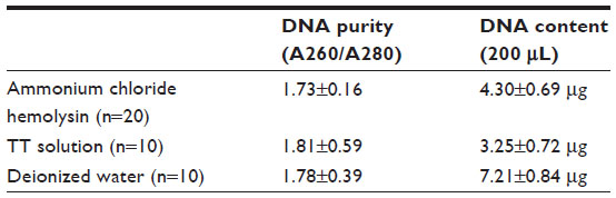 | Table 1 Genomic DNA from blood with different treatment methods |
Comparison of protease K and NaI extraction methods
The OD value for DNA extracted using the NaI method was high when measured by ultraviolet absorption. According to the calculation results of the DNA content by the confirmed equation, the obtained DNA was above 15 μg, which is more than the normal yield of human genomic DNA. Gel electrophoresis showed that the DNA content harvested by the NaI method was less than that of the protease K method (Figure 2). The reason for this might be that the iodide ion in NaI interfered with the ultraviolet absorption process. Therefore, the NaI method is not suitable for use with the magnetic microsphere extraction method.
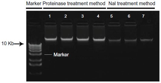 | Figure 2 Comparison of genomic DNA obtained by the proteinase K method and the NaI method. |
Optimal magnetic microsphere doses for extraction of genomic DNA from human blood
Because heme derivatives and porphyrins in blood can influence the effect of PCR amplification, they should be removed before extraction of DNA. Therefore, ammonium chloride hemolysin, Triton-Tris-HCl solution, and deionized water were used to pretreat the various blood samples. The DNA yield and recovery rate were then analyzed.
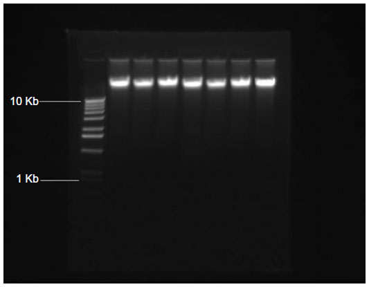
Table 1 shows that both DNA yield and recovery rate were highest when the concentration of the magnetic suspension was 0.01 mg/mL and the total quantity used was 4 mg. The average amount of DNA extracted was 7.21±0.84 μg, which was significantly different from 1–3 mg (control). The results of agarose gel electrophoresis (Figure 3) were not consistent with the DNA value measured by ultraviolet absorption, and the demonstrated qualities of seven different samples with 1–7 mg were basically fine (Figure 3).
 | Figure 3 Agarose gel electrophoresis of genomic DNA extracted from fresh blood by using various doses of magnetic microbeads. |
Table 2 shows the results for extraction of human genomic DNA from the cell lysis mixtures using a magnetic suspension concentration of 0.01 mg/mL, giving a total quantity of 1–8 mg. Both the recovery rate and yield of DNA increased with increasing doses of magnetic microspheres. It can be seen from the recovery experiment using calf thymus DNA that the magnetic microspheres adsorbed a lot of purified DNA. However, in the practical extraction application, only the right quantity of the magnetic microspheres could achieve a satisfactory yield and purity.
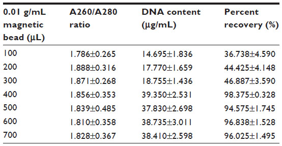 | Table 2 Genomic DNA from fresh blood extracted using various doses of magnetic microbeads |
Efficiency and purity of DNA extraction from fresh blood
The results of ultraviolet absorption spectroscopy show a typical DNA absorption peak at 260 nm for the genomic DNA extracted by the magnetic suspension method (Figure 4).
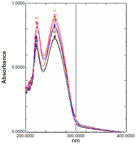 | Figure 4 Ultraviolet absorption spectra for human genomic DNA obtained from fresh blood. |
Agarose gel electrophoresis
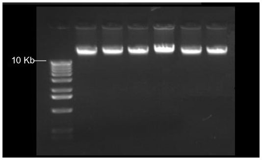
Agarose gel electrophoresis showed that the molecular weight of genomic DNA extracted by the magnetic suspension method was more than 10 kilobases with uniform brightness, suggesting that the extracted DNA was integrated and that the extraction effects were stable (Figure 5).
 | Figure 5 Agarose gel electrophoresis of genomic DNA obtained from fresh blood. |
PCR amplification in vitro
The quality of the human genomic DNA extracted from whole blood was determined by PCR. The amplified PCR fragment was the housekeeping gene, β-actin, which has a relatively conserved gene sequence and constant expression levels under various conditions. As long as there is a relatively complete human genome DNA template, β-actin can be amplified. The genomic DNA copy number can be obtained indirectly by analyzing the copy number of the amplified β-actin. The β-actin copy number is closely related to the purity of the extracted genomic DNA. Essentially, if the purity of the extracted DNA is high, β-actin can be easily amplified. Gene amplification confirmed that the extracted genomic DNA was complete and its purity was high, indicating that it can be amplified effectively in vitro (Figure 6).
 | Figure 6 Agarose gel electrophoresis of PCR products with the genomic DNA extracted from fresh blood by the magnetic microbead method as the template. |
Extracting genomic DNA from frozen blood using the magnetic microsphere method
Ultraviolet absorption spectroscopy demonstrated that the concentration of genomic DNA extracted from 200 μL of frozen blood by the magnetic microsphere method was 6.30±0.59 μg (n=30), while the concentration of genomic DNA extracted from the same frozen blood by the conventional method was only 2–3 μg. The purity of genomic DNA extracted by the magnetic microsphere method in the present study was in the range of 1.71–1.82, which is clearly superior to the value of 1.6 reported when using the traditional method.
The extracted genomic DNA samples were analyzed by gel electrophoresis. As shown in Figure 7, the molecular weight of the extracted genomic DNA was more than 10 kilobases, suggesting that the molecules of genomic DNA were extracted intact. The brightness of the extracted genomic DNA was uniform, indicating that the extraction efficiency was consistent across samples (Figure 7).
 | Figure 7 Agarose gel electrophoresis for genomic DNA extracted from dried blood. |
Extracting genomic DNA from dried blood by magnetic microspheres
Ultraviolet absorption spectroscopy demonstrated that the concentration of genomic DNA extracted from 200 μL of dried blood by the magnetic microsphere method was 2.25±0.25 μg (n=10), and the purity was 1.58–1.66. Trace blood (50 μL and 100 μL samples) could not be detected by ultraviolet absorption spectroscopy because the degree of dilution was out of range of the measurement method, so this should be detected by a more accurate method such as PCR analysis.
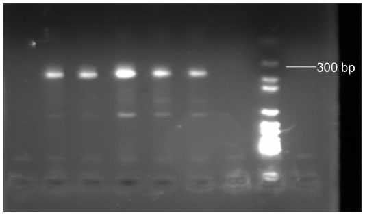
The gradient from trace amounts of DNA extracted from dried blood (50 μL, 100 μL, 200 μL) using the magnetic microsphere method was amplified in vitro. The amplified products were analyzed by agarose gel electrophoresis and compared with the molecular weight standard. The results show that the amplified fragment was of the expected size (Figure 8).
 | Figure 8 PCR results for genomic DNA extracted from dried blood. (A) 50 μL, (B) 100 μL, and (C) 200 μL. |
Comparison of genomic DNA extracted by magnetic microspheres from different blood samples
The genomic DNA content of fresh blood was high, whereas that extracted from frozen blood was low because DNA degradation had occurred and there was also loss of white cells. Further, large quantities of cells and their DNA content were lost from dried blood because of cell rupture and the grinding process, so the yield of DNA from dried blood decreased dramatically. Therefore, the state of a blood sample affects its DNA content to some degree. Other factors, including preservation time, also affect the yield and purity of DNA.
Fresh, frozen, and dried blood samples were used in this study, each at volume of 200 μL as the micro blood standard. The yield and purity of DNA extracted from the three different types of blood samples are shown in Table 1. The yield of DNA from human blood varied according to the preservation conditions and blood states sampled. The amount of genomic DNA retrieved from frozen blood was not high because of the long freezing time and low temperature, along with loss of leukocytes. Dryness, loss of water, and grinding in the dried blood samples led to less DNA being available for retrieval by the extraction process.
Discussion
Magnetic microspheres are a new functional material that has been extensively studied in recent years, has been used in the biomedicine, bioengineering, and other fields of applied research, and has attracted research attention worldwide. For magnetic microspheres to be marketable, they need to have a uniform particle size, a strong magnetic response, good stability, and a surface rich in active functional groups.1–6 How to improve the binding efficiency and specificity of biological macromolecules on the surfaces of magnetic microspheres and expand their scope of application has been the focus of intense research interest.
The present study investigated the effects of adsorbent cell lysate composition and magnetic microsphere dose on the adsorption and recovery of DNA using actual blood samples and established operating procedures. Urea–formaldehyde resin was used as the solid phase carrier, and PEG and sodium chloride were used as the adsorbents. A human whole genomic DNA extraction procedure was established, in which deionized water was used in the early stage of treatment and protease K digestion buffer was used to digest the proteins. We also studied the effects of different preservation conditions on the DNA content of blood samples, and their effects on the amount of DNA that could be extracted. We mainly used 200 μL traces of fresh whole blood, cryopreserved blood, and dried blood to extract and purify genomic DNA. Ultraviolet spectroscopy was used for detection of DNA purity and content, the integrity of the extracted DNA was examined by agarose gel electrophoresis, and the DNA obtained was used as a template for PCR amplification. The phenol chloroform extraction method was also used to extract genomic DNA from the reference sample. The results show that the magnetic bead method was better than the conventional method.
The mechanism by which genomic DNA is extracted by the magnetic microspheres also warrants discussion. Urea–formaldehyde resin matrix magnetic microspheres specifically bind target genomic DNA in an external magnetic field using their surface affinity ligands, by affinity adsorption, washing, and elution. The novel urea–formaldehyde resin magnetic microspheres developed in the present study could also be used as part of multifunctional automatic magnetic separation equipment.
In addition, the innovative magnetic microsphere method using urea–formaldehyde resin as the matrix and adsorbent was improved in the present study to decrease hemochrome and protein contamination, which significantly improves the yield and purity of DNA, and the results obtained are better than those previously reported in the literature and for commercial kits.
The A260/A280 ratios of the genomic DNA extracted from fresh whole blood, frozen blood, and dry blood using the magnetic microsphere method were in the ranges of 1.81–1.92, 1.71–1.82, and 1.56–1.64, respectively. The average yields of DNA from 200 μL of fresh whole blood, frozen blood, and dry blood were 7.21±0.84 μg, 6.30±0.59 μg, and 2.15±0.25 μg, respectively. All genomic DNA extracted from the blood samples had the typical absorption peak for DNA by ultraviolet detection. The gel electrophoresis results showed that the DNA extracted had a high degree of intactness and could be used for efficient PCR analysis. The genomic DNA extracted from 50 μL, 100 μL, and 200 μL gradient ultra-trace dried blood was used in the PCR amplification analysis with human-specific primers, and the results showed that the application effect was generally good.
Our experiments confirm that porous magnetic microspheres can be prepared by the template method. These microspheres have strong magnetic responses, a uniform particle size distribution, and a large specific surface area, which can be successfully used for the separation and purification of human whole blood genomic DNA. Being able to extract human blood genomic DNA with magnetic microspheres from blood preserved under different conditions has great advantages, in that the extracted genomic DNA is intact and of high quality, and can be used in molecular biology testing such as PCR. The biggest advantage of human whole blood genomic DNA extracted in this way is that it is efficient, convenient, fast, and automatic, although further investigations are necessary.
As early as 1979, DNA was purified using a glass method with NaI, which is efficient and rapid, and allows DNA of high quality to be obtained. Purified DNA obtained by this method can be used in restriction enzyme analysis and mapping experiments.12 Boom et al13 developed a rapid, reliable, and simple method for extraction of both RNA and DNA from human urine and serum on a small scale by using size-fractionated silica particles in 1990. The DNA extracted by Boom et al13 could only be used efficiently as a substrate for restriction endonucleases and DNA ligase but not in PCR because it contained unknown components that prevented analysis. Another reason was that there was often contamination of trace doses of extracted DNA from contemporary samples, leading to unreliable results.13 The silica-based extraction method is mainly used for purification of DNA from ancient bone samples, has high extraction efficiency, and is also fast and simple to perform. Moreover, the quality of DNA extracted from such bone samples is good and meets the requirements for further analysis, especially PCR.14 Tanaka et al15 investigated the characteristics of DNA on aminosilane-treated magnetic nanoparticles, and efficient recovery of DNA was achieved for PCR.
The data indicate that protic and aprotic solvents play an important role in the purification of DNA obtained by magnetic nanoparticles. Further, DNA extracted by this method can be used in subsequent restriction enzyme digestion and PCR experiments, which are better than previous methods.15 Charge switch nucleic acid purification technology is an important method for DNA extraction, and is already commercialized as the nonorganic solvent extraction method. However, it is expensive and not efficient for extraction of human genomic DNA from fresh whole blood, frozen blood, dried blood, or trace blood.
The method used in our present study is superior to other published methods. First, the urea–formaldehyde resin matrix magnetic microspheres we prepared have a strong magnetic response, good stability, a uniform particle size distribution, and a large specific surface area. Second, they are more efficient in extracting human genomic DNA from frozen blood, dried blood, and trace blood, with different morphologies under different storage conditions by magnetic microsphere methods than traditional magnetic microsphere methods in previous studies. Third, the genomic DNA extracted by the magnetic microsphere method is intact and of high quality, and meets the requirements for PCR. Fourth, the cost of preparation of urea–formaldehyde resin matrix magnetic microsphere is lower than the costs detailed in previous studies. Fifth, the urea–formaldehyde resin matrix magnetic microsphere method can be used for high-throughput extraction of human blood genomic DNA.
Our study has some limitations. First, porous magnetic microspheres need to be prepared, which requires additional time and added expense. Second, it is necessary to explore and prepare magnetic microspheres with a stable structure, controllable granularity, high saturation magnetization, low nonspecific binding, a biocompatible superparamagnetic separation carrier to improve the coupling efficiency of surface groups, optimization of process conditions, reduction of cost, and realization of scale production in further research. Third, it is important to expand the field of application of the magnetic microsphere method to other biological separation protocols, such as virus-specific nucleic acid probe hybridization separation and separation of immune marker cells. Fourth, the magnetic microsphere method needs to be tested for its ability to extract DNA from different types of samples such as bone, tissue, hair, and contaminated tissues.16
Conclusion
A magnetic microsphere suspension was prepared and used to extract genomic DNA from fresh whole, frozen, dried, and trace blood. The results of this study build a good foundation for research and development of high-throughput and rapid extraction of genomic DNA from various types of blood samples.
Acknowledgment
This work was supported by a grant from the National Natural Science Foundation of China.
Disclosure
The authors report no conflicts of interest in this work.
References
Cho YK, Lee JG, Park JM, Lee BS, Lee Y, Ko C. One-step pathogen specific DNA extraction from whole blood on a centrifugal microfluidic device. Lab Chip. 2007;7:565–573. | |
Hansen WL, Bruggeman CA, Wolffs PF. Pre-analytical sample treatment and DNA extraction protocols for the detection of bacterial pathogens from whole blood. Methods Mol Biol. 2013;943:81–90. | |
Li J, Liu Q, Alsamarri H, Lounsbury JA, Haversitick DM, Landers JP. Label-free DNA quantification via a ‘pipette, aggregate and blot’ (PAB) approach with magnetic silica particles on filter paper. Lab Chip. 2013;13:955–961. | |
Keeley B, Stark A, Pisanic TR 2nd, et al. Extraction and processing of circulating DNA from large sample volumes using methylation on beads for the detection of rare epigenetic events. Clin Chim Acta. 2013;425:169–175. | |
Ma C, Li C, Wang F, et al. Magnetic nanoparticles-based extraction and verification of nucleic acids from different sources. J Biomed Nanotechnol. 2013;9:703–709. | |
Patramool S, Bernard E, Hamel R, et al. Isolation of infectious chikungunya virus and dengue virus using anionic polymer-coated magnetic beads. J Virol Methods. 2013;193:55–61. | |
He N, Wang F, Ma C, et al. Chemiluminescence analysis for HBV-DNA hybridization detection with magnetic nanoparticles based DNA extraction from positive whole blood samples. J Biomed Nanotechnol. 2013;9:267–273. | |
Shan Z, Zhou Z, Chen H, et al. PCR-ready human DNA extraction from urine samples using magnetic nanoparticles. J Chromatogr B Analyt Technol Biomed Life Sci. 2012;881–882:63–68. | |
Duarte GR, Price CW, Littlewood JL, et al. Characterization of dynamic solid phase DNA extraction from blood with magnetically controlled silica beads. Analyst. 2010;135:531–537. | |
Zhang Y, Kuang M, Zhang L, Yang P, Lu H. An accessible protocol for solid-phase extraction of N-linked glycopeptides through reductive amination by amine-functionalized magnetic nanoparticles. Anal Chem. 2013;85:5535–5541. | |
Yuan Y, Zhang W, Yan R, et al. Identification of a novel 14-3-3zeta binding site within the cytoplasmic domain of platelet glycoprotein Ibalpha that plays a key role in regulating the von Willebrand factor binding function of glycoprotein Ib-IX. Circ Res. 2009;105:1177–1185. | |
Vogelstein B, Gillespie D. Preparative and analytical purification of DNA from agarose. Proc Natl Acad Sci U S A. 1979;76:615–619. | |
Boom R, Sol CJ, Salimans MM, Jansen CL, Wertheim-van Dillen PM, van der Noordaa J. Rapid and simple method for purification of nucleic acids. J Clin Microbiol. 1990;28:495–503. | |
Höss M, Pääbo S. DNA extraction from Pleistocene bones by a silica-based purification method. Nucleic Acids Res. 1993;21:3913–3914. | |
Tanaka T, Sakai R, Kobayashi R, Hatakeyama K, Matsunaga T. Contributions of phosphate to DNA adsorption/desorption behaviors on aminosilane-modified magnetic nanoparticles. Langmuir. 2009;25:2956–2961. | |
Li G, Shen B, He N, Ma C, Elingarami S, Li Z. Synthesis and characterization of Fe3O4@SiO2 core-shell magnetic microspheres for extraction of genomic DNA from human whole blood. J Nanosci Nanotechnol. 2011;11:10295–10301. |
 © 2014 The Author(s). This work is published and licensed by Dove Medical Press Limited. The
full terms of this license are available at https://www.dovepress.com/terms
and incorporate the Creative Commons Attribution
- Non Commercial (unported, 3.0) License.
By accessing the work you hereby accept the Terms. Non-commercial uses of the work are permitted
without any further permission from Dove Medical Press Limited, provided the work is properly
attributed. For permission for commercial use of this work, please see paragraphs 4.2 and 5 of our Terms.
© 2014 The Author(s). This work is published and licensed by Dove Medical Press Limited. The
full terms of this license are available at https://www.dovepress.com/terms
and incorporate the Creative Commons Attribution
- Non Commercial (unported, 3.0) License.
By accessing the work you hereby accept the Terms. Non-commercial uses of the work are permitted
without any further permission from Dove Medical Press Limited, provided the work is properly
attributed. For permission for commercial use of this work, please see paragraphs 4.2 and 5 of our Terms.