Back to Journals » International Journal of Nanomedicine » Volume 21
Extracellular Vesicles as Emerging Drug Delivery Platforms in Triple-Negative Breast Cancer: A Systematic Review
Authors Tian J ![]() , Chin YZ
, Chin YZ ![]() , Ng MH
, Ng MH ![]() , Pang KL, Law JX
, Pang KL, Law JX
Received 25 September 2025
Accepted for publication 25 November 2025
Published 8 January 2026 Volume 2026:21 570252
DOI https://doi.org/10.2147/IJN.S570252
Checked for plagiarism Yes
Review by Single anonymous peer review
Peer reviewer comments 2
Editor who approved publication: Professor Eng San Thian
Jiao Tian,1,2 Yun Zhi Chin,3 Min Hwei Ng,1 Kok Lun Pang,4 Jia Xian Law1
1Department of Tissue Engineering and Regenerative Medicine, Faculty of Medicine, Universiti Kebangsaan Malaysia, Kuala Lumpur, Malaysia; 2Department of Clinical Medicine, Zunyi Medical and Pharmaceutical College, Zunyi, Guizhou, People’s Republic of China; 3Division of Applied Biomedical Science and Biotechnology, School of Health Science, IMU University, Kuala Lumpur, Malaysia; 4Jeffrey Cheah School of Medicine and Health Sciences, Monash University Malaysia, Subang Jaya, Selangor, Malaysia
Correspondence: Jia Xian Law, Email [email protected]
Abstract: Breast cancer is the most prevalent malignancy among women, with triple-negative breast cancer (TNBC) being the most aggressive subtype. The lack of standard targeted therapies necessitates reliance on chemotherapy, which often suffers from poor targeting and significant off-target toxicity. This systematic review evaluates the therapeutic potential of drug delivery systems based on extracellular vesicles (EVs) for TNBC using 17 studies from 2020 to 2024 published in Scopus, PubMed, and Web of Science. The review discusses the diverse therapeutic cargos delivered by EVs, including chemotherapeutic agents and nucleic acids, while emphasizing the need for standardized protocols to facilitate EV isolation and characterization. Key findings provide both in vitro and in vivo evidence of the efficacy of EV-based therapies against TNBC cell lines and animal models, alongside insights into engineering strategies that enhance targeting specificity. Despite the promising findings, challenges remain in clinical translation, such as technical limitations and inconsistent reporting. The results underscore the potential of EVs to revolutionize TNBC treatment strategies, paving the way for more effective therapeutic options.
Keywords: exosome, breast cancer, drug delivery system, precision medicine, chemotherapy
Introduction
Breast cancer is the most prevalent cancer in women globally, and triple-negative breast cancer (TNBC) is its most aggressive and metastatic subtype.1–4 Since targeted receptors are absent, chemotherapy is the primary treatment strategy.5–9 However, TNBC has high rates of recurrence, poor prognosis, and unfavorable survival outcomes.10–14 Clinical efficacy of chemotherapeutics is hampered by several limitations, including poor tumor targeting, rapid systemic clearance, and substantial off-target toxicity.15–21
Precision medicine has been developed recently and represents a paradigm transformation in oncology to provide more efficient and customized treatment approaches.22–27 In this context, extracellular vesicles (EVs) have garnered increasing attention as a novel drug delivery platform.21,28–32 EVs are nanoscale vesicles found in almost all kinds of cells in the body and are important as mediators of intercellular communication through the transport of nucleic acids (DNAs and RNAs), proteins, and lipids.33–37 Due to their intrinsic biocompatibility, low immunogenicity, natural targeting ability, and adaptability, EVs are excellent candidates for precision medicine.29,38–40 Increasing evidence supports this potential, demonstrating their ability to selectively deliver therapeutic payloads to target cells, thereby enhancing anti-tumor efficacy while minimizing systemic toxicity and off-target effects.41–47
This systematic review examines the therapeutic potential of EV-based drug delivery systems in TNBC, reflecting the growing interest and promising applications of EV-based therapeutics in cancer treatment. Based on an assessment of 17 eligible studies, this review provides: (1) an evaluation of the in vitro and in vivo efficacy of EV-based therapeutic agents in TNBC cell lines and models; (2) an overview of the engineering strategies used to develop EV-based therapeutics, with emphasis on targeting specificity and delivery efficiency; (3) a detailed analysis of EV characteristics, including cellular origin, isolation and purification techniques, characterization methods, and storage conditions; (4) a quality assessment of the included studies, focusing on adherence to the MISEV2023 guidelines and in vivo validation; and (5) a translational perspective highlighting clinical challenges and opportunities informed by insights from nanotechnology, pharmacology, and cancer biology to guide future clinical applications. Overall, this review aims to provide a systematic synthesis of the current state of the art and to underscore key directions for advancing EV-based delivery systems as precision therapeutics for TNBC.
Methods
This systematic review was conducted using Scopus, PubMed, and Web of Science (WOS) databases to identify relevant publications from the past five years (2020–2024). Access to these databases was provided by the National University of Malaysia (UKM). The search query used was “extracellular vesicles OR exosomes” AND “drug delivery systems” AND “triple-negative breast cancer”, formulated based on terms obtained from the Medical Subject Headings (MeSH) database. Common terms identified from several relevant published studies were also incorporated into the search strategy. All search records, including titles, keywords, and abstracts, were downloaded and imported into EndNote. Duplicate records were removed before further screening. The initial screening excluded review articles, followed by an evaluation of titles, abstracts, and keywords for relevance to the topic. Subsequently, the full-text articles were retrieved and assessed for eligibility. The final screening was conducted based on the predefined inclusion and exclusion criteria. Inclusion criteria comprised articles reporting the use of extracellular vesicles (EVs) or exosomes (EXOs) as drug delivery systems for TNBC in in vitro, animal, or human studies. Exclusion criteria included review articles, conference abstracts, non–peer-reviewed publications, and studies not published in English. Subsequently, the in vivo studies were assessed for risk of bias using SYRCLE’s Risk of Bias (RoB) tool for animal studies. The review protocol was conducted in accordance with the Preferred Reporting Items for Systematic Reviews and Meta-Analyses (PRISMA) guidelines and has been registered in the International Prospective Register of Systematic Reviews (PROSPERO) under ID: CRD420250637660.
A total of 151 records were retrieved from three databases: Scopus (62), PubMed (38), and WOS (51). After removing 45 duplicates and 41 review articles, 65 records remained for further analysis. Subsequent screening based on titles, abstracts, and keywords excluded 38 records, leaving 27 articles for full-text assessment. Of these, 10 articles were excluded according to the predefined inclusion and exclusion criteria, resulting in a final set of 17 articles for data extraction and analysis. Among these, 6 studies reported only in vitro outcomes, 11 studies included both in vitro and in vivo outcomes, and none reported clinical outcomes. Article selection and data extraction were independently performed by two reviewers, with the corresponding author consulted to resolve discrepancies and reach consensus, thereby minimizing selection bias. The summary of the article selection protocol is reported in Figure 1.
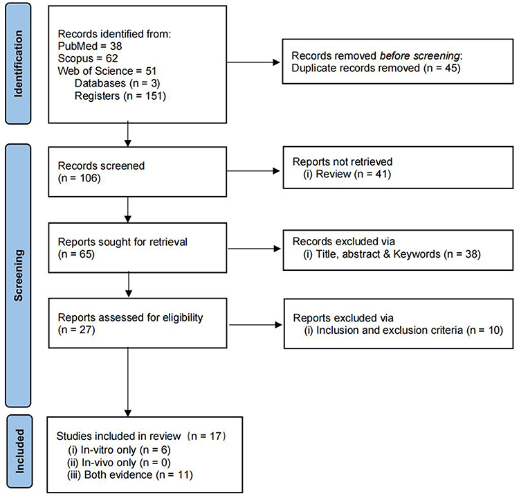 |
Figure 1 PRISMA flow chart summarizing the study selection process. |
Results and Discussion
EV-Based Therapeutic Delivery Systems for TNBC: in vitro and in vivo Evidence
This section summarizes the experimental outcomes of EV-based delivery systems in TNBC, focusing on the antitumor effects observed in in vitro cell culture and in vivo animal model studies (Table 1). It provides insight into the therapeutic efficacy and translational potential of EV-based platforms.
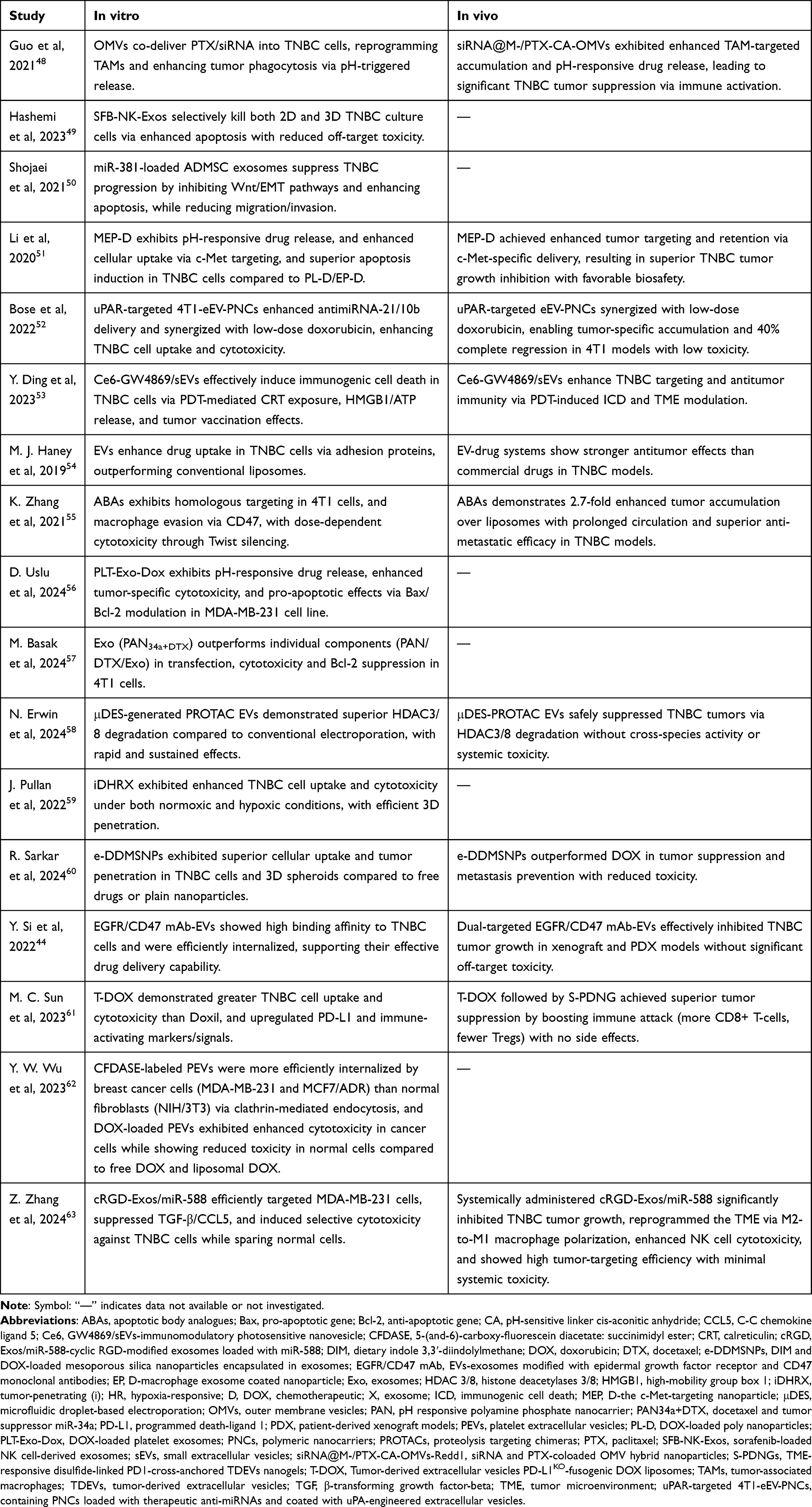 |
Table 1 Overview of EV-Based Drug Delivery in TNBC: In Vitro and In Vivo Outcomes |
All 17 studies included in this review conducted in vitro experiments to evaluate the antitumor efficacy of EV-based delivery systems against various TNBC cell lines. As summarized in Table 2, the delivery platform demonstrated effectiveness against a broad spectrum of TNBC cell lines, including human cell lines (MDA-MB-231, MDA-MB-468, HCC1806, HCC1937, BT-20) and the murine 4T1 line. Among these, the most utilized cell lines were MDA-MB-231 (11 studies)44,49–51,54,56,58–60,62,63 and 4T1 (6 studies).48,52,53,55,57,61
 |
Table 2 Summary of in Vivo Evaluation of EV-Based Delivery Systems for TNBC |
In vitro assessment of cellular uptake and cytotoxicity represents a critical initial step in evaluating the therapeutic potential of EV-mediated delivery systems. To assess uptake and targeting efficiency, 13 studies44,48–52,54,55,57,59,61–63 employed fluorescent labeling combined with confocal microscopy or flow cytometry to monitor internalization of EV-loaded cargo by TNBC cells. Notably, Pullan et al59 and Hashemi et al49 established three-dimensional (3D) cell culture models in vitro to further confirm the cellular uptake of EV-loaded drugs under physiologically relevant conditions. Cytotoxicity assays were the most common method used to quantify anticancer effects, including MTT50,51,54,56,57,60,61 and Cell Counting Kit-8 (CCK-8).48,52,53,55,62,63 Other studies used cell viability assays such as CellTiter-Blue52 and CellTiter-Glo.44 Furthermore, apoptosis induction in the cancer cells treated with EV-loaded drugs was confirmed using Annexin V/PI staining or the terminal deoxynucleotidyl transferase dUTP nick end labeling (TUNEL) assay.50,51,56,60 Collectively, these in vitro studies showed efficient cellular uptake, selective cytotoxicity toward TNBC cells, and apoptosis induction. However, the lack of systemic biological factors limits the translational relevance of in vitro models. Therefore, further investigations involving in vivo models and biodistribution studies are essential to validate the efficacy and specificity of EV-based delivery systems within the complexities of living organisms.
Biodistribution assessment is a crucial parameter for validating the in vivo tumor targeting efficacy of EV-based drug delivery systems. Among the 17 studies included, ten used various strategies to evaluate the biodistribution of the delivery platform in animals or vital organs.44,48,51–55,58,61,63 Seven of these employed both in vivo and ex vivo fluorescence or bioluminescence imaging, while Zhang et al55 used ex vivo imaging only, and Sun et al61 and Zhang et al63 performed in vivo imaging without ex vivo validation. Intravenous injection (i.v.) was the most common administration route (9 studies), with Erwin et al58 using intraperitoneal injection (i.p.). Notably, Haney et al54 compared biodistribution outcomes across i.v., i.p., and intratumoral (i.t.) routes. A range of fluorescent dyes was used for imaging, with DiR being the most frequently used,58,61,63 followed by Cy551,55 and BODIPY,48 Ce6,53 DiD,54 Cy7,44 and ICG.52 These dyes are generally classified into two groups: fluorescent imaging dyes, such as BODIPY, Cy5, Cy7, DiD, DiR, and ICG, and photosensitizers used for photodynamic therapy, such as Ce6. Although imaging techniques, administration routes, and dye selections varied, all ten studies consistently showed that EV-based delivery systems effectively targeted TNBC cells. Off-target distribution was minimal in each case.
A total of 11 studies conducted in vivo experiments to examine the efficacy of EV-based drug delivery against TNBC. Details of the in vivo studies, including animal models, gender, group, sample size, administration routes, and injection frequencies, are provided in Table 2. Diverse mouse models, such as BALB/c mice,48,52–54,60,61 BALB/c nude mice,55,63 BALB/cJ mice,44 nude mice,51,54 and NOD SCID gamma (NSG) mice,44,58 were utilized to establish tumor-bearing models in these studies. Among these in vivo studies, nine studies delivered therapeutic agents to animal models via i.v. tail vein injections.44,48,51–55,61,63 Alternative routes of administration, including i.t.60 and i.p.58 were also utilized to deliver cargoes entrapped in EVs to mouse models. Compared to i.v. and i.p., i.t. appears to require fewer injections. Sarkar et al60 administered therapeutic agents intratumorally to models with only one dose, whereas other studies employed multiple injections. All 11 in vivo studies demonstrated impressive antitumor effects of the EV-based drug delivery platform in TNBC animal models, shown by decreased tumor size and weight, as well as reduced side effects indicated by less body weight loss and higher survival rates. Across all 11 studies, one evaluated the antitumor effects only by tumor size.54 One study measured the expression of TNBC tumor-specific proteins by Western blot to evaluate the targeting antitumor effects of the EV-based therapy.58 Nine employed histopathology to further validate qualitatively and quantitatively the in vivo anticancer effects.44,48,51–53,55,60,61,63 All nine studies used Hematoxylin & Eosin (H&E) staining to evaluate the anti-tumor effect, safety, and anti-metastatic effect of EV-based therapeutics by visually observing changes in tumor tissue size, apoptosis of main organs in the body, and structural changes in common metastatic organs.44,48,51–53,55,60,61,63 Si et al45 and Zhang et al63 also utilized immunohistochemistry (IHC) to demonstrate the visualization and quantification of specific proteins associated with antitumor effects. Furthermore, some studies utilized immunofluorescent staining60 and flow cytometry61,63 to examine immune-related aspects of EV-based agents.
The integration of advanced imaging techniques with quantitative analysis and histopathological assessment revealed both the therapeutic potential and safety profile of EV-based drug delivery systems, establishing a strong basis for clinical translation.
Therapeutic Cargos and Engineering Strategies in EV-Based Delivery Systems
This section provides a comprehensive overview of EV based delivery systems in TNBC, highlighting the diverse therapeutic cargos, including chemotherapeutic drugs, nucleic acid therapeutics and bioactive compounds, delivered via EVs, as well as the engineering strategies employed for cargo loading and EV modification, as reported across the 17 studies (Table 2). Notably, nine studies reported improved anti-tumor efficacy with EV-encapsulated chemotherapeutics, while six focused on nucleic acid delivery and another six on bioactive molecules. Chemotherapeutics were most common, with doxorubicin (DOX),51,54,56,59–62 paclitaxel (PTX),48,54 docetaxel (DTX),57 and sorafenib (SFB)49 frequently featured. The nucleic acid therapeutics included siRNAs (eg, Redd1-siRNA, anti-Twist siRNA), miRNA mimics (miR-34a, miR-381-3p, miR-588), and anti-miRNAs (anti-miR-21, anti-miR-10b). Another group of studies explored EVs carrying bioactive molecules such as Ce6/GW4869 photosensitizers, saponin, DIM, Ver-A, YX968 PROTACs, and polymeric nanoparticles. This diversity of loaded cargos reflects the adaptability and potential of EV-based systems in TNBC treatment.
Regarding cargo loading techniques, electroporation was the most commonly employed method (used in 9 studies).48–50,53,55,56,58,59,63 Notably, Erwin et al58 introduced a continuous-flow microfluidic droplet-based electroporation (μDES) approach, which achieved superior loading efficiency and long-term stability compared to conventional electroporation. Alternative loading techniques used in the reviewed studies included incubation,44,48,57,60,62 co-extrusion51,52,61 as well as sonication.54
To further enhance delivery specificity and efficacy, EV modification strategies were employed in eight studies. These included both genetic engineering of donor cells48,52,55,61 and surface functionalization techniques, such as lipid59 or polymer insertion.63 Si et al44 developed dual-targeted mAb-EVs (EGFR/CD47), which showed selective uptake by both human and murine TNBC cells and improved therapeutic efficacy. Li et al51 constructed PLGA nanoparticle-coated EVs, enhancing delivery precision and stability.
Each generating approach presents distinct advantages and limitations. Therefore, continued innovation and comparative evaluation of these strategies are essential to optimize the efficacy, specificity, and translational potential of EV-based drug delivery systems for TNBC.
Characteristics of the Included Study
A comprehensive understanding of both the methodological and biological characteristics of EVs is critical for evaluating their therapeutic application in TNBC, as demonstrated by the detailed examination of the 17 studies included in this systematic review. These studies were analyzed with respect to EV origin, isolation and purification techniques, characterization approaches, and storage conditions, parameters that play a pivotal role in determining the quality, consistency, and translational potential of EV-based drug delivery systems. The following section summarizes the key features and methodological differences among the included studies.
A wide range of EV sources identified across these 17 included studies reflects the innovative and diverse strategies being adopted in the field of TNBC drug delivery. All the studies used mammalian-derived EVs except Guo et al48 that utilized outer membrane vesicles (OMVs) derived from engineered E. coli (BL21) bacteria. Among the studies used mammalian-derived EVs, six studies44,52,55,58,60,61 employed tumor cell-derived EVs, four from TNBC cells (including murine 4T152,55,61 and human MDA-MB-231 lines58) and two from other malignant tumor types.44,60 Immune cell-derived EVs were reported in four studies, ie, human NK cells,49 murine RAW 264.7 macrophages,54,57 and macrophage51 (origin not specified). Three studies utilized stem cell-derived EVs, specifically from human adipose-derived stem cells (hADSCs)50 and mesenchymal stem cells (hMSCs),63 and murine bone marrow-derived stem cells (mBMSCs),53 showcasing their regenerative and targeting potential. The remaining three studies explored less conventional sources, ie, human platelets56,62 and raw bovine milk.59
EVs derived from different cell types possess distinct physicochemical and biological properties, which can significantly influence their targeting behavior and the responses of recipient cells.64 Numerous studies have demonstrated that tumor cell-derived EVs exhibit strong homologous targeting capability, which may be attributed to the specific membrane proteins inherited from parental cancer cells that selectively facilitate drug uptake by homologous tumor cells.52,55,58,61 For instance, Zhang et al55 conjugated the toxic protein saporin and anti-twist siRNA to 4T1 cell-derived exosomes (ABAs) and co-incubated them with RAW264.7 macrophages to investigate their anti-phagocytic behavior. The results showed that ABAs inherited the anti-phagocytic properties from parental tumor cells and effectively evaded macrophage uptake. Furthermore, in vivo studies confirmed that autologous tumor-derived EVs suppressed lung metastasis by enhancing the expression of Twist and promoting epithelial-mesenchymal transition (EMT). Similarly, Erwin et al58 loaded proteolysis targeting chimera (PROTAC) molecules into MDA-MB-231-derived EVs, which promoted the degradation of histone deacetylases HDAC3/8, leading to activation of tumor suppressor genes and significant antitumor effects. In another study, Sun et al61 utilized 4T1 cell-derived EVs (TDEVs) to enhance the immunogenic cell death (ICD) effect of doxorubicin (DOX). The treatment increased HMGB1 release and calreticulin (CRT) exposure on the cell surface, thereby enhancing the immunogenicity of 4T1 tumor cells.
EVs derived from immune cells can effectively enhance immunotherapy efficacy while minimizing systemic toxicity.65 These vesicles also mimic immune cell functions due to the inherent tumor-targeting abilities of their surface proteins. For example, Basak et al57 designed macrophage-derived exosomes co-delivering docetaxel (DTX) and miR-34a (PAN34a+DTX) and the exosomes were found to inhibit 4T1 tumor growth and promoted apoptosis by upregulating inflammatory cytokines (TNF-α and IFN-γ) and downregulating the anti-apoptotic gene BCL-2. Li et al51 further modified macrophage-derived exosomes with a c-Met-targeting peptide (MEP-D) for TNBC therapy. In vitro, MEP-D exhibited the highest cellular uptake and apoptotic induction among tested groups. In vivo TUNEL staining confirmed extensive tumor cell apoptosis in the MEP-D-treated group, consistent with in vitro findings. Natural killer (NK) cell-derived EVs have also shown promise. Hashemi et al49 reported that SFB-loaded NK-EVs demonstrated high tumor selectivity in both 2D and 3D cultures, significantly downregulated VEGF-A expression, and upregulated p53 and caspase-3, thereby enhancing tumor cell apoptosis.
Stem cells are considered ideal sources of EVs because of their self-renewal and regenerative properties.66 Stem cell-derived EVs have been widely explored as therapeutic delivery vehicles targeting TNBC cells.50,53,63 Shojaei et al50 employed adipose-derived MSCs (ADMSC) exosomes to deliver a miR-381 mimic to MDA-MB-231 cells, resulting in increased apoptosis and inhibited proliferation, migration, and invasion. Zhang et al63 successfully constructed MSC-derived exosomes loaded with miR-588 and modified with cRGD peptides (cRGD-Exos/miR-588). The engineered vesicles exhibited potent TNBC tumor targeting and strong antitumor efficacy by remodeling the tumor microenvironment (TME), reprogramming tumor-associated macrophages (TAMs), and reducing immunosuppression. Interestingly, Ding et al53 encapsulated the photosensitizer Ce6 and GW4869 into bone marrow MSC-derived small EVs to generate immunomodulatory photosensitized nanovesicles. These constructs improved the tumor immune microenvironment and demonstrated strong tumor-targeting capability both in vitro and in vivo.
EVs derived from platelets also display considerable potential in tumor-targeted drug delivery.67 Uslu et al56 and Wu et al62 demonstrated that doxorubicin-loaded platelet-derived EVs (DOX-PEVs) effectively targeted and enhanced the cytotoxicity of DOX toward MDA-MB-231 breast cancer cells. Moreover, Wu et al62 showed that PEVs and DOX-PEVs could be sterilized by filtration and stored frozen without procoagulant risk or alteration of surface markers, supporting their suitability for clinical translation.
Notably, Guo et al48 developed a dual-drug delivery system based on Gram-negative bacteria-derived outer membrane vesicles (siRNA@M-/PTX-CA-OMVs) co-loading paclitaxel (PTX) and Regulated in Development and DNA Damage Response 1 (Redd1)-siRNA. This system effectively repolarized TAMs, remodeled the TME, and activated antitumor immunity to suppress TNBC progression in vitro and in vivo.
Collectively, these findings underscore that EVs from various sources, ie, tumor cells, immune cells, stem cells, platelets, or bacteria, exhibit unique immunomodulatory properties and targeting behaviors. EV-based delivery systems represent a promising and versatile platform for cancer immunotherapy, providing controlled regulation of immune responses and enhanced therapeutic selectivity. Furthermore, emerging studies have revealed that EVs can also serve as carriers of radiosensitizers to improve the precision and controllability of radiotherapy.68–71 A thorough and ongoing understanding of the biological characteristics and therapeutic potential of EVs from different origins is essential for advancing their use in TNBC treatment and facilitating clinical translation.
The isolation and purification methods of EVs varied notably. As summarized in Table 3 and visualized in Figure 2, differential centrifugation (DC) is the most commonly employed technique, with 12 studies44,48,51–54,56–59,62,63 utilizing ultracentrifugation (UC) for EV isolation, followed by commercial isolation kits49,50,60 and extrusion,55,61,62 each reported in 3 studies. Additionally, another physical disruption techniques, ie, freeze–thaw cycles, were reported in 1 study.62 Regarding purification strategies, UC emerged as the most frequently employed technique, reported in 5 studies.52,56–58,63 Several combined approaches were also described, such as UC with microfiltration (MF),53,54,59 differential centrifugation (DC) with MF,62 and a multi-step protocol integrating ultrafiltration (UF), MF, and UC.48 In addition, single-method strategies, including DC,50,61 MF,51 UF,55 and size exclusion chromatography (SEC),44 were also noted, whereas 2 studies49,60 did not specify their purification techniques.
 |
Table 3 Summary of Isolation, Characterization and Purification Methods, Storage Conditions, and Functional Analysis of EVs |
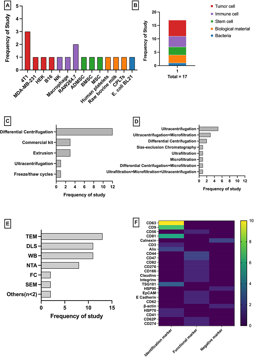 |
Figure 2 Overview of reviewed studies. (A) distribution of source, (B) type of source, (C) isolation method, (D) purification methods, (E) characterization methods, and (F) common biological markers. |
Despite their widespread use, current isolation and purification methods often struggle to achieve both high specificity and high recovery, creating challenges for standardization, reproducibility, and scalability in therapeutic applications. This limitation is particularly relevant as interest grows in EV-based drug delivery systems, especially for oncology treatment. To address this gap, there’s an urgent need to develop novel approaches that can efficiently produce clinical-grade EVs while maintaining purity and scalability.
All studies included in this review reported EVs with a size distribution typically ranging from 20 to 300 nm, as assessed by various techniques including nanoparticle tracking analysis (NTA), dynamic light scattering (DLS), and atomic force microscopy (AFM). Morphology and membrane integrity were typically assessed by transmission electron microscopy (TEM), scanning electron microscopy (SEM), and AFM. In addition, Western blotting (WB) and flow cytometry (FC) were widely used to detect EV-specific surface and cytosolic markers, while protein concentration was generally measured by the bicinchoninic acid (BCA) assay. The most frequently assessed EV markers included classical identification markers (eg, CD63, CD9, CD81, Alix, TSG101, HSP70, CD3, CD41) and functional markers (eg, CD56, CD44, CD47, CD62, CD82, CD276, CD166, CD274, CD62P, claudins, integrins, EpCAM, E-cadherin). Negative EV markers (eg, calnexin, HSP90, β-actin) were also used in some of the studies to examine the purity of the isolated EV samples and to rule out contamination by other cellular components. As illustrated in Figure 2F, a heatmap was generated based on 17 included studies to visualize the frequency of marker detection. Each value represents the number of studies that reported the corresponding marker. The analysis revealed that classical tetraspanins (CD63, CD81, and CD9) and endosomal markers (TSG101 and Alix) were most consistently identified across studies, highlighting their reliability as canonical EV markers. In contrast, functional surface molecules such as CD44, integrins, and EpCAM showed lower and more variable detection frequencies, suggesting differences in EV origin, isolation approaches, and experimental aims among studies. Appropriate characterization, including quantifying EV content, verifying EV presence, and assessing potential contamination from non-EV components, is essential for the reliability of EV-based research. However, the field continues to face substantial challenges in EVs characterization, primarily due to the lack of universally accepted markers, and EVs naturally vary in size, content, and origin. The Minimal Information for Studies of Extracellular Vesicles (MISEV) 2023 guidelines highlight that no single method can fully characterize EVs, so combining multiple complementary techniques is strongly advised to overcome individual limitations and ensure rigorous, reproducible, and comparable results across studies. Storage conditions for EVs were also examined in this review, revealing that only eight out of 17 studies clearly stated storing EVs at −80 °C before use,44,50,54,55,59,60,62,63 while the remaining nine did not report any storage details.
It is well recognized that various factors, such as storage temperature, duration, freezing and thawing procedures, storage containers, and the number of freeze-thaw cycles, can significantly affect the structural integrity, composition, and biological activity of EVs. Considering these concerns, the MISEV 2023 guidelines urge researchers to thoroughly document all storage-related parameters during EV isolation and preservation, which is crucial for enhancing study reproducibility, transparency, and minimizing storage-induced changes in EV samples.
The reviewed studies demonstrate the wide range of methods used in EV research, with differences in EV sources, isolation techniques, characterization approaches, and storage conditions potentially leading to inconsistent findings and reduced comparability between studies. Therefore, the MISEV 2023 guidelines emphasize the importance of adopting standardized protocols and comprehensive reporting to ensure reproducibility and improve data quality.
Quality Assessment of the Included Study
This section evaluates the overall quality and consistency of the included studies, with a focus on in vivo experimental design, reporting standards, and methodological transparency, as well as adherence to the EV characterization and purity criteria outlined in the MISEV2023 guidelines.
The SYRCLE’s RoB tool adopted from Hooijman 201472 was utilized to evaluate the risk of bias in the selected studies as shown in Table 4. Among the 11 studies reviewed, 8 (72.7%) explicitly mentioned the randomization of animal groups,44,48,51,52,54,55,61,63 but 3 (27.3%) did not, resulting in an unclear risk of sequence generation.53,58,60 The baseline characteristics, such as age, gender, species, and group size, were provided before treatment in 5 studies (27.3%),48,53,55,60,61 but the remaining 6 studies showed a high risk due to the lack of one or two essential details.44,51,52,54,58,63 None of the studies disclosed information regarding allocation concealment, random housing, blinding of interventions or caregivers, random outcome assessment, or blinding of outcome assessment; therefore, these potential biases were classified as unclear. Regarding attrition and reporting bias, all studies have a low risk of bias, as they reported complete data and used statistical methods for data analysis. All studies showed low risk of other biases by disclosing ethics approval, potential conflicts of interest, and the roles of collaborating parties. Generally, most of the elements evaluated showed an unknown risk of bias.
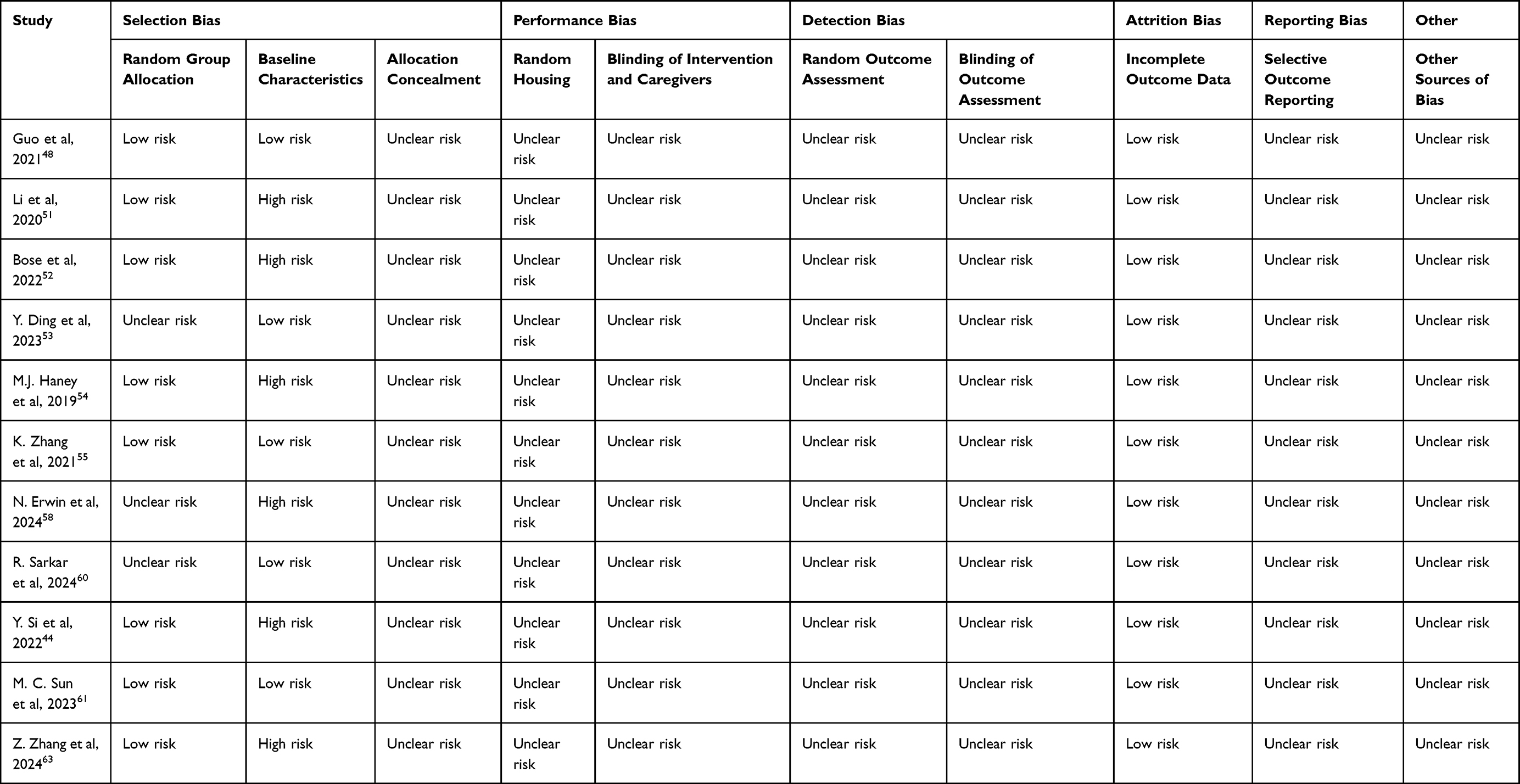 |
Table 4 SYRCLE’s Risk Tool of Bias Assessment |
In addition to evaluating the in vivo study design, this review also assessed the characterization and purity of EV across all 17 included studies based on the MISEV 2023 guidelines. According to the MISEV2023 guidelines, integrating multiple complementary techniques is recommended for EV characterization, including assessing the quantity and presence of EV and evaluating non-EV components. A detailed summary of adherence to MISEV2023 criteria is provided in Table 3. Specifically, 13 studies fulfilled the essential criteria for EV characterization through the quantitation of EV and identification of at least two positive EV protein markers and non-EV protein markers.
In contrast, four studies lacked sufficient evidence of multiparametric characterization, raising concerns about accurately identifying EVs. Regarding purity, only 6 studies adequately addressed the presence of non-EV components or used recommended orthogonal strategies, while the majority (11 studies) did not meet the MISEV2023 purity requirements. This indicates a subset of studies that consider the potential presence of contaminants. These findings reveal a significant standardization gap in EV research, underscoring the urgent need to raise awareness and improve adherence to standardized guidelines to ensure data reliability and comparability across studies.
Challenges and Future Considerations of EVs as a Drug Delivery System
The landscape of EV-based drug delivery research for TNBC shows a globally expanding yet regionally concentrated trend, with most of the 17 included studies originating from Asia, particularly China, followed by the United States and several European countries, highlighting a strong research focus in Asia and North America, with China and the USA as leading contributors (Figure 3). Temporally, research outputs remained steady from 2020 to 2024, with 3 to 4 studies published annually, suggesting an ongoing development phase rather than a short-term research boom in EV-based therapeutic strategies.
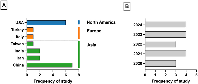 |
Figure 3 Distribution of review studies for publication by (A) region and (B) year. |
Despite growing preclinical evidence supporting the therapeutic potential of EV-based platforms, major challenges hinder clinical translation, including heterogeneous isolation and purification methods that often fail to achieve both high yield and purity, limiting reproducibility and scalability. Additionally, the lack of universally accepted EV markers and standardized characterization protocols complicates quality assessment, and few studies have examined whether EVs maintain stability and bioactivity during storage and transport. Furthermore, the existing cargo loading methods, such as electroporation and sonication, need to be refined to achieve efficient and controlled cargo loading for maximizing the therapeutic potential of EV-based drug delivery systems. Another major challenge is the rapid clearance by the mononuclear phagocyte system (MPS) and the potential off-target accumulation of EV-loaded drugs following systemic administration. Thus, more pharmacokinetics and biodistribution studies in vivo are needed to standardize the in vivo experimental protocol, including the optimal dosing and delivery route of EV-based drug delivery platforms in TNBC. In addition, the inconsistency in reporting of methodologies and outcomes remains a crucial concern. To ensure the transparency and reproducibility of data, it would be essential to adhere to the MISEV 2023 guidelines for standardized reporting. On the translational front, issues such as large-scale production, storage stability, immunogenicity, and biodistribution variability must be systematically addressed to bridge the gap between preclinical research and clinical application. Although some studies explored engineering strategies, such as those modified with targeting ligands or loaded with potent therapeutic agents to enhance delivery precision, the safety and long-term effects of these modifications remain largely unknown.
Future research should focus on harmonizing advanced techniques, developing robust and standardized manufacturing protocols, and expanding multi-center in vivo validations. These steps are crucial for advancing clinical development.
Beyond Drug Delivery – Other Roles of EVs in Cancer Immunotherapy
EVs play a complex role in cancer immunotherapy as they can either promote or suppress tumor progression.73,74 Their dualistic effects on tumor progression is strongly related to their cellular origin and molecular cargo. Tumor-derived EVs are known to carry immunosuppressive molecules such as programmed cell death 1 ligand 1 (PD-L1),75–77 FAS ligand (FASLG),78,79 MHC class I polypeptide-related sequence A (MICA),80 and TGF-β81 that can inhibit cytotoxic T lymphocyte activation and differentiation, impair NK cell and dendritic cell function, and promote the expansion of regulatory T cells, thereby fostering an immunosuppressive tumor microenvironment. Through these mechanisms, tumor-derived EVs contribute to immune escape, tumor growth, and resistance to immunotherapy. On the other hand, immune cell-derived EVs (eg, macrophages,51,54 dendritic cells,82,83 NK cells49) or engineered EVs may deliver anti-cancer agents (eg, miRNAs,48,50,52,55,57,63 cytotoxic proteins,55,58 chemotherapy drugs48,49,54,56,57,59–62) to activate the immune system and boost the immune response against cancer, thereby sensitizing cancer cells to anti-cancer therapies. Therefore, it is important to understand the complex interplay between the EVs and the tumor for rational design of EV-based therapeutic platforms for the treatment of cancer. A comprehensive consideration of their immunomodulatory effects will be critical to harnessing their full potential in cancer treatment, ie, to maximize anti-tumor efficacy while minimizing the risk of immune-related adverse events.
Conclusion
In conclusion, this review highlights the promise of EVs as a novel drug delivery system for TNBC. Analysis of the included studies demonstrates that EV-based therapeutics enhance the efficacy of anti-cancer agents while minimizing adverse effects. Both in vitro and in vivo evidence support the effectiveness of EV-mediated delivery systems in selectively targeting TNBC cells. However, challenges such as heterogeneous isolation methods, lack of standardized characterization protocols, and issues with stability and bioactivity during storage hinder clinical translation. Future research should focus on addressing these challenges, adhering to MISEV 2023 guidelines for reporting, and developing robust manufacturing protocols. Ultimately, EV-based drug delivery systems hold significant potential to improve outcomes for patients with TNBC and advance personalized cancer therapy.
Ethics Approval
Ethics approval is not required for this systematic review.
Consent for Publication
All authors read and approved the published version of the manuscript.
Acknowledgments
Figures 1–3 were produced using GraphPad Prism 10. The authors acknowledge the use of an AI tool (ChatGPT, GPT-4o) for language refinement but confirm that they fully authored, synthesized, researched, reviewed, and verified the content, taking full responsibility for its accuracy.
Author Contributions
All authors made a significant contribution to the work reported, whether that is in the conception, study design, execution, acquisition of data, analysis and interpretation, or in all these areas; took part in drafting, revising or critically reviewing the article; gave final approval of the version to be published; have agreed on the journal to which the article has been submitted; and agree to be accountable for all aspects of the work.
Funding
This research was funded by grants from the Faculty of Medicine, Universiti Kebangsaan Malaysia (FF-2025-063 and FF-2025-185).
Disclosure
The authors report no conflicts of interest.
References
1. Agelidis A, Ter-Zakarian A, Jaloudi M. Triple-negative breast cancer on the rise: breakthroughs and beyond. Breast Cancer. 2025;17:523–22.
2. Siegel RL, Kratzer TB, Giaquinto AN, Sung H, Jemal A. Cancer statistics, 2025. CA Cancer J Clin. 2025;75(1):10–45. doi:10.3322/caac.21871
3. Wang D, Yang Y, Rong W, et al. Natural history and prognostic nomogram of untreated triple negative breast cancer based on SEER database. Sci Rep. 2025;15(1):23347. doi:10.1038/s41598-025-07114-2
4. Bray F, Laversanne M, Sung H, et al. Global cancer statistics 2022: GLOBOCAN estimates of incidence and mortality worldwide for 36 cancers in 185 countries. CA Cancer J Clin. 2024;74(3):229–263. doi:10.3322/caac.21834
5. Kagihara JA, Shagisultanova E, Afghahi A, Diamond JR. Moving towards targeted therapies for triple-negative breast cancer. Curr Breast Cancer Rep. 2021;13(3):216–226. doi:10.1007/s12609-021-00416-0
6. Shaath H, Elango R, Alajez NM. Molecular classification of breast cancer utilizing long non-coding RNA (lncRNA) transcriptomes identifies novel diagnostic lncRNA panel for triple-negative breast cancer. Cancers. 2021;13(21):5350. doi:10.3390/cancers13215350
7. Ramamoorthy P, Dandawate P, Jensen RA, Anant S. Celastrol and triptolide suppress stemness in triple negative breast cancer: notch as a therapeutic target for stem cells. Biomedicines. 2021;9(5):482. doi:10.3390/biomedicines9050482
8. Leon-Ferre RA, Goetz MP. Advances in systemic therapies for triple-negative breast cancer. BMJ. 2023;381:e071674. doi:10.1136/bmj-2022-071674
9. Korde LA, Somerfield MR, Carey LA, et al. Neoadjuvant chemotherapy, endocrine therapy, and targeted therapy for breast cancer: ASCO guideline. J Clin Oncol. 2021;39(13):1485–1505. doi:10.1200/JCO.20.03399
10. Waks AG, Winer EP. Breast cancer treatment: a review. JAMA. 2019;321(3):288–300. doi:10.1001/jama.2018.19323
11. Li X, Yang J, Peng L, et al. Triple-negative breast cancer has worse overall survival and cause-specific survival than non-triple-negative breast cancer. Breast Cancer Res Treat. 2017;161(2):279–287.
12. Abdul Aziz AA, Md Salleh MS, Ankathil R. Clinicopathological and prognostic characteristics of Malaysian triple negative breast cancer patients undergoing TAC chemotherapy regimen. Int J Breast Cancer. 2020;2020:8424365. doi:10.1155/2020/8424365
13. Meng X, Cai Y, Chang X, Guo Y. A novel conditional survival nomogram for monitoring real-time prognosis of non-metastatic triple-negative breast cancer. Front Endocrinol. 2023;14:1119105. doi:10.3389/fendo.2023.1119105
14. Xiong N, Wu H, Yu Z. Advancements and challenges in triple-negative breast cancer: a comprehensive review of therapeutic and diagnostic strategies. Front Oncol. 2024;14:1405491. doi:10.3389/fonc.2024.1405491
15. Koirala M, DiPaola M. Overcoming cancer resistance: strategies and modalities for effective treatment. Biomedicines. 2024;12(8):1801. doi:10.3390/biomedicines12081801
16. Eslami M, Memarsadeghi O, Davarpanah A, Arti A, Nayernia K, Behnam B. Overcoming chemotherapy resistance in metastatic cancer: a comprehensive review. Biomedicines. 2024;12(1):183. doi:10.3390/biomedicines12010183
17. de M Cravo DL, Pimentel PAB, Garcia APV, et al. Comparative analysis of chemotherapy resistance mechanisms in humans and companion animals. Veterinary Sci. 2025;12(8):747. doi:10.3390/vetsci12080747
18. Khan MM, Yalamarty SSK, Rajmalani BA, Filipczak N, Torchilin VP. Recent strategies to overcome breast cancer resistance. Crit Rev Oncol/Hematol. 2024;197:104351. doi:10.1016/j.critrevonc.2024.104351
19. Dhanyamraju PK. Drug resistance mechanisms in cancers: execution of pro-survival strategies. J Biomed Res. 2024;38(2):95–121. doi:10.7555/JBR.37.20230248
20. Khan Y, Hussain MS, Ramalingam PS, et al. Exploring extracellular RNA as drivers of chemotherapy resistance in cancer. Mol Biol Rep. 2025;52(1):142. doi:10.1007/s11033-025-10263-2
21. Gharavi AT, Irian S, Niknejad A, Parang K, Salimi M. Harnessing exosomes as a platform for drug delivery in breast cancer: a systematic review for in vivo and in vitro studies. Mol Ther Oncol. 2024;32(2):200800. doi:10.1016/j.omton.2024.200800
22. Batool S, Sohail S, Ud Din F, et al. A detailed insight of the tumor targeting using nanocarrier drug delivery system. Drug Deliv. 2023;30(1):2183815. doi:10.1080/10717544.2023.2183815
23. Lammers T. Nanomedicine tumor targeting. Adv Mater. 2024;36(26):2312169. doi:10.1002/adma.202312169
24. Leng G, Duan B, Liu J, et al. The advancements and prospective developments in anti-tumor targeted therapy. Neoplasia. 2024;56:101024. doi:10.1016/j.neo.2024.101024
25. Bai J, Gao Y, Zhang G. The treatment of breast cancer in the era of precision medicine. Cancer Biol Med. 2025;22(4):322–347. doi:10.20892/j.issn.2095-3941.2024.0510
26. Hossain F, Majumder S, David J, Miele L. Precision medicine and triple-negative breast cancer: current landscape and future directions. Cancers. 2021;13(15):3739. doi:10.3390/cancers13153739
27. Foldi J, Geyer CE. Precision medicine for metastatic TNBC: the FUTURE is now. Cell Res. 2023;33(7):491–492. doi:10.1038/s41422-023-00815-1
28. Rehman FU, Liu Y, Zheng M, Shi B. Exosomes based strategies for brain drug delivery. Biomaterials. 2023;293:121949. doi:10.1016/j.biomaterials.2022.121949
29. Kim HI, Park J, Zhu Y, Wang X, Han Y, Zhang D. Recent advances in extracellular vesicles for therapeutic cargo delivery. Exp Mol Med. 2024;56(4):836–849. doi:10.1038/s12276-024-01201-6
30. Lu X, Fan S, Cao M, et al. Extracellular vesicles as drug delivery systems in therapeutics: current strategies and future challenges. J Pharm Investig. 2024;54(5):785–802. doi:10.1007/s40005-024-00699-2
31. Yun JH, Noh YR, Yoo S, et al. Harnessing extracellular vesicles for targeted drug delivery in ovarian cancer. Pharmaceutics. 2025;17(4):528. doi:10.3390/pharmaceutics17040528
32. Ragni E. Extracellular vesicles: recent advances and perspectives. FBL. 2025;30(6):36405. doi:10.31083/FBL36405
33. Berumen Sánchez G, Bunn KE, Pua HH, Rafat M. Extracellular vesicles: mediators of intercellular communication in tissue injury and disease. Cell Commun Signaling. 2021;19(1):104. doi:10.1186/s12964-021-00787-y
34. Petroni D, Fabbri C, Babboni S, Menichetti L, Basta G, Del Turco S. Extracellular vesicles and intercellular communication: challenges for in vivo molecular imaging and tracking. Pharmaceutics. 2023;15(6):1639. doi:10.3390/pharmaceutics15061639
35. Mosquera-Heredia MI, Morales LC, Vidal OM, et al. Exosomes: potential disease biomarkers and new therapeutic targets. Biomedicines. 2021;9(8):1061. doi:10.3390/biomedicines9081061
36. Welsh JA, Goberdhan DCI, O’Driscoll L, et al. Minimal information for studies of extracellular vesicles (MISEV2023): from basic to advanced approaches. J Extracell Vesicles. 2024;13(2):e12404. doi:10.1002/jev2.12404
37. Takahashi A, Okada R, Nagao K, et al. Exosomes maintain cellular homeostasis by excreting harmful DNA from cells. Nat Commun. 2017;8(1):15287. doi:10.1038/ncomms15287
38. Wu P, Zhang B, Ocansey DKW, Xu W, Qian H. Extracellular vesicles: a bright star of nanomedicine. Biomaterials. 2021;269:120467. doi:10.1016/j.biomaterials.2020.120467
39. He J, Ren W, Wang W, et al. Exosomal targeting and its potential clinical application. Drug Deliv Transl Res. 2022;12(10):2385–2402. doi:10.1007/s13346-021-01087-1
40. Parada N, Romero-Trujillo A, Georges N, Alcayaga-Miranda F. Camouflage strategies for therapeutic exosomes evasion from phagocytosis. J Adv Res. 2021;31:61–74. doi:10.1016/j.jare.2021.01.001
41. Cheng Q, Dai Z, Smbatyan G, Epstein AL, Lenz HJ, Zhang Y. Eliciting anti-cancer immunity by genetically engineered multifunctional exosomes. Mol Ther. 2022;30(9):3066–3077. doi:10.1016/j.ymthe.2022.06.013
42. Feng C, Xiong Z, Wang C, et al. Folic acid-modified Exosome-PH20 enhances the efficiency of therapy via modulation of the tumor microenvironment and directly inhibits tumor cell metastasis. Bioact Mater. 2021;6(4):963–974. doi:10.1016/j.bioactmat.2020.09.014
43. Peng Q, Zhang J, Ye X, et al. Robust delivery of RIG-I agonists using extracellular vesicles for anti-cancer immunotherapy. J Extracellular Vesicles. 2022;11(6):e12277. doi:10.1002/jev2.12187
44. Si Y, Chen K, Ngo HG, et al. Targeted EV to deliver chemotherapy to treat triple-negative breast cancers. Pharmaceutics. 2022;14(1):146. doi:10.3390/pharmaceutics14010146
45. Xie X, Lian S, Zhou Y, et al. Tumor-derived exosomes can specifically prevent cancer metastatic organotropism. J Control Release. 2021;331:404–415. doi:10.1016/j.jconrel.2021.01.030
46. Zhou Y, Yamamoto Y, Takeshita F, Yamamoto T, Xiao Z, Ochiya T. Delivery of miR-424-5p via extracellular vesicles promotes the apoptosis of MDA-MB-231 TNBC cells in the tumor microenvironment. Int J Mol Sci. 2021;22(2):844. doi:10.3390/ijms22020844
47. Nguyen cao TG, Kang JH, Kim W, et al. Engineered extracellular vesicle-based sonotheranostics for dual stimuli-sensitive drug release and photoacoustic imaging-guided chemo-sonodynamic cancer therapy. Theranostics. 2022;12(3):1247–1266. doi:10.7150/thno.65516
48. Guo Q, Li X, Zhou W, et al. Sequentially triggered bacterial outer membrane vesicles for macrophage metabolism modulation and tumor metastasis suppression. ACS Nano. 2021;15(8):13826–13838. doi:10.1021/acsnano.1c05613
49. Hashemi ZS, Ghavami M, Kiaie SH, et al. Novel delivery of sorafenib by natural killer cell-derived exosomes enhanced apoptosis in triple-negative breast cancer. Nanomedicine. 2023;18(5):437–453. doi:10.2217/nnm-2022-0237
50. Shojaei S, Hashemi SM, Ghanbarian H, Sharifi K, Salehi M, Mohammadi-Yeganeh S. Delivery of miR-381-3p mimic by mesenchymal stem cell-derived exosomes inhibits triple negative breast cancer aggressiveness; an in vitro study. Stem Cell Rev Rep. 2021;17(3):1027–1038. doi:10.1007/s12015-020-10089-4
51. Li S, Wu Y, Ding F, et al. Engineering macrophage-derived exosomes for targeted chemotherapy of triple-negative breast cancer. Nanoscale. 2020;12(19):10854–10862. doi:10.1039/D0NR00523A
52. Bose RJ, Kumar US, Garcia‐Marques F, et al. Engineered cell‐derived vesicles displaying targeting peptide and functionalized with nanocarriers for therapeutic microRNA delivery to triple‐negative breast cancer in mice. Adv Healthcare Mat. 2022;11(5):2101387. doi:10.1002/adhm.202101387
53. Ding YN, Ding HY, Li H, et al. Photosensitive small extracellular vesicles regulate the immune microenvironment of triple-negative breast cancer. Acta Biomater. 2023;167:534–550. doi:10.1016/j.actbio.2023.06.004
54. Haney MJ, Zhao Y, Jin YS, et al. Macrophage-derived extracellular vesicles as drug delivery systems for triple negative breast cancer (TNBC) therapy. J Neuroimmune Pharmacol. 2020;15(3):487–500. doi:10.1007/s11481-019-09884-9
55. Zhang K, Fu H, Xing C, et al. “Don’t eat me/eat me”-combined apoptotic body analogues for efficient targeted therapy of triple-negative breast cancer. J Mater Chem B. 2021;9(40):8472–8479. doi:10.1039/D1TB01116B
56. Uslu D, Abas BI, Demirbolat GM, Cevik O. Effect of platelet exosomes loaded with doxorubicin as a targeted therapy on triple-negative breast cancer cells. Mol Divers. 2024;28(2):449–460. doi:10.1007/s11030-022-10591-6
57. Basak M, Kulkarni M, Narisepalli S, Chitkara D, Mittal A. Exosomal fragment enclosed polyamine-salt nano-complex for co-delivery of docetaxel and mir-34a exhibits higher cytotoxicity and apoptosis in breast cancer cells. Sci Rep. 2024;14(1):21669. doi:10.1038/s41598-024-72226-0
58. Erwin N, De U, Xiao Y, et al. Proteolysis targeting chimera extracellular vesicles for therapeutic development treating triple negative breast cancer. 2024.
59. Pullan J, Dailey K, Bhallamudi S, et al. Modified bovine milk exosomes for doxorubicin delivery to triple-negative breast cancer cells. ACS Appl Bio Mater. 2022;5(5):2163–2175. doi:10.1021/acsabm.2c00015
60. Sarkar R, Biswas S, Ghosh R, et al. Exosome-sheathed porous silica nanoparticle-mediated co-delivery of 3,3’-diindolylmethane and doxorubicin attenuates cancer stem cell-driven EMT in triple-negative breast cancer. J Nanobiotechnology. 2024;22(1):285. doi:10.1186/s12951-024-02518-0
61. Sun M, Shi W, Wu Y, et al. Immunogenic nanovesicle‐tandem‐augmented chemoimmunotherapy via efficient cancer‐homing delivery and optimized ordinal‐interval regime. Adv Sci. 2023;10(1):2205247. doi:10.1002/advs.202205247
62. Wu YW, Lee DY, Lu YL, et al. Platelet extracellular vesicles are efficient delivery vehicles of doxorubicin, an anti-cancer drug: preparation and in vitro characterization. Platelets. 2023;34(1):2237134. doi:10.1080/09537104.2023.2237134
63. Zhang Z, Luo X, Xue X, et al. Engineered exosomes carrying miR-588 for treatment of triple negative breast cancer through remodeling the immunosuppressive microenvironment. IJN. 2024;19:743–758. doi:10.2147/IJN.S440619
64. Goo J, Lee Y, Lee J, Kim IS, Jeong C. Extracellular vesicles in therapeutics: a comprehensive review on applications, challenges, and clinical progress. Pharmaceutics. 2024;16(3):311. doi:10.3390/pharmaceutics16030311
65. Du S, Guan Y, Xie A, et al. Extracellular vesicles: a rising star for therapeutics and drug delivery. J Nanobiotechnol. 2023;21(1):231. doi:10.1186/s12951-023-01973-5
66. Lee CS, Lee M, Na K, Hwang HS. Stem cell-derived extracellular vesicles for cancer therapy and tissue engineering applications. Mol Pharm. 2023;20(11):5278–5311. doi:10.1021/acs.molpharmaceut.3c00376
67. Muttiah B, Ng SL, Lokanathan Y, Ng MH, Law JX. Beyond blood clotting: the many roles of platelet-derived extracellular vesicles. Biomedicines. 2024;12(8):1850. doi:10.3390/biomedicines12081850
68. Zhu D, Lyu M, Huang Q, et al. Stellate plasmonic exosomes for penetrative targeting tumor NIR-II thermo-radiotherapy. ACS Appl Mater Interfaces. 2020;12(33):36928–36937. doi:10.1021/acsami.0c09969
69. Zhu D, Liu Z, Li Y, Huang Q, Xia L, Li K. Delivery of manganese carbonyl to the tumor microenvironment using tumor-derived exosomes for cancer gas therapy and low dose radiotherapy. Biomaterials. 2021;274:120894. doi:10.1016/j.biomaterials.2021.120894
70. Duo Y, Zhu D, Sun X. Patient-derived microvesicles/AIE luminogen hybrid system for personalized sonodynamic cancer therapy in patient-derived xenograft models. Biomaterials. 2021;272:120755. doi:10.1016/j.biomaterials.2021.120755
71. Zhu D, Zhang T, Li Y, et al. Tumor-derived exosomes co-delivering aggregation-induced emission luminogens and proton pump inhibitors for tumor glutamine starvation therapy and enhanced type-I photodynamic therapy. Biomaterials. 2022;283:121462. doi:10.1016/j.biomaterials.2022.121462
72. Hooijmans CR, Rovers MM, De Vries RB, Leenaars M, Ritskes-Hoitinga M, Langendam MW. SYRCLE’s risk of bias tool for animal studies. BMC Med Res Methodol. 2014;14(1):43. doi:10.1186/1471-2288-14-43
73. Yamauchi T, Moroishi T. The Yin and Yang of tumour-derived extracellular vesicles in tumour immunity. J Biochem. 2021;169(2):155–161. doi:10.1093/jb/mvaa132
74. Ahn M, Mun JG, Han Y, Seo JH. Cancer cell-derived extracellular vesicles: a potential target for overcoming tumor immunotherapy resistance and immune evasion strategies. Front Immunol. 2025;16. doi:10.3389/fimmu.2025.1601266
75. Su D, Tsai H, Xu Z, et al. Exosomal PD‐L1 functions as an immunosuppressant to promote wound healing. J Extracellular Vesicle. 2020;9(1):1709262. doi:10.1080/20013078.2019.1709262
76. Chen G, Huang AC, Zhang W, et al. Exosomal PD-L1 contributes to immunosuppression and is associated with anti-PD-1 response. Nature. 2018;560(7718):382–386. doi:10.1038/s41586-018-0392-8
77. Zhang W, Ou M, Yang P, Ning M. The role of extracellular vesicle immune checkpoints in cancer. Clin Exp Immunol. 2024;216(3):230–239. doi:10.1093/cei/uxae026
78. Andreola G, Rivoltini L, Castelli C, et al. Induction of lymphocyte apoptosis by tumor cell secretion of FasL-bearing microvesicles. J Exp Med. 2002;195(10):1303–1316. doi:10.1084/jem.20011624
79. Ma F, Vayalil J, Lee G, Wang Y, Peng G. Emerging role of tumor-derived extracellular vesicles in T cell suppression and dysfunction in the tumor microenvironment. J Immunother Cancer. 2021;9(10):e003217. doi:10.1136/jitc-2021-003217
80. Ashiru O, Boutet P, Fernández-Messina L, et al. Natural killer cell cytotoxicity is suppressed by exposure to the human NKG2D ligand MICA*008 that is shed by tumor cells in exosomes. Cancer Res. 2010;70(2):481–489. doi:10.1158/0008-5472.CAN-09-1688
81. Hou PP, Chen HZ. Extracellular vesicles in the tumor immune microenvironment. Cancer Lett. 2021;516:48–56. doi:10.1016/j.canlet.2021.05.032
82. Li J, Li J, Peng Y, Du Y, Yang Z, Qi X. Dendritic cell-derived exosomes loaded neoantigens for personalized cancer immunotherapies. J Control Release. 2023;353:423–433. doi:10.1016/j.jconrel.2022.11.053
83. Shpigelman J, Rao K. Dendritic cell-derived extracellular vesicles as therapeutic cancer vaccines: mechanisms and optimization strategies. Immunology. 2025;1–13.
 © 2026 The Author(s). This work is published and licensed by Dove Medical Press Limited. The
full terms of this license are available at https://www.dovepress.com/terms
and incorporate the Creative Commons Attribution
- Non Commercial (unported, 4.0) License.
By accessing the work you hereby accept the Terms. Non-commercial uses of the work are permitted
without any further permission from Dove Medical Press Limited, provided the work is properly
attributed. For permission for commercial use of this work, please see paragraphs 4.2 and 5 of our Terms.
© 2026 The Author(s). This work is published and licensed by Dove Medical Press Limited. The
full terms of this license are available at https://www.dovepress.com/terms
and incorporate the Creative Commons Attribution
- Non Commercial (unported, 4.0) License.
By accessing the work you hereby accept the Terms. Non-commercial uses of the work are permitted
without any further permission from Dove Medical Press Limited, provided the work is properly
attributed. For permission for commercial use of this work, please see paragraphs 4.2 and 5 of our Terms.