Back to Journals » Clinical, Cosmetic and Investigational Dermatology » Volume 15
Erythrodermic Psoriasis in a Patient with Plaque Psoriasis Who Presented with Symptoms of Niacin Deficiency: A Special Case Report
Authors Wu H, Shen Y, Zhang L, Yang M, Ren Y, Mao F, Wu Z
Received 15 June 2022
Accepted for publication 14 September 2022
Published 30 September 2022 Volume 2022:15 Pages 2097—2100
DOI https://doi.org/10.2147/CCID.S378591
Checked for plagiarism Yes
Review by Single anonymous peer review
Peer reviewer comments 2
Editor who approved publication: Dr Jeffrey Weinberg
Hao Wu, Yanna Shen, Li Zhang, Mouzhe Yang, Yougang Ren, Feng Mao, Zhongxiao Wu
Department of Dermatology, Ningbo No.6 Hospital, Ningbo, People’s Republic of China
Correspondence: Zhongxiao Wu, Department of Dermatology, Ningbo No.6 Hospital, No. 1059, Zhongshan East Road, Ningbo, People’s Republic of China, Email [email protected]
Abstract: Psoriasis is relatively common in clinical practice, whereas niacin deficiency is relatively rare. We describe the clinical case of a patient with plaque psoriasis for over 20 years who also had a concomitant latent tuberculosis infection. After secukinumab and anti-tuberculosis treatment for 1 year, the psoriatic rash mostly resolved, but atypical symptoms of niacin deficiency suddenly appeared. The patient’s symptoms rapidly subsided after experimental treatment with niacin. After 2 weeks, the patient suddenly developed an erythroderma-like rash, manifesting as large areas of erythema and plaque psoriasis throughout the body. The patient was admitted to the hospital and treated with an anti-inflammatory, biologic adalimumab, tripterygium glycoside, and sodium thiosulfate. The patient was discharged after a week. This case suggests the need for caution and to look out for the emergence of new symptoms when treating patients with moderate-to-severe plaque psoriasis, especially with biologics.
Keywords: psoriasis, niacin deficiency, case report
Introduction
Psoriasis is a chronic inflammatory disease characterized by erythematous plaques, with pathological manifestations of incomplete keratinization, hyperkeratosis, Munro’s microabscesses in the stratum corneum, papillae vasodilation, and inflammatory cell infiltration. In the outpatient clinic, it is not difficult to diagnose psoriasis based on typical rash characteristics. However, sometimes it must be differentiated from seborrheic dermatitis, pityriasis rubra pilaris, ringworm, and other diseases. Niacin deficiency is a rare nutritional deficiency disorder that can simultaneously involve multiple systems and clinically manifests as dermatitis, diarrhoea, and dementia; thus, it is also known as “3D syndrome”.1 However, very few patients exhibit the complete triad, with 33% of patients reportedly having dermatitis as the only clinical manifestation.2 Niacin deficiency may appear clinically as a sunburn-like rash, but it is unclear whether there is a connection between the two and whether niacin deficiency can induce erythrodermic psoriasis. Herein, we report a case in which the symptoms of niacin deficiency suddenly appeared after the nearly complete treatment-induced resolution of the psoriatic rash. This was subsequently followed by an erythroderma-like rash. We hope that the preliminary results of this case will serve as a reference for future studies.
Case Presentation
The patient was an older male weighing 80 kg with recurrent episodes of erythematous rash and plaque formation throughout the body for over 20 years. The patient visited our hospital in November 2019 (Figure 1) and was diagnosed with severe plaque psoriasis. Subcutaneous injections of 300 mg secukinumab were then administered monthly. Concurrently, 0.45 g of rifampin and 0.3 g of isoniazid were administered orally four times daily as anti-tuberculosis treatment. A re-examination in June 2020 showed that the systemic rash had generally resolved (Figure 2). The T-spot test was found negative; hence, the anti-tuberculosis treatment was suspended. In January 2021, the T-spot test was again positive, and anti-tuberculosis treatment was resumed after consultation with the Department of Infectious Disease. An examination in August 2021 revealed a scald-like rash on the buttocks and back (Figure 3) accompanied by redness, swelling, heat, pain, occasional nausea and vomiting, poor appetite, and delayed response. On August 27, 2021, the patient was switched to subcutaneous injections of adalimumab, 80 mg on the first day, followed by 40 mg every 2 weeks, and 0.1 g of oral azithromycin one time daily. The rash continued to worsen over 4 days (Figure 4). The patient’s medical history was ascertained in detail, and we learned that the patient had a long history of alcohol consumption and had travelled with intense sun exposure before the onset of the disease. We suspected niacin deficiency. However, since there were no tests for the detection of niacin levels available in our hospital, we presumptively administered 0.3 g of oral niacin three times daily, discontinued antibiotic treatment, and continued administration of adalimumab and anti-tubercular drugs. The patient was re-examined 10 days later; he was found to have significant regression of the redness and swelling, with large patches of residual pigment deposition (Figure 5). Two weeks later, the patient developed extensive erythema and desquamation on the whole body with severe itching; hence, he was admitted to the hospital for observation. On the basis of the original treatment plan, we added Tripterygium wilfordii polyglycosides tablets 10 mg orally three times daily, and sodium thiosulfate 0.64 g intravenously for symptomatic treatment. After that, the patient’s condition improved and was discharged after 1 week.
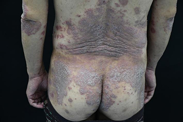 |
Figure 1 Large dark red patches can be seen on the back of the buttocks, with a large number of silver-white scales attached to them, most of which are fused into pieces, which are map-shaped and annular, and local skin lesions show lichen-like changes. |
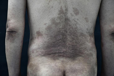 |
Figure 2 Erythematous scales on the buttocks and back basically subsided, leaving hazel pigmentation. |
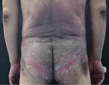 |
Figure 3 A large, diffuse dark erythema on the back of the buttocks with thickened skin and bright red margins, scales and hyperpigmentation. |
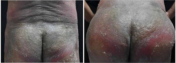 |
Figure 4 The erythema infiltration of the buttocks was aggravated, and part of the skin was pink and the pain was severe. |
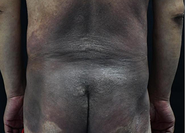 |
Figure 5 The redness and swelling subsided, the pain was relieved, and a large area of pigmentation remained. |
Discussion
Pellagra is a nutritional disorder caused by the deficiency of niacin or its precursor tryptophan and can simultaneously involve multiple systems. Clinically, it manifests as (i) symmetrical photosensitive rash, (ii) gastrointestinal symptoms, and (iii) neuropsychiatric dysfunction. The rash resembles a sunburn at the early stages of the disease, starting with acute onset erythema. This gradually transforms into an acute exudative rash on the exposed areas, such as the hands, face, neck, and chest, with pruritus, a burning sensation, and sometimes accompanied by blistering. Gastrointestinal symptoms include loss of appetite, nausea, vomiting, epigastric discomfort, abdominal pain, and increased salivation. Neuropsychiatric symptoms include depression, memory loss, and hallucinations. Niacin, also known as vitamin B3, is obtained from the diet and is abundant in meat, fish, nuts, eggs, grains, rice, and milk.3 Niacin deficiency is rare in people with a regular, healthy daily diet and is more common in developing countries than in developed countries; it persists in certain populations in developed countries, such as those with alcoholism, anorexia nervosa, malnutrition, HIV infection, and with the use of certain medications such as antiepileptics, azathioprine, and isoniazid.4 The disease tends to occur in summer and autumn and tends to recur. Among the various causes of niacin deficiency reported in domestic and international literature, alcoholism accounts for the largest proportion, followed by medications, insufficient dietary intake, malabsorption, metabolic disorders, excessive loss, and various unknown causes.5,6 The diagnosis of the present case was confirmed primarily based on medical history, clinical presentation, and experimental treatment. After a variety of symptomatic treatments did not yield significant results, a detailed history was elicited from the patient and his family, and we learned that the patient had a long history of alcohol consumption and had travelled with prolonged sun exposure before the onset of the disease. This history, typical skin lesions, flat affect, and delayed response made us consider the possibility of niacin deficiency. Although the symptoms were atypical, they subsided rapidly after experimental administration of oral niacin. The history of long-term alcohol consumption, oral anti-tuberculosis medication, and the use of biologics could be the primary causes of niacin deficiency in this case. One other case of niacin deficiency has been reported in China, but it is unclear whether the deficiency was caused by an adverse reaction to oral isoniazid or by loss of appetite after administering anti-tuberculosis medication.7 Individual cases of concomitant niacin deficiency in patients with psoriasis have been reported in China and abroad.8,9 Niacin deficiency caused by the use of biologics has not been reported, and it remains to be determined whether the relationship between psoriasis and niacin deficiency is causal or coincidental.
Conclusion
In this case report, we could not confirm whether a definite link exists between niacin deficiency, biologics, and psoriasis. However, the case suggests the need for caution when using biologics and to look out for the emergence of new symptoms when treating patients with moderate-to-severe plaque psoriasis. Additionally, timely and accurate judgments in eliciting a patient’s medical history and assessing responses to drug treatment are needed.
Data Sharing Statement
The authors confirm that the data supporting the findings of this study are available within the article.
Ethics Approval and Informed Consent
Informed consent for publication of the case details and associated images was obtained from the patient, and all procedures were performed in accordance with the Helsinki Declaration. Institutional approval was not required to publish the case details.
Consent for Publication
The patient has viewed the article contents and has provided consent for its publication in the journal.
Funding
This paper did not receive funding from any source, institutional, private, and/or corporate.
Disclosure
The authors report no conflicts of interest in this work.
References
1. Crook MA. The importance of recognizing pellagra (niacin deficiency) as it still occurs. Nutrition. 2014;30(6):729–730. doi:10.1016/j.nut.2014.03.004
2. Patterson. Practical Skin Pathology: A Diagnostic Approach: A Volume in the Pattern Recognition Series. Elsevier Health Sciences; 2013.
3. lópez M, Olivares JM, Berrios GE. Pellagra encephalopathy in the context of alcoholism: review and case report. Alcohol Alcohol. 2014;49(1):38–41. doi:10.1093/alcalc/agt070
4. de Oliveira Alves A, Bortolato T, Bernardes Filho F. Pellagra. J Emerg Med. 2018;54(2):238–240. doi:10.1016/j.jemermed.2017.10.010
5. Li R, Yu K, Wang Q, et al. Pellagra secondary to medication and alcoholism: a case report and review of the literature. Nutr Clin Pract. 2016;31(6):785–789. doi:10.1177/0884533616660991
6. Redzic S, Gupta V. Niacin deficiency. In: StatPearls. Treasure Island (FL): StatPearls Publishing; 2022.
7. Jianliang C. A case of niacin deficiency caused by oral isoniazid. Chin Pract Med. 2012;27:197–198. doi:10.14163/j.cnki.11-5547/r.2012.27.122
8. Zhimei Z. Psoriasis vulgaris with niacin deficiency: a case report. Dermatol Vener Dis. 2001;2:31.
9. Thami GP, Kaur S, Kanwar AJ. Delayed reactivation of haloperidol induced photosensitive dermatitis by methotrexate. Postgrad Med J. 2002;78(916):116–117. doi:10.1136/pmj.78.916.116
 © 2022 The Author(s). This work is published and licensed by Dove Medical Press Limited. The
full terms of this license are available at https://www.dovepress.com/terms
and incorporate the Creative Commons Attribution
- Non Commercial (unported, 3.0) License.
By accessing the work you hereby accept the Terms. Non-commercial uses of the work are permitted
without any further permission from Dove Medical Press Limited, provided the work is properly
attributed. For permission for commercial use of this work, please see paragraphs 4.2 and 5 of our Terms.
© 2022 The Author(s). This work is published and licensed by Dove Medical Press Limited. The
full terms of this license are available at https://www.dovepress.com/terms
and incorporate the Creative Commons Attribution
- Non Commercial (unported, 3.0) License.
By accessing the work you hereby accept the Terms. Non-commercial uses of the work are permitted
without any further permission from Dove Medical Press Limited, provided the work is properly
attributed. For permission for commercial use of this work, please see paragraphs 4.2 and 5 of our Terms.