Back to Journals » International Journal of Nanomedicine » Volume 21
Emodin-Based Drug Delivery Systems: Therapeutic Applications in Inflammatory Diseases
Authors Wang D, He Z, Li J, Chen Z
Received 16 December 2025
Accepted for publication 28 March 2026
Published 8 April 2026 Volume 2026:21 589401
DOI https://doi.org/10.2147/IJN.S589401
Checked for plagiarism Yes
Review by Single anonymous peer review
Peer reviewer comments 3
Editor who approved publication: Professor Lijie Grace Zhang
Dan Wang,1,* Zihao He,1,2,* Jie Li,1 Zehong Chen1,2
1Department of Pharmacy, Union Hospital, Tongji Medical College, Huazhong University of Science and Technology, Wuhan 430022, People’s Republic of China; 2Hubei Key Laboratory of Natural Active Polysaccharides, Union Hospital, Tongji Medical College, Huazhong University of Science and Technology, Wuhan, Hubei 430022, People’s Republic of China
*These authors contributed equally to this work
Correspondence: Zehong Chen, Email [email protected]
Abstract: Inflammatory diseases pose a major global health challenge, encompassing a wide range of chronic conditions driven by persistent inflammation. Emodin is a natural anthraquinone compound extracted from traditional Chinese medicinal herbs, exhibiting remarkable anti-inflammatory properties by regulating multiple inflammation-related signaling pathways. Despite its promising applications in the treatment of inflammatory diseases, issues such as poor water solubility, low bioavailability, rapid metabolism, and potential toxicity have limited its clinical translation. This review systematically reviews drug delivery systems (DDS) designed to overcome these limitations, with a focus on technological platforms including nanoparticles, liposomes, microspheres, nanocapsules, self-emulsifying systems, and microbubbles. Studies have shown that these platforms, through stimulus-responsive mechanisms and various targeting strategies, can significantly enhance emodin’s solubility, stability, targeted delivery, and sustained-release effects, thereby improving bioavailability, reducing systemic toxicity, and strengthening anti-inflammatory efficacy. This review emphasizes the analysis of the design principles, mechanisms of action, and translational prospects of each system, while discussing challenges such as biocompatibility, stability, and scalable production, to fully exploit emodin’s multi-target therapeutic potential and provide valuable references for researchers engaged in the development of anti-inflammatory DDS.
Keywords: inflammatory diseases, emodin, drug delivery systems
Introduction
Inflammatory diseases encompass a diverse array of chronic conditions characterized by persistent inflammation, including rheumatoid arthritis (RA), inflammatory bowel disease (IBD), and psoriasis. These disorders affect hundreds of millions worldwide, posing substantial risks of disability and mortality.1–3 According to the Global Burden of Disease (GBD) study, the global prevalence of RA in 2020 was 17.6 million cases, with an age-standardized rate of 208.8 per 100,000 population.4 RA causes progressive joint destruction and deformity, leading to long-term disability, chronic pain, and premature death. Projections indicate that by 2050, the global RA population will increase to 31.7 million. The economic burden of IBD is escalating, with annual healthcare costs in developed countries often exceeding tens of thousands of dollars per patient due to hospitalization, medication, and surgery, further driven by the high cost of biologics.5 In psoriasis management, prolonged topical steroid use risks skin atrophy, while immunosuppressants like cyclosporine and methotrexate may cause hepatic and renal toxicity.6,7 Although biologics demonstrate efficacy in select patients, their high costs and severe adverse effects, such as heightened infection risk, remain significant clinical hurdles.8,9 With aging populations and evolving lifestyles, the global burden of inflammatory diseases is expected to rise over the next decade, highlighting the imperative for innovative therapeutic approaches to mitigate this public health challenge.
Given the multifaceted nature of inflammatory diseases and the shortcomings of current treatments, natural products have emerged as promising drug candidates. Emodin (EMO), a prototypical anthraquinone derived from traditional Chinese herbs such as Rheum palmatum, Polygonum cuspidatum, and other Polygonaceae species, has been utilized in traditional Chinese medicine for millennia to treat constipation, inflammation, and infections. Ancient pharmacopeias, such as the Shennong Bencao Jing, document emodin-containing plants for their laxative, blood-activating, and heat-clearing properties, with applications in digestive and inflammatory disorders.10–12 Emodin exhibits broad-spectrum bioactivities, including robust anti-inflammatory effects mediated by suppressing mediators like TNF-α and IL-6,13 modulating the NLRP3 inflammasome, and alleviating oxidative stress pathways, thereby showing therapeutic promise in RA and IBD.14,15 Notably, emodin shares comparable pleiotropic anti-inflammatory mechanisms with extensively studied phytochemicals such as curcumin and resveratrol. Structurally, it belongs to the anthraquinone family, sharing a core scaffold with Rhein and aloe-emodin but differing in substituent groups which influence its specific bioactivity. Although emodin possesses therapeutic potential paralleling these well-known compounds, it encounters the same translational barriers typical of hydrophobic natural products. Its in vivo efficacy is largely curtailed by inherent physicochemical limitations, including poor aqueous solubility, extensive first-pass metabolism, and low bioavailability.16–19 High doses may induce nephrotoxicity, hepatotoxicity, and reproductive toxicity, particularly in oncology, where emodin’s cytotoxicity targets cancer cells but risks harming normal tissues.20,21 These constraints underscore the need for advanced drug delivery systems (DDS) to optimize emodin’s pharmacokinetics and minimize toxicity, thereby unlocking its potential for inflammatory disease management.
DDS represent an innovative paradigm to augment emodin’s therapeutic efficacy by surmounting solubility and bioavailability barriers through tailored pharmacokinetics and targeting.22,23 DDS offer multifunctional advantages; for example, nanocarriers encapsulate drugs efficiently, enhancing emodin’s solubility and stability while mitigating first-pass metabolism and rapid clearance, thus markedly improving bioavailability.24 Moreover, DDS enable targeted delivery, as exemplified by hyaluronic acid-modified nanoparticles that selectively engage macrophages, promoting M2 polarization and site-specific release at inflamed tissues, thereby reducing systemic toxicity and amplifying local effects.25 In inflammatory contexts, DDS sustain drug release; thermosensitive rectal hydrogels, for instance, target inflamed prostate tissue, ensuring retention and prolonged delivery, which enhances epithelial uptake and attenuates glandular inflammation.26 In chronic non-bacterial prostatitis models, triple-targeted nanoparticle-laden emodin hydrogels exhibit superior anti-inflammatory, antioxidant, and anti-fibrotic properties, significantly ameliorating inflammation and fibrosis. Another hallmark of DDS is synergy; co-delivery of emodin with siRNA targeting Kelch-like ECH-associated protein 1 facilitates concurrent administration of anti-inflammatory agents and gene therapeutics, preserving stability and bioavailability for RA treatment.25 Collectively, DDS bridge emodin’s multitarget anti-inflammatory potential to clinical utility, offering novel strategies for IBD and periodontitis, and accelerating bench-to-bedside translation.
This review systematically synthesizes recent advances in emodin-based DDS for inflammatory diseases, emphasizing design principles, therapeutic value, and translational prospects to inform optimization of emodin’s efficacy. A comprehensive literature search was conducted using keywords such as “emodin”, “drug delivery”, “liposome”, “nanoparticle”, and “inflammation” across databases including Web of Science, PubMed, Google Scholar, and ScienceDirect, spanning 2015–2025. It delineates various types of DDS, including nanoparticles, liposomes and microspheres, with an assessment of their mechanisms for improving solubility, bioavailability, and targeting. This work aims to provide a foundational reference for researchers and clinicians to expedite the clinical advancement of emodin.
Sources and Structure of Emodin
Emodin, a secondary metabolite, is primarily sourced from traditional Chinese medicinal plants, existing in free or glycosidic forms within roots, stems, leaves, or fruits.27 Predominant sources include Polygonaceae species such as Rheum palmatum L., Rheum tanguticum Maxim. ex Balf., and Rheum officinale Baill., with rhizome content ranging from 0.5% to 2%.28,29 Additional sources encompass Polygonum cuspidatum Sieb. et Zucc. and Polygonum multiflorum Thunb.,30,31 where processed P. multiflorum yields free anthraquinone levels of 0.20%-0.54%. Aloe vera contains aloe-emodin, a structural analog, at 0.1–0.5% in leaf pulp. Other origins include Fagopyrum esculentum Moench, Cassia obtusifolia L., and Fabaceae like Cassia tora L.32,33 These plants are mainly distributed in Asia, particularly China and Japan, and feature in herbal formulations like Dahuang Huanglian Xiexin Decoction.34
Emodin exhibits a characteristic anthraquinone scaffold, with its systematic name designated as 6-methyl-1,3,8-trihydroxyanthraquinone (Figure 1).28 The molecule adopts a tricyclic planar conformation, incorporating several key substitution sites: the hydroxyl moiety at C-3, the anthraquinone core predominantly involving C-2 and C-4, and the methyl substituent at C-6. Owing to its extensive nonpolar anthraquinone framework and intramolecular hydrogen bonding, emodin demonstrates negligible solubility in water while exhibiting marginal solubility in solvents such as diethyl ether, chloroform, and benzene.35,36 This profound aqueous insolubility represents a primary challenge that must be overcome in the development of DDS.
 |
Figure 1 Chemical structure of emodin. |
Anti-Inflammatory Mechanisms of Emodin
The anti-inflammatory mechanisms of emodin are intricate and multifaceted, encompassing the modulation of diverse cellular signaling pathways and inflammatory mediators (Figure 2). At its core, emodin exerts its effects by attenuating pivotal pro-inflammatory signaling cascades, consequently diminishing the biosynthesis and secretion of inflammatory mediators.
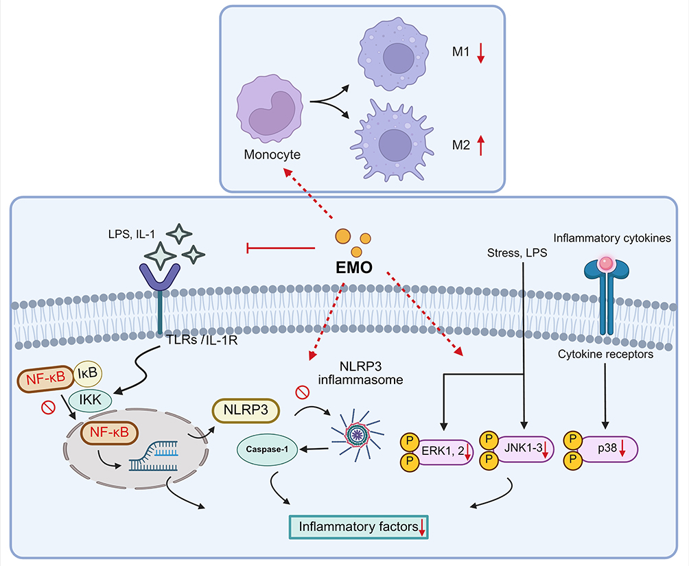 |
Figure 2 Schematic diagram of the anti-inflammatory mechanism of emodin. Emodin (EMO) exerts pleiotropic anti-inflammatory effects by suppressing pivotal pro-inflammatory cascades and modulating immune cell phenotypes. EMO blocks the activation of the NF-κB pathway by impeding IKK-mediated IκB degradation, curtails NLRP3 inflammasome assembly and subsequent caspase-1 maturation, and inhibits the phosphorylation of the MAPK family (ERK1/2, JNK1-3, and p38). Red downward arrows (↓) indicate the down-regulation or inhibition of protein phosphorylation, molecular assembly, or the secretion of inflammatory factors. The red upward arrow (↑) signifies the promotion of M2 macrophage polarization. Red prohibition symbols (⊘) and T-bars (⊣) represent the direct blocking of signaling transitions or ligand-receptor interactions. Dashed red lines trace the multi-target regulatory pathways of emodin within the cellular environment. Image created with BioRender.com, with permission. |
Inhibition of the NF-κB Signaling Pathway
Nuclear factor-κB (NF-κB) serves as a cardinal transcription factor that orchestrates the expression of numerous pro-inflammatory genes, thereby assuming a central position in inflammatory cascades.37,38 Emodin potently abrogates the activation of the NF-κB pathway. Activation of this pathway facilitates the transcriptional upregulation of pro-inflammatory cytokines, including TNF-α, IL-6, and IL-1β. By impeding the phosphorylation of IκB kinase (IKK), emodin precludes the nuclear translocation of NF-κB, thus curtailing the secretion of inflammatory mediators.14,39,40
Modulation of the MAPK Signaling Pathway
The mitogen-activated protein kinase (MAPK) signaling pathway constitutes another essential conduit for inflammatory signal transduction.41–43 Emodin attenuates the phosphorylation of key MAPK family constituents, such as p38, JNK, and ERK1/2, thereby interrupting the propagation of inflammatory signals. Notably, through suppression of the JNK pathway, emodin ameliorates pulmonary edema and inflammatory cell infiltration in conditions like acute lung injury.44,45
Inhibition of NLRP3 Inflammasome Activation
The NLRP3 inflammasome represents a critical element within the innate immune framework, wherein its activation precipitates caspase-1 maturation and the copious liberation of IL-1β and IL-18, thereby intensifying inflammatory responses and inducing pyroptosis.46,47 Extensive investigations have substantiated that emodin proficiently curtails NLRP3 inflammasome activation, consequently mitigating the efflux of inflammatory cytokines such as IL-1β and IL-18.48,49 Furthermore, emodin augments the Nrf2 pathway while diminishing reactive oxygen species (ROS) levels to alleviate oxidative stress, constituting an additional pivotal mechanism for restraining NLRP3 inflammasome activation.50
Regulation of Macrophage Polarization and Immune Response
Macrophages undergo polarization into pro-inflammatory M1 and anti-inflammatory M2 phenotypes in response to environmental stimuli, thereby dynamically modulating the magnitude and persistence of inflammatory responses. M1 macrophages predominantly drive the onset and maintenance of inflammation while facilitating pathogen elimination, whereas M2 macrophages promote the resolution of inflammation, tissue regeneration, and immune tolerance during the advanced stages of the inflammatory process.51,52 Emodin facilitates the polarization of macrophages toward the anti-inflammatory M2 phenotype, thereby enhancing the secretion of anti-inflammatory cytokines (eg, TGF-β) and expediting the resolution of inflammation alongside tissue regeneration, as observed in scenarios such as diabetic wound healing.53
To address the specific challenges posed by emodin, including its pronounced hydrophobicity arising from the planar anthraquinone core and intramolecular hydrogen bonding, susceptibility to rapid first-pass hepatic metabolism via glucuronidation and sulfation, extremely low oral bioavailability, and dose-dependent nephrotoxicity and hepatotoxicity, various emodin-loaded DDS have been strategically engineered (summarized in Table 1). These platforms are not generic carriers. Rather, they are deliberately tailored to emodin’s physicochemical and pharmacokinetic profile through lipophilic cores or lipid bilayers for efficient encapsulation of the hydrophobic anthraquinone scaffold, protective matrices or sustained-release mechanisms to shield the molecule from metabolic degradation and prolong systemic exposure, and ligand-mediated or stimulus-responsive targeting to inflamed tissues and macrophages to achieve therapeutic concentrations while minimizing off-target toxicity. The rationale for each platform and its emodin-specific advantages are discussed in the following sections and comparatively summarized in Table 2.
 |
Table 1 Comprehensive Overview of DDS for Emodin in Inflammatory Diseases Models |
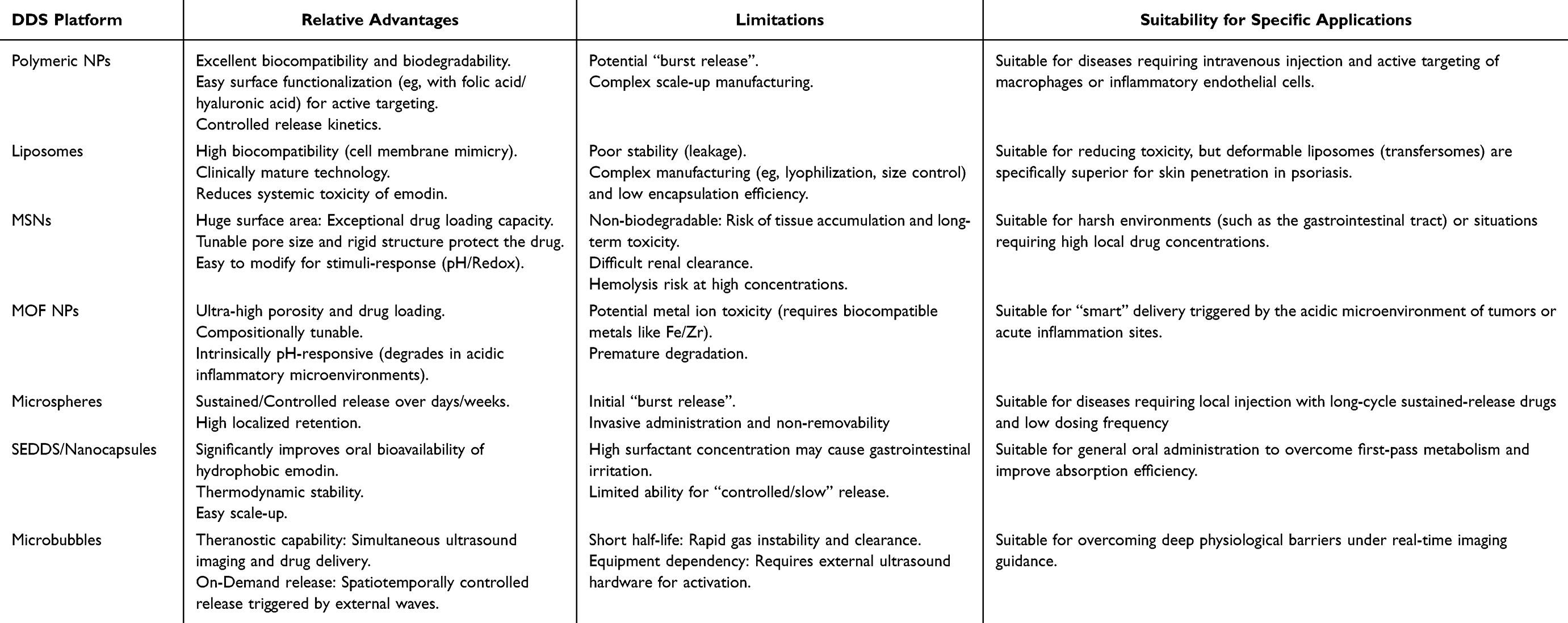 |
Table 2 Comparative Analysis of Emodin-Based DDS Platforms |
Nanoparticles
Nanoparticles (NPs) represent a leading platform for emodin delivery. These systems encapsulate the drug within nanoscale carriers, which can transport it to specific tissues or cells via tailored mechanisms.64,65 This approach not only enhances drug stability and prolongs its in vivo circulation but also minimizes off-target toxicity. Common fabrication methods include solvent evaporation, emulsion diffusion, and self-assembly.66–68 Furthermore, NPs can be engineered to respond to stimuli such as pH, reactive oxygen species (ROS), or temperature, enabling controlled drug release.69,70 In inflammatory models, NP-based delivery significantly improves emodin’s bioavailability and concurrently suppresses the expression of pro-inflammatory cytokines, including TNF-α and IL-6.
Polymeric Nanoparticles
Polymeric nanoparticles are typically fabricated from amphiphilic macromolecules. These molecules self-assemble in an aqueous environment to form “core-shell” architectures, characterized by hydrophilic coronas and hydrophobic cores, resulting in micellar nanostructures that serve as nanocarriers for drugs.71–73 Polymeric nanoparticles are particularly suitable for emodin because their amphiphilic core-shell architecture provides a hydrophobic core that efficiently sequesters the lipophilic anthraquinone moiety, dramatically improving aqueous solubility and protecting the drug from premature hepatic metabolism, while the hydrophilic shell prolongs circulation time and reduces systemic toxicity. Prevalent constituents for polymeric nanoparticles include natural biopolymers such as chitosan (CS) and dextran, or synthetic ones like polylactic acid (PLA, Figure 3A) and poly(lactic-co-glycolic acid) (PLGA, Figure 3B). These amphiphilic molecules coalesce into micro- to nanoscale, core-shell aggregates for drug incorporation. As conduits for small-molecule therapeutics, such polymeric matrices safeguard their cargo against premature degradation and enable controlled, targeted release. Furthermore, functionalization strategies for drug loading can be refined into covalent and non-covalent paradigms. Covalent conjugation exploits labile linkages (eg, ester, amide, disulfide, hydrazone) to form robust drug-polymer adducts. This approach prevents premature drug leakage during transit, releasing the active pharmacophore only in response to specific physiological cues such as pH shifts, enzymatic activity, or reductive environments.74 Such covalent strategies elevate encapsulation efficacy and governance, albeit with potential ramifications for pharmacological potency.75 Non-covalent modalities hinge on hydrophobic or electrostatic affinities, proffering operational simplicity and economic viability sans structural perturbation of the drug; archetypal polymers like PLGA and PCL ensnare lipophilic agents, or harness ionic attractions for charged entities, notwithstanding attenuated loading yields that may be ameliorated through surfactant adjuncts or functional appendages.76
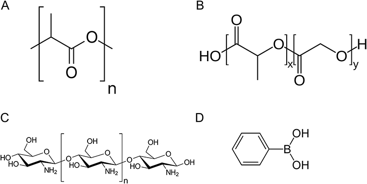 |
Figure 3 Structures of PLA (A), PLGA (B) (x is the number of lactic acid units and y is number of glycolic acid units), chitosan (C) and phenylboronic acid (D). |
PLA
PLA represents an FDA-sanctioned biodegradable macromolecule sourced from sustainable substrates like corn-derived starch. As a comparatively lipophilic entity, it is eminently apt for sequestering hydrophobic moieties akin to emodin.77 PLA undergoes gradual in vivo hydrolysis to yield lactic acid. Both PLA and its hydrolytic derivative, lactic acid, manifest non-toxicity in human physiology, evincing superior biocompatibility devoid of immunogenic provocation or inflammatory sequelae.78 By appending hydrophilic segments such as polyethylene glycol (PEG) to PLA, yielding diblock copolymers (eg, PEG-PLA), nanoparticulate hydrophilicity is augmented, thereby prolonging circulatory longevity and mitigating macrophage-mediated clearance.79 Moreover, derivatization of PEG termini with specific ligands empowers active targeting, channeling therapeutics to pathological loci such as neoplastic or inflamed domains. For example, Liu et al synthesized an adenosine-decorated PLA-b-PEG nanosystem, wherein PLA constitutes the hydrophobic nucleus and PEG the hydrophilic envelope.80 Adenosine is appended to the PEG extremity through copper-catalyzed azide-alkyne cycloaddition (CuAAC), transmuting the rudimentary PLA-PEG bilayer into a biofunctionalized ensemble. This adenosine adornment confers specificity toward inflammatory compartments expressing adenosine receptors (A2A and A2B). In osteoarthritic models, this configuration fosters nanoparticulate sequestration and focal accrual within articular spaces, curtailing systemic dissemination.
Furthermore, Liu et al prepared monomethoxy polyethylene glycol-polylactic acid-chitosan-2-mercaptobenzimidazole (mPEG-PLA-CS-MBI) copolymers, subsequently impregnated with emodin to constitute the emodin-NP platform.54 This construct embodies a core-shell motif predicated on mPEG-PLA-CS-MBI nanoparticles harboring emodin. It comprises a diblock copolymeric amalgam with a hydrophilic sheath of mPEG and CS, and a hydrophobic PLA domain for accommodating the sparingly soluble emodin, augmented by MBI thiolation to bolster mucoadhesion. The findings revealed that emodin-NP ameliorated renal functionality in 5/6 nephrectomized rodents, attenuated renal interstitial fibrogenesis, diminished serum IL-1β, IL-6, and lipopolysaccharide (LPS) concentrations, fortified intestinal barrier integrity, suppressed the enteric TLR4/MyD88/NF-κB cascade, and rectified gut microbial disequilibrium.
PLGA
PLGA denotes a biodegradable lipophilic polymer that undergoes protracted in vivo hydrolysis, culminating in fragmentation into lactic and glycolic acid monomers. Accordingly, PLGA demonstrates exemplary biocompatibility, eschewing substantive inflammatory or immunogenic retorts.81,82 Copolymerization with hydrophilic entities like PEG engenders amphiphilic block copolymers (eg, PEG-PLGA), broadening the encapsulation repertoire to encompass both hydrophilic and hydrophobic cargos. Additionally, modulation of the lactic-to-glycolic acid proportion (LA/GA) and molecular mass permits tailoring of degradative kinetics and drug efflux dynamics. PLGA nanoparticles are especially advantageous for emodin delivery owing to their tunable degradation kinetics and amphiphilic nature after PEGylation, which match emodin’s requirement for sustained release to overcome rapid metabolism and low bioavailability while allowing surface functionalization for macrophage-targeted delivery, thereby concentrating the drug at inflamed sites and minimizing nephro- and hepatotoxicity. Surface adornment of PLGA nanoparticles with ligands (eg, immunoglobulins, peptidic motifs) facilitates precision targeting to discrete cellular or tissular niches. Yu et al conjugated PEG to the PLGA surface to form PEGylated nanoparticles (PEG-PLGA NPs).83 For augmented specificity, these NPs have been conjugated with the Adipo8 (Ap) aptamer, a DNA ligand exhibiting avid binding to mature white adipocytes, thereby enabling adipocyte-selective conveyance. Presently, PLGA garners endorsement from the U.S. Food and Drug Administration (FDA) and the European Medicines Agency (EMA) for deployment in pharmacodelivery architectures and regenerative scaffolds.84 Ergo, PLGA-NPs emerge as auspicious vectors for mitigating inflammatory pathologies.
Wang et al employed PLGA as the core matrix of the nano-delivery system and incorporated the pH-sensitive enteric polymer Eudragit S100 along with montmorillonite (MMT) to fabricate EMO/PSM NPs.10 Eudragit S100 remains impervious in acidic milieus, averting untimely drug liberation in proximal gastrointestinal segments, yet solubilizes amid elevated colonic pH to expedite site-selective discharge. MMT augments NP anchorage and mucoadhesion in distal enteric tracts and inflamed colonic epithelia, engendering localized pharmacoreservoirs. The findings revealed that EMO/PSM NPs markedly surpass unbound EMO in assuaging inflammation, oxidative burden, and barrier compromise, thereby fostering mucosal restitution.
CS
Chitosan (CS), as shown in Figure 3C, constitutes a naturally occurring cationic polysaccharide procured via chitin deacetylation, susceptible to in vivo enzymatic (eg, lysozymal) and hydrolytic degradation into innocuous, assimilable monosaccharidic residues. Chitosan’s polycationic character engenders robust electrostatic engagements with anionic mucosal interfaces, thereby heightening nanoparticulate mucoadhesion and extending drug sojourn at absorptive loci.85 Chitosan can form complexes with drugs to enhance their stability and solubility in water.86,87 Nguyen et al synthesized CS-EMO NPs using the ion cross-linking method, significantly enhancing the solubility and biological activity of emodin.88 Extending this, Liu et al developed a hydrogel by incorporating sodium alginate, another natural polysaccharide. This platform enables localized drug delivery, which maintains high drug concentrations at target sites through prolonged release.89
Phenylboronic Acid (PBA)
PBA (Figure 3D) polymer is a class of nanocarriers with distinctive reactivity. When employed as functionalized polymeric nanoparticles, it enables the conferment of unique properties to the nanosystems. PBA forges reversible boronate esters with cis-diol-bearing entities (eg, polyphenolics, saccharides, glycoproteins).85 This interplay is modulated by extrinsic factors like pH and glycemia.90 PBA-functionalized polymeric nanoparticles foster multifaceted associations with emodin through lipophilic and boronate modalities, attaining elevated encapsulation quotients. Phenylboronic acid-decorated nanoparticles (PBA-NPs) were synthesized from the 3-((acrylamido)methyl) phenylboronic acid homopolymer (PBAH) via solvent displacement.91 The resulting NPs possess compact, hydrophobic interiors that efficiently load emodin through PBA-diol synergistic interactions, achieving an encapsulation efficiency (EE) of 78% and a drug loading content (LC) of 2.1%. Attributable to the acid-sensitive boronate linkage, a decrease in environmental pH from 7.4 to 5.0 can trigger the dissociation of the boronate ester bond, thereby accelerating the release of the drug from PBA-emodin-NPs.
Chemically Modified and Functionalized Nanoparticles
To further enhance emodin loading efficiency, controlled release, and active targeting, chemically modified amphiphilic polymers have been developed as a distinct functional strategy. Chemically modified amphiphilic polymers refer to a class of polymers formed by chemically synthesizing and covalently linking hydrophilic and hydrophobic segments or groups together. These systems exploit covalent and non-covalent interactions to conjugate or encapsulate the hydrophobic anthraquinone scaffold of emodin, providing superior protection against rapid hepatic metabolism and enabling stimulus-responsive or ligand-mediated delivery that conventional polymeric matrices alone cannot achieve. Such polymers typically exhibit a block architecture, exemplified by classic A-B type (eg, mPEG-PLA) or A-B-A type structures.92 When exposed to an aqueous medium, these polymers spontaneously self-assemble into nanoparticles with a core-shell morphology, known as polymer micelles.72 Wang et al constructed an amphiphilic polymer through chemical modification by chemically linking the hydrophobic drug Rhein (RH) to the chain of originally fully hydrophilic hyaluronic acid (HA), creating an HA-RH amphiphilic polymer.93 The RH-derived hydrophobic domain accommodated supplementary lipophilic cargos like emodin. The resultant HA-RH/EMO NPs encapsulated RH and EMO in defined stoichiometries and, upon enshroudment within yeast cell wall microparticles (YPs), navigated to cellular targets to modulate enteric microbiota and Th17/Treg homeostasis, thereby affording synergistic remediation of ulcerative colitis (UC).
Nanoparticle Encapsulants
Biomimetic and encapsulant-based nanoparticle systems represent another independent strategy for emodin delivery. These platforms utilize natural biological materials or cell-membrane coatings to improve biocompatibility, mucosal adhesion, immune evasion, and macrophage-specific targeting, while shielding emodin from premature metabolic degradation. YPs, which are composed of β-1,3-D-glucan, possess a robust peptidoglycan layer and a cavernous, porous “nest-like” architecture. This unique structure confers exceptional colon targeting ability and high payload capacity. Furthermore, YPs can effectively encapsulate various functionalized nanoparticles through either electrostatic adsorption or layer-by-layer self-assembly.94–96 The glucan-derived exoskeleton of YPs exhibits high acid resistance, which protects the encapsulated cargo from gastric degradation and premature drug release, thereby promoting its accumulation in the colon. YPs are readily recognized by macrophages and M cells, facilitating trans-epithelial transport and enhancing cellular uptake of emodin, thus achieving immune-directed delivery.97 Moreover, their permeable matrix enables sustained drug release through gradual diffusion, prolonging therapeutic exposure while reducing adverse effects. Given their natural origin from yeast, YPs demonstrate excellent biosafety, comply with FDA dietary standards, and hold strong translational potential.
Mesoporous Material-Based Nanoparticles
Mesoporous materials are distinguished in DDS by their unique structural characteristics. Salient attributes include high specific surface area, tunable porosity, well-defined pore topologies, and excellent biocompatibility coupled with high stability.98,99 These properties enable them to efficiently host a wide range of drug molecules, from hydrophilic to hydrophobic, and provide controlled release. For example, mesoporous silica nanoparticles (MSNs) can be functionalized with various groups (eg, amino, carboxyl, or polymers) to achieve stimulus-responsive release (eg, to pH, temperature, or enzymes), thereby enhancing delivery precision and efficacy.100,101 Therefore, MSNs can accommodate emodin’s bulky hydrophobic structure at exceptionally high loading capacities; their rigid matrix protects the drug from metabolic enzymes and can be engineered with pH- or redox-responsive gates that trigger release precisely in the acidic, oxidative microenvironment of inflamed tissues, thereby improving bioavailability and reducing off-target toxicity.
Mesoporous Silica Nanoparticles
Mesoporous silica deployed in emodin-loaded systems principally encompasses MCM-41 and SBA-15 types.
MCM-41 type: MCM-41 delineates an ordered mesoporous scaffold with porosities ranging 2 ~ 6.5 nm arrayed in hexagonal (p6mm) symmetry presented in Figure 4A, templated by cationic amphiphiles, inaugural disclosure by Mobil Oil researchers in 1992.102 MCM-41 is one of the most extensively studied drug delivery materials. Dos Santos et al constructed an MCM-41 type mesoporous silica nanoparticle DDS containing emodin.103 Findings substantiated that MCM-41 molecular sieves (specific surface area: 655 m2/g) could load 202 mg of emodin per gram of carrier. The resulting particles, with mean diameters of 100–150 nm, released over 85% of the drug over 48 hours at pH 8. However, MCM-41 is characterized by slender pore partitions and exhibits limited hydrothermal stability.
 |
Figure 4 Schematic representation of the pore structures in ordered mesoporous silicas: MCM-41 (A) and SBA-15 (B).104 |
SBA-15 type: SBA-15 (as shown in Figure 4B) manifests orderly two-dimensional hexagonal pore dispositions akin to MCM-41, yet with amplified porosities (typically 5–15 nm or greater), thereby conferring superior hydrothermal and mechanical fortitude relative to MCM-41.104 Tamara et al synthesized SBA-15 through a standard sol-gel process followed by calcination, and subsequently loaded emodin into the pre-formed SBA-15 nanoparticles using the solvent impregnation method.104 In the nanoparticle system of SBA-15 and emodin with different ratios, the emodin content can reach up to 36.4%. Emodin accretes within SBA-15 as crystalline or vitreous polymorphs in nanopore conduits, evading gastric acidic interplay and catabolism, ensued by languid liberation. Moreover, SBA-15 shields emodin against photolytic decay.
Encapsulating poorly water-soluble drug molecules within high-surface-area mesoporous silica represents a key strategy for enhancing drug dissolution. Furthermore, functionalizing the mesoporous substrate can fine-tune both the drug loading capacity and its release profile. SBA-15 has been derivatized with (γ-chloropropyl)triethoxysilane (CPTES) and (3-aminopropyl)triethoxysilane (APTES). Compared to pristine SBA-15, the functionalized material loaded with emodin exhibited a lower release rate. The APTES-functionalized SBA-15 loaded with emodin showed the lowest release amount of 74.5%, even over a period of 60 hours. These observations underscore functionalized mesoporous silica as a propitious vector for sustained liberation.105
Other mesoporous silica materials with three-dimensional cubic pore frameworks, such as MCM-48, KIT-6, and FDU-12, have been rarely reported in emodin-loaded systems. This scarcity is potentially due to a mismatch between their pore dimensions and the physicochemical requirements for efficient emodin loading and release. The interconnected channel lattice of MCM-48 enhances diffusion rates, making it suitable for complex delivery environments that require rapid mass transfer or involve macromolecular interactions. KIT-6, with its large cage-like pores, offers high loading capacity, which is advantageous for delivering macromolecules. Owing to its expansive porosity, FDU-12 is well-suited for the immobilization of enzymes, proteins, and other biomolecules, as well as for applications in biosensing and biocatalysis.106
Organic Mesoporous Materials
mPDA: Mesoporous polydopamine (mPDA) has emerged as a novel carrier due to its unique biomimetic structure and multifunctionality.107 mPDA is primarily derived from dopamine self-polymerization, inheriting the biocompatibility of polydopamine (PDA) while enhancing drug loading and responsive release through its mesoporous structure.108 PDA contains abundant catechol and amine groups, which can easily connect to various molecules via covalent or coordination bonds, including targeting ligands, biomolecules, and imaging probes, thereby achieving active targeting and multifunctionality.109,110 Moreover, leveraging polydopamine’s excellent adhesion and biocompatibility, it can be applied in the treatment of orthopedic diseases and tissue repair. mPDA can be used for treating inflammatory diseases such as arthritis.111 For example, loading drugs like emodin into mPDA enables sustained and controlled drug release, effectively inhibiting the inflammatory progression of arthritis. Luo et al developed a multifunctional nanofiber hydrogel system (NFs-H@PE) for preventing postoperative abdominal adhesions.58 In this system, emodin was loaded into mPDA to achieve sustained release, thereby establishing an anti-inflammatory microenvironment that suppresses inflammatory cell activity. Simultaneously, mPDA acts as a rapid ROS scavenger to further alleviate inflammation. As a drug carrier, mPDA significantly enhances the combined ROS-scavenging and anti-inflammatory efficacy of emodin, making this system particularly suitable for managing inflammatory conditions such as abdominal adhesions.
In addition, organic-inorganic hybrid nanomaterials are highly promising for DDS, as their core advantage is the integration of organic and inorganic components’ strengths to overcome single-material limitations and achieve synergistic performance enhancements. A representative example is Periodic Mesoporous Organosilica Nanoparticles (PMONPs). Unlike traditional mesoporous silica, the PMONP framework incorporates organic bridging groups (-R-) that form Si-R-Si bonds alongside siloxane (Si-O-Si) networks, granting them both structural stability and functional versatility. Illustrating their application, Qian et al synthesized PEG-modified PMONPs co-loaded with aloe-emodin and IR820 (Aloe-emodin/IR820@PMONs-PEG) for combined tumor therapy. This design provides a valuable reference for delivering other compounds with emodin-like structures.112
Metal-Organic Framework (MOF) Nanoparticles
MOFs are crystalline, highly porous materials formed through coordination bonds between organic ligands and metal ions or clusters.113 Their key attributes include enormous specific surface areas and highly tunable pore sizes. By selecting different metal ions (eg, Fe, Zr, Zn) and organic ligands, researchers can synthesize MOF nanoparticles with precise control over their shape, size, and chemical functionality to meet diverse needs. Many MOFs, particularly Bio-MOFs built from biocompatible ligands (eg, citric acid, amino acids), also exhibit excellent biocompatibility.114 This feature, combined with the ability to engineer stimuli-responsive behavior (eg, to pH, redox potential, or temperature), enables targeted drug release at specific lesion sites such as tumors or inflammatory tissues.115 MOF nanoparticles are uniquely appropriate for emodin because their ultra-high porosity and pH-responsive degradation in acidic inflammatory microenvironments allow both high drug loading and on-demand release at the disease site, while the metal ions can be selected to minimize additional toxicity and enable macrophage membrane coating for active targeting.
Yang et al developed an emodin-loaded nanoparticle (MVs-UiO-ED) for the treatment of acute pancreatitis by integrating MOF nanocarriers with cell membrane biomimetic coating technology. The results demonstrated that the MVs-UiO-ED group exhibited the most significant improvements in amylase and lipase levels, along with markedly alleviated inflammation, edema, and necrosis.55 The encapsulation of emodin within macrophage membranes enabled MVs-UiO-ED to achieve sustained drug release, prolonging its systemic circulation while conferring inflammation-targeting capabilities for precise delivery to sites of acute pancreatitis. Moreover, the macrophage membranes can bind and neutralize inflammatory cytokines, thereby inhibiting downstream cascades and contributing to synergistic anti-inflammatory effects. This approach innovatively overcomes the immune recognition that plagues traditional MOF carriers through “biomimetic camouflage”, which enhances in vivo circulation and targeting. This “membrane-core” composite design, inspired by functional cell mimicry, represents a versatile platform for delivering anti-inflammatory or anticancer drugs. Similarly, Xie et al constructed AE-FeMn/FA nanoparticles by loading aloe-emodin into an MOF framework.116 This design leverages the intrinsic antitumor activity of aloe-emodin and the synergistic immunotherapeutic effects mediated by the co-released Fe3⁺ and Mn2⁺ metal ions.
Protein-Based Nanoparticles
Biopolymer-based nanoparticles, particularly those derived from proteins, serve as foundational building blocks for nanocarriers owing to their favorable safety profile and inherent biodegradability.117,118 As natural biomolecules, proteins exhibit exceptional biocompatibility and degrade into non-toxic metabolites that the body can readily clear. A key advantage of proteins lies in their diverse modifiable functional groups, including amino, carboxyl, hydroxyl, and thiol moieties, which facilitate covalent conjugation with targeting ligands, imaging probes, or other therapeutic agents to create multifunctional systems. The preparation of protein nanoparticles typically avoids high temperatures or toxic organic solvents, which helps preserve drug activity and stability.119 Simultaneously, controlling the crosslinking and degradation rates of the nanoparticles enables sustained and controllable drug release, thereby extending the therapeutic duration.
Siri et al developed γ-irradiation-crosslinked BSA nanoparticles (~70 nm) for emodin delivery, which enhanced macrophage immune responses while preserving albumin’s native functions.120 Advancing this concept, Pu et al utilized lactoferrin (Lf) to encapsulate emodin into nanoparticles (EMO-NP), which were then loaded into macrophage-targeting YPs to form a dual-targeting system (EMO-NYPs).56 This system promoted anti-inflammatory effects via NF-κB inhibition and intestinal mucosal repair via the MLCK/pMLC2 pathway, demonstrating superior therapeutic outcomes.
Microspheres
Microspheres are spherical multiparticulate systems (1 ~ 1000 μm) composed of proteins, biodegradable polymers (eg, PLGA, PLA), or hydrogels (eg, hyaluronic acid-calcium alginate), and are prepared via methods like solvent evaporation or emulsification.121,122 They offer advantages over nanoparticles, including higher drug loading efficiency and clinical success rates. Furthermore, their precisely tunable size, surface properties, and internal structure enable targeted delivery, improving therapeutic outcomes and reducing off-target risks. This tunability also permits the co-loading of multiple drugs and the design of responses to stimuli such as pH, temperature, or enzymes.123
Natural Polymer Microspheres
Natural polymers, such as hyaluronic acid, chitosan, alginate, and gelatin, are typically sourced from animals, plants, or microorganisms. They are highly valued for their exceptional biocompatibility and low immunogenicity.124,125 Nonetheless, their natural origin introduces inherent challenges, including batch-to-batch variations due to difficulties in controlling purity and precise chemical composition. Moreover, these materials generally possess lower mechanical strength compared to their synthetic counterparts, which can compromise the stability of the resulting microspheres.
Yan et al constructed a colon-targeted hydrogel microsphere system (HM@OAE) by encapsulating an oleic acid-emodin (OAE) complex within hyaluronic acid-calcium alginate (HA/CA) microspheres for oral delivery.60 Here, OA served as a solubilizing agent to improve emodin’s bioavailability, while the pH-responsive CA matrix ensured targeted colonic release. This system significantly surpassed free emodin in DSS-induced colitis models, restoring intestinal barrier integrity and alleviating disease symptoms through ferroptosis inhibition and anti-inflammatory effects.
Synthetic Polymer Microspheres
Synthetic polymers are chemically synthesized, enabling precise control over their physicochemical properties through monomer selection and polymerization conditions. This chemical tailorability gives polymers like PLGA, PLA, PCL, and PEG superior mechanical strength and stability, as well as the ability to finely adjust drug release behavior for sustained therapeutic outcomes. Nevertheless, such polymers may exhibit lower biocompatibility compared to natural alternatives, and their reproducible processing into microspheres generally requires sophisticated manufacturing and tight regulatory control.126,127
Chen et al prepared lung-targeted PLA microspheres loaded with emodin (ED-PLA-MS) using an organic phase dispersion-solvent diffusion technique. The resulting microspheres exhibited a narrow size distribution, with an average diameter of 9.7 ± 0.7 μm and 86% of the population falling within the 5 ~ 20 μm range. Following intravenous administration, results confirmed that ED-PLA-MS achieved primary accumulation of emodin in the lungs, demonstrating effective lung-targeting capability.128
Liposomes
Liposomes are lipid-based vesicles with one or more concentric bilayers, a structure that allows them to load both hydrophobic drugs (within the bilayer) and hydrophilic drugs (in the aqueous interior).129,130 To improve their performance, the liposome surface can be chemically tailored, eg, through PEGylation to form “stealth liposomes” with prolonged blood circulation,131 or by grafting antibodies or peptides for targeted delivery. Liposomes are particularly well-suited for emodin because the phospholipid bilayer mimics a lipophilic environment that readily incorporates the hydrophobic anthraquinone, markedly increasing solubility and stability while enabling surface modification (eg, PEG or targeting ligands) to extend circulation half-life, reduce systemic toxicity, and achieve inflammation-specific accumulation.
Sun et al developed a liposomal emodin (LE) nanodrug for two-photon imaging and mitochondria-targeted photodynamic therapy (TPE-PDT) in the treatment of psoriasis.9 Upon laser activation, LE generates ROS that specifically target mitochondrial complex IV in macrophages, leading to its degradation and subsequent inhibition of T cell cytokine production. In imiquimod-induced psoriatic mouse models, LE demonstrated significant percutaneous absorption, effectively penetrated psoriatic lesions, and markedly alleviated skin inflammation and hyperkeratosis. The therapeutic effect is achieved through mitochondria-targeted ROS generation, which inhibits inflammatory responses and helps restore immune balance.
The development of liposomal DDS must overcome multiple challenges in complex in vivo environments, including rapid clearance, inefficient targeting, poor penetration of biological barriers, and uncontrolled drug release. A major hurdle is the recognition of liposomes as foreign bodies by the reticuloendothelial system (RES), leading to their rapid phagocytosis in organs like the liver and spleen and significantly shortened blood circulation time. A key strategy to mitigate this is the conjugation of PEG to the liposome membrane, which provides steric stabilization to improve circulation longevity and reduce RES uptake.132 Another significant barrier is the blood-brain barrier (BBB), which impedes the delivery of most neurotherapeutic agents, including emodin. Studies show that functionalizing liposomes with cyclic Arg-Gly-Asp (cRGD) peptides can confer brain-targeting capabilities.133 Demonstrating this approach, Chen et al developed PEG/cRGD-modified liposomes encapsulating emodin. These engineered liposomes significantly enhanced emodin accumulation in cerebral infarct areas, thereby reducing infarct volume, neuronal loss, and associated tissue damage.61
Other DDS
Nanocapsules
Nanocapsules are vesicular systems characterized by a core-shell architecture, where active molecules are encapsulated within a central reservoir surrounded by a polymeric membrane. The core can accommodate therapeutic agents in liquid, solid, or molecular dispersion form.134 In a study by Song et al, macrophage-targeting nanocapsules were constructed by covalently grafting mannose onto chitosan to form the shell.62 The lipid core was prepared using the phase inversion temperature (PIT) method, which enabled direct loading of hydrophobic emodin into the lipid interior. Evaluations in both macrophages and severe acute pancreatitis (SAP) mouse models demonstrated that these nanocapsules reduced key inflammatory mediators and exhibited selective accumulation in pancreatic and gastrointestinal tissues. Mechanistically, they upregulated CPT1 (carnitine palmitoyltransferase 1) expression, promoted fatty acid transport, and polarized macrophages toward the M2 phenotype, collectively enhancing the bioavailability and therapeutic efficacy of emodin.
The core-shell morphology represents a distinctive advantage of nanocapsules over conventional nanoparticles in drug delivery. A critical feature of this architecture is the polymer shell thickness, which plays a determining role in formulation stability, drug protection, and release kinetics.135,136 It is widely recognized that organic solvents pose toxicity risks in pharmaceutical formulations and must be rigorously removed. However, trace residues may persist, raising potential safety concerns in clinical settings. To circumvent this issue, polymeric nanocapsules can be synthesized via monomer polymerization in the absence of organic solvents. For instance, nanocapsules have been successfully prepared through free radical polymerization of polyacrylamide and 2-aminoethyl methacrylate hydrochloride in deoxygenated buffer,137 offering a safer and more biocompatible alternative.
Self-Emulsifying Drug Delivery Systems (SEDDS)
SEDDS represent an advanced technology designed to enhance the solubility and oral bioavailability of poorly water-soluble drugs. These systems spontaneously form stable nano- or micro-emulsions upon encountering the gastrointestinal aqueous environment, thereby significantly promoting drug absorption. A typical SEDDS formulation consists of three essential components: an oil phase, a surfactant, and a co-surfactant/cosolvent.138,139 SEDDS are particularly effective for oral emodin delivery because spontaneous microemulsion formation upon contact with gastrointestinal fluids overcomes the drug’s poor aqueous solubility and extensive first-pass metabolism, substantially increasing bioavailability without the need for invasive administration. In a study by Huang et al, an emodin-loaded SEDDS (EMO-SMEDDS) was developed using Maisine 35–1 and GTCC as the oil phase, Labrasol, TPGS, and Cremophor EL as surfactants, and Transcutol P as the co-surfactant.140 The formulated EMO-SMEDDS demonstrated significantly enhanced cellular uptake in both Caco-2 cells and glomerular mesangial cells (GMCs), along with reduced drug efflux. Moreover, compared to free emodin, EMO-SMEDDS more effectively suppressed the protein expression of key renal fibrosis markers, namely fibronectin (FN), transforming growth factor-β1 (TGF-β1), and intercellular adhesion molecule-1 (ICAM-1), in the context of diabetic nephropathy, indicating its potential for mitigating disease progression.
Microbubbles
Microbubble (MB) delivery systems are ultrasound-activated platforms that serve dual diagnostic and therapeutic roles. Typically measuring 1 ~ 8 μm in diameter (smaller than red blood cells), MBs can circulate freely through the vasculature.141 Their structure consists of a gas core (eg, sulfur hexafluoride, perfluoropropane, or perfluorobutane) encapsulated by a lipid, protein, or polymer shell.142,143 While used alone as contrast agents in ultrasound imaging, their key function in drug delivery arises from the “cavitation effect” triggered by externally applied ultrasound.144 When targeted ultrasound is focused on specific tissues, circulating drug-loaded MBs rupture, enabling highly localized and precise drug release. Microbubbles provide sustained or ultrasound-triggered release that directly counters emodin’s rapid clearance and low bioavailability, while their size and surface chemistry enable localized retention or deep-tissue penetration at inflammatory foci, significantly lowering the required dose and thereby mitigating nephro- and hepatotoxicity.
Zhang et al developed a kidney-targeted MB system named Emo@KP MBs.63 They first prepared emodin-loaded phospholipid nanoparticles (Emo@KP NPs) via ethanol injection, then conjugated the surface with αKIM-1 antibody to form Emo@KP NPs. These targeted nanoparticles were subsequently mixed with sulfur hexafluoride (SF6) gas and mechanically oscillated to form stable microbubbles. Under ultrasound-targeted microbubble destruction (UTMD), Emo@KP MBs collapse and transform in situ into nanoparticles, facilitating efficient emodin release while enabling real-time imaging via contrast-enhanced ultrasound (CEUS). This integrated strategy reduces systemic side effects such as hepatotoxicity and significantly alleviates renal inflammation and fibrosis by modulating the TGF-β1/Smad pathway, offering a precise and image-guided therapeutic approach for chronic kidney disease.
Therapeutic Applications of Emodin-Loaded DDS in Inflammatory Diseases
While the physicochemical optimization of emodin-based DDS is pivotal, the ultimate validation of their utility lies in their therapeutic outcomes. Emodin-based DDS have demonstrated remarkable efficacy across a spectrum of inflammatory diseases by modulating disease-specific microenvironments. This section categorizes recent preclinical advances based on pathological targets, highlighting the scientific rationale behind selecting specific delivery platforms.
RA
RA is characterized by persistent synovial inflammation, excessive M1 macrophage infiltration, elevated levels of ROS, and progressive joint destruction.145,146 Current emodin-loaded systems predominantly exploit macrophage-targeted strategies, such as HA-functionalized Fe-based metal-organic framework nanoparticles co-delivering emodin and KEAP1 siRNA, which leverage CD44-mediated uptake to repolarize macrophages toward an anti-inflammatory M2 phenotype and reduced the levels of ROS.25
Critically, however, oral or systemic nanoparticle approaches remain suboptimal for RA due to emodin’s poor aqueous solubility and first-pass metabolism, which necessitate high dosing and risk off-target effects. Transdermal microneedle systems, widely validated for RA with drugs like methotrexate and melittin, represent an untapped opportunity for emodin.147,148 Dissolving microneedles bypass gastrointestinal degradation, achieve sustained joint penetration without needles, and reduce systemic toxicity, offering superior patient compliance compared with repeated intra-articular injections. Similarly, advanced intra-articular injectable hydrogels (eg, ROS-scavenging or lubricant hydrogels) provide prolonged joint retention and on-demand release, which current emodin formulations lack.
IBD
IBD, including UC and Crohn’s disease, involves disrupted epithelial barriers, dysregulated gut macrophages, and microbiota imbalance in an acidic, enzyme-rich colonic environment.149,150
“Colon specificity” is the core of this field. Researchers have developed nanoscale systems such as EMO/PSM NPs, whose scientific rationale lies in using pH-sensitive polymers to encapsulate emodin, ensuring the drug remains stable in the upper gastrointestinal tract and is released only in the colon where the pH increases.10 More innovative oral colon-targeted systems dominate emodin delivery, such as yeast cell wall/β-1,3-d-glucan microparticles that are phagocytosed via dectin-1 receptors and pH/enzyme-responsive hydrogel microspheres inhibiting ferroptosis.56,60,93 These are appropriately chosen because IBD pathophysiology permits exploitation of macrophage phagocytosis and colonic pH/enzyme gradients to achieve site-specific release, thereby enhancing local efficacy while circumventing emodin’s poor solubility and rapid systemic clearance.
Psoriasis and Skin Disorders
Psoriasis is characterized by excessive proliferation of keratinocytes, thickening of the epidermis, and dilation of dermal capillaries. The stratum corneum, as a tight physical barrier, significantly restricts the transdermal absorption of hydrophobic emodin, leading to poor efficacy of topical administration. Overcoming the skin barrier is one of the key research directions in this field. Recent studies have reported that liposomes enable TPE-PDT with simultaneous fluorescence imaging of psoriatic lesions, achieving selective mitochondrial accumulation and phototoxicity against hyperproliferative keratinocytes.9 This theranostic platform represents high novelty by integrating diagnostic imaging and targeted PDT in a single nanomedicine.
Notably, inflamed psoriatic lesions are characterized by significantly elevated levels of ROS and an acidic microenvironment. These features enable stimulus-responsive drug release, which may reduce off-target effects on healthy skin and support active targeting of dermal macrophages and dendritic cells involved in the IL-23/Th17 axis. Although similar responsive strategies have been employed for other therapeutic agents, their specific application for emodin delivery remains unexplored. Concurrently, recent studies have highlighted macrophage membrane-coated nanoparticles as highly promising self-homing platforms for psoriasis therapy. By exploiting the inherent inflammation-homing properties of macrophage membranes (mediated by chemokine receptors and upregulated cytokine-binding proteins), these biomimetic systems achieve selective accumulation within psoriatic lesions while simultaneously neutralizing ROS, scavenging pro-inflammatory cytokines, and reprogramming local immunity, thereby conferring significant advantages that conventional polymeric carriers cannot provide.
Renal Inflammation and Fibrosis
Renal inflammation and fibrosis represent the common final pathway of chronic kidney disease (CKD), characterized by persistent tubulointerstitial inflammation, myofibroblast activation, excessive extracellular matrix (ECM) deposition primarily driven by the TGF-β/Smad signaling pathway, elevated levels of ROS, and macrophage infiltration, ultimately leading to progressive renal function decline.
Emodin-loaded DDS have been rationally engineered to overcome the glomerular filtration barrier, emodin’s poor aqueous solubility, and its dose-dependent nephrotoxic potential. Representative systems include mesoscale nanoparticles (Em-MNPs) that exploit size- and charge-mediated kidney targeting for antifibrotic activity,151 nanoparticle-mediated colonic irrigation that attenuates renal injury and limits tubulointerstitial fibrosis in 5/6 nephrectomized rats,54 deoxycholic acid-chitosan coated liposomes combined with in situ colonic gel that markedly enhance renal delivery and fibrosis therapy,152 and anti-α8 integrin-conjugated immunoliposomes enabling targeted codelivery to mesangial cells.153 These platforms are scientifically designed because the fibrotic kidney microenvironment features altered vascular permeability and macrophage-driven inflammation, allowing nanoparticle size, surface charge, and ligand modification to promote selective renal accumulation, intracellular TGF-β pathway modulation, and reduced systemic exposure.
However, the majority of current systems still rely heavily on colonic administration routes. Although these routes effectively exploit the gut–kidney axis, they compromise long-term patient compliance and raise concerns regarding repeated mucosal exposure. Furthermore, most platforms achieve only passive renal retention and lack active targeting to key fibrogenic cells, including podocytes, tubular epithelial cells, or myofibroblasts. Macrophage membrane-coated nanoparticles and megalin/aminopeptidase N-targeted peptide systems have already been validated for other renoprotective natural compounds in CKD models and thus represent highly promising untapped strategies for emodin. These biomimetic or ligand-directed platforms would confer active inflammation homing, deeper tubular penetration, ROS-responsive on-demand release, and superior biocompatibility, thereby addressing the current limitations and markedly elevating the translational potential of emodin in renal fibrosis.
Other Inflammatory Diseases
Emodin-loaded DDS have also demonstrated efficacy in additional conditions by aligning carrier characteristics with each disease’s distinct pathological microenvironment. In acute pancreatitis, macrophage-targeted systems capitalize on the acidic and highly oxidative pancreatic milieu to promote M2 polarization through CPT1-mediated lipid metabolic reprogramming, thereby suppressing inflammatory cytokines and serum amylase levels while circumventing rapid systemic clearance.55 For chronic non-bacterial prostatitis, platforms facilitating rectal-to-prostate transport and prolonged glandular retention overcome the blood-prostate barrier and confined inflammatory niche, enabling sustained local anti-inflammatory, antioxidant, and anti-fibrotic activity.26 In postoperative abdominal adhesions, ROS-scavenging carriers incorporated into anti-swelling nanofiber hydrogel matrices precisely match the oxidative and fibrotic microenvironment to provide sustained anti-inflammatory and antibacterial effects, thereby inhibiting adhesion formation without systemic exposure.58
Challenges and Limitations
Despite considerable advances in emodin-based DDS, their therapeutic potential remains constrained by several persistent challenges.
Biocompatibility and Toxicity Concerns
Delivery systems themselves may introduce toxicity risks. After systemic administration, most nanoparticles accumulate in the mononuclear phagocyte system, potentially inducing toxic responses in the liver, spleen, and kidneys.154,155 Emodin itself poses safety challenges in that excessive doses can produce laxative effects resulting in intestinal pain, severe diarrhea, and subsequent electrolyte imbalance and dehydration.19 Furthermore, natural biological barriers limit delivery efficiency. The blood-brain barrier, for instance, exhibits selective permeability that restricts most carriers from entering the central nervous system, preventing therapeutic drug concentrations from being achieved in brain tissues.
Preparation and Stability Limitations
Emodin carriers such as nanoparticles, liposomes, and nanocapsules face stability issues during fabrication and storage, including particle aggregation, surface defects, and sensitivity to environmental conditions like pH and temperature. Liposomes are particularly prone to vesicle fusion, aggregation, lipid peroxidation, and hydrolysis during storage, leading to drug leakage or degradation. While passive targeting is achievable, active targeting requires complex surface modifications (eg, PEGylation, ligand conjugation), which increases preparation complexity and cost.
Scale-Up and Manufacturing Hurdles
Translating laboratory successes to industrial production presents significant difficulties, including high costs, complex manufacturing processes for nanoparticles and microspheres, and a lack of standardized protocols. Additionally, the development of these systems involves high technical barriers; complex preparation procedures are not easily monitored by conventional methods, and minor process variations can substantially impact final product quality, resulting in considerable batch-to-batch uncertainty.
As summarized in Table 2, diverse DDS platforms exhibit distinct physicochemical profiles that dictate their therapeutic suitability. Rather than seeking a universal carrier for emodin, the optimal choice must be strictly pathology-driven. For systemic inflammatory conditions like RA, stability and active targeting capabilities (typically offered by polymeric nanoparticles) are paramount to minimize off-target toxicity. Conversely, for localized pathologies such as osteoarthritis or UC, strategies should prioritize prolonged retention and controlled release (via microspheres or hydrogels) over systemic circulation. Therefore, future development should pivot from generic formulation screening to rational, disease-specific carrier design that aligns the delivery mechanism with the specific biological barriers of the target tissue.
Conclusions and Future Perspectives
Emodin, a natural anthraquinone derivative, exhibits potent anti-inflammatory activity primarily through the suppression of the NF-κB and MAPK signaling pathways, leading to reduced expression of key inflammatory mediators such as TNF-α and IL-6. It has demonstrated therapeutic efficacy in multiple disease models, including UC and osteoarthritis. However, its clinical translation has been hampered by poor aqueous solubility and rapid metabolism. To overcome these limitations, future innovations in emodin-based DDS are expected to emphasize precise targeting and multifunctional integration to improve therapeutic outcomes in inflammatory diseases.
One promising direction lies in the development of smart stimulus-responsive delivery platforms. Such “intelligent” nanocarriers or hydrogels can be engineered to react to pathological cues, such as the specific pH or enzymatic milieu of inflammatory sites, enabling spatially and temporally controlled drug release, which would enhance local efficacy while minimizing systemic exposure. Another strategy involves the design of hybrid delivery systems to achieve synergistic therapeutic effects. For instance, co-encapsulating emodin with gene-based therapeutics (eg, siRNA) within biomimetic carriers such as yeast cell wall particles or 3D-bioprinted hydrogel matrices could facilitate combined gene-drug therapy, offering new avenues for managing chronic inflammatory disorders like arthritis. In addition, theranostic nanoplatforms represent a frontier in emodin-based DDS. By integrating diagnostic imaging agents (eg, for MRI or fluorescence imaging) with emodin-loaded carriers, these systems would allow real-time visualization of inflammatory sites and monitoring of drug release kinetics, thereby supporting image-guided precision treatment. Finally, to bridge the gap between laboratory research and clinical application, future efforts must systematically address the pharmacokinetic and toxicological profiles of emodin formulations. While most current evidence remains at the preclinical stage, advancing toward regulated clinical trials will be essential to validate the safety and efficacy of these novel delivery systems in human populations.
Abbreviations
BBB, Blood-brain barrier; CKD, Chronic kidney disease; CS, Chitosan; DDS, Drug delivery systems; DL, Drug loading; DSS, Dextran sulfate sodium; EE, Encapsulation efficiency; EMO, Emodin; EMO-NP, Emodin nanoparticles; HA, Hyaluronic acid; IBD, Inflammatory bowel disease; LC, Drug loading content; LE, Liposomal emodin; LPS, Lipopolysaccharide; MB, Microbubble; MOF, Metal-organic framework; mPEG, Monomethoxy polyethylene glycol; MSNs, Mesoporous silica nanoparticles; NPs, Nanoparticles; OA, Oleic acid; PBA, Phenylboronic acid; PCL, Polycaprolactone; PDA, Polydopamine; PEG, Polyethylene glycol; PEG-PLGA, PEGylated poly(lactic-co-glycolic acid); PLA, Polylactic acid; PLGA, Poly(lactic-co-glycolic acid); PMONPs, Periodic Mesoporous Organosilica Nanoparticles; RA, Rheumatoid arthritis; RES, Reticuloendothelial system; RH, Rhein; ROS, Reactive oxygen species; SEDDS, Self-emulsifying drug delivery systems; TGF-β, Transforming growth factor beta; TLR4, Toll-like receptor 4; TNF-α, Tumor necrosis factor alpha; UC, Ulcerative colitis; YPs, Yeast cell wall microparticles.
Acknowledgments
This research did not receive any specific grant from funding agencies in the public, commercial, or not-for-profit sectors.
Author Contributions
All authors made a significant contribution to the work reported, whether that is in the conception, study design, execution, acquisition of data, analysis and interpretation, or in all these areas; took part in drafting, revising or critically reviewing the article; gave final approval of the version to be published; have agreed on the journal to which the article has been submitted; and agree to be accountable for all aspects of the work.
Disclosure
The authors declare that they have no known competing financial interests or personal relationships that could have appeared to influence the work reported in this paper.
References
1. Hracs L, Windsor JW, Gorospe J, et al. Global evolution of inflammatory bowel disease across epidemiologic stages. Nature. 2025;642:458–24. doi:10.1038/s41586-025-08940-0
2. Black RJ, Lester S, Tieu J, et al. Mortality estimates and excess mortality in rheumatoid arthritis. Rheumatology. 2023;62:3576–3583. doi:10.1093/rheumatology/kead106
3. Semenov YR, Herbosa CM, Rogers AT, et al. Psoriasis and mortality in the United States: Data from the national health and nutrition examination survey. J Am Acad Dermatol. 2021;85:396–403. doi:10.1016/j.jaad.2019.08.011
4. Black RJ, Cross M, Haile LM, et al. Global, regional, and national burden of rheumatoid arthritis, 1990–2020, and projections to 2050: a systematic analysis of the global burden of disease study 2021. Lancet Rheumatol. 2023;5:e594–e610. doi:10.1016/S2665-9913(23)00211-4
5. van Linschoten RCA, Visser E, Niehot CD, et al. Systematic review: societal cost of illness of inflammatory bowel disease is increasing due to biologics and varies between continents. Aliment Pharmacol Ther. 2021;54:234–248. doi:10.1111/apt.16445
6. Patocka J, Nepovimova E, Kuca K, Wu W. Cyclosporine A: chemistry and toxicity – a review. Curr Med Chem. 2021;28:3925–3934. doi:10.2174/0929867327666201006153202
7. Lee CH, Choe SJ, Kim DH, et al. Skin atrophy caused by topical glucocorticoids is less common in patients with atopic dermatitis than in those with psoriasis. Exp Dermatol. 2022;31:182–190. doi:10.1111/exd.14441
8. Armstrong AW, Read CP. Clinical presentation, and treatment of psoriasis: a review. JAMA. 2020;323:1945–1960. doi:10.1001/jama.2020.4006
9. Sun J, Sun A, Wang Y, et al. Liposomal Emodin: a nanomedicine for two-photon imaging and mitochondria-targeted photodynamic therapy of psoriasis. Biomaterials. 2026;325:123631. doi:10.1016/j.biomaterials.2025.123631
10. Wang D, Sun M, Zhang Y, et al. Enhanced therapeutic efficacy of a novel colon-specific nanosystem loading emodin on DSS-induced experimental colitis. Phytomedicine. 2020;78:153293. doi:10.1016/j.phymed.2020.153293
11. Zheng Y-F, Liu C-F, Lai W-F, et al. The laxative effect of emodin is attributable to increased aquaporin 3 expression in the colon of mice and HT-29 cells. Fitoterapia. 2014;96:25–32. doi:10.1016/j.fitote.2014.04.002
12. Zheng Q, Li S, Li X, Liu R. Advances in the study of emodin: an update on pharmacological properties and mechanistic basis. ChinMed. 2021;16:102. doi:10.1186/s13020-021-00509-z
13. Zhu T, Zhang W, Feng S-J, Yu H-P. Emodin suppresses LPS-induced inflammation in RAW264.7 cells through a PPARγ-dependent pathway. Int Immunopharmacol. 2016;34:16–24. doi:10.1016/j.intimp.2016.02.014
14. Cheng L, Chen J, Rong X. Mechanism of emodin in the treatment of rheumatoid arthritis. Evid Based Complement Alternat Med. 2022;2022. doi:10.1155/2022/9482570
15. Ye B, Cai X, Liang X, et al. Emodin Suppresses NLRP3/GSDMD-induced Inflammation via the TLR4/MyD88/NF-κB signaling pathway in atherosclerosis. Cardiovasc Drugs Ther. 2024;39:1289–1301. doi:10.1007/s10557-024-07659-w
16. Ban E, An SH, Park B, et al. Improved solubility and oral absorption of emodin-nicotinamide cocrystal over emodin with PVP as a solubility enhancer and crystallization inhibitor. J Pharmaceut Sci. 2020;109:3660–3667. doi:10.1016/j.xphs.2020.09.030
17. Qiu N, Zhao X, Liu Q, et al. Inclusion complex of emodin with hydroxypropyl-β-cyclodextrin: preparation, physicochemical and biological properties. J Mol Liq. 2019;289:111151. doi:10.1016/j.molliq.2019.111151
18. Teng Z-H, Zhou S-Y, Yang R-T, et al. Quantitation assay for absorption and first-pass metabolism of emodin in isolated rat small intestine using liquid chromatography-tandem mass spectrometry. Biol Pharm Bull. 2007;30:1628–1633. doi:10.1248/bpb.30.1628
19. Sharifi-Rad J, Herrera-Bravo J, Kamiloglu S, et al. Recent advances in the therapeutic potential of emodin for human health. Biomed Pharmacother. 2022;154:113555. doi:10.1016/j.biopha.2022.113555
20. Akkol EK, Tatlı II, Karatoprak GŞ, et al. Is emodin with anticancer effects completely innocent? Two sides of the coin. Cancers. 2021;13(11):2733. doi:10.3390/cancers13112733
21. Yan Y, Shi N, Han X, Li G, Wen B, Gao J. UPLC/MS/MS-based metabolomics study of the hepatotoxicity and nephrotoxicity in rats induced by Polygonum multiflorum thunb. ACS Omega. 2020;5:10489–10500. doi:10.1021/acsomega.0c00647
22. Cao X, Deng T, Zhu Q, et al. Photothermal therapy mediated hybrid membrane derived nano-formulation for enhanced cancer therapy. AAPS Pharm Sci Tech. 2023;24:146. doi:10.1208/s12249-023-02594-9
23. Ai Z, Liu B, Chen J, et al. Advances in nano drug delivery systems for enhanced efficacy of emodin in cancer therapy. Int J Pharm X. 2025;9:100314. doi:10.1016/j.ijpx.2024.100314
24. Shi Y, Li J, Ren Y, et al. Pharmacokinetics and tissue distribution of emodin loaded nanoemulsion in rats. J Drug Delivery Sci Technol. 2015;30:242–249. doi:10.1016/j.jddst.2015.10.019
25. Qin M, Chen P, Chen H, Liu F, He W, Yao E. Hyaluronic acid-anchored nanoparticles co-delivering emodin and siRNA confers protection against rheumatoid arthritis via macrophage polarization. Mater Today Bio. 2025;33:102074. doi:10.1016/j.mtbio.2025.102074
26. Ye Y, Zhong W, Luo R, et al. Thermosensitive hydrogel with emodin-loaded triple-targeted nanoparticles for a rectal drug delivery system in the treatment of chronic non-bacterial prostatitis. J Nanobiotechnol. 2024;22:33. doi:10.1186/s12951-023-02282-7
27. Izhaki I. Emodin – a secondary metabolite with multiple ecological functions in higher plants. New Phytol. 2002;155:205–217. doi:10.1046/j.1469-8137.2002.00459.x
28. Dong X, Fu J, Yin X, et al. Emodin: a review of its pharmacology, toxicity and pharmacokinetics. Phytother Res. 2016;30:1207–1218. doi:10.1002/ptr.5631
29. Meng X, An X, Zhou L, Fu B, Jia L. The isomers, aloe-emodin and emodin, possess differential inhibitory activities against CYP1B1 enzyme. Steroids. 2022;185:109055. doi:10.1016/j.steroids.2022.109055
30. Wang D, Duan J, Chen X-J, et al. Pharmacokinetic characteristics of emodin in polygoni multiflori radix praeparata. J Ethnopharmacol. 2023;303:115945. doi:10.1016/j.jep.2022.115945
31. Li J, Li W, Wang S, et al. The evaluation and molecular mechanisms of hepatotoxicity induced by trans-emodin dianthrones isolated from Polygonum multiflorum Thunb. in vitro. J Ethnopharmacol. 2025;348:119916. doi:10.1016/j.jep.2025.119916
32. Jing R, Li H-Q, Hu C-L, Jiang Y-P, Qin L-P, Zheng C-J. Phytochemical and pharmacological profiles of three Fagopyrum buckwheats. Int J Mol Sci. 2016;17(4):589. doi:10.3390/ijms17040589
33. Yang Y-C, Lim M-Y, Lee H-S. Emodin Isolated from Cassiaobtusifolia (Leguminosae) Seed Shows Larvicidal activity against three mosquito species. J Agri Food Chem. 2003;51:7629–7631. doi:10.1021/jf034727t
34. Chen N, Tian R, Xiao Y, et al. A review of the traditional uses, chemical compounds, pharmacological effects and modern clinical applications of Dahuang Huanglian Xiexin Decoction. Fitoterapia. 2025;186:106879. doi:10.1016/j.fitote.2025.106879
35. Semwal RB, Semwal DK, Combrinck S, Viljoen A. Emodin - A natural anthraquinone derivative with diverse pharmacological activities. Phytochemistry. 2021;190:112854. doi:10.1016/j.phytochem.2021.112854
36. Jayasuriya H, Koonchanok NM, Geahlen RL, McLaughlin JL, Chang C-J. Emodin, a protein tyrosine kinase inhibitor from Polygonum cuspidatum. J Natural Prod. 1992;55:696–698. doi:10.1021/np50083a026
37. Baker RG, Hayden MS, Ghosh S. NF-κB, inflammation, and metabolic disease. Cell Metab. 2011;13:11–22. doi:10.1016/j.cmet.2010.12.008
38. Zhang T, Ma C, Zhang Z, Zhang H, Hu H. NF-κB signaling in inflammation and cancer. MedComm. 2021;2:618–653. doi:10.1002/mco2.104
39. Li Q, Gao J, Pang X, Chen A, Wang Y. Molecular mechanisms of action of emodin: as an anti-cardiovascular disease drug. Front Pharmacol. 2020;11:559607. doi:10.3389/fphar.2020.559607
40. Gao J, Li Y, Chen J, et al. Emodin ameliorates acute radiation proctitis in mice by regulating AKT/MAPK/NF-κB/VEGF pathways. Int Immunopharmacol. 2024;132:111945. doi:10.1016/j.intimp.2024.111945
41. Haftcheshmeh SM, Abedi M, Mashayekhi K, et al. Berberine as a natural modulator of inflammatory signaling pathways in the immune system: focus on NF-κB, JAK/STAT, and MAPK signaling pathways. Phytother Res. 2022;36:1216–1230. doi:10.1002/ptr.7407
42. Wang S, Liu Y, Sun Q, et al. Triple cross-linked dynamic responsive hydrogel loaded with selenium nanoparticles for modulating the inflammatory microenvironment via PI3K/Akt/NF-κB and MAPK signaling pathways. Adv Sci. 2023;10:2303167. doi:10.1002/advs.202303167
43. Kaminska B. MAPK signalling pathways as molecular targets for anti-inflammatory therapy—from molecular mechanisms to therapeutic benefits. Biochimica et Biophysica Acta. 2005;1754:253–262. doi:10.1016/j.bbapap.2005.08.017
44. Li D, Zhang N, Cao Y, et al. Emodin ameliorates lipopolysaccharide-induced mastitis in mice by inhibiting activation of NF-κB and MAPKs signal pathways. Eur J Pharmacol. 2013;705:79–85. doi:10.1016/j.ejphar.2013.02.021
45. Xie P, Yan L-J, Zhou H-L, et al. Emodin protects against lipopolysaccharide-induced acute lung injury via the JNK/Nur77/c-Jun signaling pathway. Front Pharmacol. 2022;13:717271 doi:10.3389/fphar.2022.717271.
46. He Y, Hara H, Núñez G. Mechanism and regulation of NLRP3 inflammasome activation. Trends Biochem Sci. 2016;41:1012–1021. doi:10.1016/j.tibs.2016.09.002
47. Kelley N, Jeltema D, Duan Y, He Y. The NLRP3 inflammasome: an overview of mechanisms of activation and regulation. Int J Mol Sci. 2019;20(13):3328. doi:10.3390/ijms20133328
48. Fu W, Liu S-C, Xu T-X, et al. Emodin inhibits NLRP3 inflammasome activation and protects against sepsis via promoting FUNDC1-mediated mitophagy. Int J Bio Sci. 2025;21:3631. doi:10.7150/ijbs.110904
49. Dai S, Ye B, Chen L, Hong G, Zhao G, Lu Z. Emodin alleviates LPS-induced myocardial injury through inhibition of NLRP3 inflammasome activation. Phytother Res. 2021;35:5203–5213. doi:10.1002/ptr.7191
50. Gao Z, Sui J, Fan R, Qu W, Dong X, Sun D. Emodin protects against acute pancreatitis-associated lung injury by inhibiting NLPR3 inflammasome activation via Nrf2/HO-1 signaling. Drug Des Devel Ther. 2020;14:1971–1982. doi:10.2147/DDDT.S247103
51. Murray PJ. Macrophage polarization. Annual Rev Physiol. 2017;79:541–566. doi:10.1146/annurev-physiol-022516-034339
52. Liu Y-C, Zou X-B, Chai Y-F, Yao Y-M. Macrophage polarization in inflammatory diseases. Int J Bio Sci. 2014;10:520–529. doi:10.7150/ijbs.8879
53. Chen C, Lin Z, Liu W, et al. Emodin accelerates diabetic wound healing by promoting anti-inflammatory macrophage polarization. Eur J Pharmacol. 2022;936:175329. doi:10.1016/j.ejphar.2022.175329
54. Lu Z, Ji C, Luo X, et al. Nanoparticle-mediated delivery of emodin via colonic irrigation attenuates renal injury in 5/6 nephrectomized rats. Front Pharmacol. 2021;11:606227. doi:10.3389/fphar.2020.606227
55. Yang L, Liu X, Yang J, et al. Biomimetic delivery of emodin via macrophage membrane-coated UiO-66-NH2 nanoparticles for acute pancreatitis treatment. Biochem Biophys Res Commun. 2024;702:149649. doi:10.1016/j.bbrc.2024.149649
56. Pu X, Ye N, Lin M, et al. β-1,3-d-Glucan based yeast cell wall system loaded emodin with dual-targeting layers for ulcerative colitis treatment. Carbohydr Polym. 2021;273:118612. doi:10.1016/j.carbpol.2021.118612
57. Gattis A, Hinojosa A, Ismail M, Keshamouni VG, Kanapathipillai M. A preliminary investigation into the activity and toxicity of an amyloid-based Emodin formulation. Toxicon. 2025;257:108308. doi:10.1016/j.toxicon.2025.108308
58. Luo W, Pan X, Ren C, et al. Anti-swelling and ROS-scavenging nanofiber hydrogel to prevent postoperative abdominal adhesion. Chem Eng J. 2025;516:164202. doi:10.1016/j.cej.2025.164202
59. Wang Y, Tang B, Zhou M, et al. Core–shell codelivery nanocarrier synergistically regulates cartilaginous immune microenvironment for total meniscus replacement. ACS Nano. 2025;19:15474–15490. doi:10.1021/acsnano.4c16158
60. Yan D, Wei Y, Ye X, et al. Colon-targeted hydrogel microsphere system encapsulating oleic acid–emodin for crohn’s disease treatment via ferroptosis inhibition. ACS Appl Mater Interfaces. 2025;17:13676–13689. doi:10.1021/acsami.4c22525
61. Chen -Y-Y, Gong Z-C, Zhang -M-M, Huang Z-H. Brain-targeting emodin mitigates ischemic stroke via inhibiting AQP4-mediated swelling and neuroinflammation. Transl Stroke Res. 2024;15:818–830. doi:10.1007/s12975-023-01170-4
62. Song H, Zhang J, Lou N, et al. Emodin nanocapsules inhibit acute pancreatitis by regulating lipid metabolic reprogramming in macrophage polarization. Phytomedicine. 2024;130:155763. doi:10.1016/j.phymed.2024.155763
63. Zhang J, Xie X, Li Y, et al. Emo@KP MBs modulates the TGF-β1/Smad signaling pathway by in situ micro-nano conversion to reduce renal inflammation and fibrosis caused by unilateral ureteral obstruction. Int J Nanomed. 2025;20:3731–3747. doi:10.2147/IJN.S499550
64. Wilczewska AZ, Niemirowicz K, Markiewicz KH, Car H. Nanoparticles as drug delivery systems. Pharmacol Rep. 2012;64:1020–1037. doi:10.1016/S1734-1140(12)70901-5
65. Mishra D, Hubenak JR, Mathur AB. Nanoparticle systems as tools to improve drug delivery and therapeutic efficacy. J Biomed Mater Res Part A. 2013;101:3646–3660. doi:10.1002/jbm.a.34642
66. Vauthier C, Bouchemal K. Methods for the preparation and manufacture of polymeric nanoparticles. Pharm Res. 2009;26:1025–1058. doi:10.1007/s11095-008-9800-3
67. Staff RH, Landfester K, Crespy D. Recent advances in the emulsion solvent evaporation technique for the preparation of nanoparticles and nanocapsules. Hierarch Macromol Struct. 2013;262:329–344 doi:10.1007/12_2013_233.
68. Chaudhary SA, Patel DM, Patel JK, Patel DH. Solvent emulsification evaporation and solvent emulsification diffusion techniques for nanoparticles. Emerg Technol Nanoparticle Manufact. 2021;2021:287–300 doi:10.1007/978-3-030-50703-9_12.
69. Yang J, Des Rieux A, Malfanti A. Stimuli-responsive nanomedicines for the treatment of non-cancer related inflammatory diseases. ACS Nano. 2025;19:15189–15219. doi:10.1021/acsnano.5c00700
70. Dutta RK, Nenavathu BP, Gangishetty MK, Reddy AVR. Studies on antibacterial activity of ZnO nanoparticles by ROS induced lipid peroxidation. Colloids Surf B. 2012;94:143–150. doi:10.1016/j.colsurfb.2012.01.046
71. Begines B, Ortiz T, Pérez-Aranda M, et al. Polymeric nanoparticles for drug delivery: recent developments and future prospects. Nanomaterials. 2020;10(7):1403. doi:10.3390/nano10071403
72. Perumal S, Atchudan R, Lee W. A review of polymeric micelles and their applications. Polymers. 2022;14(12):2510. doi:10.3390/polym14122510
73. Ghezzi M, Pescina S, Padula C, et al. Polymeric micelles in drug delivery: an insight of the techniques for their characterization and assessment in biorelevant conditions. J Control Release. 2021;332:312–336. doi:10.1016/j.jconrel.2021.02.031
74. Gavriel AG, Sambrook MR, Russell AT, Hayes W. Recent advances in self-immolative linkers and their applications in polymeric reporting systems. Polym Chem. 2022;13:3188–3269 doi:10.1039/D2PY00414C.
75. Xu Y, Chen J, Ding J, et al. Synthetic polymers for drug, gene, and vaccine delivery. Polymer Sci Technol. 2025;1:171–220. doi:10.1021/polymscitech.5c00010
76. Ke X, Ng VWL, Ono RJ, et al. Role of non-covalent and covalent interactions in cargo loading capacity and stability of polymeric micelles. J Control Release. 2014;193:9–26. doi:10.1016/j.jconrel.2014.06.061
77. Tyler B, Gullotti D, Mangraviti A, Utsuki T, Brem H. Polylactic acid (PLA) controlled delivery carriers for biomedical applications. Adv Drug Delivery Rev. 2016;107:163–175. doi:10.1016/j.addr.2016.06.018
78. Reshma CS, Remya S, Bindu J. A review of exploring the synthesis, properties, and diverse applications of poly lactic acid with a focus on food packaging application. Int J Biol Macromol. 2024;283:137905. doi:10.1016/j.ijbiomac.2024.137905
79. Xiao RZ, Zeng ZW, Zhou GL, Wang JJ, Li FZ, Wang AM. Recent advances in PEG–PLA block copolymer nanoparticles. Int J Nanomed. 2010;5:1057–1065. doi:10.2147/IJN.S14912
80. Liu X, Corciulo C, Arabagian S, Ulman A, Cronstein BN. Adenosine-functionalized biodegradable PLA-b-PEG nanoparticles ameliorate osteoarthritis in rats. Sci Rep. 2019;9:7430. doi:10.1038/s41598-019-43834-y
81. Makadia HK, Siegel SJ. Poly Lactic-co-Glycolic Acid (PLGA) as biodegradable controlled drug delivery carrier. Polymers. 2011;3:1377–1397. doi:10.3390/polym3031377
82. Puricelli C, Gigliotti CL, Stoppa I, et al. Use of poly lactic-co-glycolic acid nano and micro particles in the delivery of drugs modulating different phases of inflammation. Pharmaceutics. 2023;15(6):1772. doi:10.3390/pharmaceutics15061772
83. Yu B, Pu Y, Liu J, et al. Targeted delivery of emodin to adipocytes by aptamer-functionalized PEG-PLGA nanoparticles in vitro. J Drug Delivery Sci Technol. 2020;57:101739. doi:10.1016/j.jddst.2020.101739
84. Hua Y, Su Y, Zhang H, et al. Poly(lactic-co-glycolic acid) microsphere production based on quality by design: a review. Drug Delivery. 2021;28:1342–1355. doi:10.1080/10717544.2021.1943056
85. Elgadir MA, Uddin MS, Ferdosh S, Adam A, Chowdhury AJK, Sarker MZI. Impact of chitosan composites and chitosan nanoparticle composites on various drug delivery systems: a review. J Food Drug Anal. 2015;23:619–629. doi:10.1016/j.jfda.2014.10.008
86. Wan S, Sun Y, Sun L, Tan F. Chitosan microparticles for oral bioavailability improvement of the hydrophobic drug curcumin. Die Pharmazie. 2012;67:525–528 doi:10.1691/ph.2012.1124.
87. Zhu Y, Gu Z, Liao Y, et al. Improved intestinal absorption and oral bioavailability of astaxanthin using poly (ethylene glycol)-graft-chitosan nanoparticles: preparation, in vitro evaluation, and pharmacokinetics in rats. J Sci Food Agric. 2022;102:1002–1011. doi:10.1002/jsfa.11435
88. Nguyen HT, Le A-T, Nguyen TT, Huy TQ, Nguyen TTT. The role of chitosan in enhancing the solubility and antibacterial activity of emodin against drug-resistant bacteria. J Biomater Sci Poly Ed. 2024;35:109–125. doi:10.1080/09205063.2023.2268964
89. Liu J, Xiao S, Huang C, et al. Emodin nanoparticles-loaded injectable hydrogel with deep tumor penetration for triple-negative breast cancer therapy. Biomat Adv. 2025;177:214380. doi:10.1016/j.bioadv.2025.214380
90. Zhao D, Xu J-Q, Yi X-Q, et al. pH-activated targeting drug delivery system based on the selective binding of phenylboronic acid. ACS Appl Mater Interfaces. 2016;8:14845–14854. doi:10.1021/acsami.6b04737
91. Wang B, Chen L, Sun Y, et al. Development of phenylboronic acid-functionalized nanoparticles for emodin delivery. J Mat Chem B. 2015;3:3840–3847. doi:10.1039/C5TB00065C
92. Cui S, Yu L, Ding J. Thermogelling of amphiphilic block copolymers in water: ABA type versus AB or BAB type. Macromolecules. 2019;52:3697–3715. doi:10.1021/acs.macromol.9b00534
93. Wang Y, Zhong S, Yang K, et al. β 1,3-d-glucan particles-based “nest” protected co-loaded Rhein and Emodin regulates microbiota and intestinal immunity for ulcerative colitis treatment. Int J Biol Macromol. 2024;260:128818. doi:10.1016/j.ijbiomac.2023.128818
94. Mokhtari S, Jafari SM, Khomeiri M, Maghsoudlou Y, Ghorbani M. The cell wall compound of Saccharomyces cerevisiae as a novel wall material for encapsulation of probiotics. Food Res Int. 2017;96:19–26. doi:10.1016/j.foodres.2017.03.014
95. Zhou X, Zhang X, Han S, et al. Yeast microcapsule-mediated targeted delivery of diverse nanoparticles for imaging and therapy via the oral route. Nano Lett. 2017;17:1056–1064. doi:10.1021/acs.nanolett.6b04523
96. Soto ER, Ostroff GR. Characterization of multilayered nanoparticles encapsulated in yeast cell wall particles for DNA delivery. Bioconjug Chem. 2008;19:840–848. doi:10.1021/bc700329p
97. Ren T, Gou J, Sun W, et al. Entrapping of nanoparticles in yeast cell wall microparticles for macrophage-targeted oral delivery of cabazitaxel. Mol Pharmaceut. 2018;15:2870–2882. doi:10.1021/acs.molpharmaceut.8b00357
98. Benzigar MR, Talapaneni SN, Joseph S, et al. Recent advances in functionalized micro and mesoporous carbon materials: synthesis and applications. Chem Soc Rev. 2018;47:2680–2721. doi:10.1039/C7CS00787F
99. Chen Y, Chen H, Shi J. In vivo bio-safety evaluations and diagnostic/therapeutic applications of chemically designed mesoporous silica nanoparticles. Adv Mater. 2013;25:3144–3176. doi:10.1002/adma.201205292
100. Chen X, Cheng Y, Pan Q, et al. Chiral nanosilica drug delivery systems stereoselectively interacted with the intestinal mucosa to improve the oral adsorption of insoluble drugs. ACS Nano. 2023;17:3705–3722. doi:10.1021/acsnano.2c10818
101. Liu B, Liu W, Xu M, et al. Drug delivery systems based on mesoporous silica nanoparticles for the management of hepatic diseases. Acta Pharmaceutica Sinica B. 2025;15:809–833. doi:10.1016/j.apsb.2024.12.015
102. Zhao XS, Lu GQ, Millar GJ. Advances in mesoporous molecular sieve MCM-41. Ind Eng Chem Res. 1996;35:2075–2090. doi:10.1021/ie950702a
103. Dos Santos IMD, Pereira SP, Mezacasa AV, et al. Emodin-containing MCM-41 type mesoporous silica nanoparticle drug delivery system. Theoret Exp Chem. 2020;56:174–182. doi:10.1007/s11237-020-09649-z
104. Karczmarska A, Laskowska W, Stróż D, Pawlik K. Inside the framework: structural exploration of mesoporous silicas MCM-41, SBA-15, and SBA-16. Materials. 2025;18(15):3597. doi:10.3390/ma18153597
105. Jangra S, Chhokar V, Tomer VK, Sharma AK, Duhan S. Influence of functionalization type on controlled release of emodin from mesoporous silica. J Porous Mater. 2016;23:1047–1057. doi:10.1007/s10934-016-0162-7
106. Knežević NŽ, Durand J-O. Large pore mesoporous silica nanomaterials for application in delivery of biomolecules. Nanoscale. 2015;7:2199–2209. doi:10.1039/C4NR06114D
107. Zhu M, Shi Y, Shan Y, et al. Recent developments in mesoporous polydopamine-derived nanoplatforms for cancer theranostics. J Nanobiotechnol. 2021;19:387. doi:10.1186/s12951-021-01131-9
108. Ma H, Peng J, Zhang J, et al. Frontiers in preparations and promising applications of mesoporous polydopamine for cancer diagnosis and treatment. Pharmaceutics. 2023;15(1):15. doi:10.3390/pharmaceutics15010015
109. Ngema LM, Acter S, Adeyemi SA, et al. Mesoporous polydopamine nano-bowls demonstrate a high entrapment efficiency and pH-responsive release of paclitaxel for suppressing A549 lung cancer cell proliferation in vitro. Pharmaceutics. 2024;16(12):1536. doi:10.3390/pharmaceutics16121536
110. Das MK, Sarma A, Deka T. Polydopamine-Based Simple and Versatile Surface Modification of Polymeric Nano Drug Carriers. Surface Modification of Nanoparticles for Targeted Drug Delivery. 2019;369–389. doi: 10.1007/978-3-030-06115-9_19
111. Wang Y, Ge W, Ma Z, et al. Use of mesoporous polydopamine nanoparticles as a stable drug-release system alleviates inflammation in knee osteoarthritis. APL Bioeng. 2022;6:26101. doi:10.1063/5.0088447
112. Qian C, Wang M, Wang L, et al. Functionalized periodic mesoporous organosilica nanoparticles for combinational chemo-photothermal antitumor therapy. ACS Appl Nano Mater. 2022;5:9646–9656. doi:10.1021/acsanm.2c01863
113. Khafaga DSR, El-Morsy MT, Faried H, et al. Metal–organic frameworks in drug delivery: engineering versatile platforms for therapeutic applications. RSC Adv. 2024;14:30201–30229. doi:10.1039/D4RA04441J
114. Pooresmaeil M, Namazi H. Hyaluronic acid functionalized citric acid dendrimer/UiO-66-COOH as a stable and biocompatible platform for daunorubicin delivery. Int J Biol Macromol. 2024;268:131590. doi:10.1016/j.ijbiomac.2024.131590
115. Mhettar P, Kale N, Pantwalawalkar J, Nangare S, Jadhav N. Metal-organic frameworks: drug delivery applications and future prospects. Admet Dmpk. 2024;12:27–62. doi:10.5599/admet.2057
116. Xie H, Yi X, Huang K, et al. MOF-mediated aloe-emodin delivery enhances hepatocellular carcinoma immunotherapy via pyroptosis and immunosuppressant synergy. Int J Nanomed. 2025;20:11647–11667. doi:10.2147/IJN.S533216
117. Jacob J, Haponiuk JT, Thomas S, Gopi S. Biopolymer based nanomaterials in drug delivery systems: a review. Mater Today Chem. 2018;9:43–55. doi:10.1016/j.mtchem.2018.05.002
118. Hong S, Choi DW, Kim HN, Park CG, Lee W, Park HH. Protein-based nanoparticles as drug delivery systems. Pharmaceutics. 2020;12(7):604. doi:10.3390/pharmaceutics12070604
119. Tarhini M, Greige-Gerges H, Elaissari A. Protein-based nanoparticles: from preparation to encapsulation of active molecules. Int J Pharm. 2017;522:172–197. doi:10.1016/j.ijpharm.2017.01.067
120. Siri M, Ruocco MJF, Achilli E, et al. Effect of structure in ionised albumin based nanoparticle: characterisation, Emodin interaction, and in vitro cytotoxicity. Mater Sci Eng C. 2019;103:109813. doi:10.1016/j.msec.2019.109813
121. Kadam N, Suvarna V. Microsphere: a brief review. Asian J Biomed Pharmaceut Sci. 2015;5:13. doi:10.15272/ajbps.v5i47.713
122. Yawalkar AN, Pawar MA, Vavia PR. Microspheres for targeted drug delivery- A review on recent applications. J Drug Delivery Sci Technol. 2022;75:103659. doi:10.1016/j.jddst.2022.103659
123. Lee YJ, Kim MS. Advances in drug-loaded microspheres for targeted, controlled, and sustained drug delivery: potential, applications, and future directions. Biomed Pharmacother. 2025;189:118244 doi:10.1016/j.biopha.2025.118244.
124. Bian H, Song F, Wang S, et al. Matrix vesicle-inspired delivery system based on nanofibrous chitosan microspheres for enhanced bone regeneration. Mater Today Bio. 2025;30:101448. doi:10.1016/j.mtbio.2025.101448
125. Satchanska G, Davidova S, Petrov PD. Natural and synthetic polymers for biomedical and environmental applications. Polymers. 2024;16(8):1159. doi:10.3390/polym16081159
126. Zheng J, Chen R, Hao J, et al. Design and preparation of hydrogel microspheres for spinal cord injury repair. J Biomed Mater Res Part A. 2024;112:2358–2371. doi:10.1002/jbm.a.37788
127. Ji YB, Park JY, Kang Y, et al. Scaffold printing using biodegradable poly(1,4-butylene carbonate) ink: printability, in vivo physicochemical properties, and biocompatibility. Mater Today Bio. 2021;12:100129. doi:10.1016/j.mtbio.2021.100129
128. Chen X, Yang Z, Sun R, et al. Preparation of lung-targeting, emodin-loaded polylactic acid microspheres and their properties. Int J Mol Sci. 2014;15(4):6241–6251. doi:10.3390/ijms15046241
129. Sercombe L, Veerati T, Moheimani F, Wu SY, Sood AK, Hua S. Advances and challenges of liposome assisted drug delivery. Front Pharmacol. 2015;6:286. doi:10.3389/fphar.2015.00286
130. Hua S, Wu SY. The use of lipid-based nanocarriers for targeted pain therapies. Front Pharmacol. 2013;4:143. doi:10.3389/fphar.2013.00143
131. Laura Immordino M, Dosio F, Cattel L. Stealth liposomes: review of the basic science, rationale, and clinical applications, existing and potential. Int J Nanomed. 2006;1:297–315 doi:10.2147/DIJN.1.S633.
132. Oku N, Namba Y. Long-circulating liposomes. Crit Rev Ther Drug Carrier Syst. 1994;11:231–270.
133. Miura Y, Takenaka T, Toh K, et al Cyclic RGD-linked polymeric micelles for targeted delivery of platinum anticancer drugs to glioblastoma through the blood–brain tumor barrier. ACS Nano. 2013;7:8583–8592. doi:10.1021/nn402662d
134. Yurgel V, Collares T, Seixas F. Developments in the use of nanocapsules in oncology. Braz J Med Biol Res. 2013;46:486–501. doi:10.1590/1414-431X20132643
135. Shi C, Zhong S, Sun Y, et al. Sonochemical preparation of folic acid-decorated reductive-responsive ε-poly-L-lysine-based microcapsules for targeted drug delivery and reductive-triggered release. Mater Sci Eng C. 2020;106:110251. doi:10.1016/j.msec.2019.110251
136. Bouchemal K, Couenne F, Briançon S, Fessi H, Tayakout M. Polyamides nanocapsules: modeling and wall thickness estimation. AlChE J. 2006;52:2161–2170. doi:10.1002/aic.10828
137. Villegas MR, Baeza A, Usategui A, Ortiz-Romero PL, Pablos JL, Vallet-Regí M. Collagenase nanocapsules: an approach to fibrosis treatment. Acta Biomater. 2018;74:430–438. doi:10.1016/j.actbio.2018.05.007
138. Neslihan Gursoy R, Benita S. Self-emulsifying drug delivery systems (SEDDS) for improved oral delivery of lipophilic drugs. Biomed Pharmacother. 2004;58:173–182. doi:10.1016/j.biopha.2004.02.001
139. Uttreja P, Karnik I, Adel Ali Youssef A, et al. Self-emulsifying drug delivery systems (SEDDS): transition from liquid to solid—a comprehensive review of formulation, characterization, applications, and future trends. Pharmaceutics. 2025;17(1):63. doi:10.3390/pharmaceutics17010063
140. Huang J, Gong W, Chen Z, et al. Emodin self-emulsifying platform ameliorates the expression of FN, ICAM-1 and TGF-β1 in AGEs-induced glomerular mesangial cells by promoting absorption. Eur J Pharm Sci. 2017;99:128–136. doi:10.1016/j.ejps.2016.12.012
141. Praful P, Sandeep W, Tushar B, et al. Microububble: targeted drug delivery system: a review. J Pharm Sci Rev Res. 2017;46:66–76.
142. Navarro-Becerra JA, Borden MA. Targeted microbubbles for drug, gene, and cell delivery in therapy and immunotherapy. Pharmaceutics. 2023;15(6):1625. doi:10.3390/pharmaceutics15061625
143. Omata D, Maruyama T, Unga J, et al. Effects of encapsulated gas on stability of lipid-based microbubbles and ultrasound-triggered drug delivery. J Control Release. 2019;311-312:65–73. doi:10.1016/j.jconrel.2019.08.023
144. Sanwal R, Joshi K, Ditmans M, Tsai SSH, Lee WL. Ultrasound and microbubbles for targeted drug delivery to the lung endothelium in ARDS: cellular mechanisms and therapeutic opportunities. Biomedicines. 2021;9(7):803. doi:10.3390/biomedicines9070803
145. Smolen JS, Aletaha D, McInnes IB. Rheumatoid arthritis. Lancet. 2016;388:2023–2038. doi:10.1016/S0140-6736(16)30173-8
146. Siouti E, Andreakos E. The many facets of macrophages in rheumatoid arthritis. Biochem Pharmacol. 2019;165:152–169. doi:10.1016/j.bcp.2019.03.029
147. Zhao W, Zheng L, Yang J, Ma Z, Tao X, Wang Q. Dissolving microneedle patch-assisted transdermal delivery of methotrexate improve the therapeutic efficacy of rheumatoid arthritis. Drug Delivery. 2023;30:121–132. doi:10.1080/10717544.2022.2157518
148. Song M-H, Yan Y, Chen B, et al. Advances in intra-articular injection hydrogel drug delivery systems in the treatment of rheumatoid arthritis. Pharmaceutics. 2025;17:1118. doi:10.3390/pharmaceutics17091118
149. Xavier RJ, Podolsky DK. Unravelling the pathogenesis of inflammatory bowel disease. Nature. 2007;448:427–434. doi:10.1038/nature06005
150. Dunleavy KA, Raffals LE, Camilleri M. Intestinal barrier dysfunction in inflammatory bowel disease: underpinning pathogenesis and therapeutics. Dig Dis Sci. 2023;68:4306–4320. doi:10.1007/s10620-023-08122-w
151. Tan L, Deng X, Lai X, et al. Mesoscale nanoparticles encapsulated with emodin for targeting antifibrosis in animal models. Open Chem. 2020;18:1207–1216 doi:10.1515/chem-2020-0163.
152. Xu Z, Hou Y, Sun J, et al. Deoxycholic acid-chitosan coated liposomes combined with in situ colonic gel enhances renal fibrosis therapy of emodin. Phytomedicine. 2022;101:154110. doi:10.1016/j.phymed.2022.154110
153. Sun J, Yao W, Luo X, Xu Z, Wei Y. Codelivery of emodin and diammonium glycyrrhizinate by anti-alpha8 integrin-conjugated immunoliposomes for the treatment of renal fibrosis. J Nanomater. 2020;2020:9450618. doi:10.1155/2020/9450618
154. Najahi-Missaoui W, Arnold RD, Cummings BS. Safe nanoparticles: Are we there yet?. Int J Mol Sci. 2021;22(1):385. doi:10.3390/ijms22010385
155. Weiss M, Fan J, Claudel M, et al. Density of surface charge is a more predictive factor of the toxicity of cationic carbon nanoparticles than zeta potential. J Nanobiotechnol. 2021;19:5. doi:10.1186/s12951-020-00747-7
 © 2026 The Author(s). This work is published and licensed by Dove Medical Press Limited. The
full terms of this license are available at https://www.dovepress.com/terms
and incorporate the Creative Commons Attribution
- Non Commercial (unported, 4.0) License.
By accessing the work you hereby accept the Terms. Non-commercial uses of the work are permitted
without any further permission from Dove Medical Press Limited, provided the work is properly
attributed. For permission for commercial use of this work, please see paragraphs 4.2 and 5 of our Terms.
© 2026 The Author(s). This work is published and licensed by Dove Medical Press Limited. The
full terms of this license are available at https://www.dovepress.com/terms
and incorporate the Creative Commons Attribution
- Non Commercial (unported, 4.0) License.
By accessing the work you hereby accept the Terms. Non-commercial uses of the work are permitted
without any further permission from Dove Medical Press Limited, provided the work is properly
attributed. For permission for commercial use of this work, please see paragraphs 4.2 and 5 of our Terms.