
Back to Journals » International Journal of Women's Health » Volume 17
Elevated Homocysteine Levels and Endothelial Dysfunction in Unexplained Recurrent Spontaneous Abortion
Authors Qi L, Yang Y, Xie X, Xiang L, Zhang S, Li X  , Zhou L, Zhao D, Dong N, Zhang H, Zhang H
Received 11 November 2024
Accepted for publication 2 February 2025
Published 17 February 2025 Volume 2025:17 Pages 429—437
DOI https://doi.org/10.2147/IJWH.S503677
Checked for plagiarism Yes
Review by Single anonymous peer review
Peer reviewer comments 2
Editor who approved publication: Dr Vinay Kumar
Lin Qi,1,2,* Yong Yang,1,* Xiaofang Xie,1 Liping Xiang,3 Shuyun Zhang,3 Xue Li,3 Ling Zhou,3 Dan Zhao,1 Ningzheng Dong,2,4,5 Haifang Zhang,1 Hong Zhang3
1Department of Clinical Laboratory, the second Affiliated Hospital of Soochow University, Suzhou, Jiangsu, 215000, People’s Republic of China; 2State Key Laboratory of Radiation Medicine and Protection, Soochow University, Suzhou, Jiangsu, 215123, People’s Republic of China; 3Department of Gynaecology and Obstetrics,The second Affiliated Hospital of Soochow University, Suzhou, Jiangsu, 215000, People’s Republic of China; 4Cyrus Tang Hematology Center, Collaborative Innovation Center of Hematology, Suzhou, Jiangsu, 215000, People’s Republic of China; 5MOH Key Laboratory of Thrombosis and Hemostasis, Jiangsu Institute of Hematology, Medical College, the First Affiliated Hospital of Soochow University, Soochow University, Suzhou, Jiangsu, 215000, People’s Republic of China
*These authors contributed equally to this work
Correspondence: Haifang Zhang, Department of Clinical Laboratory, The Second Affiliated Hospital of Soochow University, Gusu District, Suzhou, Jiangsu, 215000, People’s Republic of China, Tel +86 18896808917, Email [email protected] Hong Zhang, Department of Gynaecology and Obstetrics, The Second Affiliated Hospital of Soochow University, Gusu District, Suzhou, Jiangsu, 215000, People’s Republic of China, Tel +86 13814882907, Email [email protected]
Background: Recurrent spontaneous abortion, a common condition in reproductive medicine, often arises from complex factors and lacks specific treatments. While studies have associated folate metabolism abnormalities with poor embryonic development, research on methionine metabolism, particularly homocysteine levels, has been limited.
Methods: In this study, we analyzed blood samples from women with RSA and those with normal pregnancies.
Results: We observed elevated homocysteine levels in women with RSA, which were correlated with increased total plasma microparticles, endothelial microparticles (EMPs), and free plasma DNA. Furthermore, we exposed human umbilical vein endothelial cells (HUVECs) to varying concentrations of homocysteine (0, 0.5, 1, 2, 4, 8 mmol/L). We found that higher homocysteine levels exacerbated its cytotoxic effects on HUVECs. Flow cytometry revealed that homocysteine compromised cell membrane integrity, enhanced membrane permeability, and promoted EMPs release. Our findings suggest that elevated homocysteine levels in women with RSA may induce significant endothelial cell apoptosis, leading to endothelial dysfunction and an increase in released microparticles and free DNA, potentially contributing to miscarriages.
Conclusion: This study may contribute to understanding and exploring the underlying mechanisms of unexplained recurrent spontaneous abortion (URSA).
Keywords: unexplained recurrent spontaneous abortion, homocysteine, microparticles, vascular endothelial cells, endothelium-derived microparticles, plasma DNA
Introduction
Recurrent spontaneous abortion (RSA) is a common pathological pregnancy condition in reproductive medicine, characterized by the loss of a fetus three or more times before 20 weeks of pregnancy and affect approximately 2% of fertile women.1,2 The best available data suggest that the risk of miscarriage in subsequent pregnancies is 30% after 2 losses, compared with 33% after 3 losses among patients without a history of a live birth, and this suggests that consecutive pregnancy losses occurring twice should be medically evaluated seriously, as they significantly increase the risk of a third miscarriage.1 Several pathogenic mechanisms have been identified, including chromosomal abnormalities, endocrine disorders, reproductive tract anatomical anomalies, immune system regulation disorders, and pre-thrombotic conditions.3–6 Clinically, approximately 50% of recurrent pregnancy failures remain etiologically unidentified, classified as unexplained recurrent spontaneous abortion (URSA). Treatment for these URSA cases is challenging due to the lack of specific treatments.7 Therefore, exploring the underlying mechanisms of URSA and seeking effective diagnostic and therapeutic methods is crucial for patients.
Homocysteine (Hcy), also known as hyperhomocysteinemia, is a sulfur-containing four-carbon α-amino acid primarily derived from dietary methionine. It serves as an important intermediate product in the metabolic process of methionine and does not participate in protein synthesis. In the blood, Hcy predominantly binds to plasma proteins, mainly albumin, through disulfide bonds, forming homocysteine dimers.8 In 1969, McCully hypothesized that high levels of homocysteine in the blood could contribute to disease after discovering homocysteine in autopsies of individuals who died from hereditary diseases.9
In the human body, approximately half of homocysteine (Hcy) is converted into cysteine by lyase enzymes, yielding by-products such as sulfate, pyruvate, and water. In healthy adults, Hcy levels in the blood range from about 5 to 15 μmol/L. When Hcy levels exceed 15 μmol/L, it indicates hyperhomocysteinemia. Elevated levels of homocysteine are recognized as significant risk factors for arterial and venous thrombosis, as well as atherosclerosis. Homocysteine can directly or indirectly impair vascular endothelial cell function, promote vascular smooth muscle cell proliferation, enhance platelet activity, and facilitate thrombus formation, thereby being recognized as an independent risk factor for cardiovascular diseases.10–12
In recent years, hyperhomocysteinemia has been found to be related to adverse pregnancy outcome, such as preeclampsia, preterm birth, stillbirth, and so on.13,14 However, whether Hcy is involved in the occurrence of repeated abortion has not been fully clarified. Therefore, in the present study, we aimed to understand the association between plasma homocysteine levels and unexplained recurrent miscarriage. We examined plasma Hcy concentration, total microparticle and endothelial microparticle (EMP), and plasma DNA levels in patients with unexplained recurrent abortion, and investigated the effects of homocysteine on vascular endothelial cells.
Materials and Methods
Study Population
This study was approved by the Ethic Committee of The Second Affiliated Hospital of Soochow University (JD-LK-2018-030-03), and all participants provided written informed consent.
The subjects included in this study were all patients who visited the Obstetrics and Gynecology Center of the Second Affiliated Hospital of Soochow University from November 1, 2018 to October 15, 2019.
RSA diagnosis was based on medical history, ultrasound imaging examination, and blood testing. The study included RSA patients (n=40, age: 31.2 ± 5.8 years; gestational age: 64.2 ± 6.7 days) and clinically normal pregnancies (n=40, age: 27.5 ± 4.3 years; gestational age: 54.4 ± 4.1 days), who had given birth to healthy offspring without a history of spontaneous abortion. All pregnant women were confirmed by elevated plasma HCG level, gestational sac, and yolk sac in ultrasound images. In the normal pregnancy group, women with a history of spontaneous abortion, embryo development cessation, basic diseases, or signs of threatened abortion such as abdominal pain and vaginal bleeding were excluded. In the RSA group, subjects with the following abnormalities were excluded: (1) abnormal genital tract anatomy; (2) history of basic diseases (Table 1) (3) positive autoantibodies: anti-nuclear antibody, anti-phospholipid antibody, and others; (4) abnormal levels of thyroid hormone and positive anti-thyroid antibody; (5) prethrombotic pathologic status; (6) positive vaginal secretions for mycoplasma, chlamydia, and ureaplasma urealyticum.
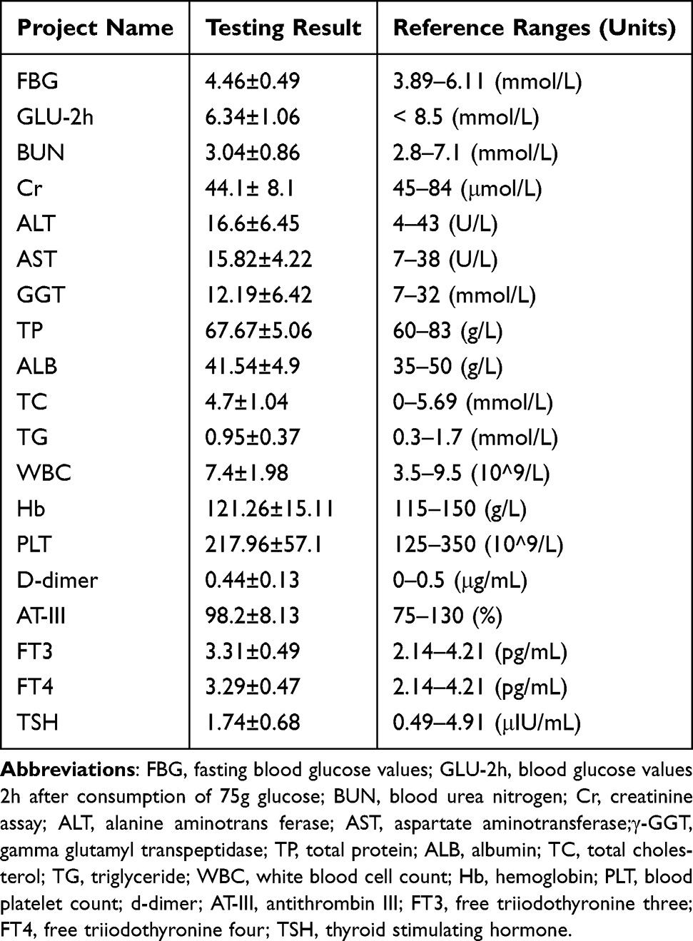 |
Table 1 Blood Test Items in Patients with Unexplained Recurrent Miscarriage |
Plasma Samples Preparation
Venous blood samples were collected from fasting participants into tubes containing 3.2% sodium citrate (v/v, anticoagulant:blood = 1:9) and centrifuged at 4°C, at 3000 rpm for 10 minutes. Plasma was extracted, aliquoted, and stored at –80°C until use.
Examination of Plasma Hcy Levels
Plasma homocysteine levels were determined using the homocysteine determination kit (Shanghai Fosun Changzheng Medical Science) according to the manufacturer’s instructions.
Analysis of Plasma Microparticle
The plasma from the two groups was centrifuged, and the supernatant was collected as MPs-poor plasma. Anti-endothelial cell antigen monoclonal antibodies (CD31-PE; cat: 555448 or CD62e-PE; cat: 551145, BD Biosciences) were incubated at room temperature in the dark for 30 minutes. Samples without any antibodies were used as negative controls. Flow-Count Fluorospheres were added to each reaction system. The number of MPs was measured using a flow cytometer (Cytomics FC 500, Beckman Coulter).15
Homocysteine Diluent Configuration
Homocysteine powder (Sigma, cat: H9633) was weighed and diluted with PBS solution. The solution was thoroughly mixed and completely dissolved. Bacteria were removed by passing through a 0.22 μm filter in an ultra-clean environment. A solution with a concentration of 16 mmol/L was prepared, and homocysteine solution concentrations of 0.5, 2, 4, and 8 mmol/L were prepared. These solutions were stored in the refrigerator at −20°C in the dark.
Effect of Homocysteine on HUVEC Cells
Human umbilical vein endothelial cells (HUVEC) obtained from ATCC were cultured in RPMI 1640 medium with 10% fetal bovine serum at 37°C and 5% CO2. To evaluate the effect of homocysteine, HUVEC cells were seeded at a density of 2×10⁵ cells/mL in a 6-well plate (1.5 mL per well) and incubated in a 37°C, 5% CO2 cell incubator. The following day, when the cell density reached approximately 90%, different concentrations of Hcy solution (0, 0.5, 1.0, 2, 4, 8 mmol/L) were added and incubated for 24 hours at 37°C in a 5% CO2 incubator. Subsequently, the toxicity of homocysteine solution on human endothelial cells was assessed using the CCK8 assay (CCK-8; Japan; cat: CK04).
Apoptosis of Hcy Treated HUVEC Cells
HUVEC cells were seeded at a density of 2×10⁵ cells/mL in 6-well plates. Subsequently, 1.5 mL of Hcy solution with different concentrations (0, 0.5, 1.0, 2, 4, 8 mmol/L) was added and incubated for 24 hours. The HUVEC cells were then digested with trypsin and 1640 medium containing serum to terminate digestion. The cell suspension was transferred to a 1.5 mL centrifuge tube and centrifuged for 10 minutes. The supernatant was discarded, and the bottom 50 µL of cells were retained and washed twice with PBS. The cells were then fixed with 70% alcohol and placed at −20°C overnight. The following day, 10 µL of 1 mg/mL PI staining solution (Abcam, ab40776) was added, followed by the addition of 400 µL of 1×PBS at room temperature in the dark for 30 minutes.
Statistical Analysis
Data were analyzed and graphed using SPSS 19.0 and Prism 9.5 (GraphPad) software. The normality and equal variance of the data were assessed using Levene’s test and Kolmogorov–Smirnov test, respectively. If the data passed the normality and equal variance tests, Student’s t-test was used for two-group comparisons, and one-way analysis of variance was used for comparisons among three or more groups. If the data did not pass the normality or equal variance test, the Mann–Whitney test was used for two independent sample comparisons, and the Kruskal–Wallis test with Mann–Whitney test and Bonferroni correction was used for multiple comparisons. Correlations were analyzed using Pearson’s correlation coefficient. Results were expressed as means ± standard deviation, and p-values < 0.05 were considered statistically significant.
Results
Plasma Homocysteine Level Increased in Women with Recurrent Abortion
We detected plasma Hcy levels in normal pregnant women and patients with recurrent abortion. As shown in Figure 1, plasma Hcy levels in women with recurrent abortion (8.43 ± 0.35 μmol/L, n=40) were significantly higher than those in normal pregnant women (6.05 ± 0.2 μmol/L, n=40) (p < 0.0001), it is very statistically significant differences in the variables for women with recurrent miscarriages in the two data sets.
 |
Figure 1 Analysis of plasma Hcy levels in women with recurrent abortion and normal pregnant women; ****p < 0.0001, it is a significant statistical difference. |
Plasma Total-MPs and Endothelial-MPs in Women with Recurrent Abortion
Plasma samples were collected from 30 normal pregnant women and 30 patients with recurrent abortion, and the total MPs in plasma were detected by FCM. The total number of MPs in the plasma of normal pregnant women and women with recurrent abortion was 26.70 ± 8.787 and 84.69 ± 27.20, respectively. The total plasma MPs of women with recurrent abortion were significantly higher than those of women with normal pregnancy (p = 0.002) (Figure 2A).
 |
Figure 2 Recurrent miscarriage and normal pregnant women were detected by FCM (A) analysis of the total MPs (B) analysis of CD31+ EMPs in plasma. Correlations between plasma Hcy levels and the total MPs (C) or CD31+ EMP number (D) of plasma in women with recurrent miscarriage. |
Using the cell surface marker CD31+ to indicate apoptotic and activated endothelial cell-derived microparticles, we found that the number of endothelium-derived MPs in the plasma of women with recurrent abortion was also significantly higher than that of normal pregnant women (n = 30, p = 0.009) (Figure 2B). Moreover, significant correlations were found between plasma Hcy levels and total MP number (Figure 2C) or EMP number (Figure 2D). These data indicate that the elevated number of total MPs and EMPs in women with recurrent miscarriage are related to plasma Hcy levels, suggesting that enhanced plasma Hcy levels are an important stimulating factor for EMP production.
Increased Plasma Free DNA Levels in Women with Recurrent Miscarriage
To determine whether plasma free DNA was elevated in women with recurrent abortion, we measured the plasma free DNA concentration using the nucleic acid-bound fluorescent dye PicoGreen. The plasma free DNA concentrations in normal pregnancy and recurrent abortion were 46.57 ± 2.44 μg/mL and 57.82 ± 2.64 μg/mL, respectively. The level of plasma free DNA in women with recurrent abortion was significantly higher than that in normal pregnant women (Figure 3A). A significant correlation was found between plasma cell-free DNA and plasma Hcy in RSA women (Figure 3B). These results suggest that high levels of plasma Hcy may promote DNA release from activated neutrophils.
 |
Figure 3 (A) Analysis of plasma DNA levels in normal pregnant women and women with recurrent miscarriage. (B) Correlations between plasma Hcy levels and the plasma DNA levels in women with recurrent miscarriage. |
Toxic Effects of Homocysteine Solution on HUVECs Cells
Given the observed elevation of plasma Hcy and EMPs in women with recurrent miscarriage, and the significant correlation between plasma Hcy and EMPs, we investigated the effect of homocysteine on endothelial cells. HUVECs were treated with varying concentrations of Hcy (0, 0.5, 1, 2, 4, 8 mmol/L) for 24 hours, and the toxic effect of homocysteine on HUVECs was assessed using the CCK8 assay. The results showed that Hcy treatment significantly affected the survival rate of HUVEC cells, and this effect was dose-dependent, suggesting that Hcy had a toxic effect on HUVEC cells (Figure 4A). We also assessed the integrity of HUVEC cells using PI staining and flow cytometry. Compared to the control group, the percentage of PI-positive cells gradually increased in the Hcy-stimulated groups, which were 1.424 ± 0.47%, 15.28 ± 1.04%, 23.11 ± 1.67%, 26.85 ± 1.56%, and 41.36 ± 1.96%, respectively (Figure 4B). These results indicate that Hcy could disrupt the integrity of HUVEC membranes and increase the permeability of the cell membrane.
 |
Figure 4 Different concentrations of homocysteine solution were applied to HUVECs cells (A) Observe the cytotoxicity of HUVEC cells by CCK8 experiment, and (B) the apoptotic of HUVEC cells was detected by PI staining and flow cytometry;**p < 0.01, it is a significant statistical difference. |
Homocysteine Stimulates HUVEC to Release Increased EMPs
To investigate the effect of homocysteine on HUVECs, HUVECs were stimulated with different concentrations of Hcy (0, 0.5, 1, 2, 4, 8 mmol/L) for 24 hours. The cell supernatant was collected, and the number of EMPs released by HUVECs was detected by flow cytometry. Compared with the normal control group (0 mmol/L), the number of EMPs released by HUVECs after Hcy stimulation gradually increased. Moreover, the number of EMPs released by HUVECs in the Hcy groups of 1, 2, 4, and 8 mmol/L was significantly higher than that in the control group, indicating that Hcy can promote the release of EMPs by HUVEC cells (Figure 5).
 |
Figure 5 Different concentrations of homocysteine solution were applied to HUVECs cells, to analysis the number of EMPs released from HUVECs by FCM;**p < 0.01, it is a significant statistical difference. |
Discussion
Recurrent miscarriage (RSA) is a prevalent pathological condition in reproductive medicine, affecting 1–5% of couples striving for pregnancy, despite extensive efforts by clinicians and researchers to ascertain its etiology, approximately 50% of cases remain unexplained, significantly impacting patients’ lives. Currently, the etiology and pathological mechanism of half of unexplained RSA cases are multifaceted, with many attributed to endocrine disorders, metabolic abnormalities, infections, etc.16 Recently, some experts have suggested a metabolic component, homocysteine (Hcy), wherein mild to moderate hyperhomocysteinemia (HHcy) may pose a threat to women experiencing habitual abortion or placental abruption. However, the specific pathological mechanism of damage remains unclear. Homocysteine, a sulfur-containing amino acid formed during methionine metabolism, typically presents at fasting plasma concentrations of 5–15 μmol/L.17 When Hcy fails to metabolize to methionine or cysteine due to genetic or nutritional deficiencies, it accumulates systemically along with its protein-related metabolites, augmenting the Hcy-thiolactone pathway’s flux and associating with various pathological conditions in humans.18 Therefore, metabolic abnormalities, such as elevated homocysteine levels, alone or in combination, can impede pregnancy maintenance. The study we present aims to elucidate the mechanism of HHcy in patients with recurrent spontaneous abortion through case studies and basic experimental research.
Previous data suggest that homocysteine is not only a disease biomarker but also a guide for disease prevention.19 During normal pregnancy, the concentration of Hcy in the blood shows a decreasing trend, with a more pronounced decrease during 8–12 weeks of pregnancy.20,21 In our study, we observed that plasma Hcy levels in RSA patients were significantly higher than those in normal pregnant women during the same period (6–10 weeks of gestational age), suggesting a correlation between increased plasma Hcy concentration and RSA. Steegers-Theunissen et al suggested the relationship between recurrent early pregnancy loss and elevated homocysteine concentrations, and have been proven.22,23 However, the precise mechanism of action between homocysteine and miscarriage remains unclear.
Maternal vascular endothelial injury, which promotes thrombosis and affects fetal blood and oxygen supply, is also one of the pathological mechanisms of recurrent pregnancy loss.20 HHcy can also affect endothelial cell functions, such as cytoskeleton remodeling, up-regulation of adhesion molecule expression, cell barrier dysfunction, and inflammatory response.24 In our study, we observed that the elevated number of total MPs and EMPs in women with recurrent miscarriage are related to plasma Hcy levels, suggesting that enhanced plasma Hcy levels are an important stimulating factor for EMP production. However, the mechanism by which Hcy metabolism is blocked and high levels of Hcy stimulate endothelial cells leading to endothelial dysfunction and the release of EMPs remains to be verified.
In our study, we observed significantly elevated plasma Hcy concentrations in women with RSA compared to normal controls, along with a significant increase in the total number of plasma particles. Plasma MPs have various sources, such as endothelial cells, platelets, white blood cells, and red blood cells, which release MPs during activation or apoptosis.25–28 Among these plasma MPs, EMPs are released in response to endothelial cell injury, activation, and apoptosis, serving as indicators of endothelial cell injury. The cell surface antigen CD31 serves as a stabilizing marker for EMPs, with endothelial cell-derived particles represented by CD31+.29
To identify the primary source of increased particulates in the plasma of women with RSA, we detected CD31-positive MPs and found a significantly higher number of CD31+ MPs in the plasma of RSA women compared to those with normal pregnancies. These results suggest that the majority of the increased MPs in RSA women’s plasma are EMPs, released during oxidative stress or apoptosis of endothelial cells. Additionally, Our study found that an increase in plasma free DNA in women with URSA.
Our study showed that elevated levels of Hcy in women with RSA may stimulate endothelial cells, leading to endothelial dysfunction and increased release of particles as well as free DNA. To visualize the effect of Hcy on endothelial cells, we treated HUVECs with varying concentrations of Hcy. We observed a significant reduction in the survival rate of HUVECs cells following Hcy stimulation, with a concurrent increase in the number of PI-positive cells correlating with the Hcy concentration. This suggests that elevated concentrations of Hcy exert cytotoxic effects on HUVECs, compromising cell membrane integrity, increasing membrane permeability, promoting the release of EMPs from endothelial cells, and potentially contributing to embryonic developmental arrest and miscarriage.
Conclusion
Our findings indicated that plasma Hcy levels were significantly elevated in women with recurrent miscarriages compared to those with normal pregnancies. Additionally, the total number of MPs and EMPs, as well as plasma free DNA, were also significantly increased in women with RSA. In vitro experimental results showed that the toxic effect of Hcy on HUVECs was enhanced with the increase in Hcy concentration. Our findings demonstrated that high levels of Hcy in women with RSA may stimulate apoptosis of endothelial cells, which in turn leads to endothelial dysfunction, releasing microparticles and free DNA. This affects embryo development, and may be related to embryo development arrest and abortion. Therefore, the present study primarily elucidates the influencing factors and the role of homocysteine in the pathogenesis of recurrent miscarriage. Additionally, it provides novel insights and therapeutic targets for the early diagnosis and prevention of recurrent miscarriage.
Data Sharing Statement
All data generated or analyzed during this study are included in this article.
Ethics Approval and Consent to Participate
This work has been carried out in accordance with the Declaration of Helsinki (2000) of the World Medical Association. This study was approved by the Ethic Committee of The Second Affiliated Hospital of Soochow University (JD-LK-2020-041-02), and all participants provided written informed consent.
Author Contributions
All authors made a significant contribution to the work reported, whether that is in the conception, study design, execution, acquisition of data, analysis and interpretation, or in all these areas; took part in drafting, revising or critically reviewing the article; gave final approval of the version to be published; have agreed on the journal to which the article has been submitted; and agree to be accountable for all aspects of the work.
Funding
This study was supported by the Science and Technology Program of Suzhou (SKY2022005) to Hong Zhang. This study was supported by the Science and Technology Program of Suzhou (SKY2021007) to Haifang Zhang.
Disclosure
The authors declare that they have no competing interests.
References
1. Ford HB, Schust DJ. Recurrent pregnancy loss: etiology, diagnosis, and therapy. Rev Obstet Gynecol. 2009;2(2):76–83.
2. Wang XH, Xu S, Zhou XY, et al. Low chorionic villous succinate accumulation associates with recurrent spontaneous abortion risk. Nat Commun. 2021;12(1):3428. doi:10.1038/s41467-021-23827-0
3. Lord J, McMullan DJ, Eberhardt RY, et al. Prenatal exome sequencing analysis in fetal structural anomalies detected by ultrasonography (PAGE): a cohort study. Lancet. 2019;393(10173):747–757. doi:10.1016/S0140-6736(18)31940-8
4. Matalon ST, Blank M, Ornoy A, Shoenfeld Y. The association between anti-thyroid antibodies and pregnancy loss. Am J Reprod Immunol. 2001;45(2):72–77. doi:10.1111/j.8755-8920.2001.450202.x
5. Carbonnel M, Pirtea P, de Ziegler D, Ayoubi JM. Uterine factors in recurrent pregnancy losses. Fertil Sterility. 2021;115(3):538–545. doi:10.1016/j.fertnstert.2020.12.003
6. Alecsandru D, Klimczak AM, Garcia Velasco JA, Pirtea P, Franasiak JM. Immunologic causes and thrombophilia in recurrent pregnancy loss. Fertil Sterility. 2021;115(3):561–566. doi:10.1016/j.fertnstert.2021.01.017
7. Martínez-Zamora M, Cervera R, Balasch J. Recurrent miscarriage, antiphospholipid antibodies and the risk of thromboembolic disease. Clin Revi Allergy Immunol. 2012;43(3):265–274. doi:10.1007/s12016-012-8316-0
8. Welch GN, Loscalzo J. Homocysteine and atherothrombosis. New Engl J Med. 1998;338(15):1042–1050. doi:10.1056/NEJM199804093381507
9. McCully KS. Vascular pathology of homocysteinemia: implications for the pathogenesis of arteriosclerosis. Am J Pathol. 1969;56(1):111–128.
10. Miller JW. Homocysteine - what is it good for? J Internal Med. 2021;290(4):934–936. doi:10.1111/joim.13288
11. Koklesova L, Mazurakova A, Samec M, et al. Homocysteine metabolism as the target for predictive medical approach, disease prevention, prognosis, and treatments tailored to the person. EPMA J. 2021;12(4):477–505. doi:10.1007/s13167-021-00263-0
12. Hermann A, Sitdikova G. Homocysteine: biochemistry, Molecular Biology and Role in Disease. Biomolecules. 2021;11(5):737. doi:10.3390/biom11050737
13. Serrano NC, Quintero-Lesmes DC, Becerra-Bayona S, et al. Association of pre-eclampsia risk with maternal levels of folate, homocysteine and vitamin B12 in Colombia: a case-control study. PLoS One. 2018;13(12):e0208137. doi:10.1371/journal.pone.0208137
14. Liu C, Luo D, Wang Q, et al. Serum homocysteine and folate concentrations in early pregnancy and subsequent events of adverse pregnancy outcome: the Sichuan Homocysteine study. BMC Pregnancy Childbirth. 2020;20(1):176. doi:10.1186/s12884-020-02860-9
15. Mooberry MJ, Key NS. Microparticle analysis in disorders of hemostasis and thrombosis. Cytometry A. 2016;89(2):111–122. doi:10.1002/cyto.a.22647
16. Rai R, Regan L. Recurrent miscarriage. Lancet. 2006;368(9535):601–611. doi:10.1016/S0140-6736(06)69204-0
17. Von Woon E, Greer O, Shah N, Nikolaou D, Johnson M, Male V. Number and function of uterine natural killer cells in recurrent miscarriage and implantation failure: a systematic review and meta-analysis. Human Reproduction Update. 2022;28(4):548–582. doi:10.1093/humupd/dmac006
18. Jakubowski H. Homocysteine Modification in Protein Structure/Function and Human Disease. Physiol Rev. 2019;99(1):555–604. doi:10.1152/physrev.00003.2018
19. Smith AD, Refsum H. Homocysteine - from disease biomarker to disease prevention. J Internal Med. 2021;290(4):826–854. doi:10.1111/joim.13279
20. Walker MC, Smith GN, Perkins SL, Keely EJ, Garner PR. Changes in homocysteine levels during normal pregnancy. Am J Clin Exp Obstet Gynecol. 1999;180(3):660–664. doi:10.1016/S0002-9378(99)70269-3
21. Dai C, Fei Y, Li J, Shi Y, Yang X. A Novel Review of Homocysteine and Pregnancy Complications. Biomed Res Int. 2021;2021:6652231. doi:10.1155/2021/6652231
22. Nelen WL, Blom HJ, Steegers EA, den Heijer M, Thomas CM, Eskes TK. Homocysteine and folate levels as risk factors for recurrent early pregnancy loss. Obstetrics Gynecol. 2000;95(4):519–524. doi:10.1016/s0029-7844(99)00610-9
23. Qiu X, Gao F, Qiu Y, et al. Association of maternal serum homocysteine concentration levels in late stage of pregnancy with preterm births: a nested case-control study. J Maternal Fetal Neonatal Med. 2018;31(20):2673–2677. doi:10.1080/14767058.2017.1351534
24. Chatterjee B, Fatima F, Seth S, Sinha Roy S. Moderate Elevation of Homocysteine Induces Endothelial Dysfunction through Adaptive UPR Activation and Metabolic Rewiring. Cells. 2024;13(3):214. doi:10.3390/cells13030214
25. Jan M, Cueto R, Jiang X, et al. Molecular processes mediating hyperhomocysteinemia-induced metabolic reprogramming, redox regulation and growth inhibition in endothelial cells. Redox Biol. 2021;45:102018. doi:10.1016/j.redox.2021.102018
26. Kim Y, Goodman MD, Jung AD, et al. Microparticles from aged packed red blood cell units stimulate pulmonary microthrombus formation via P-selectin. Thromb Res. 2020;185:160–166. doi:10.1016/j.thromres.2019.11.028
27. Burnouf T, Goubran HA, Chou ML, Devos D, Radosevic M. Platelet microparticles: detection and assessment of their paradoxical functional roles in disease and regenerative medicine. Blood Rev. 2014;28(4):155–166. doi:10.1016/j.blre.2014.04.002
28. Wang JG, Aikawa E, Aikawa M. Leukocyte-derived microparticles as proinflammatory mediators in atherosclerosis. J Am College Cardiol. 2013;62(16):1442–1445. doi:10.1016/j.jacc.2013.04.054
29. Li B, Huang Q, Lin C, et al. Increased circulating CD31+/CD42b-EMPs in Perthes disease and inhibit HUVECs angiogenesis via endothelial dysfunction. Life Sci. 2021;265:118749. doi:10.1016/j.lfs.2020.118749
 © 2025 The Author(s). This work is published and licensed by Dove Medical Press Limited. The
full terms of this license are available at https://www.dovepress.com/terms
and incorporate the Creative Commons Attribution
- Non Commercial (unported, 3.0) License.
By accessing the work you hereby accept the Terms. Non-commercial uses of the work are permitted
without any further permission from Dove Medical Press Limited, provided the work is properly
attributed. For permission for commercial use of this work, please see paragraphs 4.2 and 5 of our Terms.
© 2025 The Author(s). This work is published and licensed by Dove Medical Press Limited. The
full terms of this license are available at https://www.dovepress.com/terms
and incorporate the Creative Commons Attribution
- Non Commercial (unported, 3.0) License.
By accessing the work you hereby accept the Terms. Non-commercial uses of the work are permitted
without any further permission from Dove Medical Press Limited, provided the work is properly
attributed. For permission for commercial use of this work, please see paragraphs 4.2 and 5 of our Terms.