
Back to Journals » Clinical, Cosmetic and Investigational Dermatology » Volume 19
EGFR Inhibitor-Induced Delayed Wound Healing Following PICC-port Implantation: Three Cases and a Literature Review
Authors Liu M  , Zhong B , Lin D , Huang S , Lin G , Lin X, Jiang S
Received 21 April 2026
Accepted for publication 8 July 2026
Published 22 July 2026 Volume 2026:19 617447
DOI https://doi.org/10.2147/CCID.S617447
Checked for plagiarism Yes
Review by Single anonymous peer review
Peer reviewer comments 2
Editor who approved publication: Prof. Dr. Rungsima Wanitphakdeedecha
Mianzhu Liu,1,* Bihua Zhong,2,* Danzhu Lin,3 Shaoman Huang,4 Genfa Lin,5 Xiujia Lin,1 Shaoru Jiang6
1Department of Peritoneal Dialysis, Jieyang People’s Hospital, Jieyang, Guangdong, People’s Republic of China; 2Second Department of Medical Oncology, Jieyang Cancer Center, Jieyang People’s Hospital, Jieyang, Guangdong, People’s Republic of China; 3Nursing Department, Jieyang People’s Hospital, Jieyang, Guangdong, People’s Republic of China; 4Department of Healthcare-Associated Infection Control, Jieyang People’s Hospital, Jieyang, Guangdong, People’s Republic of China; 5Department of Anesthesiology and Operating Room, Jieyang People’s Hospital, Jieyang, Guangdong, People’s Republic of China; 6Gynecology and Reproductive Medicine Center, Jieyang People’s Hospital, Jieyang, Guangdong, People’s Republic of China
*These authors contributed equally to this work
Correspondence: Shaoru Jiang, Gynecology and Reproductive Medicine Center, Jieyang People’s Hospital, Jieyang, Guangdong, People’s Republic of China, Email [email protected]
Abstract: Epidermal growth factor receptor (EGFR) inhibitors are widely used in the treatment of cancer and are often associated with toxicities, such as rash and xerosis. However, their impact on wound healing after PICC-port placement is not well understood. This report presents three cases in which EGFR inhibitor therapy was associated with significantly delayed wound healing following PICC-port implantation. Wound complications, including slough formation, infection, periwound skin damage and skin dehiscence, were observed in all three cases. Despite standard wound care, healing remained delayed until EGFR inhibitor therapy was discontinued. In each case, cessation of the EGFR inhibitor was critical for resolving the wound healing complications, and continuous debridement with advanced dressings facilitated eventual closure. These cases highlight the need for careful perioperative management when combining EGFR inhibitors with surgical procedures, particularly in the context of PICC-port placement. The consistent temporal relationship between drug withdrawal and healing initiation across all three cases suggests a contributory role of EGFR inhibitors. Larger prospective studies are needed to further clarify the relationship between EGFR inhibitor therapy and wound healing, and to inform optimal perioperative management in patients receiving these agents.
Keywords: EGFR inhibitor, delayed wound healing, PICC-port complication, wound management, debridement, catheters, neoplasms
Introduction
Epidermal growth factor receptor (EGFR) inhibitors are widely used in the treatment of various cancers, including lung, breast, and colorectal cancer.1 While these targeted therapies are effective, they are associated with unique toxicities, such as rash,1 paronychia,2 and xerosis.3 Beyond these well-recognized adverse effects, emerging evidence from animal models and experimental studies suggests that EGFR inhibitors may also impair wound healing.4–6 However, the clinical relevance of this finding, particularly in the context of surgical or invasive procedures, remains to be elucidated.
Peripherally inserted central catheter (PICC)-ports are commonly used in cancer patients for intravenous chemotherapy and parenteral nutrition.7 PICC-port placement involves a surgical incision, and delayed wound healing at the insertion site can lead to infection, patient discomfort, and unplanned device removal.8–10 Known risk factors for delayed wound healing include infection, malnutrition, underlying disease, and certain medications.11,12 Whether EGFR inhibitor therapy specifically affects wound healing after PICC-port implantation has not been investigated.
In this report, we describe three cases of significantly delayed wound healing following PICC-port placement in cancer patients receiving EGFR inhibitor therapy. These cases highlight a previously underrecognized complication and underscore the need for careful perioperative management when combining EGFR inhibitors with surgical procedures. A review of the relevant literature is also provided.
Case Presentations
The characteristics of the three cases are summarized in Table 1. A detailed description of each case is presented below.
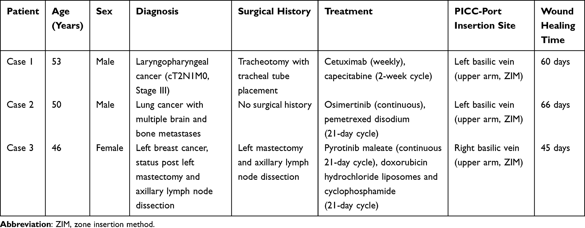 |
Table 1 Patient Characteristics |
Case 1
A 53-year-old man with a 5-year history of laryngopharyngeal cancer (cT2N1M0 Stage III) presented with invasion of the thyroid cartilage necessitating tracheotomy and placement of a tracheal tube. A combined treatment regimen consisting of capecitabine and cetuximab was prescribed. The capecitabine regimen consisted of a 2-week cycle of oral administration followed by a 1-week discontinuation period. Cetuximab was administered intravenously once weekly. The patient had completed 10 cycles of cetuximab and 4 cycles of capecitabine before this admission. The patient had a low body mass index (BMI) of 17.6, indicating underweight. Considering the potential risk of malnutrition, a decision was made to establish vascular access for parenteral nutrition. A PICC-port was then inserted into the patient’s left basilic vein in the upper arm. On the day of the procedure, he received his 11th intravenous dose of cetuximab.
By day 14 after implantation of the PICC-port, delayed wound healing with a layer of slimy yellow material was noted (Figure 1A). This material was identified as slough according to the definition provided in a previous report.13 Erythema and roughness of the periwound skin were also noted.
 |
Figure 1 Wound healing in case 1. (A) Delayed wound healing with a layer of slimy yellow slough noted 14 days post-port implantation, with erythema and roughness of the periwound skin. (B) Secondary suturing of the wound 21 days after implantation. (C) Recurrence of yellow slough, minor cracking, and adjacent epidermal denudation 7 days after secondary suturing (day 28 after implantation). (D) Wound closure with scab formation indicating healing 60 days after implantation. Abbreviation: PICC, peripherally inserted central catheter. |
Laboratory data (Table 2) showed a C-reactive protein (CRP) level of 45.48 mg/L (reference 0.00–6.00), while the white blood cell count was within normal limits at 5.73 × 10⁹/L. In addition, the patient had mild hypoalbuminemia (albumin 36.6 g/L; reference 40–55). Coagulation parameters and other biochemistry parameters were unremarkable.
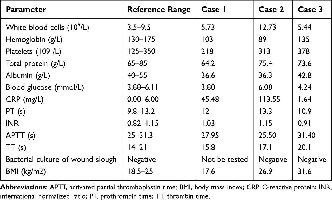 |
Table 2 Results of Laboratory Tests Performed in the Three Cases at the Time of Impaired Wound Healing |
From day 14 post-implantation, the wound was managed by daily cleaning with normal saline and disinfection with iodophor. The slough was scraped off daily with a scalpel but returned the following day. Repeated sharp debridement caused pain and discomfort to the patient, so Urgo hydrogel (Urgo Medical, Chenôve, France) was applied to promote autolytic debridement and alleviate the pain associated with the procedure. When the wound remained unhealed on day 21 after implantation of the PICC-port, secondary suturing was performed (Figure 1B) and Urgo hydrogel was discontinued. Seven days later, minor cracking, yellow slough, and adjacent epidermal denudation were noted (Figure 1C). To manage the wound, Urgo hydrogel was restarted, and a silver alginate dressing was added.
At day 40, the wound was still unhealed, and the patient was noted to have bleeding from the pharyngeal mucosa. The physician attributed the delayed wound healing and mucosal bleeding to cetuximab and elected to suspend its administration. After discontinuation, the slough gradually decreased, and the wound progressively healed. Complete healing required 60 days (Figure 1D). Key events are summarized in Table 3.
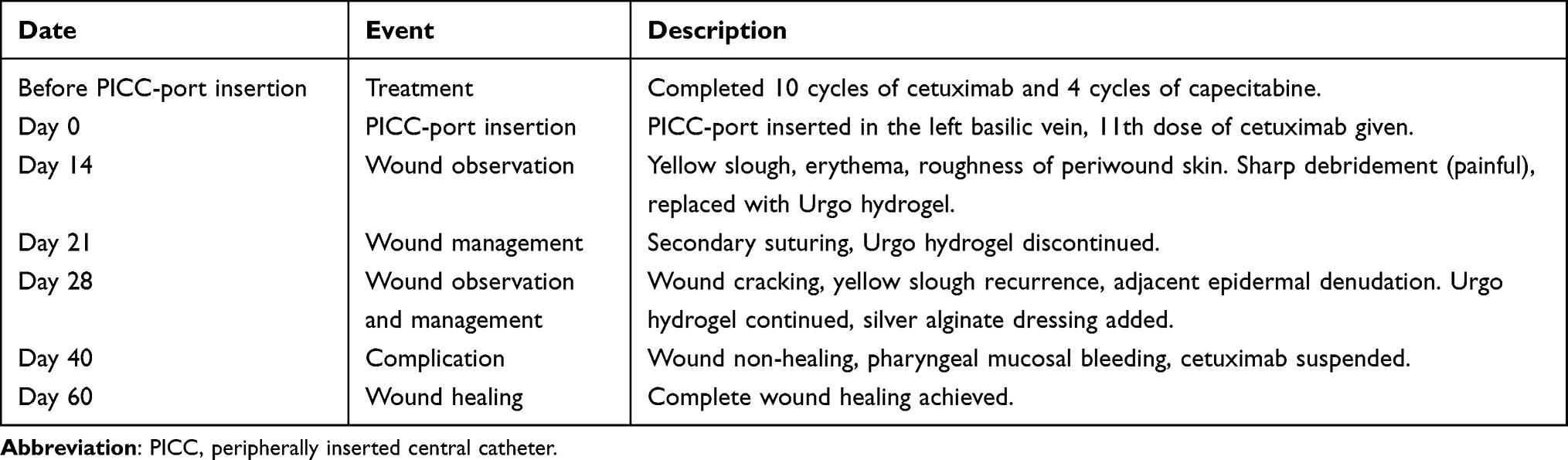 |
Table 3 Timeline of Events in Case 1 |
Case 2
A 50-year-old man with a history of lung cancer, multiple brain metastases, and metastatic bone disease was admitted to our hospital for a scheduled chemotherapy and targeted agent regimen. The prescribed treatment comprised continuous oral osimertinib and pemetrexed disodium on a 21-day cycle, of which he had completed 9 cycles before admission. Because the patient’s peripheral veins were compromised, a PICC-port was implanted. The left basilic vein was selected for access, with the reservoir positioned in the upper arm. After implantation, the incision was routinely cleaned with chlorhexidine gluconate.
On day 7 after implantation of the PICC-port, yellow slough and skin dehiscence appeared at the wound, venipuncture, and port-skin puncture sites (Figure 2A). By day 14, the wound dehiscence had worsened, with obvious skin separation (Figure 2B). The wound also demonstrated clear signs of local infection, including redness at the edges, purulent discharge, and local warmth.
 |
Figure 2 Wound healing in case 2. (A) Yellow slough at the wound, venipuncture, and port puncture sites 7 days after PICC-port implantation. (B) Worsening of wound dehiscence with obvious skin separation, along with redness, 14 days after implantation. (C) Purulent discharge, granulation tissue growth, reduction in wound size and inflammation by day 30. (D) Complete healing of the wound 66 days post-implantation. Abbreviation: PICC, peripherally inserted central catheter. |
Laboratory data (Table 2) showed an elevated CRP level (113.55 mg/L) and white blood cell count (12.73 × 10⁹/L), while microbial cultures of the slough were negative. Other parameters were within normal limits.
From day 7 onwards, given the inflammatory features, Urgo hydrogel was applied to remove the slough and promote autolytic debridement, and the wound was covered with a silver alginate dressing, which was changed once daily.
At day 21 after implantation, the patient elected to discontinue osimertinib due to financial reasons. After discontinuation, fresh granulation tissue gradually grew at the base of the wound. By day 30 post-implantation, the wound had decreased in size and the inflammation had diminished (Figure 2C). Complete healing of the wound, venipuncture, and port sites required 66 days (Figure 2D). Table 4 summarizes the key events and interventions for this case.
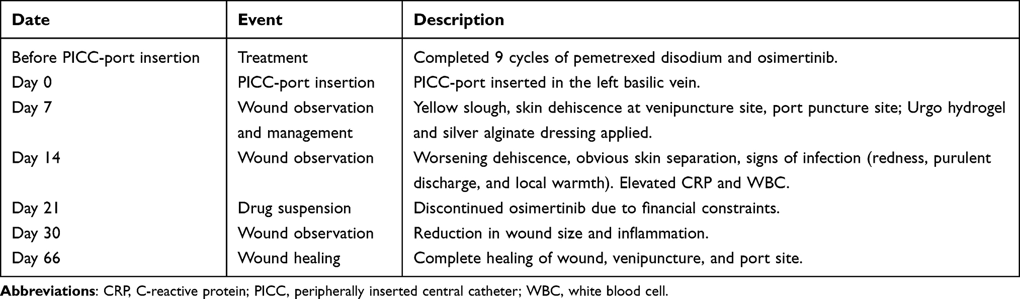 |
Table 4 Timeline of Events in Case 2 |
Case 3
A 46-year-old woman who had undergone left mastectomy and axillary lymph node dissection for breast cancer 1 month earlier was admitted to our oncology department for a combined treatment regimen consisting of chemotherapy (doxorubicin hydrochloride liposomes and cyclophosphamide, 21-day cycle) and targeted therapy with pyrotinib maleate given continuously on a 21-day cycle. A standard PICC-port insertion procedure was performed, and the incision was routinely cleaned with chlorhexidine gluconate. Two days after implantation, the patient started her intravenous chemotherapy, with concomitant oral administration of pyrotinib maleate.
By day 10 after PICC-port implantation, mild epidermal dehiscence with thin, superficial white slough was noted at the wound site, along with marginal erythema and periwound hyperpigmentation (Figure 3A). By day 14, the dehiscence persisted, with periwound skin damage and a small amount of yellow slough covering the wound bed (Figure 3B). The surrounding skin showed patchy hyperpigmentation, along with a single blister.
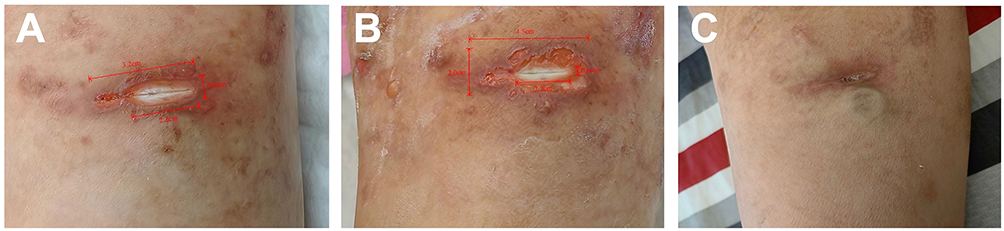 |
Figure 3 Wound healing in case 3. (A) Mild epidermal dehiscence with thin, superficial white slough, marginal erythema, and periwound hyperpigmentation 10 days after PICC-port implantation. (B) Persistent dehiscence with periwound skin damage, yellow slough, patchy hyperpigmentation, and a single blister 14 days after implantation (C) Complete healing of the wound at 45 days post-implantation. |
Microbial culture of the slough was negative (Table 2), consistent with the finding in Case 2. Other laboratory parameters were within normal limits, apart from a mildly elevated platelet count (378 × 109/L).
On day 10 after PICC-port implantation, the wound was treated with Urgo hydrogel and a nano-silver dressing for topical antibacterial effect.14 By day 14, in view of the periwound skin damage and to avoid further maceration (a known risk associated with Urgo hydrogel according to the product information), the Urgo hydrogel was discontinued. The wound was then managed with daily gentle sharp debridement using a scalpel to remove the slough, while the nano-silver dressing was maintained.
Learning from the previous cases, the physician considered that the condition of the wound and skin was related to the dermal toxicity of pyrotinib maleate. After taking informed consent from the patient, pyrotinib maleate was suspended for 14 days, starting from day 10 after PICC-port implantation.
By day 24, the periwound skin damage had resolved and the epidermal dehiscence had gradually decreased in size, and pyrotinib maleate was resumed. The wound was fully closed by day 45 after discontinuation and implementation of the above-mentioned wound care measures (Figure 3C). Key events in this case are summarized in Table 5.
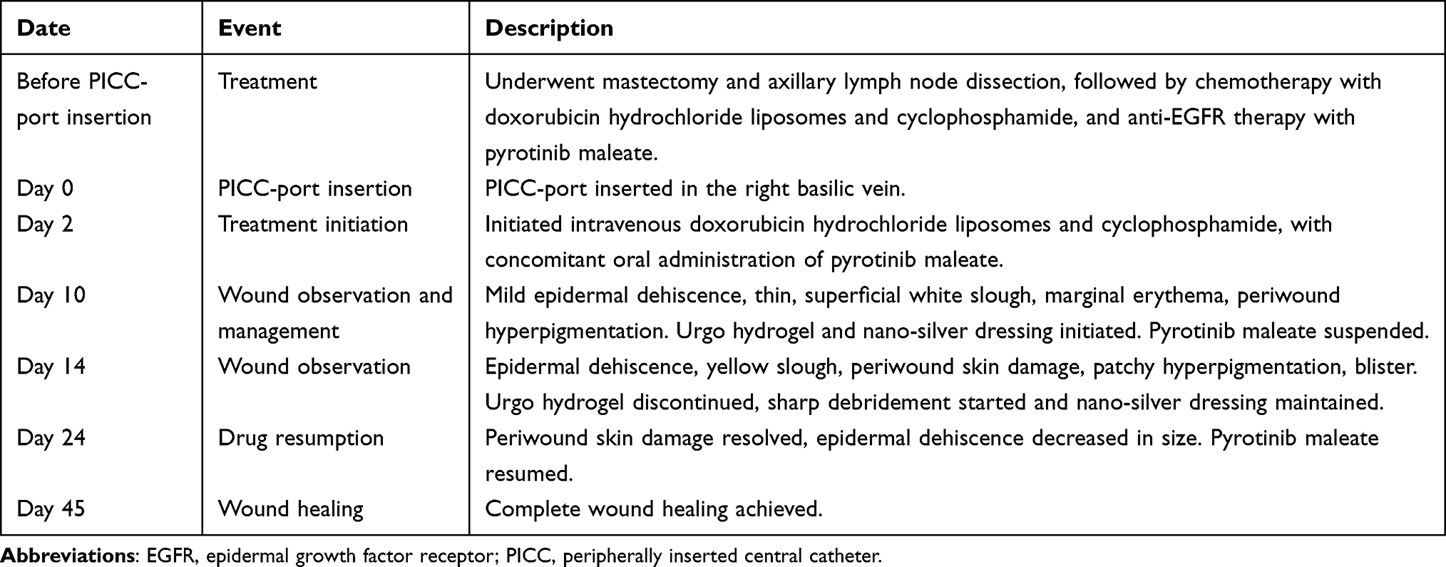 |
Table 5 Timeline of Events in Case 3 |
Discussion
Biological Rationale
EGFR is highly expressed in basal keratinocytes, hair follicles, and sweat glands of the skin.15 When a tissue injury occurs, expression of EGFR increases transiently, promoting healing by regulation of acute inflammation, stimulation of keratinocyte migration and proliferation, and enhancement of angiogenesis (Figure 4).16 Inhibition of EGFR disrupts these processes, leading to impaired migration of keratinocytes and delayed re-epithelialization, which is a critical step in wound closure and restoration of the integrity of the skin.17 This ultimately results in compromised barrier function and delayed wound healing (Figure 4). Therefore, it is biologically plausible that patients receiving EGFR inhibitor therapy may experience delayed healing after surgical procedures such as PICC-port implantation.
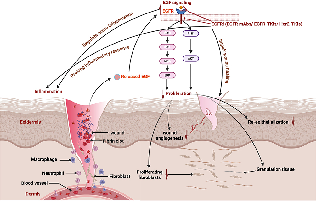 |
Figure 4 Mechanisms via which epidermal growth factor receptor inhibitors impair wound healing [created in BioRender. Liu, M. (2026) https://BioRender.com/qvvf96c]. Solid arrows (↓) indicate signal activation or biological promotion (arrow direction for layout only); upward arrows (↑) indicate increase, prolongation, or upregulation; downward arrows (↓) indicate decrease, reduction, or downregulation; blunted arrows (⊣) indicate inhibition or blockade. Normal wound healing (upper and middle sections): EGF released binds to the EGFR, activating downstream pathways (↓), leading to increased (↑) proliferation, re-epithelialization, formation of granulation tissue, and angiogenesis, while resolving inflammation. Effect of EGFR inhibitors (lower section): EGFR inhibitors block (⊣) EGFR signaling, resulting in decreased (↓) proliferation and angiogenesis, prolonged (↑) inflammation, impaired granulation tissue, and delayed re-epithelialization (↓), ultimately causing chronic, non-healing wounds. Abbreviations: EGF, epidermal growth factor; EGFR, epidermal growth factor receptor. |
Interpretation
All three patients received a distinct EGFR-targeted therapy, namely, cetuximab, osimertinib, or pyrotinib (Figure 5), either before or immediately after placement of a PICC-port without a washout period, and all developed chronic wound features, including persistent slough formation, rash, and epidermal damage. The wound healing times were markedly prolonged to 60, 66, and 45 days, respectively.
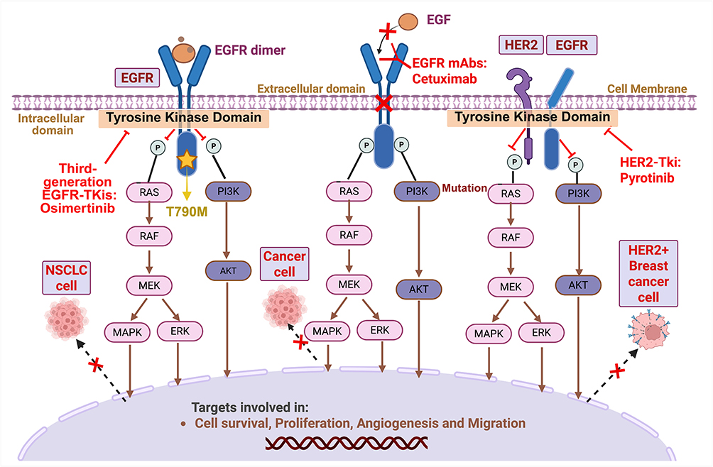 |
Figure 5 Mechanisms of action of three epidermal growth factor receptor inhibitors: cetuximab, osimertinib, and pyrotinib [created in BioRender. Liu, M. (2026) https://BioRender.com/hef75t6]. Red crosses (×) indicate inhibition or blockade; blunted arrows (⊣) indicate irreversible inhibition or blockade. Cetuximab binds to the extracellular domain of EGFR, thereby blocking (×) activation of EGFR and downstream signaling. Osimertinib acts on the intracellular kinase domain of mutant EGFR, irreversibly inhibiting (⊣) kinase activity. Pyrotinib targets the intracellular kinase domains of HER2 and EGFR, irreversibly blocking (⊣) phosphorylation and downstream signaling. Each of these inhibitors suppresses pathways involved in cell survival, proliferation, angiogenesis, and migration. Abbreviations: HER2, human epidermal growth factor receptor 2; EGFR, epidermal growth factor receptor. |
The three cases also differed in several clinical characteristics. Case 1 had low BMI and hypoalbuminemia, which are known confounders for impaired healing. Nevertheless, despite persistently poor nutritional status, the wound started healing following discontinuation of cetuximab. Case 2 had elevated inflammatory markers and clinical signs of infection, but the microbial culture was negative. This discrepancy is consistent with the known cutaneous toxicity of EGFR inhibitors, which can induce sterile, neutrophil‑rich inflammation rather than true bacterial infection.18–20 It is therefore plausible that the inflammatory features were related to drug-induced skin toxicity, although a false‑negative culture cannot be completely ruled out. In Case 3, nutritional parameters were normal, yet delayed healing still occurred and resolved only after pyrotinib withdrawal. This indicates that, in this patient, the observed healing pattern was not driven by nutritional deficiency but rather to drug exposure.
Despite these differences between the cases, the consistent temporal relationship between drug cessation and initiation of healing across all three cases further supports a contributory role of EGFR inhibitors, although causality cannot be definitively established from a retrospective case series.
Existing Evidence and Controversy
The clinical evidence concerning the impact of EGFR inhibitors on wound healing remains mixed. Some preclinical evidence indicates that EGFR inhibitors delay wound healing in animal models and cell cultures.4,5,21 Clinical case reports have also described delayed healing of the corneal epithelium and worsening of chronic surgical wounds in patients receiving cetuximab.22,23 A review of 63 tyrosine kinase inhibitors identified that 24 agents had implications for wound healing, with inhibition of EGFR being a key contributing mechanism.24 Together, these lines of evidence point to a biologically and clinically plausible concern that EGFR inhibitors might impair wound repair in clinical settings.
However, the clinical evidence remains controversial. Govindan et al25 reported no delay in skin wound healing in four patients who underwent surgery for lung cancer while taking gefitinib. A review similarly found insufficient clinical evidence to support the claim that EGFR inhibitors impair wound healing.26 Furthermore, a national consensus statement on cutaneous adverse events associated with targeted therapies does not specifically list delayed wound healing as a complication.20 Similarly, a recent retrospective study of patients who underwent cutaneous surgery while on EGFR inhibitors identified only one case of wound dehiscence and necrosis of transplanted nasal cartilage among 31 procedures.27
Importantly, the evidence summarized above comes from preclinical models, case reports, literature reviews, and consensus statements.4,20–28 However, none of these study types comes from prospective controlled trials specifically examining wound healing in patients receiving EGFR inhibitors. Furthermore, the available reports vary considerably across subjects studied, wound characteristics, and drug regimens. This marked heterogeneity, together with the paucity of robust prospective data, is a major source of the current uncertainty. In this context, our three cases provide additional real-world observations that help bridge the gap between biological plausibility and clinical practice.
Implications for Clinical Management
Based on our experience with these three cases, we propose several practical implications for clinicians managing impaired wound healing in patients receiving EGFR inhibitor therapy.
First, active and continuous debridement is essential for removal of recurring slough and preparation of the wound bed.29 With constant interference from anti-EGFR agents, healing of the epidermal skin is impaired, leading to persistent slough. Slough consists of fibrin, pus, leucocytes, dead cells, microorganisms, and proteinaceous material,13 which can prolong inflammation, attract bacteria, and delay healing.11,30 In case 1, secondary suturing failed to prevent recurrent slough because epithelial proliferation and angiogenesis remained impaired. Instead, daily debridement combined with Urgo hydrogel facilitated autolytic debridement, removed necrotic tissue, and relieved pain.13,29
Second, temporary discontinuation of the EGFR inhibitor should be considered when optimal wound care fails to achieve healing. In all three of our cases, healing progressed only after drug suspension. While some authors suggest basing the interruption period on the drug’s half-life,31 we found that the clinical condition of the wound may be a more practical guide, considering that the effects in tissue can persist beyond the calculated half-life. The decision to stop therapy requires careful risk-benefit assessment and thorough discussion with the patient and family, given the potential risk for disease progression (case 2 developed a new metastasis during the suspension period). Careful timing of anti-EGFR therapy relative to elective procedures such as PICC-port implantation may help mitigate this risk.
Third, infection control remains challenging. Although microbiological cultures were negative in cases 2 and 3, open wounds and slough compromise the skin barrier, creating a portal for potential entry of bacteria.32,33 Furthermore, delayed healing itself can be a sign of infection. Given the risk of secondary infection, we used silver alginate in case 1 and 2, and nano-silver dressings in case 3, as these are commonly used in managing chronic or inflamed wounds.34
Limitation and Future Research
This retrospective case series has several limitations. The small sample size, lack of a control group, and potential confounding factors (eg, malnutrition in case 1) prevent drawing of definitive causal inferences. Moreover, the possibility that healing would have occurred with continued EGFR inhibitor therapy given sufficient time and standard wound care alone cannot be ruled out. Prospective studies with larger sample sizes and control groups are needed to establish a causal relationship and to define optimal perioperative management strategies, including the ideal washout period for an EGFR inhibitor before an elective surgical procedure.
Conclusion
This report presents three cases of markedly delayed wound healing after PICC-port implantation in patients receiving EGFR inhibitor therapy. Healing times ranged from 45 to 66 days, far exceeding the expected 1–2 weeks, and all three patients had chronic wound features, including persistent slough, rash, and epidermal damage. Wound healing progressed only after discontinuation of the EGFR inhibitor in all cases.
Clinicians should be aware that EGFR inhibitor therapy may increase the risk of delayed wound healing following PICC-port placement or similar procedures. When healing is impaired, active wound management, including debridement, appropriate dressings, and careful consideration of temporary drug suspension, may be beneficial. Any decision to interrupt anti-EGFR therapy must balance the potential benefit of wound healing against the risk of cancer progression and should be made with informed patient consent.
Abbreviations
APTT, activated partial thromboplastin time; BMI, body mass index; CRP, C-reactive protein; EGF, epidermal growth factor; EGFR, epidermal growth factor receptor; INR, international normalized ratio; PICC, peripherally inserted central catheter; PT, prothrombin time; TM, tympanic membrane; WBC, white blood cell; ZIM, zone insertion method.
Data Sharing Statement
The authors confirm that all the data supporting the findings of this study are included in this article.
Ethical Approval and Informed Consent
This research was approved by the Institutional Ethical Committee of Jieyang People’s Hospital (approval number 2024055) and carried out in accordance with the Declaration of Helsinki. Institutional approval was required and obtained for the publication of the case details. All patients provided written informed consent for publication of their cases and the accompanying images.
Acknowledgments
We are grateful to the patients mentioned in this report for allowing us to publish the details of their cases. We also thank Liwen Bianji (Edanz) (www.liwenbianji.cn) for editing the English text of a draft of this manuscript.
Author Contributions
All authors made a significant contribution to the work reported, including in the conception, study design, execution, acquisition of data, analysis and interpretation, or in all of these areas; took part in drafting, revising, or critically reviewing the article; gave final approval of the version to be published; have agreed on the journal to which the article has been submitted; and agree to be accountable for all aspects of the work.
Funding
The authors received financial support for the research, authorship, and publication of this article from the Medical Science and Technology Research Foundation of Guangdong Province (grant number A2024766) and the Jieyang Science and Technology Program (grant number ylws2024074).
Disclosure
The authors report no conflicts of interest in this work.
References
1. Ayati A, Moghimi S, Salarinejad S, Safavi M, Pouramiri B, Foroumadi A. A review on progression of epidermal growth factor receptor (EGFR) inhibitors as an efficient approach in cancer targeted therapy. Bioorg Chem. 2020;99:103811. doi:10.1016/j.bioorg.2020.103811
2. Segaert S, Van Cutsem E. Clinical signs, pathophysiology and management of skin toxicity during therapy with epidermal growth factor receptor inhibitors. Ann Oncol. 2005;16(9):1425–11. doi:10.1093/annonc/mdi279
3. Agero AL, Dusza SW, Benvenuto-Andrade C, Busam KJ, Myskowski P, Halpern AC. Dermatologic side effects associated with the epidermal growth factor receptor inhibitors. J Am Acad Dermatol. 2006;55(4):657–670. doi:10.1016/j.jaad.2005.10.010
4. Schagen J, Sly PD, Fantino E. Characterizing well-differentiated culture of primary human nasal epithelial cells for use in wound healing assays. Lab Invest. 2018;98(11):1478–1486. doi:10.1038/s41374-018-0100-1
5. Kaftan H, Reuther L, Miehe B, Hosemann W, Herzog M. The influence of inhibition of the epidermal growth factor receptor on tympanic membrane wound healing in rats. Growth Factors. 2010;28(4):286–292. doi:10.3109/08977191003620238
6. Pilcher BK, Dumin J, Schwartz MJ, et al. Keratinocyte collagenase-1 expression requires an epidermal growth factor receptor autocrine mechanism. J Biol Chem. 1999;274(15):10372–10381. doi:10.1074/jbc.274.15.10372
7. Augustin AM, Kertels O, Wiegering V, Thurner A, Kickuth R. Percutaneous implantation of peripherally inserted totally implantable venous access systems in the forearm in adolescent patients. Pediatr Radiol. 2022;52(8):1550–1558. doi:10.1007/s00247-022-05321-x
8. Xu HP, Chen R, Zhang HY. A prospective study of arm port catheterization in 770 breast cancer patients. Chin Nurs Manag. 2020;20(12):1864–1868. doi:10.3969/j.issn.1672-1756.2020.12.022
9. Shiono M, Takahashi S, Takahashi M, Yamaguchi T, Ishioka C. Current situation regarding central venous port implantation procedures and complications: a questionnaire-based survey of 11,693 implantations in Japan. Int J Clin Oncol. 2016;21(6):1172–1182. doi:10.1007/s10147-016-1003-z
10. Versey Z, da Cruz Nizer WS, Russell E, et al. Biofilm-innate immune interface: contribution to chronic wound formation. Front Immunol. 2021;12:648554. doi:10.3389/fimmu.2021.648554
11. Angel D. Slough: what does it mean and how can it be managed. Wound Pract Res. 2019;27(4):164–167. doi:10.33235/wpr.27.4.164-167
12. Guo S, Dipietro LA. Factors affecting wound healing. J Dent Res. 2010;89(3):219–229. doi:10.1177/0022034509359125
13. Percival SL, Suleman L. Slough and biofilm: removal of barriers to wound healing by desloughing. J Wound Care. 2015;24(11):498,500–3,506–10. doi:10.12968/jowc.2015.24.11.498
14. Lin H, BoLatai A, Wu N. Application progress of nano silver dressing in the treatment of diabetic foot. Diabetes Metab Syndr Obes. 2021;14:4145–4154. doi:10.2147/DMSO.S330322
15. Nanney LB, Magid M, Stoscheck CM, King LE. Comparison of epidermal growth factor binding and receptor distribution in normal human epidermis and epidermal appendages. J Invest Dermatol. 1984;83(5):385–393. doi:10.1111/1523-1747.ep12264708
16. Lupu I, Voiculescu VM, Bacalbasa N, Prie BE, Cojocaru I, Giurcaneanu C. Cutaneous adverse reactions specific to epidermal growth factor receptor inhibitors. J Med Life. 2015;8 Spec Issue(Spec Issue):57–61.
17. Kim DY, Kang YH, Kang MK. Umbelliferone preserves skin epidermal barrier integrity in diabetes-induced keratinocytes by regulating actin dynamics and suppressing ROS-mediated apoptosis. Nutr Diab. 2025;15(1):51. doi:10.1038/s41387-025-00407-5
18. Fabbrocini G, Panariello L, Caro G, Cacciapuoti S. Acneiform rash induced by EGFR inhibitors: review of the literature and new insights. Skin Appendage Disord. 2015;1(1):31–37. doi:10.1159/000371821
19. Billi AC, Sarkar MK, Gudjonsson JE. When bugs and drugs conspire: driving acneiform skin toxicity. J Clin Invest. 2020;130(3):1090–1092. doi:10.1172/JCI133787
20. Grávalos C, Sanmartín O, Gúrpide A, et al. Clinical management of cutaneous adverse events in patients on targeted anticancer therapies and immunotherapies: a national consensus statement by the Spanish academy of dermatology and venereology and the Spanish society of medical oncology. Clin Transl Oncol. 2019;21(5):556–571.
21. Kaftan H, Reuther L, Miehe B, Hosemann W, Herzog M. Delay of tympanic membrane wound healing in rats with topical application of a tyrosine kinase inhibitor. Wound Repair Regen. 2008;16(3):364–369. doi:10.1111/j.1524-475X.2008.00375.x
22. Foerster CG, Cursiefen C, Kruse FE. Persisting corneal erosion under cetuximab (Erbitux) treatment (epidermal growth factor receptor antibody). Cornea. 2008;27(5):612–614. doi:10.1097/ICO.0b013e318166f483
23. Medek K, Koelblinger P, Koller J, et al. Wound healing deficits in severe generalized recessive dystrophic epidermolysis bullosa along anticancer treatment with cetuximab. J Dtsch Dermatol Ges. 2019;17(4):448–450.
24. Nahm WJ, Falanga V. The adverse impact of tyrosine kinase inhibitors on wound healing and repair. Int Wound J. 2025;22(4):e70513. doi:10.1111/iwj.70513
25. Govindan R, Behnken D, Read W, McLeod H. Wound healing is not impaired by the epidermal growth factor receptor-tyrosine kinase inhibitor gefitinib. Ann Oncol. 2003;14(8):1330–1331. doi:10.1093/annonc/mdg352
26. Shah DR, Dholakia S, Shah RR. Effect of tyrosine kinase inhibitors on wound healing and tissue repair: implications for surgery in cancer patients. Drug Safety. 2014;37(3):135–149. doi:10.1007/s40264-014-0139-x
27. Robinson MA, Patel AB, Wilmas KM, Thomas V, Heberton M. Cutaneous surgery outcomes in patients on epidermal growth factor receptor inhibitors. Dermatologic Surg. 2021;47(11):1519–1521. doi:10.1097/DSS.0000000000003214
28. Nakamura Y, Sotozono C, Kinoshita S. The epidermal growth factor receptor (EGFR): role in corneal wound healing and homeostasis. Exp Eye Res. 2001;72(5):511–517. doi:10.1006/exer.2000.0979
29. Strohal R, Dissemond J, O’Brien J, Piaggesi A, Rimdeika R, Young T. An updated overview and clarification of the principle role of debridement. EWMA document: debridement. J Wound Care. 2013;22(Sup1):S1–S52. doi:10.12968/jowc.2013.22.Sup1.S1
30. Rodrigues M, Kosaric N, Bonham CA, Gurtner GC. Wound healing: a cellular perspective. Physiol Rev. 2018.
31. Scappaticci FA, Fehrenbacher L, Cartwright T, et al. Surgical wound healing complications in metastatic colorectal cancer patients treated with bevacizumab. J Surg Oncol. 2005;91(3):173–180. doi:10.1002/jso.20301
32. Soueidy C, Skaff S, Stephan F, Kattan J. Cetuximab severe cutaneous toxicity. a gateway for bacteremia: case report. Anticancer Drugs. 2023;34(1):187–189. doi:10.1097/CAD.0000000000001312
33. Powers JG, Higham C, Broussard K, Phillips TJ. Wound healing and treating wounds: chronic wound care and management. J Am Acad Dermatol. 2016;74(4):607–625. doi:10.1016/j.jaad.2015.08.070
34. Finley PJ, Huckfeldt RE, Walker KD, Shornick LP. Silver dressings improve diabetic wound healing without reducing bioburden. Wounds. 2013;25(10):293–301.
 © 2026 The Author(s). This work is published and licensed by Dove Medical Press Limited. The
full terms of this license are available at https://www.dovepress.com/terms
and incorporate the Creative Commons Attribution
- Non Commercial (unported, 4.0) License.
By accessing the work you hereby accept the Terms. Non-commercial uses of the work are permitted
without any further permission from Dove Medical Press Limited, provided the work is properly
attributed. For permission for commercial use of this work, please see paragraphs 4.2 and 5 of our Terms.
© 2026 The Author(s). This work is published and licensed by Dove Medical Press Limited. The
full terms of this license are available at https://www.dovepress.com/terms
and incorporate the Creative Commons Attribution
- Non Commercial (unported, 4.0) License.
By accessing the work you hereby accept the Terms. Non-commercial uses of the work are permitted
without any further permission from Dove Medical Press Limited, provided the work is properly
attributed. For permission for commercial use of this work, please see paragraphs 4.2 and 5 of our Terms.