
Back to Journals » Drug Design, Development and Therapy » Volume 19
Effects of Different Application Routes of Ozone on Testicular Tissue in Rats with Spinal Cord Ischemia and Reperfusion Injury
Authors Küçük A  , Köksal Z , Yeşil S, Şengel N, Sezen ŞC , Güneş I , Atlı M , Kip G, Acar SG, Özer A , Kavutçu M, Arslan M
Received 20 April 2025
Accepted for publication 8 August 2025
Published 18 August 2025 Volume 2025:19 Pages 7089—7098
DOI https://doi.org/10.2147/DDDT.S528786
Checked for plagiarism Yes
Review by Single anonymous peer review
Peer reviewer comments 2
Editor who approved publication: Dr Tuo Deng
Ayşegül Küçük,1 Zeynep Köksal,2 Süleyman Yeşil,3 Necmiye Şengel,4 Şaban Cem Sezen,5 Işın Güneş,6 Muharrem Atlı,5 Gülay Kip,7 Seda Gökgöz Acar,8 Abdullah Özer,9 Mustafa Kavutçu,8 Mustafa Arslan7,10,11
1Department of Physiology, Kütahya Health Sciences University Faculty of Medicine, Kütahya, Turkey; 2Department Anesthesiology and Reanimation, Ankara 29 Mayıs State Hospital, Ankara, Turkey; 3Department of Urology, Gazi University Faculty of Medicine, Ankara, Turkey; 4Department of Oral and Maxillofacial Surgery (Anesthesiology and Reanimation Specialist), Gazi University Faculty of Dentistry, Ankara, Turkey; 5Department of Histology and Embryology, Kırıkkale University Faculty of Medicine, Kırıkkale, Turkey; 6Department of Anesthesiology and Reanimation, Erciyes University Faculty of Medicine, Kayseri, Turkey; 7Department of Anesthesiology and Reanimation, Gazi University Faculty of Medicine, Ankara, Turkey; 8Department of Medical Biochemistry, Gazi University Faculty of Medicine, Ankara, Turkey; 9Department of Cardiovascular Surgery, Gazi University Faculty of Medicine, Ankara, Turkey; 10Life Sciences Application and Research Center, Gazi University, Ankara, Turkey; 11Laboratory Animal Breeding and Experimental Research Center (GÜDAM), Gazi University, Ankara, Turkey
Correspondence: Işın Güneş, Department of Anesthesiology and Reanimation, Erciyes University Faculty of Medicine, Köşk Mahallesi, Dedeefendi Sokak, Melikgazi/Kayseri, 38030, Turkey, Tel +90 533 226 74 44, Email [email protected]
Background: Spinal cord ischemia-reperfusion injury (IRI) is a serious condition that can develop after spinal and thoracoabdominal surgeries or spinal cord traumas. The aim of this study was to investigate the effects of different administration routes of ozone on testicular tissue in rats with spinal cord ischemia and reperfusion injury.
Methods: Rats were divided into five groups (n:5): control (C), ischemia-reperfusion (IR), IR-rectal ozone (IR+RO), IR-intratechal ozone (IR+ITO), and IR-intraperitoneal (i.p) ozone (IR+IPO). Ozone–oxygen mixture was administered 30 minutes before midline laparotomy: 1 mg/kg (50 μg/mL) by rectal insufflation to the IR+RO group, 20 μL (20 μg/mL) intratechally to the IR+ITO group, and 0.7 μg/kg (50 μg/mL) intraperitoneally to the IR+IPO group. The spinal cord IR model was established. The testicular tissue was collected for histopathological and biochemical analyses.
Results: The Malondialdehyde (MDA) levels and Paraoxonase-1 (PON1) activities were significantly lower in the IR+RO, IR+ITO, and IR+IPO groups than in the IR group. Catalase (CAT) was significantly higher in the IR+ITO and IR+IPO groups than in the IR group. While the Cosentino score was significantly higher in the IR group than in the C group (p< 0.001), it was significantly lower in the IR+RO, IR+ITO and IR+IPO groups than in the IR group (p< 0.001, all).
Conclusion: Ozone can regulate the negative effects of IRI by regulating cellular oxidative stress mechanisms. This effect can be achieved most effectively on testis tissue in the spinal cord IRI model through intrathecal and intraperitoneal administration.
Keywords: spinal cord, ischemia reperfusion, testis, ozone, rat
Introduction
Spinal cord ischemia-reperfusion injury is a serious complication that can develop especially after aortic surgeries.1 Increased oxidative stress and inflammation after I/R can cause tissue damage not only in the spinal cord but also in many distant organs.2 Testicular tissue can also be affected by changes in the circulatory system and is sensitive to damage caused by oxidative stress.3
It is known that ozone therapy reduces oxidative stress by activating antioxidant systems.4 Ozone (O3) therapy reduces chronic oxidative stress by regulating cellular redox balance and causes the production of messengers that reach all cells in the body.5 These messengers can increase the O2 distribution capacity in the tissues by stimulating the bone marrow to produce excess erythrocytes. Thus, both antioxidant defenses are increased and pentose phosphate and glycolysis pathways are activated. At the same time, these messengers can promote the release of stem cells from the bone marrow for the regeneration of ischemic organs.6 However, it is not fully known whether the effects on testicular tissue after spinal cord IR vary according to the route of administration.7 A study on rabbits has shown that ozone administered via rectal insufflation has a healing effect on spinal cord IRI.8 Another study on rats has shown that intraperitoneal and intratesticular ozone is therapeutic in rats with testicular ischemia reperfusion injury.9 However, there is no study in the literature examining the effect of ozone on distant organs in rats with spinal cord IRI. In this study, the effects of ozone administered in different ways on testicular tissue in rats with spinal cord IR injury were investigated.
Materials and Methods
Animals and the Experimental Protocol
This study was approved by Gazi University Animal Experiments Local Ethics Committee. (Approval No: G.Ü.E.T-25-016) 30 Wistar albino rats, 2–2.5 months old and weighing 200–250 g, were used in the study. Animals were kept at 25°C ± 2°C and 55 ± 5% humidity on a 12-hour day/12-hour night cycle. Animals had free access to water and food.
The animals were randomly divided into five groups (n:6): control (C), ischemia-reperfusion group (IR), IR rectal ozone group (IR+RO), IR intratechal ozone group (IR+ITO), and the IR intraperitoneal (i.p) ozone group (IR+IPO). A medical ozone generator was used, whose mixed gas concentration (oxygen/ozone) could be controlled with an ultraviolet spectrophotometer at 254 nm. Disposable silicone-treated polypropylene syringes (ozone-resistant) were used to ensure the retention of O3 (Figure 1).
 |
Figure 1 Graphical representation of the experimental method. |
After 2 hours of fasting, all rats were anesthetized with intramuscular 50 mg/kg ketamine hydrochloride (Ketax® bottle, Vem İlaç San. Tic. A.Ş., İstanbul, Turkey) and 10 mg/kg xylazine hydrochloride (Alfazyne® bottle 2%, Ege Vet). The animals were placed on the surgical table in the supine position. Only laparotomy was applied to the control group. In the IR groups, 30 minutes of ischemia was applied to the infrarenal abdominal aorta using an atraumatic microvascular clamp and then the clamp was removed and reperfused for 120 minutes. The spinal cord IR model and duration were determined according to previous studies.10,11
Thirty minutes before midline laparotomy, 1 mg/kg (50 µg/mL) of ozone-oxygen mixture was administered to the IR+RO group by rectal insufflation12,13 and 0.7 µg/kg (50 µg/mL) to the IR+IPO group intraperitoneally. In the IR+ITO group, intrathecal administration was performed by direct lumbar puncture between the L5 and L6 vertebrae using a 30-gauge, ½-inch needle at the level of the cauda equina of the rats 30 minutes before laparotomy. When a sudden slight movement of the tail was observed, 20 µg/mL of the ozone-oxygen mixture was administered within 30 seconds and the needle was withdrawn after waiting for 15 seconds.11,14
At the end of the experiment, testicular tissue samples were collected for biochemical and histopathological analysis. Rats were sacrificed under deep anesthesia by intracardiac blood collection.
Biochemical Evaluations
Testicular tissues were washed with cold NaCl solution (0.154 M) to remove blood contamination and then homogenized in a Diax 900 (Heidolph Instruments GmbH&Co KG, Schwabach, Germany) at 1,000 U for approximately 3 min. After centrifugation at 10,000 g for approximately 60 min, the upper clear layer was removed.
Chemicals
All reagents were purchased from Sigma (Sigma-Aldrich Corp, St. Louis, MO, USA) and Merck (Merck KgaA, Darmstadt, Germany).
TBARS assay (CAS Number:122–31-6, Sigma-Aldrich, Lot: MKBH2096V) is based on the reaction of malondialdehyde with TBA, which forms a pink pigment with an absorption maximum at 532 nm in acidic pH and 1,1,3,3-tetraethoxypropane was used as a standard MDA solution. Different concentrations of TEP (CAS No: 122–31-6) solution was prepared at different concentrations freshly in 0.1 M pH 7 TRIS-HCl (CAS No: 1185−53-1) buffer solution from concentrated TEP.
CAT activity is based on the measurement of absorbance decrease due to H2O2 (Sigma- Aldrich H1009, CAS Number 7722–84-1) consumption at 240 nm as described by Aebi.
Paraoxonase (PON) activity was measured as the rate of paraoxon (Sigma CAS 311–45-5 C10H14NO6P) hydrolysis by monitoring the absorbance increase at 405 nm and 25 °C. A mixture of 1.0 mM paraoxon and 1.0 mM CaCl2 (CAS No: 10043–52-4) in Tris/HCl buffer (pH 8.0, 100 mM) was used.
Malondialdehyde (MDA) levels were measured using the Van Ye method.15 A reaction with thiobarbituric acid at 90°C–100°C was used. The sample was mixed with cold 20% (w/v) trichloroacetic acid at room temperature, and the precipitate was centrifuged for 10 min at 3,000 rpm and room temperature to form a pellet. Then, a portion of the supernatant was placed in an equal volume of 0.6% (w/v) thiobarbituric acid in a boiling water bath for 30 min. After cooling, the absorbances of the sample and the blank were read at 532 nm.
Catalase (CAT) activity was measured using the Aebi H method. B.16 Paraoxonase-1 (PON-1) activity was measured as the rate of paraoxon hydrolysis by monitoring the absorbance increase at 405 nm and 25 °C. A mixture of 1.0 mM paraoxon and 1.0 mM CaCl2 in Tris/HCl buffer (pH 8.0, 100 mM) was used.17 CAT and PON-1 activities were given in IU/mg protein. The amount of protein, for example, was determined using the Lowry O method, and bovine serum albumin (BSA) was used as the standard protein.18
Histopathological Analysis
Testicular tissue samples were fixed in 10% neutral formaldehyde at room temperature, dehydrated, and embedded in paraffin. 4-μm-thick sections were cut from paraffin blocks. Sections were deparaffinized in xylene, changed three times for 10 min each, and rehydrated in a decreasing ethanol series. Tissue samples were stained with H&E for 10 min. Captures were obtained using a Nikon Eclipse 80i light microscope (Nikon Corporation) and evaluated according to the grading system proposed by Cosentino et al19 (Table 1). In addition, the diameters (μm) of 50 randomly selected circular seminiferous tubules per sample were measured, and the mean diameter of the seminiferous tubule was calculated.
 |
Table 1 Cosentino Classification of Testicular Damage19 |
Statistical Analysis
Statistical Package for the Social Sciences (IBM, Armonk, NY, USA) 20.0 was used for the statistical analysis. The data distribution was examined using the Shapiro–Wilk test then analyzed with the ANOVA test and the Tukey HSD post hoc test. The results were expressed as mean ± standard error . p < 0.05 was considered statistically significant.
Results
Biochemical Results
Testis tissue MDA levels, CAT, and PON1 enzyme activities were found to be significantly different between the groups (p < 0.001, p = 0.020, p < 0.001, respectively) (Table 2).
The MDA levels were higher in the IR group than in the C group (p < 0.001), and lower in the IR+RO, IR+ITO, and IR+IPO groups than in the IR group (p < 0.001, p=0.001, p<0.001, respectively) (Table 2).
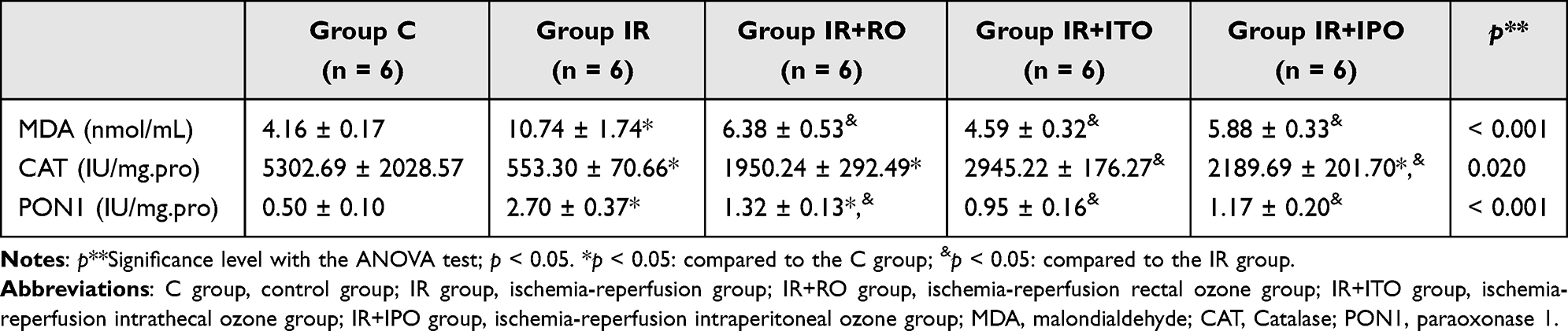 |
Table 2 Rat Testis Tissue Oxidant Status Parameters [Mean ± SE] |
Catalase enzyme activities were lower in the IR, IR+RO and IR+IPO groups than in the C group (p <0.001, p=0.017 and p = 0.025, respectively), and higher in the IR+ITO and IR+IPO groups than in the IR group (p = 0.002 and p = 0.04, respectively) (Table 2).
PON1 enzyme activities were significantly higher in the IR and IR+RO groups than in the C group (p <0.001, p=0.014, respectively) and significantly lower in the IR+RO, IR+ITO, and IR+IPO groups than in the IR group (p <0.001, all groups) (Table 2).
Histopathological Results
Cosentino score was found to be significantly different between the groups (p<0.001). Cosentino score was found to be significantly higher in the IR group compared to the C group (p<0.001), and significantly lower in the IR+RO, IR+ITO and IR+IPO groups compared to the IR group (p<0.001, all), (Table 3, Figures 2–6).
 |
Table 3 Histopathological Findings of Testicular Tissue [Mean ± SE] |
 |
Figure 2 Normal tetstis tissue, C group, H&Ex100, scale bar 50 µm. Abbreviations: S, septum connective tissue; L, Leydig cells; ST, seminiferous tubules; Sp, spermatid; Sc, spermatogenic cells; Sr, Sertoli cells; Sg, spermatogonium; v, vacuolization; →**, spermatozoa. |
 |
Figure 3 Ischemia-reperfusion (IR) group, H&Ex100, scale bar 50 µm. Abbreviations: S, septum connective tissue; L, Leydig cells; ST, seminiferous tubules; Sp, spermatid; Sc, spermatogenic cells; Sr, Sertoli cells; Sg, spermatogonium; inf, inflammation (separation); →**, spermatozoa; ► irregular seminiferous epithelium; conj, congestion; ♦, interstitial edema. |
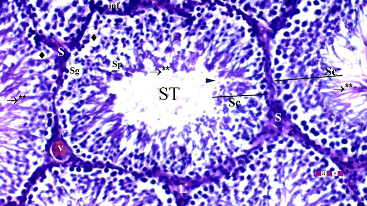 |
Figure 4 Ischemia-reperfusion rectal ozone (IR+RO) group, H&Ex100, scale bar 50 µm. Abbreviations: S, septum connective tissue; L, Leydig cells, ST, seminiferous tubules; Sp, spermatid; Sc, spermatogenic cells; Sg, spermatogonium; inf, inflammation (separation); →**, spermatozoa; ► irregular seminiferous epithelium; v, vacuolization (hemorrhagic); ♦, interstitial edema. |
 |
Figure 5 Ischemia-reperfusion intrathecal ozone (IR+ITO) group, H&Ex100, scale bar 50 µm. Abbreviations: S, septum connective tissue; L, Leydig cells; ST, seminiferous tubules; Sc, spermatogenic cells; Sg, spermatogonium; inf, inflammation (separation); →**, spermatozoa; ► irregular seminiferous epithelium; v, vacuolization; ♦, interstitial edema. |
 |
Figure 6 Ischemia-reperfusion intraperitoneal ozone (IR+IPO) group, H&Ex100, scale bar 50 µm. Abbreviations: ST, seminiferous tubules; Sp, spermatid; Sc, spermatogenic cells; Sg, spermatogonium; inf, inflammation (separation); →**, spermatozoa; ► irregular seminiferous epithelium; ♦, interstitial edema. |
Degenerated intratubular germinal epithelial cells were found to be significantly different between the groups (p=0.016). Degenerated intratubular germinal epithelial cells were found to be more in the IR group than in the C group (p=0.002), and significantly lower in the IR+ITO and IR+IPO groups than in the IR group (p=0.007, both) (Table 3, Figure 2–6).
Interstitial edema/peritubular congestion was significantly different between the groups (p=0.041). Interstitial edema/peritubular congestion was seen more in the IR group compared to the C group (p=0.005). Interstitial edema/peritubular congestion was significantly lower in the IR+ITO, and IR+IPO groups compared to the IR group (p=0.016, both), (Table 3, Figures 2–6).
Vacuolation of tubular epithelium of spermatogonia and Sertoli cells were found to be significantly different between the groups (p=0.015). Vacuolation of tubular epithelium of spermatogonia and Sertoli cells were seen more in the IR group compared to the C group (p=0.003). Vacuolation of tubular epithelium of spermatogonia and Sertoli cells were found to be significantly lower in the IR+RO, IR+ITO and IR+IPO groups compared to the IR group (p=0.011, p=0.03, p=0.037, respectively), (Table 3, Figure 2–6).
Buckled basement membrane thickening (deformed-malformed) was significantly different between the groups (p=0.027). Buckled basement membrane thickening (deformed-malformed) was seen more in the IR group compared to the C group (p=0.002). Buckled basement membrane thickening (deformed-malformed) was significantly lower in the IR+ITO and IR+IPO groups compared to the IR group (p=0.020, both), (Table 3, Figures 2–6). Mononuclear cell infiltrations (MNL inflammation) found to be similar between groups (p=0.242), (Table 3, Figures 2–6).
Discussion
Spinal cord IR injury is a serious condition that can occur as a result of various clinical conditions (eg aortic surgeries, spinal traumas, vascular pathologies) and can lead to irreversible damage to nerve cells and systemic inflammatory response.20–22 However, the effects of spinal cord IR injury on peripheral organs have been relatively less investigated. The findings obtained in this study indicate that spinal cord IR injury causes inflammation, oxidative stress, and structural changes in testicular tissue. This study evaluates the histopathological and biochemical effects of spinal cord ischemia-reperfusion (IR) injury on testicular tissue and the potential of ozone therapy to attenuate these effects. Our results show that spinal cord IR injury causes significant tissue damage and oxidative stress-related changes in testicular tissue, and that ozone therapy significantly attenuates these changes.
According to the Cosentino score, degenerative intratubular germinal epithelial cells, interstitial edema/peritubular congestion, tubular epithelial vacuolization, and basement membrane thickening were significantly increased in the IR group. These changes indicate that the testicular tissue has suffered severe structural damage and the microenvironment suitable for spermatogenesis has been disrupted. In particular, the disruption of the blood-testis barrier may increase the intratesticular inflammatory response and cause the loss of germ cells.
There are several possible mechanisms for the apparent IR damage in testicular tissue:
- Effect on Neuronal Connections: The spinal cord regulates testicular functions via sympathetic and parasympathetic nerves. Disruption of neuronal transmission after IR damage may lead to changes in testicular blood flow and disruptions in spermatogenesis.23 In particular, the hypothalamus-pituitary-gonadal (HPG) axis may be affected, leading to deterioration in testicular functions.24
- Increase in Systemic Inflammatory Response: Increase in the release of proinflammatory cytokines (TNF-α, IL-6, IL-1β) after IR damage may trigger inflammation in the testicles.25 Although mononuclear cell infiltration (MNL) did not show a significant difference in our study, the effects of inflammatory cytokines overlap with histopathological damage.
- Increase in Oxidative Stress: The significant increase in MDA levels in the IR group indicates that lipid peroxidation and oxidative stress in the testicular tissue increased. The significant decrease in catalase (CAT) enzyme activity, an important component of the antioxidant system, in the IR group suggests that intracellular defense mechanisms are inadequate and free radicals accumulate in the testicular tissue and cause damage.
These findings indicate that spinal cord IR injury may negatively affect testicular functions and that this effect may occur through mechanisms such as inflammation, oxidative stress and disruption of neural transmission.
In our study, it was shown that ozone therapy applied by different routes (rectal, intrathecal, intraperitoneal) significantly reduced the negative effects of spinal cord IR injury on the testis. Degenerative germinal cell ratio, tubular vacuolization and interstitial edema levels were significantly reduced compared to the IR group.
Ozone treatment reduced lipid peroxidation by decreasing MDA levels and supported cellular antioxidant defense by increasing CAT activity. These protective effects of ozone therapy can be explained by several mechanisms. One of these is the reduction of oxidative stress; when applied at low doses, ozone can strengthen cellular defense by increasing the expression of antioxidant enzymes (SOD, CAT, GSH-Px).4 In our study, we found that CAT activity increased significantly in the IR+ITO and IR+IPO groups compared to the IR group.
Ozone therapy can suppress the inflammatory response by inhibiting NF-κB pathways and increasing anti-inflammatory cytokines (IL-10).26 Rectal and intrathecal ozone applications are thought to alleviate inflammation in testicular tissue.
Ozone can increase oxygen delivery to tissues by increasing erythrocyte deformability and improving endothelial function.27 Improvement of testicular microcirculation can increase the resistance of cells to IR damage by increasing their oxygenation.28
Considering these mechanisms, it is seen that ozone therapy has the strongest effect especially through intrathecal29 and intraperitoneal application. The greater effectiveness of intrathecal application may be related to its local anti-inflammatory and antioxidant effects on the spinal cord.
Clinical Significance and Future Studies: This study shows that spinal cord IR damage may have significant effects on testicular functions. It may be important to evaluate testicular functions especially in cases of infertility developing after spinal cord surgeries, aortic aneurysm surgeries and spinal traumas.
Although ozone therapy has the potential to be a protective agent for testicular functions with its antioxidant and anti-inflammatory effects, some issues need to be clarified before clinical applications:
- Most effective Ozone dose and administration route: High-dose ozone may have a pro-oxidant effect. The effectiveness and safety of intrathecal ozone application should be investigated in further studies.
- Effects on long-term testicular functions: The long-term effects of ozone therapy on sperm parameters and fertility should be evaluated.
- Human studies: Data obtained in animal models should be extrapolated to human studies.
Conclusion
This study shows that spinal cord IR injury causes significant histopathological and biochemical changes in testicular tissue and that ozone therapy significantly attenuates these changes. Ozone therapy can be considered as a potential therapeutic agent for the prevention of spinal cord-induced testicular damage. However, further studies are needed to determine the most appropriate dose, duration and method of administration.
Data Sharing Statement
The data sets used and/or analyzed during the current study are available from the corresponding author on reasonable request.
Ethics Approval and Consent to Participate
Ethical approval for the study was obtained from Animal Research Committee of Gazi University (Ankara, Turkey; approval no. G.Ü.ET-25-016).
Author Contributions
All authors made a significant contribution to the work reported, whether that is in the conception, study design, execution, acquisition of data, analysis and interpretation, or in all these areas; took part in drafting, revising or critically reviewing the article; gave final approval of the version to be published; have agreed on the journal to which the article has been submitted; and agree to be accountable for all aspects of the work.
Disclosure
The authors declare that they have no competing interests.
References
1. Wan IY, Angelini GD, Bryan AJ, Ryder I, Wan Underwood MJ, Innes YP. Prevention of spinal cord ischaemia during descending thoracic and thoracoabdominal aortic surgery. Eur J Cardiothorac Surg. 2001;19(2):203–213. doi:10.1016/S1010-7940(00)00646-1
2. Ulus AT, Yavas S, Sapmaz A, et al. Effect of conditioning on visceral organs during indirect ischemia/reperfusion injury. Ann Vasc Surg. 2014;28(2):437–444.3. doi:10.1016/j.avsg.2013.06.027
3. Ge L, Wei LH, Du CQ, et al. Hydrogen-rich saline attenuates spinal cord hemisection-induced testicular injury in rats. Oncotarget. 2017;8(26):42314. doi:10.18632/oncotarget.15876
4. Sagai M, Bocci V. Mechanisms of action involved in ozone therapy: is healing induced via a mild oxidative stress? Med Gas Res. 2011;1:1–18. doi:10.1186/2045-9912-1-29
5. Viebahn-Haensler R, León Fernández OS. Ozone in medicine. The low-dose ozone concept and its basic biochemical mechanisms of action in chronic inflammatory diseases. Int J Mol Sci. 2021;22(15):7890. doi:10.3390/ijms22157890
6. Bocci V, Zanardi I, Travagli V. Potentiality of oxygen-ozonetherapy to improve the health of aging people. Current Aging Sci. 2010;3(3):177–187. doi:10.2174/1874609811003030177
7. Bocci V. Scientific and medical aspects of ozone therapy. State of the art. Archiv Med Res. 2006;37:425–435. doi:10.1016/j.arcmed.2005.08.006
8. Akman T, Aras AB, Şehitoğlu MH, et al. The ameliorative effect of ozone therapy on spinal cord ischemia in rabbits. Ann Clin Anal Med. 2020;11(3):221–226.
9. Mete F, Tarhan H, Celik O, et al. Comparison of intraperitoneal and intratesticular ozone therapy for the treatment of testicular ischemia-reperfusion injury in rats. Asian J Androl. 2017;19(1):43–46. doi:10.4103/1008-682X.171570
10. Şengel N, Köksal Z, A.d D, et al. Effects of dexmedetomidine administered through different routes on kidney tissue in rats with spinal cord ischaemia-reperfusion injury. Drug Des Devel Ther. 2022;16:2229–2239. doi:10.2147/DDDT.S361618
11. Zhang P, Zhang Q, Liu X, et al. Tanshinone protects against spinal cord ischemia-reperfusion injury by inhibiting JNK activity. Comput Intell Neurosci. 2022;2022:7619797. doi:10.1155/2022/7619797
12. Wang L, Chen H, Liu X-H, et al. Ozone oxidative preconditioning inhibits renal fibrosis induced by ischemia and reperfusion injury in rats. Exp Ther Med. 2014;8(6):1764–1768. doi:10.3892/etm.2014.2004
13. Wang Z, Han Q, Guo Y-L, X-H X-HL, Qiu T. Effect of ozone oxidative preconditioning on inflammation and oxidative stress injury in rat model of renal transplantation. Acta Cir Bras. 2018;33(3):238–249. doi:10.1590/s0102-865020180030000006
14. T.l Y, Hobo S, Peters C, et al. Preclinical toxicity screening of intrathecal oxytocin in rats and dogs. Anesthesiology. 2014;120(4):951–961. doi:10.1097/ALN.0000000000000148
15. Van Ye TM, Roza AM, Pieper GM, Henderson J, Johnson CP, Adams MB. Inhibition of intestinal lipid peroxidation does not minimize morphologic damage. J Surg Res. 1993;55:553–558. doi:10.1006/jsre.1993.11832
16. Aebi H. Catalase. Method Enzym Anal Elsev. 1974;1974:673–684.
17. Brites FD, Verona J, Schreier LE, Fruchart JC, Castro GR, Wikinski RL. Paraoxonase 1 and platelet-activating factor acetylhydrolase activities in patients with low hdl-cholesterol levels with or without primary hypertriglyceridemia. Arch Med Res. 2004;35:235–240. doi:10.1016/j.arcmed.2004.02.002
18. Lowry O, Rosebrough N, Farr AL, Randall R. Protein measurement with the Folin phenol reagent. J Biol Chem. 1951;193:265–275. doi:10.1016/S0021-9258(19)52451-6
19. Cosentino MJ, Nishida M, Rabinowitz R, Cockett AT. Histopathology of prepubertal rat testes subjected to various durations of spermatic cord torsion. J Androl. 1986;7:23–31. doi:10.1002/j.1939-4640.1986.tb00862.x
20. Simon F, Oberhuber A. Ischemia and reperfusion injury of the spinal cord: experimental strategies to examine postischemic paraplegia. Neural Regen Res. 2016;11(3):414–415. doi:10.4103/1673-5374.179050
21. Fan YD, Zhu ML, Geng D, Zhou K, Du GJ, Wang ZL. The study on pathological mechanism and solution method for spinal cord ischemia reperfusion injury. Eur Rev Med Pharmacol Sci. 2018;22(13):4063–4068. doi:10.26355/eurrev_201807_15394
22. Tokmak M, Yuksel Y, Sehitoglu MH, et al. The neuroprotective effect of syringic acid on spinal cord ischemia/reperfusion injury in rats. Inflammation. 2015;38:1969–1978. doi:10.1007/s10753-015-0177-2
23. Gürer B, Kertmen H, Kasim E, et al. Neuroprotective effects of testosterone on ischemia/reperfusion injury of the rabbit spinal cord. Injury. 2015;46(2):240–248. doi:10.1016/j.injury.2014.11.002
24. Dalmasso C, Loria AS. Neuroendocrine Control of the Vascular System. Cardiovascular Neuroendocrinology. Cham: Springer International Publishing; 2023:43–81.
25. Unsal V, Kolukcu E, Gevrek F, Firat F. Sinapic acid reduces ischemia/reperfusion injury due to testicular torsion/detorsion in rats. Andrologia. 2021;53(8):e14117. doi:10.1111/and.14117
26. Zeng J, Lei L, Zeng Q, et al. Ozone therapy attenuates NF-κB-mediated local inflammatory response and activation of Th17 cells in treatment for psoriasis. Int J Bio Sci. 2020;16(11):1833. doi:10.7150/ijbs.41940
27. Akbudak IH, Kucukatay V, Kilic-Erkek O, Ozdemir Y, Bor-Kucukatay M. Investigation of the effects of major ozone autohemotherapy application on erythrocyte deformability and aggregation. Clin Hemorheol Microcirc. 2019;71(3):365–372. doi:10.3233/CH-180417
28. Colli LG, Belardin LB, Echem C, et al. Systemic arterial hypertension leads to decreased semen quality and alterations in the testicular microcirculation in rats. Sci Rep. 2019;9(1):11047. doi:10.1038/s41598-019-47157-w
29. Wang J, Wu M, Lin X, Li Y, Fu Z. Low-concentration oxygen/ozone treatment attenuated radiculitis and mechanical allodynia via PDE2A-cAMP/cGMP-NF-κB/p65 signaling in chronic radiculitis rats. Pain Res Manag. 2018;13:5192814.
 © 2025 The Author(s). This work is published and licensed by Dove Medical Press Limited. The
full terms of this license are available at https://www.dovepress.com/terms
and incorporate the Creative Commons Attribution
- Non Commercial (unported, 4.0) License.
By accessing the work you hereby accept the Terms. Non-commercial uses of the work are permitted
without any further permission from Dove Medical Press Limited, provided the work is properly
attributed. For permission for commercial use of this work, please see paragraphs 4.2 and 5 of our Terms.
© 2025 The Author(s). This work is published and licensed by Dove Medical Press Limited. The
full terms of this license are available at https://www.dovepress.com/terms
and incorporate the Creative Commons Attribution
- Non Commercial (unported, 4.0) License.
By accessing the work you hereby accept the Terms. Non-commercial uses of the work are permitted
without any further permission from Dove Medical Press Limited, provided the work is properly
attributed. For permission for commercial use of this work, please see paragraphs 4.2 and 5 of our Terms.