Back to Journals » Diabetes, Metabolic Syndrome and Obesity » Volume 13
Effect of Rubus anatolicus Leaf Extract on Glucose Metabolism in HepG2, CRI-D2 and C2C12 Cell Lines
Authors Safarzad M, Marjani A, Saghaeian Jazi M, Qujeq D ![]() , Mir SM, Marjani M, Nezhadebrahimi Kaldehi A
, Mir SM, Marjani M, Nezhadebrahimi Kaldehi A
Received 5 January 2020
Accepted for publication 1 April 2020
Published 14 April 2020 Volume 2020:13 Pages 1109—1116
DOI https://doi.org/10.2147/DMSO.S244850
Checked for plagiarism Yes
Review by Single anonymous peer review
Peer reviewer comments 2
Editor who approved publication: Prof. Dr. Juei-Tang Cheng
Mahdieh Safarzad,1 Abdoljalal Marjani,1 Marie Saghaeian Jazi,1 Durdi Qujeq,2 Seyed Mostafa Mir,2 Majid Marjani,3 Abbas Nezhadebrahimi Kaldehi1
1Metabolic Disorders Research Center, Department of Biochemistry and Biophysics, Gorgan Faculty of Medicine, Golestan University Medical Sciences, Gorgan, Iran; 2Cellular and Molecular Biology Research Center, Health Research Institute, Department of Clinical Biochemistry, Student Research Committee, Babol University of Medical Science, Babol, Iran; 3Faculty of Pharmacy, Eastern Mediterranean University, Famagusta 99628, Turkey
Correspondence: Abdoljalal Marjani
Metabolic Disorders Research Center, Department of Biochemistry and Biophysics, Faculty of Medicine, Golestan University of Medical Sciences, Gorgan, Golestan Province 4934174515, Iran
Tel +98 171 4421651
Fax +98 171 4440225
Email [email protected]
Introduction: The aim of this study was to assess the effects of Rubus anatolicus on glucose metabolism in HepG2, CRI-D2 and C2C12 cell lines.
Materials and Methods: R. anatolicus was collected in Golestan province, Iran. Three different cell lines HepG2 (human liver cell), CRI-D2 (mice pancreatic cell) and C2C12 (rat myoblast) were used for cell culture experiments. Cell viability was measured using MTT assay. Cells were treated with various concentrations of the extract (6.25– 400 μg/mL) and then the extracellular glucose level and intracellular glycogen content were measured using colorimetric methods. The insulin level of the culture medium was measured using the ELISA method.
Results: Our findings showed that R. anatolicus extract enhances glucose uptake and consumption by all three cell lines. The R. anatolicus extract exposure also elevated cellular glycogen content in HepG2 and C2C12 cells (for 200 and 100 μg/mL) significantly. We found a significant increase in glucose uptake and consequently higher stimulation of insulin secretion in CRI-D2 cell pancreatic cells treated with R. anatolicus extract.
Conclusion: The R. anatolicus appears to activate glucose uptake and cellular glycogen synthesis probably by activating the glycogenesis or inhibition of glycogenolysis pathways. The extract enhances insulin secretion in the pancreatic cells by increased glucose uptake.
Keywords: Rubus anatolicus, glucose metabolism, HepG2, CRI-D2, C2C12, cell lines
Introduction
Diabetes mellitus is a public health problem worldwide affecting near half a billion people life, and it has been estimated that the number of people suffering from diabetes will reach 700 million by 2045.1,2
Type 2 Diabetes Mellitus (T2DM) is a serious and one of the most prevalent metabolic disorders. T2DM is characterized by the inability to control blood glucose due to insulin resistance, inadequate insulin secretion, and excessive or inappropriate glucagon secretion. T2DM increases the risk of serious complications such as hyperglycemia, hypertension, dyslipidemia and obesity.3–5 The liver contributes to blood glucose control, storage and utilization. It also provides glucose to both insulin insensitive and insulin-sensitive tissues; in other words, liver plays an important role in glucose homeostasis.6
Phytotherapy is one way of protecting or preventing metabolic disease. The plants with phytotherapy capability are usually rich in Phytochemicals such as terpenoids (iridoids, mono- and sesquiterpenes, saponins), phenolic compounds (phenylpropanoids, flavonoids, catechins, rosmarinic acid, polyketides, tannins) and polysaccharides.7–9
Phytochemicals that stimulate glucose uptake in the liver might play an important role in the amelioration of diabetes. Discovery of the new hypoglycemic phytochemicals with few or no side effects is important to improve the quality of life for patients with T2DM.10
Studies show that natural products, fruit bioactive and polyphenolic compounds could prevent and manage chronic metabolic conditions, such as metabolic syndrome, type 2 diabetes with little or no side-effects.11,12 So it seems that identification and evaluation of the medicinal plants with antioxidant and antidiabetic properties can be used to expand new therapeutic arsenal against diabetes disease.
Accordingly, some fruit and plant which contains polyphenolic compounds such as Rubus may be candidate as hypoglycemic agent.
Rubus anatolicus or Elm-leaf blackberry (Focke, ie, Focke ex Hausskn) is a perennial shrub, belonging to the Rosaceae family and Rosoiedeae subfamily with more than 740 species around the world, and are classified in 12 or 15 subgenera.13–15 Eight shrub species of the Rubus have been recognized in forest and non-forest areas of Iran, which R. anatolicus is one of them. Western Asia, northwest of Himalaya, Kashmir, Chitral, Pakistan, Afghanistan, Turkmenistan, Caucasus and the Balkan Peninsula are the habitats of this plant.16,17
R. anatolicus is a wild plant that grows in abundance in Golestan province, Northeast of Iran. R. anatolicus leaves and roots have many traditional uses. The leaves of this plant are used for treating certain health conditions such as diarrhea, bloody mucus, hematuria, diabetes, anemia, dysphonia, cutaneous conditions, and gynecological disorders, also it is used as a strong anti-inflammatory, antiseptic and against oral or ophthalmic infections.18–20
This plant contains antioxidants, anthocyanins, vitamins, minerals, folic acid, tannin flavonol, glycosides, and ellagitannins. The antioxidant effect of R. anatolicus leaves is due to high phenolic compounds, which have antioxidant and free radical scavenging activities.21–24
Free radicals and oxidative stress that results from overproduction of reactive oxygen species (ROS) are closely associated with a number of disease states such as hyperglycemia and metabolic disorders including diabetes mellitus.25 Therefore, it is reasonable that dietary antioxidants, especially polyphenolic compounds that present in wild plants can neutralize free radicals and oxidative stress.26,27
As previous studies have reported, phenolic-enriched extracts from food or plant could regulate glucose balance in the body by regulating glucose hemostasis enzymes such as AMPK/Akt/GSK-3β.27–29
It also suggested that Blackberry phenolic compounds have the potential to control postprandial glucose levels by delaying glucose absorption, because this compound is effective in inhibiting alpha-amylase and alpha-glucosidase activities in vitro. These are the key enzymes that catalyze the final step in the digestive process of carbohydrates in mammals.30,31 In addition, some studies have shown that blackberry extract showed high stimulation of glucose uptake in the HepG2 cell line.25,32
By considering this fact that medical plant contains phenolic compounds which can affect or regulate glucose homeostasis, we assess effects of Rubus Anatolicus leave extract on glucose metabolism in HepG2 (human liver hepatocellular carcinoma), CRI-D2 (an insulinoma cell line from rat) and C2C12 (Mus musculus myoblast) cell lines.
Materials and Methods
Chemicals and Reagents
RPMI-1640 and Dulbecco’s modified Eagle’s medium (DMEM), fetal bovine serum (FBS), Trypsin-EDTA and MTT (2, 3-bis-(2-methoxy-4-nitro-5-sulfophenyl)-2H-tetrazolium-5-carboxanilide) were purchased from Bio-Idea Company (Tehran. Iran). BSA was purchased from Solarbio (Life Sciences). Penicillin–streptomycin was purchased from Gibco Life Technologies (Paisley, UK). Glucose oxidase was purchased from Pars Azmon Company (Tehran, Iran). The insulin radioimmunoassay kit was purchased from Mercodia. The protein assay reagent was obtained from Bio-Rad (Copenhagen, Denmark). The chemicals including sulfuric acid, ethanol, Na2So4, KOH, all were purchased from Merk (Germany).
Preparation of R. anatolicus Extract
R. anatolicus leaf was locally collected in Golestan province (North East of Iran), on September and October 2018 from various localities throughout Golestan province, and was rinsed with water and then were dried under the sunlight for 72 h. The dried leaf was ground to powder using an electric blender and then was sieved. The R. anatolicus leaf powder was kept in dark condition prior to extraction. For extraction, 20 g of the R. anatolicus leaf powder was added to 200 mL of methanol (80%) for 48 h on a shaker. The mixture was filtered and the extract was evaporated to dryness under reduced pressure at 40°C by a rotary evaporator, and then, the concentrated extract was dried in a freeze dryer.33,34 The R. anatolicus powder was dissolved in DMSO 4% and stored at −20°C until use.
Cell Lines and Cell Culture
For testing the antidiabetic properties of the R. anatolicus extract, we used the HepG2 (human liver hepatocellular carcinoma), CRI-D2 (an insulinoma cell line originated from rat) and C2C12 (Mus musculus myoblast) cell lines that all were provided from Pasteur Institute of Iran. These cells were cultured in DMEM (for CRI-D2, C2C12) or RPMI-16 (for HepG2) supplemented with 10% fetal bovine serum, 100U/mL penicillin, 100 lg/mL streptomycin. All cell cultures were maintained at 37◦C in a humidified atmosphere of 5% CO2.
Cytotoxicity Tests
For the cell viability evaluation, HepG2, C2C12 and CRI-D2 cells were seeded at 1 × 10⁴ cells/mL in 96-well culture plates and incubated for 24 h at 37°C in a humidified atmosphere containing 5% CO2. R. anatolicus extract was added at 6.25–400 μg/mL and incubated for a further 24 h. MTT assay was also used as a cell viability test. MTT (working solution was 5 mg/mL in PBS) was added to the cells and incubated for 4 h at 37°C in a humidified atmosphere containing 5% CO2; cells were then solubilized in 100% DMSO for 5 min. The absorbance was measured using ChroMate® micro-plate reader at 570 nm.
Glucose Consumption
HepG2 cell line was grown in RPMI 1640; CRI-D2 and C2C12 were grown in DMEM/F12, containing 10% fetal bovine serum. The glucose consumption assay was performed according to Liu et al36. Briefly; cells were seeded in 96-well plates with eight wells left as blanks. After cell lines reached about 80% to 90% confluence, cells were washed twice with PBS. The mediums were replaced by DMEM/RPMI 1640 supplemented with 0.2% BSA and R. anatolicus extract at various concentrations (400, 200, 100, 50, 25, 12.5, 6.2 µg/mL) and DMSO was the solvent control. Finally, after 24 h, the medium was removed and its glucose concentrations were determined by the glucose oxidase method. The amount of cell glucose consumption (GC) was calculated from the glucose concentrations of the cell containing wells subtracted from the cell-free blank wells. Briefly, 10 µL of the medium in every well was mixed with 1000 µL glucose oxidase/peroxidase reagent from the kit. After being reacted for 10 min at 37◦c, the absorbance of the mixture was measured at 546 nm by the Jenway 6105 UV/VIS spectrophotometer.
Insulin Release
The CRI-D2 cells were seeded at a density of 106 cells/well in a 6-well plate in DMEM supplemented with 10% fetal bovine serum (FBS). On day 3 or 4, the medium was replaced with the new one containing R. anatolicus extract at various concentrations (400, 200, 100, 50 and 25µg/mL) and DMSO as solvent control. After 24-h incubation, the medium was collected and the insulin concentrations were determined by Mercodia Rat Insulin ELISA radioimmunoassay (Mercodia AB, Sweden), a solid two-phase immunoassay. It is based on direct sandwich technique, in which two monoclonal antibodies are directed against separate antigenic determinants on the insulin molecule. All the insulin secretion primary outcomes were normalized to total cell protein content by the Bradford reagent. The value obtained was indicated as µg of insulin per milligram of protein.
Glycogen Measurement
HepG2 and C2C12 cells were cultured in 6-well plate (5×10³ and 3 × 10⁵ cells/mL, respectively), When cells reached confluence, the mediums were replaced by DMEM/RPMI 1640 supplemented with R. anatolicus extract at various concentrations (200, 100, 50, 25 µg/mL and DMSO as solvent control). After 24-h incubation, cells harvested for glycogen and total protein measurement. In order to prevent spontaneous glycogenolysis, the cells were thawed at the moment of glycogen extraction. Glycogen was extracted from the cells and measured according to M. Rousset et al method.35 The relative levels of glycogen content of HepG2 and C2C12 cells were normalized to the protein content by the Bradford reagent. The value obtained was indicated as µg of glycogen per milligram of protein.
Statistical Analysis
Statistical analysis was performed via GraphPad Prism 6 software (GraphPad Software, Inc., La Jolla, CA). One-way analysis of variance (ANOVA) was performed to determine the significant differences between study groups. Significant differences are shown as *p<0.05, **p<0.01, ***p<0.001 and ****p<0.0001.
Results
Cell Viability Test MTT Assay
To determine the cytotoxicity of R. anatolicus extract at various concentrations, HepG2,CRI-D2 and C2C12 cells were incubated with different concentrations of R. anatolicus leaf extract (from 0 to 400 μg/mL) for 24 h. MTT assay was performed to test for cell viability. As it is shown in Figure 1, the leaf extract of R. anatolicus at all concentrations from 0 to 400 μg/mL not only had a cytotoxic effect on cell viability but also increased cell growth as compared to control (0.1% DMSO). For the next analysis, R. anatolicus extract at concentrations of 200 to 6.25 μg/mL was selected for glucose consumption assays.
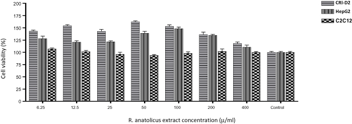 |
Figure 1 Effects of increasing concentrations of R. anatolicus extract on HepG2, CRI-D2 and C2C12 cell lines (6.25–400 mg/mL and control). MTT OD was measured after 24-h incubation with different concentrations of R. anatolicus extract. |
Effects of R.anatolicus Extract on Glucose Consumption and Glycogen Content in HepG2 and C2C12 Cell Lines
In the present study, R. anatolicus leaf extract was investigated for its role in the stimulation of glucose uptake in mice skeletal muscle cells (C2C12) and human hepatic cell (Hep G2). The extracellular glucose concentrations of HepG2 and C2C12 cell lines were measured after 24 h treating with 400, 200, 100, 50, 25, 12.5, 6.25 µg/mL of R. anatolicus leaf extract and 0.1% DMSO as control (Figure 2). As illustrated in Figure 2A and C, R. anatolicus extract exhibited a significant effect on glucose consumption in two cell lines. In order to understand the reason for the extracellular glucose consumption in HepG2 and C2C12 cells, we measured the glycogen content of these cells after R. anatolicus extract treatment. In relation to the control, the glycogen content in Hep-G2 cells was significantly increased for the 200, 100, 50 and 25 μg/mL (Figure 2B). In C2C12 cells, we saw a significant increase in glycogen content just for 200 μg/mL, and 100 μg/mL had no effect on C2C12 cell glycogen content. On the other hand, the glycogen content was significantly decreased for the 50 and 25 μg/mL (Figure 2D).
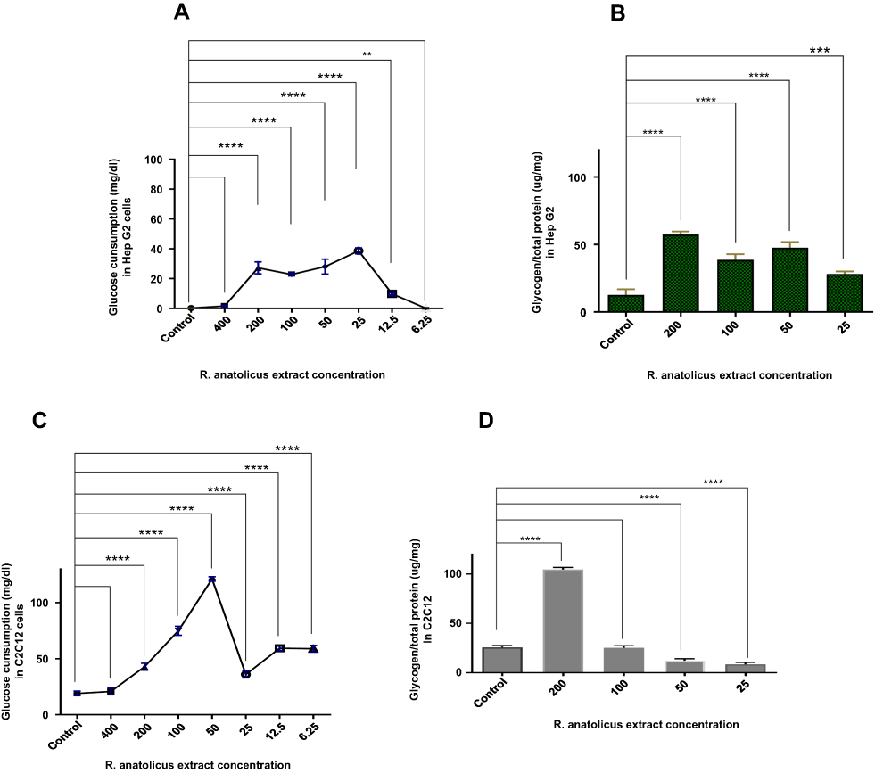 |
Figure 2 Effect of R. anatolicus extract on extracellular glucose consumption and cellular Glycogen content. (A) and (B) show the Effects of R. anatolicus extract on glucose consumption and glycogen content in HepG2 cells, respectively. And Figure 2C and 2D show the effects of R. anatolicus extract on glucose consumption and glycogen content in C2C12 cells, respectively. GCs were measured after 24-h incubation with 400–6.25 µg/mL R. anatolicus extract or control and glycogen content were measured after 24-h incubation with 200–25 µg/mL R. anatolicus extract or control Data are means ± SEM. **p<0.01, ***p<0.001 and ****p<0.0001. |
Effects of R.anatolicus Extract on Glucose Consumption and Insulin Secretion in CRI-D2 Cells
In the present study, we also investigated the effect of the R. anatolicus extract on glucose consumption and insulin secretion in the CRI-D2 cell line. The extracellular glucose concentrations of CRI-D2 cells were measured after 24 h treating with 400, 200, 100, 50, 25, 12.5 and 6.25 µg/mL of R. anatolicus extract and 0.1% DMSO as control (Figure 3A). As it is shown in Figure 3A, all concentrations of R. anatolicus extract (except 12.5 µg/mL) showed a significant effect on glucose consumption in the CRI-D2 cell. In order to understand the reason for extracellular glucose consumption in the CRI-D2 cell line, the level of insulin secretion from these cells was measured 24 h after R. anatolicus extract treatment (Figure 3B). As shown in 3B, among the different concentrations of R. anatolicus extract (200, 100, 50, 25 µg/mL), the maximum insulin secretion was observed in 200, 50 and 25 μg/mL, respectively. But insulin secretion was not significantly increased in 400 and 100 μg/mL.
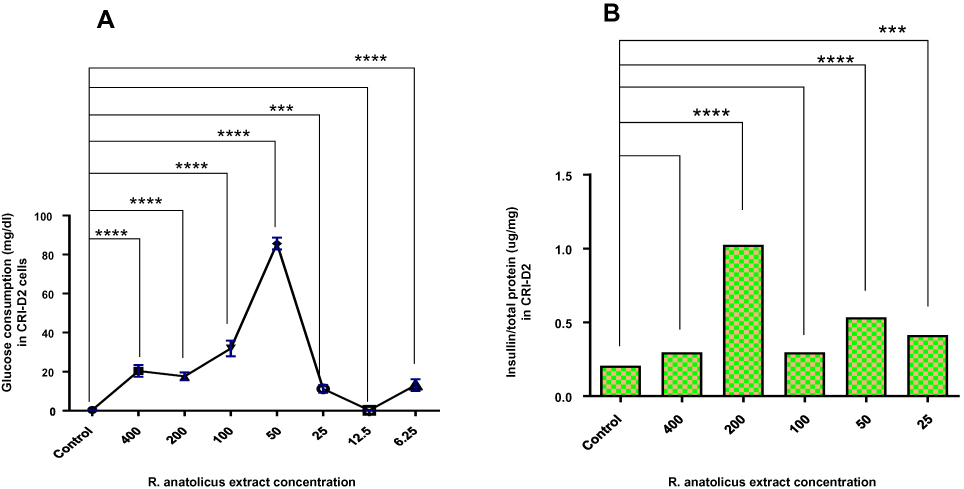 |
Figure 3 Effect of R. anatolicus extract on glucose consumption and insulin secretion in CRI-D2 cells. GC (A) and insulin secretion (B) were measured after 24-h incubation with 400–6.25 µg/mL R. anatolicus extract or control. Data are means ± SEM. p<0.05, p<0.01, ***p<0.001 and ****p<0.0001. |
Discussion
It has been reported that phytochemicals such as phenolic compounds attracted attention for developing new antidiabetic drugs. By considering this fact that phytochemicals (esp. polyphenols and flavonoid) have antidiabetic potential, so identifying and evaluating medicinal plants by antioxidant and antidiabetic properties can be used for expanding new therapeutic against diabetes disease.
As Rubus species contain polyphenolic compounds,22,24 in this study we investigate the effect of R.anatolicus leaf extract on glucose metabolism.
Our results demonstrated R. anatolicus leaf extract stimulates glucose consumption in HepG2, C2C12 and CRI-D2 cell lines (Figure 2A, 2C and 3A), with the maximum effect in C2C12 cells. This indicates that R. anatolicus extract stimulates glucose uptake and utilization in studied cells from different origins of muscle, hepatic and pancreatic tissues.
One of the possible mechanisms for enhanced glucose consumption by the cells in the presence of R. anatolicus extract could be the high content of phenolic compounds (such as phenolic acids, flavonols, flavanols, anthocyanins, proanthocyanidins and ellagitannins) in this plant. High concentrations of phenolic content resulted in increased uptake and consumption of glucose by the cells32,36 It has been shown that high phenolic component in R. anatolicus could promote AMPK and Akt phosphorylation. Activated AMPK (p-AMPK) via an insulin-independent mechanism, regulate glucose uptake in cells37,38 and suppress the hepatic glucose production by PEPCK and G6Pase modulation.39 Furthermore, AKT suppresses gluconeogenesis in the hepatocyte by deactivating the expression of phosphoenolpyruvate carboxykinase (PEPCK) and glucose-6-phosphatase (G6Pase).40
Flavonoid is another phenolic compound in this plant which can regulate glucose level in cells mainly by increasing GLUT-2 expression in pancreatic β cells and flavonoid also can upregulate and promote translocation of GLUT-4 via PI3K/AKT, CAP/Cb1/TC10 and AMPK pathways.41
As it is shown in Figure 2B, in HepG2 cells by increasing in R. anatolicus extract concentration, glycogen content approximately increased when compared to the control. We assumed that an increase in glucose consumption could lead to an increase in glycogen synthesis. As R. anatolicus extract enhanced glucose uptake and glycogen synthesis in HepG2 and C2C12 cell, it seems that R. anatolicus extract may display insulin-like effects in muscle and liver cells.
Some study illustrates that anthocyanins can stimulate the glucose uptake in the liver and skeletal muscle cells by stimulating AMP-activated protein kinase (AMPK).42,43 AMPK upregulates glucose transporter 4 (GLUT 4) in skeletal muscle, suppresses glucose production and increases glycogenesis.44 In addition to the effect of R. anatolicus extract on glucose uptake and glycogenesis in HepG2, C2C12 cells, our result shows this extract stimulates insulin secretion in pancreatic cells (CRI-D2 cell). As shown in Figure 3B, R. anatolicus extract (esp. 200, 100, 50, 25 µg/mL), significantly increased insulin secretion in CRI-D2 cell, it seems that R. anatolicus extract by stimulating glucose uptake in the pancreatic cell (CRI-D2) causes insulin secretion. As a previous study showed, when glucose enters the pancreatic β- cells by GLUT, this glucose, through glycolysis and the Krebs cycle, produces ATP. Elevation of cytoplasmic ATP or ATP/ADP ratio causes closing ATP-sensitive K+ channels, membrane depolarization and opening L-type voltage-dependent Ca2+ channels. The increase in cytosolic free Ca2+ concentration increases the rate of insulin exocytosis.45,46 In a study that was conducted by Jouad et.al, they assay the hypoglycemic effect of Rubus fructicosis L (RFL) in normal and streptozotocin-induced diabetic rats. They did not see any significant changes in plasma insulin concentrations after treatment with the aqueous extract of RFL both in normal and diabetic rats. So they declared that RFL decreases blood glucose levels independent of elevation of insulin secretion, at least for the doses used in their study.47 Furthermore, some research reported that consumption of fruit and vegetable rich in antioxidant, polyphenol and anthocyanins (such as berries, tea) decreased the incidence of type 2 diabetes.28
In Martin de Bock study, their results show that olive leaf polyphenols improved insulin secretion in individuals who received this extract.48 As previously mentioned, Rubus species contain polyphenolic compounds; such as anthocyanins. It has been shown that other phenolic compounds including anthocyanins and anthocyanidins induced insulin secretion from pancreatic β-cells (INS-1832/13 cell).49 One of the reasons that we can categorize R. anatolicus as an antidiabetic herbal remedy is because of its effect on insulin secretion.
Conclusion
In this study, the effects of R. anatolicus leaf extract on glucose, insulin and glycogen levels in different cell lines were evaluated. The results showed that the extract of R. anatolicus leaf in liver, muscle and pancreas cells increased cellular glucose uptake leading to an elevated level of stored glycogen in muscle and liver cells and higher insulin secretion in the pancreas cell line. Our finding suggests that R. anatolicus leaf extract could be a potential candidate phytochemical in diabetes mellitus treatment and protection.
Acknowledgment
The authors would like to thank the Research Deputy of Golestan University of Medical Sciences for financial support. The corresponding author wishes to thank Miss Mahdieh Safarzad for her sincere help.
Ethics and Consent Statement
The ethic committee of the Golestan University of Medical Sciences approved the study (with ethics number: IR.GOUMS.REC.1395.282).
Funding
This work has been supported by the Research Deputy of Golestan University of Medical Science.
Disclosure
The authors declared no conflicts of interest.
References
1. Organization WH. Global Report on Diabetes: Executive Summary. World Health Organization; 2016.
2. Saeedi P, Salpea P, Karuranga S, et al. Mortality attributable to diabetes in 20–79 years old adults, 2019 estimates: results from the international diabetes federation diabetes atlas. Diabetes Res Clin Pract;2020:108086. doi:10.1016/j.diabres.2020.108086
3. Skyler JS. Diabetes mellitus: pathogenesis and treatment strategies. J Med Chem. 2004;47(17):4113–4117. doi:10.1021/jm0306273
4. Drucker DJ, Nauck MA. The incretin system: glucagon-like peptide-1 receptor agonists and dipeptidyl peptidase-4 inhibitors in type 2 diabetes. Lancet. 2006;368(9548):1696–1705. doi:10.1016/S0140-6736(06)69705-5
5. Khardori R. Type 2 diabetes mellitus: practice essentials, background, pathophysiology. Medscape. 2017.
6. Strik BC. Berry crops: worldwide area and production systems. Berry Fruit. 2007;1:3–51.
7. Teng H, Yuan B, Gothai S, Arulselvan P, Song X, Chen L. Dietary triterpenes in the treatment of type 2 diabetes: to date. Trends Food Sci Technol. 2018;72:34–44. doi:10.1016/j.tifs.2017.11.012
8. Kwinana-Mandindi T. Phytochemical and antioxidant composition of selected local wild plants in south africa: consideration of alternative nutrients for health promotion.
9. Wink M. Modes of action of herbal medicines and plant secondary metabolites. Medicines. 2015;2(3):251–286. doi:10.3390/medicines2030251
10. Wild S, Roglic G, Green A, Sicree R, King H. Global prevalence of diabetes: estimates for the year 2000 and projections for 2030. Diabetes Care. 2004;27(5):1047–1053. doi:10.2337/diacare.27.5.1047
11. Jacques PF, Cassidy A, Rogers G, Peterson JJ, Meigs JB, Dwyer JT. Higher dietary flavonol intake is associated with lower incidence of type 2 diabetes. J Nutr. 2013;143(9):1474–1480. doi:10.3945/jn.113.177212
12. Cassidy A, Rogers G, Peterson JJ, Dwyer JT, Lin H, Jacques PF. Higher dietary anthocyanin and flavonol intakes are associated with anti-inflammatory effects in a population of US adults. Am J Clin Nutr. 2015;102(1):172–181. doi:10.3945/ajcn.115.108555
13. Hummer KE. Rubus diversity. HortScience. 1996;31(2):182–183. doi:10.21273/HORTSCI.31.2.182
14. Park JH, Oh S-M, Lim SS, et al. Induction of heme oxygenase-1 mediates the anti-inflammatory effects of the ethanol extract of Rubus coreanus in murine macrophages. Biochem Biophys Res Commun. 2006;351(1):146–152. doi:10.1016/j.bbrc.2006.10.008
15. Dehaan R, Louis J, Wilson A, Hall A, Rumbachs R. Discrimination of blackberry (Rubus fruticosus sp. agg.) using hyperspectral imagery in Kosciuszko National Park,NSW, Australia. ISPRS J Photogramm Remote Sens. 2007;62(1):13–24. doi:10.1016/j.isprsjprs.2007.01.004
16. Mozaffarian V. A Dictionary of Iranian Plant Names: Latin, English, Persian. Farhang Mo’aser; 1996.
17. Akhani H. Flora Iranica: facts and figures and a list of publications by KH Rechinger on Iran and adjacent areas. Rostaniha. 2006;7(Suppl 2):19–61.
18. Ghorbanli M, Livani F, Sateyi A. An analysis of the antioxidant compounds in the alcoholic extractions and the juice of elm-leaf blackberry or Rubus anatolicus (Focke,i.e. Focke ex Hausskn.) during the ripening period. 2012;8(33):16.
19. Ghorbanli ML, Saati A, Livani F. Study of antioxidant activity and components of elm-leaved blackberry (Rubus anatolicus Focke) plant during ripening period. J Invest Appl Med Plants. 2009;2(1):1388. 25–35.
20. Khezry S. Pharmacobotany atlas (The properties of fruits,plants and vegetables). Rostam Khani. 2002.
21. Lee J. Sorbitol, Rubus fruit, and misconception. Food Chem. 2015;166:616–622. doi:10.1016/j.foodchem.2014.06.073
22. Diasamidze M, Vanidze M, Djafaridze I, Qamadadze E, Kalandia A. Phenol compounds of blackberry (Rubus caucasicus Focke and Rubus anatolicus L.) fruit and leaf. J Chem Chem Eng. 2013;7(6):539.
23. Larrosa M, García-Conesa MT, Espín JC, Tomás-Barberán FA. Ellagitannins, ellagic acid and vascular health. Mol Aspects Med. 2010;31(6):513–539. doi:10.1016/j.mam.2010.09.005
24. Oszmiański J, Wojdyło A, Nowicka P, Teleszko M, Cebulak T, Wolanin M. Determination of phenolic compounds and antioxidant activity in leaves from wild Rubus L. species. Molecules. 2015;20(3):4951–4966. doi:10.3390/molecules20034951
25. Kayama Y, Raaz U, Jagger A, et al. Diabetic cardiovascular disease induced by oxidative stress. Int J Mol Sci. 2015;16(10):25234–25263. doi:10.3390/ijms161025234
26. Pizzino G, Irrera N, Cucinotta M, et al. Oxidative stress: harms and benefits for human health. Oxid Med Cell Longev. 2017;2017.
27. Chen L, Lin X, Fan X, Qian Y, Lv Q, Teng H. Sonchus oleraceus Linn extract enhanced glucose homeostasis through the AMPK/Akt/GSK-3β signaling pathway in diabetic liver and HepG2 cell culture. Food Chem Toxicol. 2020;136:111072. doi:10.1016/j.fct.2019.111072
28. Jemai H, El Feki A, Sayadi S. Antidiabetic and antioxidant effects of hydroxytyrosol and oleuropein from olive leaves in alloxan-diabetic rats. J Agric Food Chem. 2009;57(19):8798–8804. doi:10.1021/jf901280r
29. Liu Y, Wang D, Zhang D, et al. Inhibitory effect of blueberry polyphenolic compounds on oleic acid-induced hepatic steatosis in vitro. J Agric Food Chem. 2011;59(22):12254–12263. doi:10.1021/jf203136j
30. McDougall GJ, Shpiro F, Dobson P, Smith P, Blake A, Stewart D. Different polyphenolic components of soft fruits inhibit α-amylase and α-glucosidase. J Agric Food Chem. 2005;53(7):2760–2766. doi:10.1021/jf0489926
31. Cheplick S, Kwon YI, Bhowmik P, Shetty K. Clonal variation in raspberry fruit phenolics and relevance for diabetes and hypertension management. J Food Biochem. 2007;31(5):656–679. doi:10.1111/j.1745-4514.2007.00136.x
32. Ho GTT, Nguyen TKY, Kase ET, Tadesse M, Barsett H, Wangensteen H. Enhanced glucose uptake in human liver cells and inhibition of carbohydrate hydrolyzing enzymes by Nordic berry extracts. Molecules. 2017;22(10):1806. doi:10.3390/molecules22101806
33. Ponti D, Costa A, Zaffaroni N, et al. Isolation and in vitro propagation of tumorigenic breast cancer cells with stem/progenitor cell properties. Cancer Res. 2005;65(13):5506–5511. doi:10.1158/0008-5472.CAN-05-0626
34. Arawande J, Komolafe E. Antioxidative potential of banana and plantain peel extracts on crude palm oil. Ethnobotanical Leafl. 2010;14:559–569.
35. Rousset M, Chevalier G, Rousset J-P, Dussaulx E, Zweibaum A. Presence and cell growth-related variations of glycogen in human colorectal adenocarcinoma cell lines in culture. Cancer Res. 1979;39(2Part 1):531–534.
36. Mursu J, Virtanen JK, Tuomainen TP, Nurmi T, Voutilainen S. Intake of fruit, berries, and vegetables and risk of type 2 diabetes in finnish men: the kuopio ischaemic heart disease risk factor study. Am J Clin Nutr. 2014;99(2):328–333. doi:10.3945/ajcn.113.069641
37. Lee CT, Ussher JR, Mohammad A, Lam A, Lopaschuk GD. 5′-AMP-activated protein kinase increases glucose uptake independent of GLUT4 translocation in cardiac myocytes. Can J Physiol Pharmacol. 2014;92(4):307–314. doi:10.1139/cjpp-2013-0107
38. Chen L, Lin X, Xiao J, Tian Y, Zheng B, Teng H. Sonchus oleraceus Linn protects against LPS-induced sepsis and inhibits inflammatory responses in RAW264.7 cells. J Ethnopharmacol. 2019;236:63–69. doi:10.1016/j.jep.2019.02.039
39. Mihaylova MM, Shaw RJ. The AMPK signalling pathway coordinates cell growth, autophagy and metabolism. Nat Cell Biol. 2011;13(9):1016–1023. doi:10.1038/ncb2329
40. Klover PJ, Mooney RA. Hepatocytes: critical for glucose homeostasis. Int J Biochem Cell Biol. 2004;36(5):753–758. doi:10.1016/j.biocel.2003.10.002
41. Hajiaghaalipour F, Khalilpourfarshbafi M, Arya A. Modulation of glucose transporter protein by dietary flavonoids in type 2 diabetes mellitus. Int J Biol Sci. 2015;11(5):508. doi:10.7150/ijbs.11241
42. Ho GTT, Kase ET, Wangensteen H, Barsett H. Phenolic elderberry extracts, anthocyanins, procyanidins, and metabolites influence glucose and fatty acid uptake in human skeletal muscle cells. J Agric Food Chem. 2017;65(13):2677–2685. doi:10.1021/acs.jafc.6b05582
43. Rojo LE, Ribnicky D, Logendra S, et al. In vitro and in vivo anti-diabetic effects of anthocyanins from Maqui Berry (Aristotelia chilensis). Food Chem. 2012;131(2):387–396. doi:10.1016/j.foodchem.2011.08.066
44. Takikawa M, Inoue S, Horio F, Tsuda T. Dietary anthocyanin-rich bilberry extract ameliorates hyperglycemia and insulin sensitivity via activation of AMP-activated protein kinase in diabetic mice. J Nutr. 2010;140(3):527–533. doi:10.3945/jn.109.118216
45. Komatsu M, Takei M, Ishii H, Sato Y. Glucose-stimulated insulin secretion: a newer perspective. J Diabetes Investig. 2013;4(6):511–516. doi:10.1111/jdi.12094
46. Schuit FC, Huypens P, Heimberg H, Pipeleers DG. Glucose sensing in pancreatic β-cells. Model Study Other Glucose-Regulated Cells Gut Pancreas Hypothalamus. 2001;50(1):1–11.
47. Xie X-M, Pang X-B, LI X-T. Hypoglycemic effect and mechanism of raspberry ketone on diabetic model mice [J]. Chin Pharm J. 2012;23.
48. de Bock M, Derraik JG, Brennan CM, et al. Olive (Olea europaea L.) leaf polyphenols improve insulin sensitivity in middle-aged overweight men: a randomized, placebo-controlled, crossover trial. PLoS One. 2013;8(3):e57622. doi:10.1371/journal.pone.0057622
49. Anderson RA, Polansky MM. Tea enhances insulin activity. J Agric Food Chem. 2002;50(24):7182–7186. doi:10.1021/jf020514c
 © 2020 The Author(s). This work is published and licensed by Dove Medical Press Limited. The
full terms of this license are available at https://www.dovepress.com/terms
and incorporate the Creative Commons Attribution
- Non Commercial (unported, 3.0) License.
By accessing the work you hereby accept the Terms. Non-commercial uses of the work are permitted
without any further permission from Dove Medical Press Limited, provided the work is properly
attributed. For permission for commercial use of this work, please see paragraphs 4.2 and 5 of our Terms.
© 2020 The Author(s). This work is published and licensed by Dove Medical Press Limited. The
full terms of this license are available at https://www.dovepress.com/terms
and incorporate the Creative Commons Attribution
- Non Commercial (unported, 3.0) License.
By accessing the work you hereby accept the Terms. Non-commercial uses of the work are permitted
without any further permission from Dove Medical Press Limited, provided the work is properly
attributed. For permission for commercial use of this work, please see paragraphs 4.2 and 5 of our Terms.