
Back to Journals » Clinical Interventions in Aging » Volume 21
Effect of Lesser Trochanter Fixation on Functional Outcomes Following Hemiarthroplasty for Unstable Intertrochanteric Fractures in Elderly Patients: A Retrospective Cohort Study
Authors Li Y, Zhang J  , Guo Q, Jia L, Kou W, Zhao S, Liu T, Wang Y , Wang W
Received 26 March 2026
Accepted for publication 8 July 2026
Published 20 July 2026 Volume 2026:21 612273
DOI https://doi.org/10.2147/CIA.S612273
Checked for plagiarism Yes
Review by Single anonymous peer review
Peer reviewer comments 2
Editor who approved publication: Prof. Dr. Nandu Goswami
Yongwei Li,1,* Jiahao Zhang,2,* Qin Guo,2 Le Jia,2 Wanyi Kou,3 Shenghua Zhao,2 Tun Liu,2 Yulin Wang,2 Wei Wang2
1Department of Orthopedics, The Second Affiliated Hospital of Xi’an Jiaotong University, Xi’an, Shaanxi, People’s Republic of China; 2Department of General Orthopedics, The Second Affiliated Hospital of Xi’an Jiaotong University, Xi’an, Shaanxi, People’s Republic of China; 3Department of Otolaryngology-Head and Neck Surgery, The Second Affiliated Hospital of Xi’an Jiaotong University, Xi’an, Shaanxi, People’s Republic of China
*These authors contributed equally to this work
Correspondence: Wei Wang, Department of General Orthopedics, The Second Affiliated Hospital of Xi’an Jiaotong University, No. 157 Xiwu Road, Xincheng District, Xi’an, Shaanxi, 710004, People’s Republic of China, Tel +8613384906990, Email [email protected]
Background: The necessity of lesser trochanter (LT) fixation during hemiarthroplasty for unstable intertrochanteric fractures in the elderly remains controversial. This study aimed to determine whether LT fixation influences functional recovery and clinical outcomes.
Methods: We retrospectively analyzed 105 patients (aged ≥ 65 years) who underwent cementless hemiarthroplasty for unstable intertrochanteric fractures with displaced LT fragments. Patients were divided into Fixation (n = 33) and Non-fixation (n = 72) groups. The primary outcome was the Harris Hip Score (HHS) at 12 months. Secondary outcomes included operative time, blood loss, limb length discrepancy, hip flexor strength, and complications.
Results: Mean Harris Hip Score (HHS) at 12 months did not significantly differ between the non-fixation and fixation groups (83.1 ± 12.1 vs 85.2 ± 6.9; mean difference, − 2.1 points; p = 0.477). Incidence of hip flexor weakness was also similar (20.8% vs 9.1%; p = 0.171). Multivariate analysis showed that LT fixation was not an independent predictor of postoperative HHS (β = 2.53; 95% CI, − 3.61 to 8.67; p = 0.413). However, the non-fixation group had significantly shorter operative times (124.7 ± 14.9 vs 134.0 ± 15.1 min; p = 0.004). Blood loss and complication rates were comparable between groups.
Conclusion: In this retrospective cohort, routine anatomical fixation of a displaced LT during hemiarthroplasty was not associated with superior functional recovery in elderly patients with unstable intertrochanteric fractures. Omitting fixation shortened operative time without an observed increase in complications, suggesting that non-fixation may be a reasonable operative simplification in selected patients.
Keywords: intertrochanteric fractures, hemiarthroplasty, lesser trochanter, functional recovery
Introduction
Femoral intertrochanteric fractures represent one of the most prevalent osteoporotic fractures among the elderly, contributing significantly to morbidity, loss of independence, and increased mortality. For this highly vulnerable demographic, the integration of multidisciplinary care and optimized perioperative pathways is crucial.1–3 Historically, the treatment paradigm for these injuries has heavily relied on internal fixation devices, such as intramedullary nails.4,5 However, driven by a rapidly aging global population, the prevalence and incidence of these fractures continue to rise,6,7 with epidemiological projections suggesting the annual global burden will exceed 6.26 million cases by 2050.8
Within this fracture spectrum, highly unstable patterns in geriatric patients present unique surgical challenges and elevated mortality risks.9,10 In recent years, while various optimal surgical methods remain under discussion,11 hemiarthroplasty (HA) has increasingly been adopted as a viable alternative to internal fixation for severely comminuted fractures.12,13 Compared with internal fixation, HA offers immediate mechanical stability, facilitates early weight-bearing, and eliminates the risks associated with fixation failure.14–16 Nevertheless, performing HA for intertrochanteric fractures presents specific technical hurdles, particularly when the proximal femoral anatomy is disrupted by displacement of the lesser trochanter (LT).
As the primary insertion site for the iliopsoas complex—the chief hip flexor—the LT plays a pivotal role in lower-extremity biomechanics, gait stability, and functional activities of daily living.17–19 Displacement of the LT fragment is a hallmark of unstable fracture patterns, occurring in approximately 50% of proximal femoral fractures.20 From anatomical and biomechanical perspectives, disruption of this iliopsoas attachment could theoretically impair hip flexion strength and jeopardize functional recovery, an assumption corroborated by several internal fixation studies.21–23 Furthermore, the LT fragment may contribute to the reconstruction of the posteromedial support, a structure widely regarded as a critical stabilizer.
Despite these theoretical considerations, the clinical necessity of routine LT fixation specifically during HA remains a subject of ongoing debate. Insertion of a cementless femoral stem fundamentally alters the biomechanical environment of the proximal femur, potentially attenuating the functional significance of LT integrity.24 In practice, anatomical restoration of the LT can increase surgical complexity, prolong operative time, and elevate intraoperative blood loss. Given that elderly patients frequently present with multiple comorbidities and limited physiological reserve, minimizing surgical trauma and anesthetic exposure to facilitate immediate mobilization is important. This simplified approach aligns with the core principles of Enhanced Recovery After Surgery (ERAS).25
Consequently, while concerns persist that failing to secure the LT might alter postoperative hip biomechanics, clinical evidence evaluating its impact during hemiarthroplasty remains sparse. Addressing this knowledge gap is essential for evidence-based intraoperative decision-making and may allow for the safe simplification of surgical procedures.
Accordingly, the present study sought to evaluate the clinical significance of LT fixation during HA for unstable intertrochanteric fractures in elderly patients. We hypothesized that omitting the fixation of the displaced lesser trochanter during hemiarthroplasty does not compromise postoperative functional recovery, while significantly reducing operative trauma in frail patients. By comparing functional outcomes, hip flexor strength, and perioperative parameters between fixation and non-fixation cohorts, this study aims to clarify the necessity of routine LT fixation in this surgical context.
Materials and Methods
Study Design and Ethical Approval
This study was a retrospective cohort study conducted at the Second Affiliated Hospital of Xi’an Jiaotong University. The study protocol was approved by the Institutional Review Board of the Second Affiliated Hospital of Xi’an Jiaotong University (Approval No. (2026) Ethics Review-Research No. (061)), and all procedures adhered to the principles set forth in the Declaration of Helsinki. Informed consent was waived by the ethics committee on account of the retrospective design. This study is reported in accordance with the Strengthening the Reporting of Observational Studies in Epidemiology (STROBE) guidelines.
Patient Selection
Medical records of all patients with intertrochanteric fractures treated between January 2019 and January 2025 were systematically reviewed. A total of 601 patients were assessed for eligibility. Of these, 484 patients were excluded before data collection for the hemiarthroplasty cohort because they were younger than 65 years, were managed with internal fixation (PFNA/DHS), had stable fracture patterns (AO/OTA 31-A1), had pathological fractures or previous hip surgery, or had severe neuromuscular disorders. Data were then collected from 117 patients who underwent cementless hemiarthroplasty. Before final analysis, 12 patients were excluded: 4 died during follow-up from causes unrelated to surgery, 3 declined further follow-up after postoperative infection, and 5 could not continue follow-up because they lived outside the region. No patients were excluded because of reoperation, revision, pulmonary embolism, or other major postoperative complications. Ultimately, 105 patients were included in the final analysis (Non-fixation, n = 72; Fixation, n = 33). The patient recruitment and selection process are summarized in Figure 1.
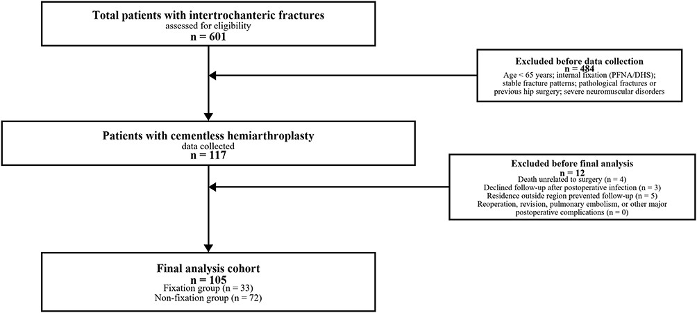 |
Figure 1 Flowchart of the patient selection process. A total of 601 patients with intertrochanteric fractures were assessed for eligibility. Overall, 484 patients were excluded before data collection for the hemiarthroplasty cohort, and data were collected from 117 patients who underwent cementless hemiarthroplasty. Before final analysis, 12 patients were excluded: 4 died during follow-up from causes unrelated to surgery, 3 declined further follow-up after postoperative infection, and 5 could not continue follow-up because they lived outside the region. No exclusions were due to reoperation, revision, pulmonary embolism, or other major postoperative complications. Finally, 105 patients were included in the final analysis (Non-fixation, n = 72; Fixation, n = 33). |
Inclusion criteria were as follows:
- Age ≥65 years at the time of surgery;
- Radiographic diagnosis of an unstable intertrochanteric fracture classified as AO/OTA type 31-A2 or 31-A3, with the lesser trochanter displaced;
- Availability of baseline, perioperative, and radiographic data for the index hospitalization;
- Hemiarthroplasty (cementless) as the definitive surgical intervention.
Patients meeting any of the following criteria were excluded from the study:
- Pathological fractures: Fractures resulting from primary or metastatic bone tumors (while elderly osteoporotic fractures were included, those with underlying malignancy were strictly excluded).
- Previous surgical history: A history of prior surgical intervention on the ipsilateral hip.
- Severe neuromuscular disorders: Co-existing conditions that significantly impair limb function or gait, such as hemiplegia or Parkinson’s disease.
- Incomplete baseline, perioperative, or radiographic records for the index hospitalization.
- Early major complications within 3 months after surgery, including severe periprosthetic infection, prosthetic dislocation, major medical events, or mortality, when these events precluded subsequent clinical and functional evaluation. These patients were excluded before construction of the 105-patient full cohort.
Surgical Technique
All procedures were performed under general or spinal anesthesia with patients in the lateral decubitus position using a standard posterolateral approach. Upon exposure of the fracture site, the fractured femoral head and free neck fragments were extracted. For the comminuted proximal femur, minimal soft tissue stripping was strictly observed to preserve the blood supply to the greater trochanter and remaining calcar. The femoral canal was sequentially broached and prepared to achieve diaphyseal or metaphyseal press-fit stability. A cementless femoral stem with a bipolar prosthetic head was then implanted. Following joint reduction, the displaced greater trochanteric fragments were anatomically reduced and secured using tension band wiring or titanium cables to reconstruct the abductor mechanism. In the fixation group, the displaced lesser trochanteric fragments were specifically reduced and stabilized using cerclage wiring (Figure 2), whereas they were left unfixed in the non-fixation group.
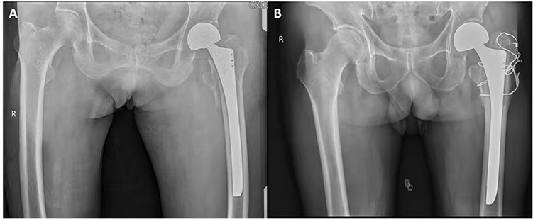 |
Figure 2 Representative postoperative radiographs of the two surgical strategies. (A) Anteroposterior pelvic radiograph of a patient in the non-fixation group, demonstrating cementless hemiarthroplasty with the displaced lesser trochanter fragment left unfixed. (B) Radiograph of a patient in the fixation group, showing the lesser trochanter fragment reduced and stabilized with cerclage wiring. Both groups achieved satisfactory prosthetic positioning and joint reduction. |
Group Definition
Intraoperative management of the displaced lesser trochanter determined group allocation. In the Fixation group, the lesser trochanter fragment was mobilized, reduced, and stabilized using cerclage wires or titanium cables. In the Non-fixation group, the displaced lesser trochanter fragment was intentionally left unfixed, relying on fibrous healing and musculotendinous adaptation. All procedures were performed by the same senior orthopedic surgeon. Surgeons followed a structured set of intraoperative clinical criteria to guide decision-making for lesser trochanter fixation. Following trial reduction and femoral stem implantation, fixation was generally omitted when the fragment was judged to be relatively stable, well aligned, and unlikely to compromise the femoral medullary canal or femoral stem stability. Fixation was performed if the fragment exhibited severe overall instability, involved the femoral calcar, or carried a risk of postoperative migration into the joint space and injury to surrounding soft tissue structures. Surgical practice patterns also evolved over time: the surgeon showed a greater tendency to perform fixation in earlier cases, whereas a preference for non-fixation was observed in more recent cases. Patient age, sex, American Society of Anesthesiologists (ASA) physical status classification, and pre-injury functional status were not formal indications for fixation. Treatment allocation was driven primarily by the morphological characteristics of fracture fragments and intraoperative fragment stability, while temporal shifts in surgical philosophy may also have impacted intraoperative decision-making. The absence of a prospective standardized allocation protocol creates a risk of residual confounding that cannot be fully eliminated.
Postoperative Management and Follow-Up
Postoperatively, active exercises of the knee and hip joints were permitted from the first postoperative day as tolerated. Assisted weight-bearing ambulation was initiated using a walker, ambulation aid, or crutches once the patient’s medical and functional condition permitted safe mobilization. Patients were encouraged to use walking aids during the first 3 months after surgery. Follow-up assessments were scheduled at 6 weeks, 3 months, 6 months, and 1 year postoperatively, and annually thereafter. Because this predominantly elderly population frequently had multiple comorbidities and variable exercise tolerance, the timing and progression of mobilization were individualized rather than imposed as identical milestones for every patient. Perioperative management included routine antibiotic prophylaxis and thromboprophylaxis with low molecular weight heparin in accordance with institutional guidance.
Outcome Measures
Data were extracted from the hospital electronic medical record system and the picture archiving and communication system (PACS) by two independent, blinded researchers, and all entries were cross-checked. The following variables were recorded: (1) demographics and baseline characteristics, including age, sex, height, weight (body mass index), fracture laterality, ASA classification, admission Barthel Index, and pre-injury Parker Mobility Score; (2) perioperative variables, comprising operative time (skin incision to wound closure), length of hospital stay, visible blood loss, and hidden blood loss (calculated using the Nadler formula and Gross equation26,27); (3) radiographic parameters assessed on standard postoperative anteroposterior pelvic radiographs, including postoperative bipolar cup abduction angle and lower limb length discrepancy (LLD); and (4) clinical follow-up data, including HHS at the final visit,28 and the Ludloff test result.29 The postoperative bipolar cup abduction angle was measured on the early postoperative anteroposterior pelvic radiograph, following Lee et al, as the angle formed by the interteardrop line and the equator line of the bipolar cup.30 This parameter describes the postoperative orientation of the bipolar cup and has been used as a biomechanical indicator of the static resultant hip contact-force direction in the frontal plane. LLD was measured as the difference in perpendicular distances from the pelvic reference line to the lesser trochanteric region on each side.31 Radiographic measurements were independently performed by two blinded orthopedic surgeons, and the mean of the two measurements was used for analysis. Interobserver reliability was assessed using the intraclass correlation coefficient (ICC), with ICCs of 0.89 for postoperative bipolar cup abduction angle and 0.87 for LLD.32 The Ludloff test was performed with the patient seated and the knees extended; a positive result was recorded if the patient experienced weakness or deep groin pain when actively lifting the heel off the examination table, indicating iliopsoas dysfunction. The occurrence of complications such as infection, dislocation, or periprosthetic fracture was also recorded. In this elderly cohort, the Ludloff test was used as a pragmatic bedside assessment because multimorbidity, limited examination tolerance, and variable follow-up compliance precluded uniform objective dynamometric testing. The test can provide an indirect clinical indication of pain-provoked iliopsoas activation and gross postoperative hip-flexor performance, but it does not quantify muscle torque or exclude subtle weakness.
Statistical Analysis
All statistical analyses were performed using Python (version 3.11) with the SciPy (version 1.11.4) and Statsmodels libraries. Continuous variables were first assessed for normality using the Shapiro–Wilk test. Normally distributed data were expressed as mean ± standard deviation and compared between groups using independent-samples t-tests. Categorical variables were expressed as frequencies and percentages (n, %) and compared using the chi-squared test or Fisher’s exact test, as appropriate. All analyses were performed in the final cohort of 105 patients unless otherwise specified. Because this was a retrospective cohort study based on all eligible patients during the study period, no a priori sample-size calculation was performed. Post-hoc power analysis was performed to evaluate the adequacy of the available sample size with two-tailed α = 0.05. For continuous outcomes, Cohen’s d was calculated to quantify standardized effect sizes. For HHS at 12 months, the observed intergroup mean difference was 2.1 points with a pooled standard deviation of 10.90, yielding Cohen’s d = 0.195 and post-hoc power < 0.8. For operative time, the observed mean difference was 9.3 min with a pooled standard deviation of 14.96, yielding Cohen’s d = 0.622 and post-hoc power > 0.8. For binary outcomes including complications and the Ludloff test, event counts were limited, leading to low statistical power to detect moderate between-group differences; Fisher’s exact test was therefore used where appropriate.
To isolate the independent effect of the surgical strategy on functional recovery, a multivariate linear regression model was constructed. The Harris Hip Score (HHS) at the 12-month follow-up was defined as the dependent variable. Based on clinical relevance and established predictive models for geriatric hip fractures,10 the fixation strategy (fixed vs non-fixed), age, ASA classification, and pre-injury Parker Mobility Score were entered into the model as independent covariates. Unstandardized regression coefficients (β) and their corresponding 95% confidence intervals (CIs) were calculated to quantify the predictive value of each factor. Cohen’s d was calculated for the primary outcome to facilitate the interpretation of effect size, where 0.2, 0.5, and 0.8 represent small, medium, and large effects, respectively.
Data visualization was generated using the Matplotlib library (version 3.8.1). Continuous data in figures are presented as means with standard deviations unless otherwise specified. Final graphical representations were exported as high-resolution (300 DPI) TIFF files to ensure publication quality. All statistical tests were two-sided, and a p-value < 0.05 was considered statistically significant.
Results
Patient Demographics and Baseline Characteristics
A total of 105 patients met the inclusion criteria and were enrolled: 72 in the non-fixation group and 33 in the fixation group. Baseline demographic and clinical characteristics are summarized in Table 1. The mean age was 80.2 ± 4.6 years in the non-fixation group and 79.1 ± 6.1 years in the fixation group (p = 0.304). No statistically significant differences were identified between the two groups in BMI, fracture laterality, ASA classification, or pre-injury Parker Mobility Score. However, admission Barthel Index was significantly lower in the fixation group than in the non-fixation group (28.0 ± 7.2 vs 33.2 ± 7.8; p = 0.002), indicating a baseline imbalance in admission functional status. Although the fixation group also had a numerically higher proportion of male patients (p = 0.085), this difference did not reach statistical significance.
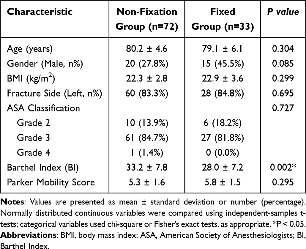 |
Table 1 Baseline Demographic and Clinical Characteristics of the Two Study Groups |
Primary Outcomes
At final follow-up, HHS revealed no statistically significant difference between the non-fixation group (83.1 ± 12.1) and the fixation group (85.2 ± 6.9; mean difference, −2.1 points; p = 0.477). The effect size was small (Cohen’s d = 0.20), indicating no clinically apparent between-group difference in this cohort (Figure 3). Iliopsoas function was evaluated using the Ludloff clinical test; the rate of a positive result was 15/72 (20.8%) in the non-fixation group and 3/33 (9.1%) in the fixation group, with no statistically significant between-group difference (p = 0.171; Figure 4). Lower limb length discrepancy was well controlled in both groups, with the majority of patients falling within the clinically acceptable absolute LLD ≤ 10 mm (p = 0.822; Figure 5).
 |
Figure 3 Comparison of postoperative Harris Hip Score (HHS) between the two groups. Box plots illustrate the medians, quartiles, and ranges of HHS scores at the final follow-up. No statistically significant between-group difference was observed (mean difference, −2.1 points; p = 0.477). |
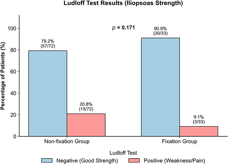 |
Figure 4 Comparison of Ludloff test results for iliopsoas function. Bar charts illustrate the proportions of patients with negative and positive Ludloff test results in the non-fixation and fixation groups. The positive Ludloff test rates were 15/72 (20.8%) and 3/33 (9.1%), respectively. Fisher’s exact test showed no statistically significant difference between groups (p = 0.171). |
 |
Figure 5 Comparison of postoperative absolute leg length discrepancy (LLD) in the full cohort (n = 105; Non-fixation, n = 72; Fixation, n = 33). Values are presented as mean ± standard deviation. The shaded area demarcates the clinically acceptable range (absolute LLD ≤ 10 mm). No statistically significant between-group difference in LLD was observed (p = 0.822). |
To further assess the independent effect of fixation strategy, a multivariate linear regression analysis was conducted. After adjusting for age, ASA classification, and pre-injury Parker Mobility Score, lesser trochanter fixation was not identified as an independent predictor of postoperative HHS (β = 2.53; 95% CI: −3.61 to 8.67; p = 0.413; Figure 6).
 |
Figure 6 Forest plot of the multivariate linear regression analysis identifying factors associated with postoperative Harris Hip Score (HHS). The plot displays the regression coefficients (Beta values) and their 95% confidence intervals (CIs) for potential influencing factors. The vertical dashed line at x = 0 represents the line of no effect. After adjustment for age, ASA classification, and pre-injury Parker score, fixation of the lesser trochanter was not an independent predictor of postoperative HHS (β = 2.53; 95% CI, −3.61 to 8.67; p = 0.413). |
Perioperative Outcomes
Postoperative radiographic outcomes are presented in Table 2. The postoperative bipolar cup abduction angle differed statistically between the non-fixation and fixation groups (25.7 ± 6.1° vs 22.4 ± 8.3°; p = 0.046). This measure was interpreted as an early postoperative descriptor of bipolar cup orientation rather than as an acetabular cup parameter. Perioperative data are also presented in Table 2. The mean operative time was significantly shorter in the non-fixation group compared with the fixation group (124.7 ± 14.9 min vs 134.0 ± 15.1 min; p = 0.004), reflecting the procedural simplification achieved by omitting the fixation step. Visible blood loss (p = 0.055), hidden blood loss (p = 0.699), and total blood loss (p = 0.554) were all comparable between groups in the full cohort (Figure 7). Length of hospital stay was also similar between groups (p = 0.128). Complications reported in the final analytic cohort were events documented after the early exclusion period. Among the 105 patients included in the final analysis, no prosthetic dislocation, periprosthetic fracture, pulmonary embolism, reoperation, revision, or surgery-related death was recorded. Because routine postoperative ultrasonographic screening was not performed, asymptomatic deep vein thrombosis (DVT) could not be reliably assessed.
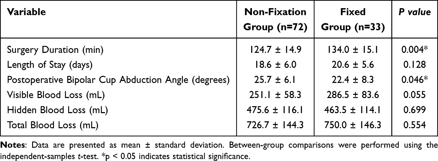 |
Table 2 Comparison of Postoperative Radiographic and Perioperative Variables Between the Non-Fixation and Fixation Groups |
 |
Figure 7 Comparison of perioperative visible, hidden, and total blood loss between the two groups in the full cohort (n = 105; Non-fixation, n = 72; Fixation, n = 33). Grouped bar charts illustrate the mean volumes of visible blood loss (recorded intraoperatively), hidden blood loss (calculated), and total blood loss. Error bars indicate the standard deviation. Statistical analysis revealed no significant differences in visible (p = 0.055), hidden (p = 0.699), or total blood loss (p = 0.554) between the non-fixation and fixation groups. |
Discussion
This retrospective cohort study investigated the functional and perioperative consequences of omitting lesser trochanter fixation during cementless hemiarthroplasty for unstable intertrochanteric fractures in a high-comorbidity elderly population. The principal finding was that no statistically significant between-group differences were observed in the Harris Hip Score or the Ludloff clinical test, while the non-fixation approach was associated with a statistically significant reduction in operative time (124.7 ± 14.9 min vs 134.0 ± 15.1 min; p = 0.004). Admission Barthel Index was lower in the fixation group, and the postoperative bipolar cup abduction angle also differed between groups. Neither finding changed the main result of the present analysis, namely that no statistically significant between-group difference was observed in the evaluated functional outcomes. Nevertheless, the lower admission Barthel Index represents a potential baseline imbalance, and the bipolar cup abduction angle is a postoperative descriptive parameter. Accordingly, given the retrospective design and limited sample size, these findings should be interpreted cautiously.
Recontextualising the Biomechanical Rationale for Fixation
The conventional argument for lesser trochanter fixation rests on a straightforward anatomical premise: the lesser trochanter is the principal insertion site of the iliopsoas complex, which functions as the dominant hip flexor and contributes to lumbopelvic stability during ambulation.17–19 Displacement of this fragment, occurring in approximately 50% of proximal femoral fractures,20 may compromise iliopsoas function through loss of its bony lever arm. This concern is supported by studies reporting reduced hip-flexion strength, delayed functional recovery, or outcome differences associated with the involvement and position of the displaced lesser trochanter.21–23 These observations provide a reasonable clinical and biomechanical rationale for fixation in selected patients, particularly when the fragment is markedly displaced or unstable.
A key consideration is that hemiarthroplasty alters the proximal femoral biomechanical environment in ways that may limit direct extrapolation from internal fixation studies. When a cementless femoral stem is implanted, it achieves immediate mechanical stability through diaphyseal or metaphyseal press-fit fixation, independent of the integrity of proximal bony fragments.14,24 The force-transmission pathway through the proximal femur is reconstituted around the prosthetic construct rather than the native bony architecture. The cementless stem bypasses the comminuted calcar and transfers weight-bearing loads distally to the intact femoral diaphysis. In this setting, the lesser trochanter fragment may function more as a periarticular soft-tissue attachment than as a load-critical bony structure.33,34
Functional Outcomes and the Capacity for Musculotendinous Adaptation
The absence of a statistically significant difference in hip flexor function after lesser trochanter non-fixation is biologically plausible. Two complementary mechanisms may help explain this finding. First, the iliopsoas tendon may possess some adaptive capacity; fibrous healing at the tendon-bone interface could be sufficient to re-establish effective force transmission in a functional range. This interpretation is consistent with the observed Ludloff test results (20.8% vs 9.1%; p = 0.171). In this elderly, multimorbid cohort, the Ludloff test offered a feasible indirect bedside indication of pain-provoked iliopsoas activation and gross postoperative hip-flexor performance when uniform dynamometric assessment was limited by examination tolerance and follow-up compliance. However, it remains a semi-quantitative screening test and cannot directly quantify muscle torque or exclude subtle between-group weakness.
Second, synergistic hip flexor compensation may contribute to preserved function. Hauschild et al35 reported that hip flexion power did not decline relative to the contralateral limb despite significant fatty infiltration of the iliopsoas following large lesser trochanter displacement, suggesting compensatory recruitment of the rectus femoris, sartorius, and tensor fasciae latae In a geriatric population whose peak functional demand rarely approaches the threshold at which subclinical iliopsoas weakness becomes manifest, this compensatory reserve may help sustain performance on validated outcome instruments. The small effect size observed in the present study is consistent with this interpretation, although it should not be interpreted as evidence that the two strategies produce identical functional outcomes.
Recent clinical evidence is also consistent with this interpretation. In 2025, Noree et al36 published a randomized controlled trial directly addressing lesser trochanter fixation in patients treated with intramedullary nailing, a population in whom the biomechanical rationale for fixation may be stronger than in hemiarthroplasty patients. Even within that context, maximum isometric hip flexor strength at twelve months did not differ significantly between wire-fixation and non-fixation groups. These findings further question the need for routine lesser trochanter fixation in hemiarthroplasty, where the prosthetic stem provides an alternative structural scaffold.
The Primacy of Operative Time Reduction in the Frail Elderly: A Geriatric Safety Perspective
Although the null finding with respect to functional outcomes is itself substantively meaningful, the perioperative dimension of this study carries arguably greater clinical weight for the geriatric population under investigation. The study cohort had a mean age exceeding 80 years and was characterized by a high burden of medical comorbidity, with over 80% of patients classified as ASA classification 3—denoting severe systemic disease that substantively limits functional capacity. This demographic profile defines a population operating with markedly attenuated physiological reserve, for whom each additional minute of surgical stress and anesthetic exposure carries disproportionate risk.37,38
The approximately 10-minute reduction in operative time achieved in the non-fixation group (124.7 ± 14.9 min vs 134.0 ± 15.1 min; p = 0.004) should not be dismissed as a marginal procedural gain. Postoperative delirium represents one of the most consequential complications of hip fracture surgery in older adults, with reported incidence rates of 28–61% in this population.37,38 It is independently associated with prolonged hospitalization, accelerated cognitive decline, increased institutionalization rates, and elevated 12-month mortality. Prolonged operative duration is an established modifiable risk factor for its development through pathways including cumulative anesthetic drug exposure, intraoperative hypotension, and systemic inflammatory activation. The elimination of a discrete operative step, with no observed worsening of functional or safety outcomes in this cohort, may therefore represent a useful risk-reduction strategy in this vulnerable population.
This reasoning is consistent with ERAS principles in geriatric orthopedic surgery.1,25 Zhu et al25 demonstrated that formal ERAS implementation in elderly intertrochanteric fracture surgery was associated with significant reductions in postoperative complications and length of stay. The non-fixation strategy described in the present study is compatible with these principles because it eliminates a step of uncertain functional benefit, reduces anesthetic duration, and does not appear to compromise early mobilization in this cohort. Given that intertrochanteric fractures constitute a substantial and growing proportion of orthopedic emergency admissions in aging populations,8 operative simplification that preserves clinical outcomes may have practical value.39,40
Limb Length Restoration: Adequacy Without Fixation
A secondary but clinically pertinent concern regarding omission of lesser trochanter fixation relates to the potential for prosthetic leg length discrepancy, given the lesser trochanter’s role as an intraoperative landmark for limb length assessment. The present data partly address this concern. Although the postoperative bipolar cup abduction angle differed statistically between groups, it was measured on early postoperative radiographs and should be interpreted as a descriptive parameter of bipolar cup orientation. Lee et al reported that bipolar cup position reflects the frontal-plane direction of the hip contact force acting on it, and the relative stability of this angle across supine, double-leg standing, and single-leg standing positions suggests that it is influenced mainly by static anatomy and muscle force-vector balance rather than transient dynamic loading alone.30 Therefore, the early postoperative between-group difference observed in the present cohort may reflect periarticular muscle imbalance or postoperative static hip geometry after LT fixation or non-fixation. However, this radiographic difference was not accompanied by a between-group difference in LLD, HHS, Ludloff test results, dislocation, or periprosthetic fracture. The majority of patients in the non-fixation cohort had an absolute LLD within 10 mm, and no statistically significant intergroup difference in LLD was observed (p > 0.05). These findings should nevertheless be interpreted cautiously because the clinical significance of the abduction-angle difference remains uncertain and requires further study.
Limitations
Several methodological limitations of the present study warrant careful consideration.
First, the retrospective observational design precludes causal inference and introduces residual confounding. Allocation reflected intraoperative assessment of fragment stability and migration risk and may also have been affected by temporal evolution of surgical practice. Although age, sex, ASA classification, and pre-injury functional status were not formal indications for fixation, the absence of prospective randomization or a fully standardized allocation protocol permits selection bias. In addition, admission Barthel Index was significantly lower in the fixation group. Although this imbalance did not change the main observed functional findings, residual confounding related to baseline functional status and unmeasured anatomical factors cannot be excluded.
Second, the present study did not employ a dedicated, validated frailty assessment instrument—such as the Clinical Frailty Scale (CFS) or the Fried Frailty Phenotype—which would have permitted precise quantification of biological vulnerability. Nevertheless, it is important to note that this limitation does not leave the frailty burden of the study population uncharacterized. The predominance of ASA classification 3—present in over 80% of both cohorts—provides a meaningful, internationally standardized proxy for physiological reserve and systemic comorbidity. ASA classification 3 denotes a patient with severe systemic disease that substantively limits functional capacity, and its near-uniform distribution across both groups attests to the high-vulnerability nature of this cohort and supports the clinical generalizability of the findings to comparably frail populations. Future studies should nonetheless incorporate validated frailty instruments prospectively.
Third, iliopsoas function was evaluated using the Ludloff clinical test, a semi-quantitative bedside examination, rather than objective dynamometric measurement of peak isometric hip-flexor torque. The test can provide a pragmatic, indirect indication of pain-provoked activation and gross postoperative hip-flexor performance, which is relevant in an elderly population with limited tolerance and variable examination compliance. Nevertheless, its sensitivity may be insufficient to detect subtle reductions in muscle force. Future prospective studies should incorporate hand-held or isokinetic dynamometry and quantitative gait analysis where feasible.
Fourth, patients without evaluable follow-up or with major events that precluded subsequent assessment were excluded before construction of the final analytic cohort. Follow-up-related exclusions included death unrelated to surgery, refusal of further follow-up after postoperative infection, and inability to continue follow-up because of residence outside the region. This may have introduced selection or attrition bias. Routine postoperative DVT screening was not performed, and asymptomatic thromboembolic events may therefore have been underdetected.
Finally, this was a single-center study with all procedures performed by a single senior surgeon, and formal adherence to a structured ERAS protocol was not prospectively documented. While surgical uniformity strengthens internal validity by eliminating inter-surgeon variability, it simultaneously limits external validity. In addition, no a priori sample-size calculation was performed because of the retrospective design. Post-hoc power analysis suggested adequate power to detect the observed moderate operative-time difference but limited power to detect small functional differences in HHS. Therefore, the absence of statistical significance should not be interpreted as evidence that the two strategies produce identical functional outcomes. Further prospective investigations with rigorous a priori sample size calculation are required to draw definitive conclusions.
Conclusion
This retrospective cohort study found that omission of lesser trochanter fixation during cementless hemiarthroplasty for unstable intertrochanteric fractures was not associated with worse postoperative hip function, iliopsoas clinical test results, or limb length restoration in elderly patients. The simplified operative strategy was associated with a statistically significant reduction in operative time without an observed increase in blood loss, hospitalization duration, or perioperative complications. The lower admission Barthel Index in the fixation group and the difference in postoperative bipolar cup abduction angle did not change the main functional findings in the present analysis; however, they remain relevant to the cautious interpretation required by the retrospective design and limited statistical power. In a population with a mean age exceeding 80 years and a predominant ASA classification 3 comorbidity burden, operative simplification may be clinically relevant, but definitive clinical recommendations should await confirmation from prospective randomized controlled trials.
Future Directions
Subsequent investigations should adopt a prospective randomized controlled design with adequate statistical power. An extended follow-up interval of 12–24 months would facilitate evaluation of long-term functional outcomes and late complications. Stratified subgroup analyses— incorporating bone density assessment using dual-energy X-ray absorptiometry (DXA) and thedegree of lesser trochanter displacement—may identify patient subpopulations from whom targeted fixation could be expected to provide benefit. The integration of objective functional assessment modalities, including instrumented gait analysis, timed up-and-go tests, and dynamometric measurement of hip flexion force, would enhance the precision of outcome evaluation. Additionally, application of a validated frailty screening tool (eg, the Clinical Frailty Scale) and formal ERAS protocol documentation would strengthen both the interpretability and the generalizability of future research in this population.
Data Sharing Statement
The datasets generated and/or analyzed during the current study are not publicly available due to hospital patient privacy regulations, but are available from the corresponding author on reasonable request.
Ethics Approval and Consent to Participate
This retrospective study was approved by the Institutional Review Board of the Second Affiliated Hospital of Xi’an Jiaotong University (Approval No. (2026) Ethics Review-Research No. (061)) All procedures adhered to the principles set forth in the Declaration of Helsinki. Given the retrospective nature of the study and the use of anonymized data, the requirement for written informed consent was waived by the Institutional Review Board.
Author Contributions
All authors made a significant contribution to the work reported, whether that is in the conception, study design, execution, acquisition of data, analysis and interpretation, or in all these areas; took part in drafting, revising or critically reviewing the article; gave final approval of the version to be published; have agreed on the journal to which the article has been submitted; and agree to be accountable for all aspects of the work. Specifically, according to the CRediT taxonomy: Jiahao Zhang: Conceptualization, Methodology, Formal Analysis, Writing – Original Draft. Qin Guo: Data Curation, Investigation, Writing – Review & Editing. Yongwei Li: Conceptualization, Supervision, Project Administration, Writing – Review & Editing.
Funding
This work was supported by the Key Research and Development Program of Shaanxi Province, Department of Science and Technology of Shaanxi Province (No. 2024SF-YBXM-207). The funder had no role in the study design, data collection, data analysis, data interpretation, manuscript preparation, or decision to submit the article for publication.
Disclosure
The authors declare that they have no competing interests in this work.
References
1. Li L, Wang L, Ma C, et al. Clinical effect of ‘fast-track management’ guided by ERAS concept under the multi-disciplinary cooperation mode of femoral intertrochanteric fracture in the elderly. Biotechnol Genet Eng Rev. 2024;40(3):2393–15. doi:10.1080/02648725.2023.2199541
2. Zarzour F, Aftabi S, Leslie WD. Effects of femoral neck width and Hip axis length on incident hip fracture risk: a registry-based cohort study. J Bone Miner Res. 2025;40(3):332–338. doi:10.1093/jbmr/zjaf019
3. Jiao S, Zhou J, Feng Z, et al. The role of neutrophil percentage to albumin ratio in predicting 1-year mortality in elderly patients with Hip fracture and external validation. Front Immunol. 2023;14:1223464. doi:10.3389/fimmu.2023.1223464
4. Wang P, Qin H, Liu Z, et al. Biomechanical analysis of different lengths of INTERTAN in the treatment of AO31A2.3 fractures: a finite element study. Comput Biol Med. 2025;194:110474. doi:10.1016/j.compbiomed.2025.110474
5. Nie SB, Zhao YP, Li JT, et al. Medial support nail and proximal femoral nail antirotation in the treatment of reverse obliquity intertrochanteric fractures: a finite-element analysis. Chin Med J. 2020;133(22):2682–2687. doi:10.1097/CM9.0000000000001031
6. Rupp M, Walter N, Pfeifer C, et al. The incidence of fractures among the adult population of Germany—an analysis from 2009 through 2019. Dtsch Arztebl Int. 2021;118(40):665–669. doi:10.3238/arztebl.m2021.0238
7. Walter N, Szymski D, Bärtl S, et al. Prevalence of fractures in the adult population of Germany: an evaluation of inpatient and outpatient cases. Dtsch Arztebl Int. 2025;122.
8. Cooper C, Campion G, Melton LJ. Hip fractures in the elderly: a world-wide projection. Osteoporos Int. 1992;2(6):285–289. doi:10.1007/BF01623184
9. Qi T, Yan Y, Qi W, et al. Hip joint-preserving strategies for treating osteonecrosis of the femoral head: from nonoperative to operative procedures. J Orthop Transl. 2025;51:256–277.
10. Hao Y, Wang R, Chen Z, et al. One-year mortality risk in older individuals with femoral intertrochanteric fracture: a tertiary center in China. BMC Geriatr. 2024;24:544. doi:10.1186/s12877-024-05159-y
11. Cheng YX, Sheng X. Optimal surgical methods to treat intertrochanteric fracture: a Bayesian network meta-analysis based on 36 randomized controlled trials. J Orthop Surg Res. 2020;15(1):402. doi:10.1186/s13018-020-01943-9
12. Hongku N, Woratanarat P, Nitiwarangkul L, et al. Fracture fixation versus hemiarthroplasty for unstable intertrochanteric fractures in elderly patients: a systematic review and network meta-analysis of randomized controlled trials. Orthop Traumatol Surg Res. 2022;108(1):102838. doi:10.1016/j.otsr.2021.102838
13. Yoo JI, Cha YH, Kim KJ, et al. Comparison between cementless and cemented bipolar hemiarthroplasty for treatment of unstable intertrochanteric fractures: systematic review and meta-analysis. Hip Pelvis. 2018;30(4):241–253. doi:10.5371/hp.2018.30.4.241
14. Martinho T, Stoffel K. Treatment of intertrochanteric femur fractures with Hip arthroplasty in older patients: a narrative review of indications and outcomes. Medicina. 2021;57(8):763. doi:10.3390/medicina57080763
15. Sniderman J, Vivekanantha P, Shah A, et al. Hemiarthroplasty for unstable intertrochanteric Hip fractures: a matched cohort study. J Arthroplasty. 2023;38(8):1522–1527. doi:10.1016/j.arth.2023.01.057
16. Paul I, Ks A, Yadav AK, Gawhale S. Hemiarthroplasty as a viable option for unstable intertrochanteric femur fractures in the elderly: a functional outcome analysis. Cureus. 2025;17(12):e77041. doi:10.7759/cureus.77041
17. Bordoni B, Varacallo M. Anatomy, bony pelvis and lower limb, iliopsoas muscle. In: StatPearls [Internet]. Treasure Island (FL): StatPearls Publishing; 2024.
18. Anderson CN. Iliopsoas: pathology, diagnosis, and treatment. Clin Sports Med. 2016;35(3):419–433. doi:10.1016/j.csm.2016.02.009
19. Rattanapakdawat P, Charoenjittichai B, Tangjaroenkamsri P. The iliopsoas muscle complex: a comprehensive review. J Anat. 2019;234(5):887–895.
20. Lamb J, Panteli M, Pneumaticos S, et al. Epidemiology of pertrochanteric fractures: our institutional experience. Eur J Trauma Emerg Surg. 2014;40:225–232. doi:10.1007/s00068-014-0375-x
21. Aprato A, Lo Baido R, Crosio A, et al. Does lesser trochanter implication affect Hip flexion strength in proximal femur fracture? Eur J Trauma Emerg Surg. 2015;41:523–529. doi:10.1007/s00068-014-0476-6
22. van der Sijp MPL, Moonen L, Schipper IB, et al. The functional effect of lesser trochanter involvement in Hip fractures: a prospective cohort study. Injury. 2020;51(11):2634–2639. doi:10.1016/j.injury.2020.09.002
23. Sun Q, Ge W, Hu H, et al. The influence of position of the displaced lesser trochanter on clinical outcome of unstable trochanteric femur fractures in the elderly. Biomed Res Int. 2018;2018:5013646. doi:10.1155/2018/5013646
24. Zhou Z, Gjyshinca B, Podvorica V, et al. Primary hemiarthroplasty for treatment of unstable pertrochanteric femoral fractures (AO/OTA Type 31 A2.3) in elderly osteoporotic patients. SICOT J. 2017;3:47. doi:10.1051/sicotj/2017001
25. Zhu W, Yan Y, Sun Y, et al. Implementation of Enhanced Recovery After Surgery (ERAS) protocol for elderly patients receiving surgery for intertrochanteric fracture: a propensity score-matched analysis. J Orthop Surg Res. 2021;16(1):469. doi:10.1186/s13018-021-02599-9
26. Nadler SB, Hidalgo JH, Bloch T. Prediction of blood volume in normal human adults. Surgery. 1962;51(2):224–232.
27. Gross JB. Estimating allowable blood loss: corrected for dilution. Anesthesiology. 1983;58(3):277–280. doi:10.1097/00000542-198303000-00016
28. Harris WH. Traumatic arthritis of the Hip after dislocation and acetabular fractures: treatment by mold arthroplasty. J Bone Joint Surg Am. 1969;51(4):737–755. doi:10.2106/00004623-196951040-00012
29. Bahr R, Mæhlum S. Clinical Guide to Sports Injuries. Champaign (IL): Human Kinetics; 2004.
30. Lee MS, Chen ACY, Kuo CH, Tai CL, Senan V, Shih CH. The position of the bipolar cup reflects the direction of the hip contact force acting on it. J Arthroplasty. 2007;22(2):189–194. doi:10.1016/j.arth.2006.02.079
31. Mahmood SS, Al-Amiry B, Mukka SS, Baea S, Sayed-Noor AS. Validity, reliability and reproducibility of plain radiographic measurements after total Hip arthroplasty. Skeletal Radiol. 2015;44(3):345–351. doi:10.1007/s00256-014-2055-7
32. Koo TK, Li MY. A guideline of selecting and reporting intraclass correlation coefficients for reliability research. J Chiropr Med. 2016;15(2):155–163. doi:10.1016/j.jcm.2016.02.012
33. Cui Q, Liu YS, Li DF, et al. Cemented Hip hemiarthroplasty clinical observations on unstable intertrochanteric fracture in elderlies. Eur J Trauma Emerg Surg. 2016;42:651–656. doi:10.1007/s00068-015-0566-0
34. Kim SY, Kim YG, Hwang JK. Cementless calcar-replacement hemiarthroplasty compared with intramedullary fixation of unstable intertrochanteric fractures. J Bone Joint Surg Am. 2005;87(10):2186–2192. doi:10.2106/JBJS.D.02768
35. Hauschild O, Ghanem N, Oberst M, et al. No difference in flexion power despite iliopsoas fatty degeneration in healed Hip fractures with large lesser trochanter displacement. Eur J Orthop Surg Traumatol. 2018;28(8):1523–1529. doi:10.1007/s00590-018-2151-9
36. Noree N, Boonyanuwat W, Limpiyakosol P, et al. To fix or not to fix? Evaluating lesser trochanter fixation in unstable intertrochanteric fractures: a randomized controlled trial. J Orthop Surg;2025. 10225536251333121. doi:10.1177/10225536251333121
37. Zhao S, Sun T, Zhang J, et al. Risk factors and prognosis of postoperative delirium in nonagenarians with Hip fracture. Sci Rep. 2023;13:2167. doi:10.1038/s41598-023-27829-4
38. de Haan E, van Rijckevorsel VAJIM, Bod P, et al. Delirium after surgery for proximal femoral fractures in the frail elderly patient: risk factors and clinical outcomes. Clin Interv Aging. 2023;18:193–203. doi:10.2147/CIA.S390906
39. Nho JH, Seo GW, Kang TW, et al. Bipolar hemiarthroplasty in unstable intertrochanteric fractures with an effective wiring technique. Hip Pelvis. 2023;35(2):99–107. doi:10.5371/hp.2023.35.2.99
40. Camurcu Y, Cobden A, Sofu H, et al. What are the determinants of mortality after cemented bipolar hemiarthroplasty for unstable intertrochanteric fractures in elderly patients? J Arthroplasty. 2017;32(10):3038–3043. doi:10.1016/j.arth.2017.04.042
 © 2026 The Author(s). This work is published and licensed by Dove Medical Press Limited. The
full terms of this license are available at https://www.dovepress.com/terms
and incorporate the Creative Commons Attribution
- Non Commercial (unported, 4.0) License.
By accessing the work you hereby accept the Terms. Non-commercial uses of the work are permitted
without any further permission from Dove Medical Press Limited, provided the work is properly
attributed. For permission for commercial use of this work, please see paragraphs 4.2 and 5 of our Terms.
© 2026 The Author(s). This work is published and licensed by Dove Medical Press Limited. The
full terms of this license are available at https://www.dovepress.com/terms
and incorporate the Creative Commons Attribution
- Non Commercial (unported, 4.0) License.
By accessing the work you hereby accept the Terms. Non-commercial uses of the work are permitted
without any further permission from Dove Medical Press Limited, provided the work is properly
attributed. For permission for commercial use of this work, please see paragraphs 4.2 and 5 of our Terms.