Back to Journals » International Journal of Nanomedicine » Volume 12
Drug delivery to solid tumors: the predictive value of the multicellular tumor spheroid model for nanomedicine screening
Authors Millard M, Yakavets I, Zorin V, Kulmukhamedova A, Marchal S, Bezdetnaya L
Received 22 July 2017
Accepted for publication 22 September 2017
Published 31 October 2017 Volume 2017:12 Pages 7993—8007
DOI https://doi.org/10.2147/IJN.S146927
Checked for plagiarism Yes
Review by Single anonymous peer review
Peer reviewer comments 2
Editor who approved publication: Dr Thomas Webster
Marie Millard,1,2 Ilya Yakavets,1–3 Vladimir Zorin,3,4 Aigul Kulmukhamedova,1,2,5 Sophie Marchal,1,2 Lina Bezdetnaya1,2
1Centre de Recherche en Automatique de Nancy, Centre National de la Recherche Scientifique UMR 7039, Université de Lorraine, 2Research Department, Institut de Cancérologie de Lorraine, Vandœuvre-lès-Nancy, France; 3Laboratory of Biophysics and Biotechnology, 4International Sakharov Environmental Institute, Belarusian State University, Minsk, Belarus; 5Department of Radiology, Medical Company Sunkar, Almaty, Kazakhstan
Abstract: The increasing number of publications on the subject shows that nanomedicine is an attractive field for investigations aiming to considerably improve anticancer chemotherapy. Based on selective tumor targeting while sparing healthy tissue, carrier-mediated drug delivery has been expected to provide significant benefits to patients. However, despite reduced systemic toxicity, most nanodrugs approved for clinical use have been less effective than previously anticipated. The gap between experimental results and clinical outcomes demonstrates the necessity to perform comprehensive drug screening by using powerful preclinical models. In this context, in vitro three-dimensional models can provide key information on drug behavior inside the tumor tissue. The multicellular tumor spheroid (MCTS) model closely mimics a small avascular tumor with the presence of proliferative cells surrounding quiescent cells and a necrotic core. Oxygen, pH and nutrient gradients are similar to those of solid tumor. Furthermore, extracellular matrix (ECM) components and stromal cells can be embedded in the most sophisticated spheroid design. All these elements together with the physicochemical properties of nanoparticles (NPs) play a key role in drug transport, and therefore, the MCTS model is appropriate to assess the ability of NP to penetrate the tumor tissue. This review presents recent developments in MCTS models for a better comprehension of the interactions between NPs and tumor components that affect tumor drug delivery. MCTS is particularly suitable for the high-throughput screening of new nanodrugs.
Keywords: nanodrug, tridimensional model, distribution, accumulation, cytotoxicity
Introduction
The complex biology of cancer highlights the urgent need of specific and effective treatments. Low-weight molecules that have been developed as antineoplastic agents since several decades have enabled progress in cancer management, gradually placing chemotherapy at a crucial position in cancer care. However, poor water solubility, unfavorable pharmacokinetics and adverse side effects of many molecules with potential antineoplastic properties have hindered their clinical applications.1 The lack of selective drug delivery to neoplastic tissues is the main reason for both systemic drug toxicity and poor efficiency. Therefore, tumor targeting is becoming a major issue in cancer therapy since it would allow to overcome side effects and to ensure full effectiveness of antineoplastic agents. In this context, nanomedicine, based on passive and/or active (or receptor-mediated) tumor targeting, yielding improved drug delivery has been considered promising.2 Recent developments in the field have succeeded in placing a significant number of anticancer nanotherapeutics in the pipeline; some of them are now investigated in advanced clinical settings.3 Despite this progress, it must be conceded that until now, few nanodrugs have been included in the arsenal of chemotherapeutics used for clinical cancer care management. Nanotherapeutics issued from promising preclinical studies often failed to achieve the expected results in patients, and as such, nanomedicine is still considered challenging for cancer therapy.4 To be efficiently delivered to the tumor, nanoparticles (NPs) must overcome different biological barriers that influence the extent of tissue penetration. Molecule diffusion through the interstitial space strongly depends on the structure and composition of the interstitial compartment. The dense extracellular matrix (ECM) mainly composed of loosely, poorly organized and interconnected collagen lattices together with polysaccharides like glycosaminoglycans (GAGs) to ensure the structural integrity of the network regulates the movement of NPs in the interstitium.5 In addition, host stromal cells including fibroblasts contribute to the production of ECM or to the capture of extravasated compounds (mononuclear phagocytes).6,7 Size of interstitial spaces along with the reduction in cell density is another feature that influences the movement of molecules in tumors.8,9 Molecule transport through vascular space is closely related to vessel morphology and to vascular organization.10,11 With the tumor growth, vessel compressions due to the development of proliferating cells or other stromal components contribute to the enhancement of blood flow and intravascular pressure irregularities.12,13 These parameters strongly disrupt oxygen, nutrient and blood-borne molecule delivery to tumor tissues.10
Extravasation of NPs >40 kDa is facilitated by the high permeability of tumor vessels. In the presence of vascular pores, from 200 to 1,200 nm in size,14 larger than those in normal blood vessels (range 50–150 nm), the fenestrated neovascular wall enhances vascular permeability with, however, important variability both spatially and temporally within a tumor or from one tumor to another.15 This particularity of tumor vessels was first described by Matsumura and Maeda in 1986. Attributed to the enhanced permeability of blood vessels along with the low clearance rate of poor lymphatic system developed by tumor tissues, this phenomenon was called enhanced permeability and retention (EPR) effect (Figure 1).16 Although the limitations of EPR effect have been extensively discussed,17 it remains the main process of passive tumor targeting by NPs, resulting in an increased drug accumulation in tumor while sparing healthy tissues.18 This selective accumulation and retention of NPs in solid tumor by EPR effect, contrary to small molecules that get out of the tumor vessels, makes the use of spherical tridimensional (3D) model highly appropriate.19,20 In fact, 3D models have expanded in the recent years reproducing better the complexity of the tumor than 2D cell culture and therefore being more representative of clinical situation.21 Among them, multicellular tumor spheroids (MCTSs) are of great interest in cancer research since some of their physiological features are close to those of avascular cancer metastases.22,23 MCTS models have been recognized suitable for studies of cell survival, cell proliferation, resistance to drug therapy, cell migration and hypoxia phenomena.24 MCTS models are also appropriate to estimate the ability of nanotherapeutics to penetrate and distribute inside tumor tissues.19,25 This review provides a comprehensive overview of the MCTS model focusing on the evaluation of nanodrugs along with the possibility to better predict its in vivo translation.
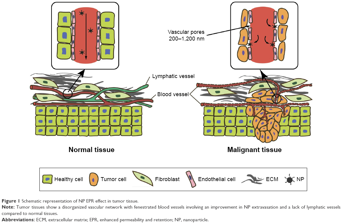 | Figure 1 Schematic representation of NP EPR effect in tumor tissue. |
MCTS model
Although 3D models cannot fully reflect the extent of tumor complexity and heterogeneity, they can be finely handled to meet specific tumor aspects. The recent review of Katt et al23 on the subject gave a valuable insight into different in vitro tumor models, with their particularities and specific use. Among them, spheroid-based models are the most widely used. Most 3D models have spherical shape, and the word “sphere” is commonly used to name any cellular aggregate. Spherical models can be obtained from fragments of tumors (tissue-derived tumor spheres and organotypic multicellular spheroids), from cancer stem cell culture (tumorospheres) or from single cancer cell suspension culture (MCTSs). All models have strengths and weaknesses. For example, fragments of tumor are suitable to study both tumor heterogeneity and the interaction between tumor and stroma, whereas tumorospheres are more appropriate for studying cancer stemness and in vivo tumorigenicity. The MCTS model is probably the most convenient to evaluate posttreatment tumor growth and cell survival, and it is therefore well adapted for high-throughput drug screening.19,25,26
Characteristics of MCTS model
Described for the first time in the early 1970s, Sutherland applied MCTS with the aim to evaluate tumor cell survival after chemotherapy. The MCTS model is an intermediate between 2D cell culture and in vivo tumor models (Figure 2). Derived from cancer cell lines known to be easily maintained in culture conditions, the 3D structure of MCTS mimics nonvascularized microtumors, which are similar to micrometastases observed in patients.27 Indeed, MCTS shares three fundamental characteristics with in vivo tumors: 1) a heterogeneous cellular growth with the presence of proliferating cells at the periphery surrounding a ring of quiescent cells and a necrosis core; 2) pH, nutriments and oxygen gradients from the periphery to the center and 3) more or less complex ECM that depends on the cell lines and the technique used for MCTS production.
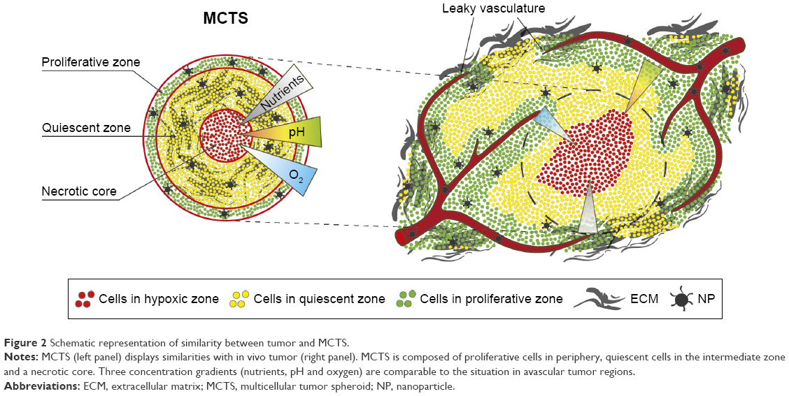 | Figure 2 Schematic representation of similarity between tumor and MCTS. |
- MCTS growth is similar to the one observed in solid tumors since the latter grows in 3D from the center to the periphery. The organization of the MCTS in three distinct layers, the outer one composed of proliferating cells and the inner one composed of quiescent cells and necrotic core, depends strongly on MCTS volume and growth rate. Below 120 μm in diameter, MCTS is well oxygenated while oxygen concentration and cell viability decrease rapidly with the spheroid growth.28,29
- The thickness of viable cell layer decreases in MCTS with low doubling time, and the fastest growing spheroids tend to have both higher oxygen consumption and pH decrease although anoxia is not necessary to obtain low pH values. These characteristics make MCTS model particularly well adapted for the evaluation of oxygen-dependent treatments such as ionizing radiation and photodynamic therapy (PDT).30–32
- Together with hypoxia, the ECM represents a crucial determinant in the tumor progression via epithelial–mesenchymal transition (EMT), tumor migration, invasion and metastasis processes. Many ECM proteins (fibronectin, laminin, collagen and GAGs) can be found in MCTS in similar proportions to those expressed in tumor in vivo.33 As a matter of fact, numerous studies reported the utility of MCTS taking into consideration the mechanisms of tumor cells’ migration and invasiveness as well as the treatments applied to prevent them.26,34
MCTS production techniques
MCTS is the 3D in vitro model that is most widely used for different purposes, such as cell proliferation, tumor growth, immune interactions, drug screening and in the case of the ECM-embedded MCTS, for cell invasion and matrix remodeling.23 The reason for this widespread use is the relative simplicity of MCTS production even though methods are becoming more and more complex with technological advances.19,26 Methodology for two commonly used techniques (liquid-overlay technique and gyratory rotation system) is based on the promotion of cell aggregates obtained from single tumor cell placed in agarose (or other inert substance)-coated flasks to prevent cell adhesion to the substrate. After 3–4 days growth, the aggregates are transferred into appropriate containers (coated petri dishes, culture plates, spinner or gyratory rotation system) where MCTS development is maintained until reaching the suitable size.35 The liquid-overlay technique offers the collection of few but more homogeneous in size MCTS, whereas spinner flask or gyratory rotation system is usually used for mass production (Table 1).23,26 The third method named hanging drop technique consists of the formation of cell droplets hanging on the underside of the lid of tissue culture dish. Cell clusters induced by gravity at the bottom of the drop grow into spheroids with a more regular size.19,23 Recently, innovative but sophisticated solutions have taken advantage of new technologies, such as microfluidics, to improve the control of size and composition of MCTS while maintaining high throughput (Table 1).19,36
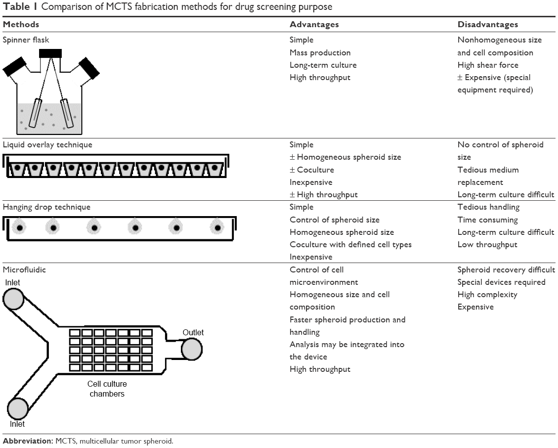 | Table 1 Comparison of MCTS fabrication methods for drug screening purpose |
To be more representative of tumor complexity, coculture of tumor cells with immune, endothelial cells or fibroblasts helps to better understand a crosstalk between cancer and stromal cells as well as the mechanisms of metastatic invasiveness.19,33,37 For example, the migration of endothelial cells into MCTS or the formation of vascular networks within spheroids is often used to assess the potential of tumor vascularization. Thus, by means of this spheroid coculture technique, factors that induce or inhibit angiogenesis can be studied.38 Scaffold-based techniques using natural (Matrigel®, hydrogels or alginate gels) or synthetic polymers as a semisolid matrix provide an excellent means to study the influence of external physical factors on the spheroid growth. Manipulating scaffolds to mimic particular physical properties of the tumor environment, such as stiffness and porosity, contributes to significant diversification of MCTS application for different and well-defined purposes.39
MCTS as a predictive model for nanodrug screening
Evaluation of drug sensitivity
Nowadays, MCTS rather than 2D cell culture-based assays are considered reliable enough for high-throughput drug screening. This model enables the selection of negative and/or positive new drug candidates, especially nanotherapeutics, thus reducing animal testing.19,40,41 Moreover, the development of MCTS-based biosensor devices is becoming widespread.36,42 Recently, Patra et al demonstrated the advantage to couple microfluidics with flow cytometry for the rapid production and viability analysis of a great number of MCTS with well-defined size, which were submitted to different drugs and their combination.36
Drug screening typically involves several steps. After spheroid formation (Table 1) and incubation with the drug for various periods of time, measurements of spheroid growth kinetics (ie, spheroid integrity, growth inhibition and regrowth after treatment) are performed.43 They are followed by the determination of cell viability (eg, acid phosphatase or lactate dehydrogenase release assays)43,44 and/or cell survival by testing the ability of a single cell to grow into a colony by clonogenic assay.40,45 Overall, MCTS is more resistant to the treatment than 2D cell culture and can recapitulate the drug resistance, observed in solid tumors.41,42,46
It must be noted that MCTS with both hypoxic and necrotic areas closely mimic specific gene expression profiles of tumors in vivo.47 Hypoxia and necrosis play key roles in the complex mechanisms (stress response, gene expression, signal transduction) of anticancer drug resistance. Spheroids have contributed to better understanding of the altered responsiveness of hypoxic tumor cells and the importance of cell–cell and cell–matrix interactions in therapeutic resistance.48 Desoize and Jardillier49 in 2000 showed that A549 human lung cancer cells, which have established contact with their microenvironment, become much less sensitive to anticancer drugs (ie, 27-fold decrease for cisplatin and 6,625 times lower sensitivity for vinblastine). In another study, it has been shown that acquisition of drug resistance by ovarian carcinoma spheroids, after treatment with Taxol, was related to culture conditions of spheroids. The use of compact spheroids, with an important E-cadherin expression, involves a low incorporation and poor diffusion of chemotherapeutic drugs. This result revealed the impact of adhesion molecule in drug resistance.48,50 Thus, MCTS is recognized to be a powerful model to assess tumor sensitivity to anticancer agents.19,25,40,41
The tumor-immune response assessed by MCTS coculturing between tumor and immune cells in order to observe the migration, interactions and cytotoxic effects of immune cells within spheroids is another contribution of MCTS to the development of new therapeutic strategies. It is important to note that immune response can be stimulated by promoting infiltration and cytotoxicity of leukocytes in MCTS.51 For example, it was demonstrated that the blocking of lactic acid secreted by tumor cells increased the migration of monocytes into MCTS.52
Evaluation of drug content and distribution in MCTS
Considering the complex tumor environment, the heterogeneous and limited dissemination of conventional anticancer drugs in tumors has been regarded as a crucial issue in cancer treatment.53–55 In comparison with their low-weight molecular payload, nanocarriers have increased size and other specific characteristics listed in the “Impact of physicochemical properties of NPs on their penetration” section below, which increase the risk of impaired distribution within the tumor. In this regard, 3D in vitro models and above all MCTS have been applied for evaluating the NP potential to accumulate and penetrate in depth the tumor tissue.20,34,56–59
Determination of free or drug-loaded NPs’ distribution and concentration in spheroids can be performed by various techniques. The distribution of gold NPs in MCTS could be precisely assessed with transmission electronic microscopy (TEM),60,61 while fluorescence microscopy was successfully applied for the detection of dye-labeled NPs. In this case, the NPs are either labeled with fluorescent probes,34,57,62–64 specific dyes or loaded with fluorescent drugs such as doxorubicin (DOX),58,65–68 Oregon Green 488 Taxol,20 or porphyrin-based photosensitizers (PSs).59,69 Estimation of drug concentration in MCTS could be done by high-performance liquid chromatography (HPLC),20 flow cytometry or chemical extraction with successive fluorescence measurements.43,44,57,64
After incubation, MCTS is collected and embedded in special medium as tissue-Tek before being cut in thin sections (~5 μm).32,57 The next step consists of examining spheroid sections by fluorescence microscopy to determine the extent of fluorescence intensity toward the spheroid center.20,34,58,59,62,64,70 NP penetration can also be evaluated in intact fixed MCTS64–66 or in intact living MCTS by using confocal laser scanning microscopy.34,71 An important lesson to draw from these experiments has been the excellence of the MCTS model to determine the main factors hindering or favoring NP penetration. Two major factors can modulate accumulation and distribution of NPs in MCTS. The first one is specific characteristics of cell lines and/or methods used to form spheroids. MCTS with lower cell density and larger extracellular space enable NP migration, whereas stroma-embedded MCTS turns out to be an appropriate model to demonstrate the barrier effect of stromal components on NP penetration.34,43
The second main factor is a design of NPs that takes into account various parameters such as size,20,60–62 composition,72 surface charge and coating of the nanocarriers.20,64 These properties are valuable indications for the optimal design of nanocarriers, allowing improved penetration in MCTS and treatment effectiveness. Surface of NPs plays a key role in its intra-spheroid repartition. Here, an attractive approach is the addition of collagenase on the NP surface to trigger ECM degradation, increasing the accessible space for NP transport and therefore improving its penetration.73 Likewise, gold NPs present an added benefit in drug delivery, thanks to the ease of fabrication and controllable surface functionality.60
Recent studies have shown that functionalization of gold NPs with tiopronin on NP surface considerably increases accumulation of NPs in MCTS and in vivo in solid xenografted tumors.61,74 Another interesting approach is to design an NP with regulated drug release to control the localization of NP inside MCTS or solid tumor. For example, Sims et al designed a PEGylated NP to target center areas, thus achieving deeper penetration into spheroids. In the same paper, the authors successfully designed the NPs coated with a cell-penetrating peptide (MPG) to target proliferative regions strongly confining NPs to periphery layers.64 Gold NP surface functionalization using multilayer formulation was also tested in 3D cells.72 Two-layer particles were synthesized with an inner layer of alkanethiol and an outer layer of phosphatidylcholine (a major component of biological membranes). Three-layer nanoshells were additionally coated with high-density lipoprotein. Both types of NPs demonstrated considerably enhanced penetration and diffusion into 3D cells compared to standard polyethylene glycol (PEG)-coated nanoshells.72
Impact of physicochemical properties of NPs on their penetration
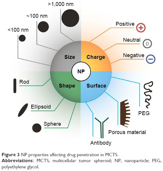
Physicochemical parameters assigned to a specific compound are important for its transport and distribution in biological media, playing a critical role in NP penetration and accumulation in solid tumor. Albeit many reviews provided insightful information about this complex issue, the contribution of each parameter is not easy to evaluate.3,17,26,75 Study of NPs’ structural properties such as size, shape, charge or surface (Figure 3) in the MCTS model provides information for understanding and predicting drug delivery to solid tumors.
 | Figure 3 NP properties affecting drug penetration in MCTS. |
- Many studies demonstrated improved penetration in MCTS for NPs with small size (<100 nm).20 For example, 50 nm-sized lipid NPs showed a considerably deeper penetration and homogeneous distribution as compared to 120 nm-sized ones.76 NPs >100 nm are too large to cross ECM interstitial spaces in spheroid73,77 and are confined to the outer layers as demonstrated with gelatin NPs (210 nm) that only penetrated ~40 μm deep.20 Micelles are generally smaller than liposomes and penetrate deeply and homogeneously in MCTS.20,62 For example, it was shown that very small-sized micelles (15 nm) penetrated faster and deeper in spheroids but over time failed to be retained and thus accumulated to the same extent than their larger counterparts (55 nm).62 Huang et al evaluated how the size of ultrasmall gold NPs could influence localization and penetration in MCTS. The authors highlighted that ultrasmall gold NPs (2 and 6 nm in diameter) cannot be observed in MCTS due to their small size.61 The penetration behavior of NPs in MCTS was revealed to be comparable to solid tumor.60 Several reports in tumor-bearing rodents demonstrated that NPs with ~100 nm in diameter are considered optimal to escape extensive clearance and to achieve vascular extravasation by the EPR effect.75,78 In fact, NPs <50 nm interact with liver cells and are poorly retained in tumor tissues.75 The same holds for NPs >300 nm, which are massively captured by RES.79
- An equally important factor is the surface charge of NPs.80 The penetration of NPs into MCTS depends strongly on their charges as shown for negatively charged quantum dots that penetrated deeper into spheroids than their positive counterparts, the latter being confined to superficial layers of spheroids.81 Other examples were given by liposomes similar in sizes but different in chemical composition that strongly influenced their surface charge. Neutral liposomes displayed important penetration capacity into spheroids, but they accumulated less than their cationic DOTAP (N-[1-(2,3(dioleoyloxy)propyl]-N,N,N-trimethylammonium methyl-sulfate) counterparts. Neutral DMPC (dimyristoyl-phosphatidylcholine):cholesterol liposomes (~−9 mV) exhibited at 100 μm from the spheroid rim 49.2% diffusion compared to 2.3% and 21.4% for cationic DMPC:DOPE (dioleoylphosphatidylcholine) liposomes (~50 mV) with respectively DC-chol (3b-[N-(N′, N′-dimethylaminoethane)-carbamoyl]cholesterol) and DOTAP components.57 In another study, the limited penetration of cationic DOTAP liposomes into spheroids was attributed to electrostatic interactions of this type of liposomes with spheroid cells.20,57 However, in the same study, DOTAP liposomes accumulated to a higher extent than neutral (DOPE) or anionic (DOPS) ones, suggesting that accumulation of NPs is not fully related to their ability to penetrate in depth into spheroid. A similar pattern of NPs’ diffusion in tumor tissue was observed in vivo.75 It must be noted that cationic liposomes targeted tumor vasculature by electrostatic binding and displayed lower ability to disseminate at distance from the vessel area in the tumor.82 It was also shown that both anionic and cationic NPs (<−20 mV and >+10 mV) are retained by tumor ECM, probably because local patches of either negative or positive charge of heparan sulfate chains are attached to the laminin/collagen IV network.83
- The shape of the nanomaterial chosen for drug delivery also plays a critical role in NP penetration and accumulation in MCTS. The aspect ratio height/diameter (H/D) of NPs impacts its transport through interstitial spaces of MCTS. For example, experimental findings showed that decreasing the aspect ratio from 0.45 to 0.30 improved NP accumulation and produced homogeneous penetration into spheroids.77 Short nanorods (400 nm in length) accumulate more rapidly and are better internalized into spheroids as compared to long nanorods (<2,000 nm in length).84 In addition, nanorods compared to nanospheres seem to diffuse more rapidly in spheroids.85 This result is in agreement with in vivo observations, showing that elongated shapes, such as rods and filaments, favor extravasation and accumulation of NPs. Distribution kinetics given for both shapes (elongated vs spherical) indicate that flexible nanorods accumulated more rapidly and penetrated more deeply in tumors as compared to nanospheres with identical hydrodynamic radius.86
- Modification of the surface coating is another factor affecting penetration of NPs. The inert PEG placed on the NP surface in order to minimize the interactions with the RES prevents cell interactions and therefore limits NP penetration into MCTS.20,57,75 Conversely, enhancement of NP penetration by using ECM-degrading enzymes was suggested. This approach was verified by using collagenase-coated NPs that produced a fourfold increase in the amount of 100 nm NPs delivered in MCTS.73 NP diffusion in MCTS could also be improved by addition of cell-penetrating peptides on the NP surface such as RDG,87,88 TAT or MPG.64,65 It has been demonstrated that MPG-modified poly(D,L-lactic-co-glycolic acid) (PLGA) NPs exhibited a greater fluorescence intensity and a higher incorporation and diffusion in HeLa spheroids compared to unmodified PLGA NPs.64
Another modification affecting the carrier surface is the addition of a targeting moiety to address the cargo to specific tissue. The addition of a ligand at the NP surface, directed against receptors exposed at the outer cell surface, is indeed a mean to improve uptake and distribution of NPs in the tumor despite the NPs’ complicated tumor microenvironment and complex surface chemistry.67 In addition, drug delivery mediated by immunotargeting was reported to result from drug internalization into the targeted cells rather than the NPs’ increased localization in the tumor.89 Therefore, the risk of early internalization into the cells of the outer rim of the spheroid is not negligible.90 Targeting approaches are particularly justified for brain cancer since >98% of drug molecules do not reach the tumor, mainly as a result of the blood–brain barrier (BBB). An attractive possibility to increase NPs’ abilities to cross the BBB and to penetrate the glioma tissue consists of the development of dual-targeting delivery systems.91 The first approach is to place at the surface of the carrier a unique ligand that targets the corresponding receptor overexpressed at the surface of both endothelial and/or cancer cells like angiopep-2, a specific ligand of low-density lipoprotein receptor-related protein (LRP).63 Another solution is to modify the carrier surface with two kinds of ligands, one could target the endothelium and the other could target tumor tissue. The third possibility is to use two kinds of ligands, the first one specific for both BBB and the cancer cells and another ligand could promote the NP penetration into the tumor parenchyma. This strategy was investigated by Zong et al with DOX liposome decorated with a transferrin (Tf) fragment to target the Tf receptor overexpressed at the surface of both endothelial and tumor cells and at the same time with cell-penetrating peptide to facilitate intracellular drug delivery in tumors. The authors demonstrated a deeper NP penetration into the cell spheroid and an improved therapeutic effect of glioma tumors in animals.65
Another possibility to achieve a higher NP penetration into MCTS was assessed by coadministration of the NP with the peptide iRGD, thus facilitating the NP extravasation and distribution.88 The coadministration of a basic liposomal formulation of Taxol together with the iRGD peptide produced a threefold increase in drug accumulation and penetration into MCTS as compared with other liposomes or micelles especially designed to improve drug delivery in MCTS.20
Drug efficacy depends on NP behavior in MCTS
The relationship that exists between the ability of NPs to accumulate/to penetrate the spheroid and drug cytotoxicity is taken more into consideration when evaluating the potential of NPs to be efficient in vivo.20,60 In this context, Table 2 resumes recent studies that used the MCTS model to evaluate drug accumulation, penetration and cytotoxicity with the aim to demonstrate the anticancer potential of new nanotherapeutics.
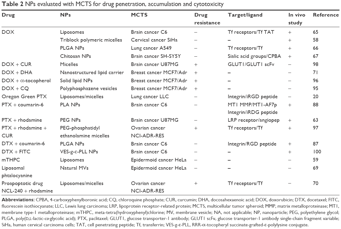 | Table 2 NPs evaluated with MCTS for drug penetration, accumulation and cytotoxicity |
In general terms, the highest cytotoxic effect in MCTS was obtained with NPs exhibiting both high penetration and accumulation. Solomon et al20 showed recently that drug accumulation is associated with greater cytotoxicity even if NPs are restricted to the spheroid periphery. Formulations that penetrate deeper are also cytotoxic, but better effects are obtained when both high accumulation and penetration can be achieved.20 As was already mentioned, drug penetration into the tumor tissue is a major concern in brain cancer management, and many studies with the aim to evaluate the capacity of new nanoformulations to penetrate the tumor tissue used MCTS from brain cancer cell lines (Table 2).
DOX, one of the most commonly used drugs in oncology, has occupied a key place in nanomedicine with several nanoformulations, which were either approved for treatment of patients or subjected to clinical investigations.92 In fact, the serious DOX-induced adverse effects in patients are a major argument to improve its selective delivery to tumors.92 The fluorescent properties of DOX molecule rend the DOX tracing quite easy in organs and cells, and therefore, DOX is considered as an excellent tool for evaluation of NPs in vivo and in vitro.93 PEGylated liposomal formulations of DOX (Doxil®/Caelyx®) were already applied in patients with satisfying results in terms of reduced cardiotoxicity, albeit no significant enhancement of therapeutic efficacy has been achieved.4 The limited tumor distribution of DOX, which was shown to be restricted to the vascular region in vivo and to the outer rim in spheroids, is detrimental to its cytotoxic potential and fully justifies the investigations aiming to increase DOX efficiency, especially by the enhancement of both DOX accumulation and distribution in cancer tissues.53,67 This issue is particularly crucial in the context of multidrug resistance (MDR) that counteracts DOX effects and implies the necessity to administrate DOX together with other chemotherapeutic drugs or MDR modulators.94 In this regard, MCTS from MDR cell lines such as breast cancer MCF7/Adr or ovarian cancer NCI-ADR-RES are useful tools to evaluate innovative solutions to restore DOX cytotoxicity as was shown with intrinsic drug-resistant glioblastoma U87MG cells.70,71,95–98 DOX-loaded nanoplatforms (targeted or nontargeted) that allow the simultaneous administration of several anticancer agents were evaluated in MCTS based on three criteria: accumulation, penetration and cytotoxic effects (Table 2).70,71,95–98 An excellent example of what can be done was the coloading of DOX with the MDR modulator curcumin (CUR) that inhibits different resistance-related pathways.94,98 Polymeric micelles loaded with DOX and/or CUR (zeta potential of −4.4 mV, 15 nm in size) were decorated with a single-chain fragment variable (scFv), smaller, more stable and less immunogenic than IgG antibody, and were addressed against the GLUT1 protein in overexpressed glioblastoma cells. The greater synergism of both drugs when coloaded in the GLUT1 scFv micelles was attributed to the increased drug delivery to cells. By using U87MG spheroids, deeper penetration of DOX into the spheroids was observed and significant cytotoxic effects were measured from 48 h to 5 days after treatment. Since much less DOX was required in monolayer cells to obtain similar cytotoxic effects, the authors pointed out the necessity to use such 3D in vitro model to evaluate the efficacy of nanoformulations.98 A similar approach was previously reported by the same authors with Tf-targeted paclitaxel (PTX) and CUR-coloaded micelles assessed in monolayer and spheroids from MDR ovarian cancer cells and thereafter in xenografted tumors.97 PTX, one of the most effective anticancer agents, is another anticancer drug for which nanovectorization has been considered of major importance. The poor water solubility of the drug significantly limited the development of intravenous PTX formulations and the use of Cremophor® EL (CrEL) as a vehicle generated serious side effects in patients. Therefore, PTX nanoformulations have been designed to improve PTX solubilization, to avoid the use of CrEL and to ensure the stability of the NPs in the blood flow. Abraxane, a PTX albumin-based natural polymer, has been approved for clinical use, and other nanoformulations continue to be developed, some of them being evaluated in ongoing clinical trials.4,99 Docetaxel (DTX), another taxane used as an anticancer drug, is in theory fourfold more potent than PTX but suffered from all disadvantages of chemotherapeutic drugs, such as low water solubility, poor ability to cross the BBB, rapid clearance from the blood and short retention time in tumor.100 For all the reasons given earlier, PTX and DTX are good candidates for nanovectorization even if, unlike DOX, they are not fluorescent molecules. To estimate their distribution in spheroids, Oregon Green PTX was used.20 More generally, fluorescent dyes such as rhodamine,63,70,97 coumarin87,88 or fluorescein isothiocyanate (FITC) are appropriate for this purpose.95
Table 2 indicates that a great variety of NPs were designed to improve the anticancer drug delivery to tumors. The production of biogenic membrane vesicles (MVs) as a drug carrier constituted a particular case. Exosomes and microvesicles, both named extracellular vesicles (EVs), are endogenous particles excreted by a great variety of cell types. EVs play an important role in intercellular communication and cancer development by delivering their biological materiel to target cells. Biocompatibility, low immunogenicity and high stability in circulation are attractive properties of EVs, and among the potential therapeutic applications of EVs, drug delivery received more and more attention.101 By using EVs charged with fusogenic liposomes (MVs), Lee et al69 showed a special way for the payload to disseminate into the spheroid due to the capacity of liposomes to fuse with the cell membrane.66 It seems that successive rounds of MV uptake, liposome delivery and de novo MV production by the cells contributed to deeper distribution of the liposomes into spheroids.66 MVs charged with the hydrophobic PS Zn phtalocyanine (ZnPc) embedded in such liposomes were tested for PDT applications. In spheroids, increased penetration and higher cell content of PS were found, suggesting that MVs delivered the fusogenic liposomes to adjacent cells from the outer rim toward the spheroid center. Taken together, these findings can explain the higher efficacy of the photodynamic treatment obtained in spheroids and in vivo as compared with those obtained with nonfusogenic liposomes.69
Because most effective PSs are lipophilic molecules with a high propensity to aggregate, encapsulation in nanocarriers can highly influence their performance in PDT. In addition, nanocarriers have the advantage to improve tumor selectivity, resulting in an increase in photochemical efficacy while sparing healthy tissues. The PS meta-tetra(hydroxyphenyl)chlorine (mTHPC; Foscan®) is a potent PS approved for the treatment of head and neck carcinoma, but its poor pharmacokinetics properties leading to skin photosensitivity considerably limited its clinical application.4 Liposomal formulations of mTHPC (Foslip®, Fospeg®) provided some improvements with regard to pharmacokinetics in tumor-bearing mice, but the capacity of the mTHPC-loaded liposomes to penetrate the tumor tissue remained unknown until recently.4,102 The high fluorescence quantum yield of mTHPC allows the accurate tracing of PS penetration into the MCTS.32,103 It was shown that irrespective of formulation (free mTHPC, Foslip® or Fospeg®), the PS fluorescence was limited to the outer rim of the spheroid with a slightly higher drug content for liposomal mTHPC formulations. The photocytotoxicity was not significantly different between formulations. Despite the external localization of the PS, photodamage were observed in the internal region of spheroids, suggesting the diffusion of the singlet oxygen, a highly cytotoxic form of reactive oxygen species produced by the photodynamic reaction.59
Of big interest is the consistency of the results obtained in spheroids with tumors established from the same cell type. On the basis of in vivo imaging in tumor-bearing mice, a strong parallel exists in the capacity of NPs to accumulate and to penetrate in cell spheroids as compared with tumor tissue.65,66,87,88 For example, England et al72 have shown that smaller citrate gold NPs exhibit enhancement of diffusion and accumulation in metastatic pancreatic adenocarcinoma spheroids (S2-VP10) compared to larger silica gold NPs. These results were also obtained in S2-VP10 orthotopic xenograft tumor and showed the predictive value of MCTS.104 The same statement holds for growth inhibition effects measured in MCTS and in vivo tumors subjected to NPs’ treatment. The similarity between the evolution of spheroid volume and tumor growth after treatment constitutes a great advantage with respect to 2D cell models.63,87,88,95
Predictive response of MCTS by mathematical models
Nowadays, mathematical modeling is an invaluable tool for understanding the impact of cellular interactions on drug movement within MCTS. Indeed, the physical–chemical parameters of drug accumulation and distribution in tumor tissues can be determined by means of computational modeling. Along with experimental studies, mathematical models provide pertinent perspectives for understanding mechanisms controlling drug penetration in spheroids and their consequences on treatment efficacy. Owing to their spherical symmetry, spheroids can be easily defined mathematically in radial dimensions. Equations obtained are of first order that greatly facilitate the analytical resolution. Many of mathematical models consist of systems of ordinary differential equations that describe the diffusional drug transport coupled with the specific cell binding. The tumor growth velocity is usually implemented through a generalized Darcy’s law,105 while Fick’s laws and Michaelis–Menten equations provide the description of NP diffusion transport and drug-cell binding.
Modeling macromolecule delivery to tumors was based either on finite element models to predict cell culture response to mitotic cell targeting drugs or on numerical solutions to simulate antibody delivery to tumor spheroids,106 thereby assuming their structural uniformity and the heterogeneous distribution of binding sites in MCTS.107,108 All these models focused mainly on the penetration of macromolecules (ie, dextrans and antibodies) in MCTS and showed strong evidence of transport barriers that impair macromolecule diffusion in MCTS. Currently, there is an increasing interest in the use of large NPs such as liposomes and polymeric NPs for drug delivery. Several groups used mathematical models to estimate diffusional and binding characteristics of cationic liposomes with variable charges and sizes.109,110 They separated parameters affecting drug transport in the MCTS into two groups: the parameters depending on liposome to cell interactions (binding constants, uptake and retention rates, termed NP–cell biointerface parameters) and the diffusivity parameters related to MCTS environment (cell and ECM density) and NP characteristics (size, shape, etc.). Gao et al109 successfully simulated diffusion of negatively charged polystyrene beads, near-neutral liposomes and positively charged liposomes with respective diameters of 20, 110 and 130 nm in 3D pharynx FaDu tumor spheroids, while Wientjes et al110 achieved excellent precision in the prediction of the diffusion of NPs with various sizes (20–135 nm) and surface charges (−49 to +44 mV) in MCTS. Finally, very recently, Curtis et al examined the cytotoxicity of novel two- and three-layer gold NPs in heterogeneously vascularized tumors for future in vivo evaluation. They established a complicated mathematical model involving mass equations describing tissue growth combined with diffusion of small molecules (drug, oxygen and cell nutrients) and parameters of drug release from NPs. Moreover, the authors summarized the experimentally estimated coefficients with known literature data to complete the precise simulation of tumor nanotherapy response.111
Two major advantages of the modeling of NPs’ penetration into MCTS could be outlined. On one hand, it provides insight into the factors that affect the distribution of particles in avascular regions of tumors, and on the other hand, modeling helps to delineate the factors related to the improvement of the design of nanotherapeutic agents. By achieving these goals, mathematical models have the potential to provide valuable information to predict therapeutic efficacy of macromolecules or nanotherapeutics.19
Conclusion
Nanomedicine constitutes a rich area of investigations designated to revolutionize anticancer therapy. Progresses achieved in the last 10 years offer new perspectives on key issues for drug delivery optimization and control, selective targeting of cancer cells and early detection of cancer molecular pathways. However, despite attractive concepts already highlighted in many studies, a lot of work should be done to benefit fully from the great potential of nanomedicine. It is of utmost importance to integrate all relevant disciplines, including mathematics, physics, chemistry, biology, pharmacology and clinical oncology, to break through the limits of monodisciplinary knowledge and to connect the huge potential of nanotechnology to the practical needs of medical oncology. It is obvious that a gap remains between in vitro evaluation and clinical translation. Several weaknesses have been identified and require now increased investment by the international scientific community to facilitate nanomedicine translation. Notably, the toxicity of nanomaterials toward human cells and tissues needs further development. Although NPs are designed for specific tumor targeting while avoiding systemic toxicity, NP degradation inside the body, accumulation in internal organs and potential interactions with biological processes remain major concerns. Efforts must also be made to adjust clinical trials to nanomedicine specificities by the careful recruitment of patients with the adequate response to this type of treatment.
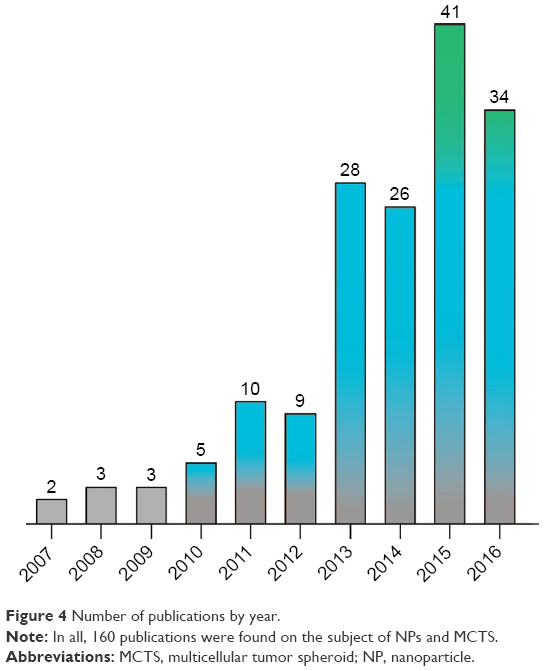
In this context, there is an urgent need to expand the available set of tools for screening high-potential nanodrugs before taking the ultimate step of clinical investigations. The key issue of relevant preclinical models to assess the progress obtained with new nanotherapeutic concepts and guide or stop the project before significant and costly investments is highly timely.112 In vitro, ex vivo and in vivo preclinical models are fundamental requirements to achieve these goals. In addition to in vitro platforms like MCTS, ex vivo organoid models of cancer or microtumors and tissue slices can be integrated into the early phase of nanodrug development.21 In vivo, non-murine models (invertebrates, nematodes or zebrafish) can also be useful for initial triage in drug screening.113 For the respect of vascularity, the in vivo model developed from the chorioallantoic membrane (CAM) of the chicken egg can be considered as an alternative to mammalian models. Grafting a tumor on the CAM is a relatively simple, rapid and inexpensive procedure, which may be used to evaluate nanodrug transport from the vessel compartment to the tumor.114 Finally, new technologies like the most recent innovation in genome engineering are promising for enriching the catalog of animal models with tumor that fully mimics clinical cancer. The advantage of such technology is to obtain spontaneous or induced tumor growth with similar microenvironment, including vascular network, stromal cells and immune reactions.21 In fact, none of the single model can provide rapid and specific information needed for clinical translation, and 2D or 3D in vitro models are still required to provide basic data on the behavior of nanodrugs in contact with the cell components. MCTS constitutes a classical in vitro 3D model, which raises nowadays a renewed interest in the field of nanodrug delivery. A bibliographic search on MEDLINE for the terms “nanoparticles” and “tumor cell spheroids” would reveal ~160 papers published over 10 years, most of them (138) over the last 5 years (Figure 4). Undoubtedly, the constant evolution of spheroid design should contribute to more and more widespread use of this in vitro model for drug screening purpose. In fact, development of scaffold-based techniques or coculture with fibroblast has allowed to closely mimic in vivo situation and microfluidic devices along with the high-yield production of homogeneous MCTS. Another crucial characteristic is the ability to produce MCTS from various cancer cell lines such as breast, colon, ovarian, brain or prostate cancers. Using MCTS to determine tumor penetration, accumulation and antitumor activity of newly designed nanotherapeutics is becoming an essential step to demonstrate their potential value for cancer treatment.
 | Figure 4 Number of publications by year. |
Acknowledgments
This work was supported by the French “Ligue Nationale contre le Cancer (CCIR-GE)” and the Institut de Cancérologie de Lorraine. Aigul Kulmukhamedova acknowledges the JCS “Center for International Programs”, Kazakhstan, for financial support.
Disclosure
The authors report no conflicts of interest in this work.
References
Zamboni W, Torchilin V, Patri A, et al. Best practices in cancer nanotechnology: perspectives from NCI nanotechnology alliance. Clin Cancer Res. 2012;18(12):3229–3241. | ||
Duncan R, Gaspar R. Nanomedicine(s) under the microscope. Mol Pharm. 2011;8(6):2101–2141. | ||
Wicki A, Witzigmann D, Balasubramanian V, Huwyler J. Nanomedecine in cancer therapy: challenges, opportunities and clinical applications. J Control Release. 2015;200:138–157. | ||
Marchal S, Hor AE, Millard M, Gillon V, Bezdetnaya L. Anticancer drug delivery: an update on clinically applied nanotherapeutics. Drugs. 2015;75(14):1601–1611. | ||
Netti P, Berk D, Swartz M, Grodzinsky A, Jain R. Role of extracellular matrix assembly in interstitial transport in solid tumors. Cancer Res. 2000;60(9):2497–2503. | ||
Davies CL, Berk D, Pluen A, Jain R. Comparison of IgG diffusion and extracellular matrix composition in rhabdomyosarcomas grown in mice versus in vitro as spheroids reveals the role of host stromal cells. Br J Cancer. 2002;86(10):1639–1644. | ||
Prabhakar U, Maeda H, Jain R, et al. Challenges and key considerations of the enhanced permeability and retention effect for nanomedicine drug delivery in oncology. Cancer Res. 2013;73(8):2412–2417. | ||
Au J, Jang S, Zheng J, et al. Determinants of drug delivery and transport to solid tumors. J Control Release. 2001;74(1–3):31–46. | ||
Junttila M, de Sauvage F. Influence of tumour micro-environment heterogeneity on therapeutic response. Nature. 2013;501:346–354. | ||
Jain R. Delivery of novel therapeutic agents in tumors: physiological barriers and strategies. J Natl Cancer Inst. 1989;81(8):570–576. | ||
Nagy J, Dvorak H. Heterogeneity of the tumor vasculature: the need for new tumor blood vessel type-specific targets. Clin Exp Metastasis. 2012;29(7):657–662. | ||
Stylianopoulos T, Martin J, Chauhan V, et al. Causes, consequences, and remedies for growth-induced solid stress in murine and human tumors. Proc Natl Acad Sci U S A. 2012;109(38):15101–15108. | ||
Padera T, Stoll B, Tooredman J, Capen D, di Tomaso E, Jain R. Pathology: cancer cells compress intratumour vessels. Nature. 2004;427(6976):695. | ||
Hobbs S, Monsky W, Yuan F, et al. Regulation of transport pathways in tumor vessels: role of tumor type and microenvironment. Proc Natl Acad Sci U S A. 1998;95(8):4607–4612. | ||
Jain R. Delivery of molecular and cellular medicine to solid tumors. Adv Drug Deliv Rev. 2012;64(suppl):353–365. | ||
Matsumura Y, Maeda H. A new concept for macromolecular therapeutics in cancer chemotherapy: mechanism of tumoritropic accumulation of proteins and the antitumor agent smancs. Cancer Res. 1986;46(12):6387–6392. | ||
Bertrand N, Wu J, Xu X, Kamaly N, Farokhzad O. Cancer nanotechnology: the impact of passive and active targeting in the era of modern cancer biology. Adv Drug Deliv Rev. 2014;66:2–25. | ||
Maeda H, Tsukigawa K, Fang J. A retrospective 30 years after discovery of the enhanced permeability and retention effect of solid tumors: next-generation chemotherapeutics and photodynamic therapy-problems, solutions, and prospects. Microcirculation. 2016;23(3):173–182. | ||
Mehta G, Hsiao A, Ingram M, Luker G, Takayama S. Opportunities and challenges for use of tumor spheroids as models to test drug delivery and efficacy. J Control Release. 2012;164(2):192–204. | ||
Solomon M, Lemera J, D’Souza G. Development of an in vitro tumor spheroid culture model amenable to high-throughput testing of potential anticancer nanotherapeutics. J Liposome Res. 2016;26(3):246–260. | ||
Gould S, Junttila M, de Sauvage F. Translational value of mouse models in oncology drug development. Nat Med. 2015;21(5):431–439. | ||
Hickman J, Graeser R, de Hoogt R, et al; IMI PREDECT Consortium. Three-dimensional models of cancer for pharmacology and cancer cell biology: capturing tumor complexity in vitro/ex vivo. Biotechnol J. 2014;9(9):1115–1128. | ||
Katt M, Placone A, Wong A, Xu Z, Searson P. In vitro tumor models: advantages, disadvantages, variables, and selecting the right platform. Front Bioeng Biotechnol. 2016;4(12):1–14. | ||
Khaitan D, Dwarakanath B. Multicellular spheroids as an in vitro model in experimental oncology: applications in translational medicine. Expert Opin Drug Discov. 2006;1(7):663–675. | ||
Patel N, Aryasomayajula B, Abouzeid A, Torchilin V. Cancer cell spheroids for screening of chemotherapeutics and drug-delivery systems. Ther Deliv. 2015;6(4):509–520. | ||
Weiswald L, Bellet D, Dangles-Marie V. Spherical cancer models in tumor biology. Neoplasia. 2015;17(1):1–15. | ||
Sutherland R. Cell and environment interactions in tumor microregions: the multicell spheroid model. Science. 1988;240(4849):177–184. | ||
Glicklis R, Merchuk J, Cohen S. Modeling mass transfer in hepatocyte spheroids via cell viability, spheroid size, and hepatocellular functions. Biotechnol Bioeng. 2004;86(6):672–680. | ||
Langan L, Dodd N, Owen S, Purcell W, Jackson S, Jha A. Direct measurements of oxygen gradients in spheroid culture system using electron parametric resonance oximetry. PLoS One. 2016;11(2):1–13. | ||
Foster T, Hartley D, Nichols M, Hilf R. Fluence rate effects in photodynamic therapy of multicell tumor spheroids. Cancer Res. 1993;53(6):1249–1254. | ||
Buffa F, West C, Byrne K, Moore J, Nahum A. Radiation response and cure rate of human colon adenocarcinoma spheroids of different size: the significance of hypoxia on tumor control modelling. Int J Radiat Oncol Biol Phys. 2001;49(4):1109–1118. | ||
Coutier S, Mitra S, Bezdetnaya L, et al. Effects of fluence rate on cell survival and photobleaching in metaTetra-(hydroxyphenyl)chlorin-photosensitized Colo 26 multicell tumor spheroids. Photochem Photobiol. 2001;73(3):297–303. | ||
Kim S, Lee E, Kuh H. Co-culture of 3D tumor spheroids with fibroblasts as a model for epithelial-mesenchymal transition in vitro. Exp Cell Res. 2015;335(2):187–196. | ||
Priwitaningrum D, Bondé J, Sridhar A, et al. Tumor stroma-containing 3D spheroid arrays: a tool to study nanoparticle penetration. J Control Release. 2016;244(pt B):257–268. | ||
Santini M, Rainaldi G. Three-dimensional spheroid model in tumor biology. Pathobiology. 1999;67(3):148–157. | ||
Patra B, Peng C, Liao W, Lee C, Tung Y. Drug testing and flow cytometry analysis on a large number of uniform sized tumor spheroids using a microfluidic device. Sci Rep. 2016;6:21061. | ||
Schreiber-Brynzak E, Klapproth E, Unger C, et al. Three-dimensional and co-culture models for preclinical evaluation of metal-based anticancer drugs. Invest New Drugs. 2015;33(4):835–847. | ||
Timmins N, Dietmair S, Nielsen L. Hanging-drop multicellular spheroids as a model of tumour angiogenesis. Angiogenesis. 2004;7(2):97–103. | ||
Thoma C, Zimmermann M, Agarkova I, Kelm J, Krek W. 3D cell culture systems modeling tumor growth determinants in cancer target discovery. Adv Drug Deliv Rev. 2014;69–70:29–41. | ||
Hirschhaeuser F, Walenta S, Mueller-Klieser W. Efficacy of catumaxomab in tumor spheroid killing is mediated by its trifunctional mode of action. Cancer Immunol Immunother. 2010;59(11):1675–1684. | ||
Friedrich J, Seidel C, Ebner R, Kunz-Schughart L. Spheroid-based drug screen: considerations and practical approach. Nat Protoc. 2009;4(3):309–324. | ||
Edmondson R, Broglie J, Adcock A, Yang L. Three-dimensional cell culture systems and their applications in drug discovery and cell-based biosensors. Assay Drug Dev Technol. 2014;12(4):207–218. | ||
Mikhail A, Eetezadi S, Allen C. Multicellular tumor spheroids for evaluation of cytotoxicity and tumor growth inhibitory effects of nanomedicines in vitro: a comparison of Docetaxel-loaded block copolymer micelles and Taxotere. PLoS One. 2013;8(4):e62630. | ||
Perche F, Torchilin V. Cancer cell spheroids as a model to evaluate chemotherapy protocols. Cancer Biol Ther. 2012;13(12):1205–1213. | ||
Franken N, Rodermond H, Stap J, Haveman J, van Bree C. Clonogenic assay of cells in vitro. Nat Protoc. 2006;1(5):2315–2319. | ||
Karlsson H, Fryknas M, Larsson R, Nygren P. Loss of cancer drug activity in colon cancer HCT-116 cells during spheroid formation in a new 3-D spheroid cell culture system. Exp Cell Res. 2012;318(13):1577–1585. | ||
Däster S, Amatruda N, Calabrese D, et al. Induction of hypoxia and necrosis in multicellular tumor spheroids is associated with resistance to chemotherapy treatment. Oncotarget. 2017;8(1):1725–1736. | ||
Shield K, Ackland M, Ahmed N, Rice G. Multicellular spheroids in ovarian cancer metastases: biology and pathology. Gynecol Oncol. 2009;113(1):143–148. | ||
Desoize B, Jardillier J. Multicellular resistance: a paradigm for clinical resistance? Crit Rev Oncol Hematol. 2000;36(2–3):193–207. | ||
Makhija S, Taylor D, Gibb R, Gerçel-Taylor C. Taxol-induced Bcl-2 phosphorylation in ovarian cancer cell monolayer and spheroids. Int J Oncol. 1999;14(3):515–521. | ||
Hoffmann T, Schirlau K, Sonkoly E, et al. A novel mechanism for anti-EGFR antibody action involves chemokine-mediated leukocyte infiltration. Int J Cancer. 2009;124(11):2589–2596. | ||
Gottfried E, Kunz-Schughart L, Andreesen R, Kreutz M. Brave little world: spheroids as an in vitro model to study tumor-immune-cell interactions. Cell Cycle. 2006;5(7):691–695. | ||
Lankelma J, Dekker H, Luque F, et al. Doxorubicin gradients in human breast cancer. Clin Cancer Res. 1999;5(7):1703–1707. | ||
Giordano S, Morosi L, Veglianese P, et al. A registry on distal popliteal and infrapopliteal revascularization with coronary drug-eluting stents. Sci Rep. 2016;6:37027. | ||
Patel K, Trédan O, Tannock I. Distribution of the anticancer drugs doxorubicin, mitoxantrone and topotecan in tumors and normal tissues. Cancer Chemother Pharmacol. 2013;72(1):127–138. | ||
Goodman T, Chen J, Matveev K, Pun S. Spatio-temporal modeling of nanoparticle delivery to multicellular tumor spheroids. Biotechnol Bioeng. 2008;101(2):388–399. | ||
Kostarelos K, Emfietzoglou D, Papakostas A, Yang W, Ballangrud Ä, Sgouros G. Binding and interstitial penetration of liposomes within avascular tumor spheroids. Int J Cancer. 2004;112(4):713–722. | ||
Kim T, Mount C, Gombotz W, Pun S. The delivery of doxorubicin to 3-D multicellular spheroids and tumors in a murine xenograft model using tumor-penetrating triblock polymeric micelles. Biomaterials. 2010;31(28):7386–7397. | ||
Gaio E, Scheglmann D, Reddi E, Moret F. Uptake and photo-toxicity of Foscan®, Foslip® and Fospeg® in multicellular tumor spheroids. J Photochem Photobiol B. 2016;161:244–252. | ||
Huo S, Ma H, Huang K, et al. Superior penetration and retention behavior of 50 nm gold nanoparticles in tumors. Cancer Res. 2013;73(1):319–330. | ||
Huang K, Ma H, Liu J, et al. Size-dependent localization and penetration of ultrasmall gold nanoparticles in cancer cells, multicellular spheroids, and tumors in vivo. ACS Nano. 2012;6(5):4483–4493. | ||
Mikhail A, Eetezadi S, Ekdawi S, Stewart J, Allen C. Image-based analysis of the size- and time-dependent penetration of polymeric micelles in multicellular tumor spheroids and tumor xenografts. Int J Pharm. 2014;464(1–2):168–177. | ||
Xin H, Sha X, Jiang X, Zhang W, Chen L, Fang X. Anti-glioblastoma efficacy and safety of paclitaxel-loading Angiopep-conjugated dual targeting PEG-PCL nanoparticles. Biomaterials. 2012;33(32):8167–8176. | ||
Sims LB, Curtis LT, Frieboes HB, Steinbach-Rankins JM. Enhanced uptake and transport of PLGA-modified nanoparticles in cervical cancer. J Nanobiotechnology. 2016;14:33. | ||
Zong T, Mei L, Gao H, et al. Synergistic dual-ligand doxorubicin liposomes improve targeting and therapeutic efficacy of brain glioma in animals. Mol Pharm. 2014;11(7):2346–2357. | ||
Guo Y, Wang L, Lv P, Zhang P. Transferrin-conjugated doxorubicin-loaded lipid-coated nanoparticles for the targeting and therapy of lung cancer. Oncol Lett. 2015;9(3):1065–1072. | ||
Wang X, Tang H, Wang C, Zhang J, Wu W, Jiang X. Phenylboronic acid-mediated tumor targeting of chitosan nanoparticles. Theranostics. 2016;6(9):1378–1392. | ||
Lee W, Guo W, Ho V, et al. Delivery of doxorubicin and paclitaxel from double-layered microparticles: the effects of layer thickness and dual-drug vs. single-drug loading. Acta Biomater. 2015;27:53–65. | ||
Lee J, Kim J, Jeong M, et al. Liposome-based engineering of cells to package hydrophobic compounds in membrane vesicles for tumor penetration. Nano Lett. 2015;15(5):2938–2944. | ||
Pattni B, Nagelli S, Aryasomayajula B, et al. Targeting of micelles and liposomes loaded with the pro-apoptotic drug, NCL-240, into NCI/ADR-RES cells in a 3D spheroid model. Pharm Res. 2016;33(10):2540–2551. | ||
Mussi S, Sawant R, Perche F, et al. Novel nanostructured lipid carrier co-loaded with doxorubicin and docosahexaenoic acid demonstrates enhanced in vitro activity and overcomes drug resistance in MCF-7/Adr cells. Pharm Res. 2014;31(8):1882–1892. | ||
England CG, Priest T, Zhang G, et al. Enhanced penetration into 3D cell culture using two and three layered gold nanoparticles. Int J Nanomedicine. 2013;8:3603–3617. | ||
Goodman T, Olive P, Pun S. Increased nanoparticle penetration in collagenase treated multicellular spheroids. Int J Nanomedecine. 2007;2(2):265–274. | ||
Kumar A, Ma H, Zhang X, et al. Gold nanoparticles functionalized with therapeutic and targeted peptides for cancer treatment. Biomaterials. 2012;33(4):1180–1189. | ||
Ernsting M, Murakami M, Roy A, Li S. Factors controlling the pharmacokinetics, biodistribution and intratumoral penetration of nanoparticles. J Control Release. 2013;172(3):782–794. | ||
Hinger D, Navarro F, Käch A, et al. Photoinduced effects of m-tetrahydroxyphenylchlorin loaded lipid nanoemulsions on multicellular tumor spheroids. J Nanobiotechnology. 2016;14(1):68. | ||
Agarwal R, Jurney P, Raythatha M, et al. Effect of shape, size, and aspect ratio on nanoparticle penetration and distribution inside solid tissues using 3D spheroid models. Adv Healthc Mater. 2015;4(15):2269–2280. | ||
Li S, Huang L. Pharmacokinetics and biodistribution of nanoparticles. Mol Pharm. 2008;5(4):496–504. | ||
Hrkach J, Von Hoff D, Mukkaram Ali M, et al. Preclinical development and clinical translation of a PSMA-targeted docetaxel nanoparticle with a differentiated pharmacological profile. Sci Transl Med. 2012;4(128):128ra39. | ||
Stylianopoulos T, Poh M, Insin N, et al. Diffusion of particles in the extracellular matrix: the effect of repulsive electrostatic interactions. Biophys J. 2010;99(5):1342–1349. | ||
Ma H, Jiang Q, Han S, et al. Multicellular tumor spheroids as an in vivo-like tumor model for three-dimensional imaging of chemotherapeutic and nano material cellular penetration. Mol Imaging. 2012;11(6):487–498. | ||
Campbell R, Fukumura D, Brown E, et al. Cationic charge determines the distribution of liposomes between the vascular and extravascular compartments of tumors. Cancer Res. 2002;62(23):6831–6836. | ||
Lieleg O, Baumgärtel R, Bausch A. Selective filtering of particles by the extracellular matrix: an electrostatic bandpass. Biophys J. 2009;97(6):1569–1577. | ||
Zhao J, Lu H, Xiao P, Stenzel M. Cellular uptake and movement in 2D and 3D multicellular breast cancer models of Fructose-based cylindrical micelles is dependent on the rod length. ACS Appl Mater Interfaces. 2016;8(26):16622–16630. | ||
Lee K, Hubbard L, Hern S, Yildiz I, Gratzl M, Steinmetz N. Shape matters: the diffusion rates of TMV rods and CPMV icosahedrons in a spheroid model of extracellular matrix are distinct. Biomater Sci. 2013;1(6):1–12. | ||
Chauhan V, Popović Z, Chen O, et al. Fluorescent nanorods and nanospheres for real-time in vivo probing of nanoparticle shape-dependent tumor penetration. Angew Chem Int Ed. 2012;50(48):11417–11420. | ||
Shi K, Zhou J, Zhang Q, et al. Arginine-Glycine-Aspartic acid-modified lipid-polymer hybrid nanoparticles for docetaxel delivery in glioblastoma multiforme. J Biomed Nanotechnol. 2015;11(3):382–391. | ||
Gu G, Gao X, Hu Q, et al. The influence of the penetrating peptide iRGD on the effect of paclitaxel-loaded MT1-AF7p-conjugated nanoparticles on glioma cells. Biomaterials. 2013;34(21):5138–5148. | ||
Kirpotin D, Drummond D, Shao Y, et al. Antibody targeting of long-circulating lipidic nanoparticles does not increase tumor localization but does increase internalization in animal models. Cancer Res. 2006;66(13):6732–6740. | ||
Albanese A, Lam A, Sykes E, Rocheleau J, Chan W. Tumour-on-a-chip provides an optical window into nanoparticle tissue transport. Nat Commun. 2013;4:2718. | ||
Gao H, Pang Z, Jiang X. Targeted delivery of nano-therapeutics for major disorders of the central nervous system. Pharm Res. 2013;30(10):2485–2498. | ||
Cagel M, Grotz E, Bernabeu E, Moretton M, Chiappetta D. Doxorubicin: nanotechnological overviews from bench to bedside. Drug Discov Today. 2017;22(2):270–281. | ||
Mohan P, Rapoport N. Doxorubicin as a molecular nanotheranostic agent: effect of doxorubicin encapsulation in micelles or nanoemulsions on the ultrasound-mediated intracellular delivery and nuclear trafficking. Mol Pharm. 2010;7(6):1959–1973. | ||
Iyer A, Singh A, Ganta S, Amiji M. Role of integrated cancer nanomedicine in overcoming drug resistance. Adv Drug Deliv Rev. 2013;65(13–14):1784–1802. | ||
Xu J, Zhu X, Qiu L. Polyphosphazene vesicles for co-delivery of doxorubicin and chloroquine with enhanced anticancer efficacy by drug resistance reversal. Int J Pharm. 2016;498(1–2):70–81. | ||
Oliveira M, Aryasomayajula B, Pattni B, Mussi S, Ferreira L, Torchilin V. Solid lipid nanoparticles co-loaded with doxorubicin and a-tocopherol succinate are effective against drug-resistant cancer cells in monolayer and 3-D spheroid cancer cell models. Int J Pharm. 2016;512(1):292–300. | ||
Sarisozen C, Abouzeid A, Torchilin V. The effect of co-delivery of paclitaxel and curcumin by transferrin-targeted PEG-PE-based mixed micelles on resistant ovarian cancer in 3-D spheroids and in vivo tumors. Eur J Pharm Biopharm. 2014;88(2):539–550. | ||
Sarisozen C, Dhokai S, Tsikudo E, Luther E, Rachman I, Torchilin V. Nanomedicine based curcumin and doxorubicin combination treatment of glioblastoma with scFv-targeted micelles: in vitro evaluation on 2D and 3D tumor models. Eur J Pharm Biopharm. 2016;108:54–67. | ||
Bernabeu E, Cagel M, Lagomarsino E, Moretton M, Chiappetta D. Paclitaxel: what has been done and the challenges remain ahead. Int J Pharm. 2017;526(1–2):474–495. | ||
Xu H, Mao K, Lu C, et al. An injectable acellular matrix scaffold with absorbable permeable nanoparticles improves the therapeutic effects of docetaxel on glioblastoma. Biomaterials. 2016;107:44–60. | ||
Stremersch S, De Smedt S, Raemdonck K. Therapeutic and diagnostic applications of extracellular vesicles. J Control Release. 2016;244(pt B):167–183. | ||
Reshetov V, Lassalle H, François A, et al. Photodynamic therapy with conventional and PEGylated liposomal formulations of mTHPC (temoporfin): comparison of treatment efficacy and distribution characteristics in vivo. Int J Nanomedecine. 2013;8:3817–3831. | ||
Yakavets I, Yankovsky I, Millard M, et al. The alteration of temoporfin distribution in multicellular tumor spheroids by β-cyclodextrins. Int J Pharm. 2017;529(1–2):568–575. | ||
England CG, Huang JS, James KT, Zhang G, Gobin AM, Frieboes HB. Detection of phosphatidylcholine-coated gold nanoparticles in orthotopic pancreatic adenocarcinoma using hyperspectral imaging. PLoS One. 2015;10(6):e0129172. | ||
Macklin P, McDougall S, Anderson ARA, Chaplain MAJ, Cristini V, Lowengrub J. Multiscale modelling and nonlinear simulation of vascular tumour growth. J Math Biol. 2009;58(4–5):765–798. | ||
Ward J, King J. Mathematical modelling of drug transport in tumour multicell spheroids and monolayer cultures. Math Biosci. 2003;181(2):177–207. | ||
Graff C, Wittrup K. Theoretical analysis of antibody targeting of tumor spheroids: importance of dosage for penetration, and affinity for retention. Cancer Res. 2003;63(6):1288–1296. | ||
Wenning L, Murphy R. Coupled cellular trafficking and diffusional limitations in delivery of immunotoxins to multicell tumor spheroids. Biotechnol Bioeng. 1999;62(5):562–575. | ||
Gao Y, Li M, Chen B, et al. Predictive models of diffusive nanoparticle transport in 3-dimensional tumor cell spheroids. AAPS J. 2013;15(3):816–831. | ||
Wientjes M, Yeung B, Lu Z, Wientjes M, Au J. Predicting diffusive transport of cationic liposomes in 3-dimensional tumor spheroids. J Control Release. 2014;192:10–18. | ||
Curtis LT, England CG, Wu M, Lowengrub J, Frieboes HB. An interdisciplinary computational/experimental approach to evaluate drug-loaded gold nanoparticle tumor cytotoxicity. Nanomedicine (Lond). 2016;11(3):197–216. | ||
Hare JI, Lammers T, Ashford MB, Puri S, Storm G, Barry ST. Challenges and strategies in anti-cancer nanomedicine development: an industry perspective. Adv Drug Deliv Rev. 2017;108:25–38. | ||
Day C, Merlino G, Van Dyke T. Preclinical mouse cancer models: a maze of opportunities and challenges. Cell. 2015;163(1):39–53. | ||
Vargas A, Zeisser-Labouèbe M, Lange N, Gurny R, Delie F. The chick embryo and its chorioallantoic membrane (CAM) for the in vivo evaluation of drug delivery systems. Adv Drug Deliv Rev. 2007;59(11):1162–1176. |
 © 2017 The Author(s). This work is published and licensed by Dove Medical Press Limited. The
full terms of this license are available at https://www.dovepress.com/terms
and incorporate the Creative Commons Attribution
- Non Commercial (unported, 3.0) License.
By accessing the work you hereby accept the Terms. Non-commercial uses of the work are permitted
without any further permission from Dove Medical Press Limited, provided the work is properly
attributed. For permission for commercial use of this work, please see paragraphs 4.2 and 5 of our Terms.
© 2017 The Author(s). This work is published and licensed by Dove Medical Press Limited. The
full terms of this license are available at https://www.dovepress.com/terms
and incorporate the Creative Commons Attribution
- Non Commercial (unported, 3.0) License.
By accessing the work you hereby accept the Terms. Non-commercial uses of the work are permitted
without any further permission from Dove Medical Press Limited, provided the work is properly
attributed. For permission for commercial use of this work, please see paragraphs 4.2 and 5 of our Terms.