Back to Journals » International Medical Case Reports Journal » Volume 19
Dicephalus Tetrabrachius Parapagus Conjoined Twins: A Case Report from Orotta National Maternity Referral Hospital, Eritrea
Authors Ghirmay R, Abdu N ![]() , Zerezghi H
, Zerezghi H
Received 15 January 2026
Accepted for publication 24 March 2026
Published 29 March 2026 Volume 2026:19 596524
DOI https://doi.org/10.2147/IMCRJ.S596524
Checked for plagiarism Yes
Review by Single anonymous peer review
Peer reviewer comments 2
Editor who approved publication: Dr Xudong Zhu
Ruth Ghirmay,1 Nuru Abdu,2 Habteselassie Zerezghi1
1Department of Obstetrics and Gynecology, Orotta National Referral Maternity and Teaching Hospital, Asmara, Eritrea; 2Medicine Information Services Unit, Pharmacy Services Division, Department of Medical Services, Ministry of Health, Asmara, Eritrea
Correspondence: Nuru Abdu, Medicine Information Services Unit, Pharmacy Services Division, Department of Medical Services, Ministry of Health, Asmara, Eritrea, Tel +291-7262928, Email [email protected]
Background: Conjoined twins (CT) are exceptionally rare congenital anomalies, with an estimated incidence of 1 in 50,000 to 200,000 births, and they pose substantial diagnostic and therapeutic challenges. When the condition is not identified before labour, the rigid fetal mass may result in absolute mechanical obstruction, frequently leading to severe maternal complications, including uterine rupture. This risk is particularly pronounced in low-resource settings, where delayed presentation and limited access to timely surgical intervention further increase the likelihood of adverse maternal outcomes.
Case Summary: A 26-year-old woman (gravida 4, para 3) was referred to Orotta National Maternity Referral Hospital from a peripheral clinic with features of severe obstructed labour and evolving haemodynamic compromise. Intrapartum assessment confirmed the stillbirth of conjoined twins with dicephalus tetrabrachius parapagus morphology, characterized by fusion at the lower torso and pelvis. Initial laboratory investigations demonstrated significant anaemia.
Intervention and Outcome: An emergency laparotomy was performed to relieve the obstruction, and the fetal mass was delivered through a classical uterine incision. Intraoperative findings revealed an extensive uterine rupture, involving approximately half the uterine length and extending into the left broad ligament. Owing to the severity of the rupture and the need for rapid and definitive haemorrhage control, a life-saving subtotal hysterectomy was performed. The patient received blood transfusions and comprehensive postoperative care and was successfully stabilised and recovered from the event.
Conclusion: This case illustrates a rare and life-threatening presentation of undiagnosed dicephalus parapagus conjoined twins resulting in obstructed labour and uterine rupture in an unscarred uterus. It highlights the crucial role of early and adequate antenatal screening and emphasizes that prompt, definitive surgical management, such as subtotal hysterectomy, remains central to maternal survival in catastrophic obstetric emergencies, particularly in resource-limited settings complicated by massive haemorrhage.
Keywords: conjoined twins, thoracopagus twins, obstructed labour, subtotal hysterectomy, emergency obstetric surgery, Eritrea
Introduction
Conjoined twins represent a distinct and rare form of monozygotic twinning, arising from incomplete cleavage of the zygote approximately 13–15 days after fertilization, resulting in varying degrees of embryonic fusion.1,2 Classification is based on the anatomical site of fusion, with recognized types including thoracopagus (fusion at the chest, often involving a shared heart), omphalopagus (fusion at the anterior abdominal wall, commonly with shared hepatic or gastrointestinal structures), craniopagus (cranial fusion), ischiopagus (pelvic fusion), pygopagus (sacral fusion), and dicephalus parapagus (two heads on a single thoracoabdominal trunk, sometimes with four upper limbs, termed tetrabrachius).2,3 Among these, dicephalus parapagus twins are exceptionally rare.4,5 This configuration poses substantial obstetric challenges, including a high risk of obstructed labour, potential misdiagnosis as a multiple gestation, and complex intrapartum management decisions.6 Owing to the extent of shared organs and anatomical complexity, conjoined twin pregnancies are associated with extremely high perinatal mortality, with more than half resulting in stillbirth or neonatal death within the first 24 hours of life.3,7
When antenatal diagnosis is missed, labour complicated by conjoined twins may convert a rare fetal anomaly into a severe obstetric emergency.6 Prolonged obstructed labour is a principal risk factor for uterine rupture in an otherwise unscarred uterus, a complication associated with markedly increased maternal and perinatal morbidity and mortality, particularly in settings with limited access to timely surgical intervention and blood transfusion services.7–9
The clinical course in this case reflects the convergence of three critical factors – undiagnosed complex conjoined twins, obstructed labour, and subsequent uterine rupture. The favourable maternal outcome underscores the importance of prompt, definitive surgical management and offers valuable insights for obstetric teams working in resource-limited environments.
Case Presentation
Patient Demographic and History
A 26-year-old woman (gravida 4, para 3) was referred from a peripheral clinic to Orotta National Referral Maternity Hospital following a prolonged second stage of labour. The referring peripheral clinic had no access to obstetric ultrasound, and antenatal care was primarily based on clinical examination and fetoscope monitoring. There was no documented first, second, or third trimester ultrasound examination prior to referral. Her obstetric history included three previous uncomplicated vaginal deliveries, with no history of uterine surgery, suggesting an unscarred uterus. On presentation, she exhibited acute signs of shock, and the fetal mass was partially delivered, described as “head and hands out per vagina”. The referring diagnosis suggested either a “locked twin” or a “head trapped in the perineum,” indicating severe mechanical obstruction. The combination of prolonged labour and delayed referral placed her at high risk for acute uterine compromise.
Initial Clinical Findings and Diagnosis
On arrival, the patient appeared critically ill, with unrecordable blood pressure, a pulse of 120 beats per minute, cold extremities, and an oxygen saturation of 98% on room air. She demonstrated mild conjunctival pallor without scleral icterus. Respiratory examination revealed bilateral air entry with clear breath sounds, while cardiovascular assessment showed tachycardia without murmurs or gallop rhythms. Abdominal palpation revealed generalized tenderness, with fetal parts palpable through the uterine wall. On vaginal examination, the fetal head and hands were impacted at the introitus, with partial delivery of the head and neck. The vaginal canal showed evidence of trauma, minimal bleeding, and urinary catheterization was technically challenging due to pelvic distortion caused by the impacted fetal mass.
Diagnostic Test Results
Preoperative laboratory testing revealed significant anemia (hemoglobin 9.2 g/dL, hematocrit 27.9%) consistent with both chronic multiparity and acute blood loss.10–12 Red cell distribution width was elevated at 52.1 fL, suggesting anisocytosis, likely compounded by acute hemodynamic stress. White blood cell count was within normal limits (6.83 × 10³/µL), reducing the immediate concern for systemic infection.
Surgical Management and Intraoperative Findings
The inability to deliver the complex fetal mass vaginally, coupled with the imminent risk to maternal life, necessitated immediate emergency laparotomy. A focused emergency ultrasonography demonstrated a large, complex fetal mass, precluding accurate measurement of abdominal circumference, with absent fetal cardiac activity, a finding consistent with a severe congenital anomaly, and detected hemoperitoneum. Following detailed discussion with the patient and her husband regarding the critical condition and possible life-saving interventions, informed consent was obtained, with explicit emphasis on prioritizing maternal survival.
Preoperatively, two large-bore intravenous lines were secured, and broad-spectrum intravenous antibiotics (ceftriaxone and metronidazole) were administered. An emergency laparotomy was performed via a midline vertical skin incision extending approximately 2 cm above the umbilicus, and the abdomen was entered layer by layer. Upon entering the peritoneal cavity, a large hematoma was encountered. Intraoperative findings revealed a severe uterine rupture with lateral extension into the broad ligament and adjacent uterosacral ligament; an anatomically significant finding associated with active haemorrhage.
A classical (vertical) uterine incision was performed to obtain maximum uterine exposure and facilitate rapid fetal extraction. Despite this approach, delivery of the fetal mass remained impossible due to the extent of impaction and fusion. Given the non-viability of the fetuses and the escalating risk to the mother, fetal decapitation was done as a life-saving maneuver, allowing extraction of the conjoined stillborn twins. The placenta was found to be anteriorly located and was removed by controlled cord traction.
Further inspection demonstrated a massive, ragged uterine rupture, extending longitudinally and involving approximately half the uterine length, suggesting disruption of the uterine corpus with possible lower segment involvement. The extensive tissue friability, the length of the tear, and uncontrolled haemorrhage originating from the broad ligament injury rendered primary uterine repair technically unfeasible and prohibitively risky. Consequently, a definitive life-saving subtotal hysterectomy was performed. Intraoperatively, the patient received a total of 3 litres of normal saline and 4 units of blood, along with 4 units of fresh frozen plasma.
The procedure adhered to established guidelines for managing catastrophic uterine rupture in extremis13,14 and aligns with evidence supporting subtotal hysterectomy over complex repair in such scenarios.15,16 The bladder was inspected and remained intact, and the abdomen was closed in layers after achieving hemostasis.
Anatomical Classification of Fetal Anomaly
Gross examination confirmed two distinct heads (dicephalus) and four upper limbs (tetrabrachius), with fusion along the lower torso and a shared pelvic structure [Figure 1]. This morphology is consistent with dicephalus tetrabrachius parapagus, a severe variant of lateral fusion.17,18 The rigid shared pelvis accounted for the obstructed labour and subsequent uterine rupture.
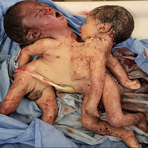 |
Figure 1 Image of dicephalus tetrabrachius parapagus conjoined twins. |
Postoperative Course
Postoperatively, the patient received aggressive fluid resuscitation, continued broad-spectrum antibiotics (ceftriaxone and metronidazole for 7 days), analgesia (diclofenac), and hemodynamic support. Anticipating ongoing anemia, additional blood products were reserved. She developed a transient paralytic ileus, managed conservatively with nil per os, intravenous fluids, electrolyte optimization, and gradual reintroduction of oral intake. Drain output decreased steadily, and the Foley catheter remained for seven days due to extensive pelvic dissection. Laboratory tests stabilized, with hemoglobin rising to borderline 10 g/dL following iron supplementation. Maternal hemodynamic stability and recovery were achieved, reflecting successful resuscitation and definitive surgical management.
Fetal Outcome
The conjoined twins were stillborn, reflecting the high perinatal mortality associated with this anomaly and the prolonged obstructed labour.3,4 The fetal outcome underscored that maternal salvage required prioritization over potential neonatal intervention, allowing all resources to focus on maternal survival.
Discussion
The twins described in this report represent the rare dicephalus tetrabrachius parapagus subtype, fused at the pelvis and lower abdomen. Parapagus twins typically share a single, complex pelvic ring (Ischiopagus) and often have shared portions of the gastrointestinal, genitourinary, and lower neural systems.17,18 Even if these twins had been born alive, the prognosis for surgical separation would have been exceedingly poor due to the likely involvement of vital structures in the lower trunk.19,20 From an obstetric perspective, the clinical significance of this anatomy is clear: the rigid, fused pelvis constitutes an unyielding mass, resulting in absolute mechanical obstruction and sustained pressure on the lower uterine segment, rendering vaginal delivery impossible.
Early antenatal diagnosis of conjoined twins is typically achieved through first- or second-trimester ultrasound; however, third-trimester anatomical surveys remain critically important, particularly in low-resource settings where earlier imaging may not be performed.21 Late detection can still allow for timely referral and planned delivery, potentially preventing severe complications such as uterine rupture. Uterine rupture in an unscarred uterus is uncommon but carries substantial maternal morbidity.22,23 In this case, high parity and prolonged obstructed labour predisposed the patient to rupture. Continuous pressure from the impacted fetal mass on the thinned lower uterine segment resulted in traumatic tearing. Extension into the broad ligament - a highly vascular region containing the parametrium and uterine vessels – was a key contributor to massive hemorrhage, making primary repair unsafe and justifying the performance of a subtotal hysterectomy.24,25
The management of massive obstetric hemorrhage, particularly in cases of uterine rupture, requires rapid and decisive damage control. In low-resource settings, where access to blood products and operating time may be limited, achieving immediate hemostasis is paramount. After unsuccessful attempts at repair, a subtotal hysterectomy (STH) was performed. STH is faster than complex uterine reconstruction, provides definitive control of bleeding, and substantially reduces operative time in critically ill patients.15,26,27 Although this approach sacrifices fertility, maternal survival takes absolute precedence. This case exemplifies “damage control obstetrics”, demonstrating that timely surgical intervention, coupled with prompt transfusion and meticulous supportive care, can achieve favourable maternal outcomes even in catastrophic emergencies.28,29
This case also underscores diagnostic challenges in resource-limited settings. The patient was referred from a peripheral health facility where ultrasound imaging was not available, and assessment relied on clinical examination and fetoscope use. As a result, the conjoined twin pregnancy remained undiagnosed until the onset of obstructed labour. Early and accurate prenatal diagnosis of conjoined twins, ideally through detailed ultrasonography or fetal MRI, is essential for timely counselling, referral, and planned delivery. Late or unmonitored pregnancies expose healthcare teams to unforeseen, complex emergencies with high maternal and fetal risk. Expanding access to high-quality antenatal screening, diagnostic imaging, and robust referral networks is critical to ensure that rare anomalies are managed by multidisciplinary teams in adequately equipped centres. In this case, maternal survival depended entirely on the immediate availability of a skilled surgical team and access to blood replacement therapy.
Moreover, a limitation of this case is the lack of detailed diagnostic imaging and formal fluid quantification upon presentation. While a rapid ultrasound detected hemoperitoneum and fetal demise, the patient’s critical instability necessitated immediate life-saving surgery, precluding comprehensive imaging documentation.
Conclusion
This case report illustrates a rare and life-threatening obstetric emergency involving undiagnosed dicephalus tetrabrachius parapagus conjoined twins, resulting in absolute mechanical obstruction, catastrophic uterine rupture, and massive hemorrhage. It underscores that in resource-limited settings, missed antenatal diagnosis of complex fetal anomalies can rapidly progress to life-threatening obstetric emergencies. It also demonstrates that, in cases of severe, ragged uterine rupture, particularly with broad ligament involvement, prompt recognition and immediate execution of subtotal hysterectomy constitute the definitive, life-saving intervention.
The favourable maternal outcome highlights the effectiveness of damage control obstetrics, combining rapid surgical intervention, timely transfusion, and attentive supportive care, even in resource-limited environments. Furthermore, this case emphasizes the critical importance of early prenatal diagnosis, high-quality antenatal surveillance, and multidisciplinary preparedness in preventing and mitigating extreme obstetric emergencies. Ultimately, maternal survival must remain the foremost priority, with fertility preservation considered secondary in life-threatening scenarios.
Ethical Approval and Consent to Participate
Ethical approval from the Ministry of Health Research Ethics and Protocol Review Committee was not required for this case report. Written informed consent was obtained from the patient for the publication of her anonymized clinical information and associated images in an international peer-reviewed journal. The preparation and reporting of this manuscript adhered strictly to the recommendations of the CARE guidelines for case reports.30
Author Contributions
All authors made substantial contributions to the conception and design, acquisition of clinical information, drafting and revising the manuscript critically for important intellectual content, approved the final version for publication, agreed on the journal of submission, and accept responsibility for all aspects of the work.
Funding
The study received no funding.
Disclosure
The authors declare no conflicts of interest in this work.
References
1. Kaufman M. The embryology of conjoined twins. Child’s Nerv Syst. 2004;20(8):508–6. doi:10.1007/s00381-004-0985-4
2. Mian A, Gabra NI, Sharma T, et al. Conjoined twins: from conception to separation, a review. Clin Anat. 2017;30(3):385–396. doi:10.1002/ca.22839
3. Spitz L, Kiely E, Pierro A. Conjoined twins. Rickham’s Neonatal Surg. 2018;457–474.
4. Mutchinick OM, Luna‐Muñoz L, Amar E, et al. Conjoined twins: a worldwide collaborative epidemiological study of the International Clearinghouse for birth defects surveillance and research. Am J Med Genet C Semin Med Genet.
5. Watanabe K, Ono M, Shirahashi M, Ikeda T, Yakubo K. Dicephalus parapagus conjoined twins diagnosed by first‐trimester ultrasound. Case Rep. Obstet. Gynecol. 2016;2016(1):8565193. doi:10.1155/2016/8565193
6. Greco PS, Pitts DA, Weadock WJ, et al. Conjoined twins: an obstetrician’s guide to prenatal care and delivery management. J Perinatol. 2021;41(10):2424–2431. doi:10.1038/s41372-021-01107-5
7. Willobee BA, Mulder M, Perez EA, et al. Predictors of in-hospital mortality in newborn conjoined twins. Surgery. 2019;166(5):854–860. doi:10.1016/j.surg.2019.06.028
8. Justus Hofmeyr G, Say L, Metin Gülmezoglu A. Systematic review: WHO systematic review of maternal mortality and morbidity: the prevalence of uterine rupture. BJOG. 2005;112(9):1221–1228. doi:10.1111/j.1471-0528.2005.00725.x
9. Desta M, Kassa GM, Getaneh T, et al. Maternal and perinatal mortality and morbidity of uterine rupture and its association with prolonged duration of operation in Ethiopia: a systematic review and meta-analysis. PLoS One. 2021;16(4):e0245977. doi:10.1371/journal.pone.0245977
10. Dagne WK, Shiferaw M, Gedfie S, et al. Modifiable risk factors for anemia in pregnancy: an umbrella review of systematic reviews and meta-analyses. BMC Pregnancy Childbirth. 2025;26. doi:10.1186/s12884-025-08531-x
11. Fite MB, Assefa N, Mengiste B. Prevalence and determinants of Anemia among pregnant women in sub-Saharan Africa: a systematic review and Meta-analysis. Arch Public Health. 2021;79(1):219. doi:10.1186/s13690-021-00711-3
12. Azzam A, Khaled H, Alrefaey AK, et al. Anemia in pregnancy: a systematic review and meta-analysis of prevalence, determinants, and health impacts in Egypt. BMC Pregnancy Childbirth. 2025;25(1):29. doi:10.1186/s12884-024-07111-9
13. Christopoulos P, Hassiakos D, Tsitoura A, Panoulis K, Papadias K, Vitoratos N. Obstetric hysterectomy: a review of cases over 16 years. J Obstetrics Gynaecol. 2011;31(2):139–141. doi:10.3109/01443615.2010.536858
14. Karayalçın R, Özcan S, Özyer Ş, Mollamahmutoğlu L, Danışman N. Emergency peripartum hysterectomy. Arch Gynecol Obstetrics. 2011;283(4):723–727. doi:10.1007/s00404-010-1451-z
15. Escobar MF, Nassar AH, Theron G, et al. FIGO recommendations on the management of postpartum hemorrhage 2022. Int J Gynecol Obstet. 2022;157:3–50. doi:10.1002/ijgo.14116
16. Organization WH. Consolidated guidelines for the prevention, diagnosis, and treatment of postpartum haemorrhage.
17. Walcutt JE, Kline-Fath BM, Ayyala RS, Lim F-Y, Nagaraj UD. Fetal MRI findings in conjoined twin pregnancies. Pediatr Radiol. 2025;1–19.
18. Alene TD, Abebe MS. A case of ischiopagus dicephalus conjoined twins with tetrabrachius bipus from Dessie, Ethiopia. Int. Med. Case Rep. J. 2022:425–429.
19. Barth R, Filly R, Goldberg J, Moore P, Silverman N. Conjoined twins: prenatal diagnosis and assessment of associated malformations. Radiology. 1990;177(1):201–207. doi:10.1148/radiology.177.1.2204966
20. Tannuri ACA, Batatinha JAP, Velhote MCP, Tannuri U. Conjoined twins–twenty years’ experience at a reference center in Brazil. Clinics. 2013;68(3):371–377. doi:10.6061/clinics/2013(03)OA14
21. Arkorful J, Agyeman EF, Nmai RA, Piersson AD. A rare case of dicephalic parapagus conjoined twins diagnosed in the third trimester. Radiol. Case Rep. 2025;20(1):846–850. doi:10.1016/j.radcr.2024.10.124
22. Gibbins KJ, Weber T, Holmgren CM, Porter TF, Varner MW, Manuck TA. Maternal and fetal morbidity associated with uterine rupture of the unscarred uterus. Am J Clin Exp Obstet Gynecol. 2015;213(3):382.e381–382.e386. doi:10.1016/j.ajog.2015.05.048
23. McEvoy A, Corbett GA, Nolan C, et al. Outcomes of uterine rupture in the setting of the unscarred compared with the scarred uterus. Obstetrics Gynecol. 2023;141(4):854–856. doi:10.1097/AOG.0000000000005108
24. Marcellin L, Delorme P, Bonnet MP, et al. Placenta percreta is associated with more frequent severe maternal morbidity than placenta accreta. Am J Clin Exp Obstet Gynecol. 2018;219(2):193.e191–193.e199. doi:10.1016/j.ajog.2018.04.049
25. Allen L, Jauniaux E, Hobson S, Papillon-Smith J, Belfort MA. FIGO consensus guidelines on placenta accreta spectrum disorders: nonconservative surgical management. Int J Gynecol Obstet. 2018;140(3):281–290. doi:10.1002/ijgo.12409
26. Lucidi A, Janiaux E, Buca D, et al. Outcome of supra-cervical compared to total hysterectomy for emergency peri-partum hemorrhage: a systematic review and meta-analysis. Am J Clin Exp Obstet Gynecol. 2025;234:321–349. doi:10.1016/j.ajog.2025.09.033
27. Thakur A, Heer M, Thakur V, Heer G, Narone J, Narone R. Subtotal hysterectomy for uterine rupture. Int J Gynecol Obstet. 2001;74(1):29–33. doi:10.1016/S0020-7292(01)00389-7
28. Jung YW, Kim J, Shin WK, et al. Outcomes and prognosis of postpartum hemorrhage according to management protocol: an 11-year retrospective study from two referral centers. World J Emerg Surg. 2024;19(1):27. doi:10.1186/s13017-024-00556-5
29. Carvajal JA, Ramos I, Kusanovic JP, Escobar MF. Damage-control resuscitation in obstetrics. J Matern Fetal Neonatal Med. 2022;35(4):785–798. doi:10.1080/14767058.2020.1730800
30. Gagnier JJ, Kienle G, Altman DG, Moher D, Sox H, Riley D. The CARE guidelines: consensus-based clinical case reporting guideline development. Global Adv Health Med. 2013;2(5):38–43. doi:10.7453/gahmj.2013.008
 © 2026 The Author(s). This work is published and licensed by Dove Medical Press Limited. The
full terms of this license are available at https://www.dovepress.com/terms
and incorporate the Creative Commons Attribution
- Non Commercial (unported, 4.0) License.
By accessing the work you hereby accept the Terms. Non-commercial uses of the work are permitted
without any further permission from Dove Medical Press Limited, provided the work is properly
attributed. For permission for commercial use of this work, please see paragraphs 4.2 and 5 of our Terms.
© 2026 The Author(s). This work is published and licensed by Dove Medical Press Limited. The
full terms of this license are available at https://www.dovepress.com/terms
and incorporate the Creative Commons Attribution
- Non Commercial (unported, 4.0) License.
By accessing the work you hereby accept the Terms. Non-commercial uses of the work are permitted
without any further permission from Dove Medical Press Limited, provided the work is properly
attributed. For permission for commercial use of this work, please see paragraphs 4.2 and 5 of our Terms.