Back to Journals » Clinical, Cosmetic and Investigational Dermatology » Volume 15
Determination of the Difference Between Men and Women Anthropometry Auricles Using Photogrammetric Method in Sundanese Ethnic Group
Authors Boesoirie SF ![]() , Handayani R, Gatera VA
, Handayani R, Gatera VA ![]() , Aroeman NA, Boesoirie TS
, Aroeman NA, Boesoirie TS
Received 14 July 2022
Accepted for publication 16 September 2022
Published 4 October 2022 Volume 2022:15 Pages 2133—2141
DOI https://doi.org/10.2147/CCID.S380115
Checked for plagiarism Yes
Review by Single anonymous peer review
Peer reviewer comments 2
Editor who approved publication: Dr Jeffrey Weinberg
Shinta Fitri Boesoirie,1 Riri Handayani,1 Vesara Ardhe Gatera,2,3 Nur Akbar Aroeman,1 Thaufiq Siddiq Boesoirie1
1Department of Otorhinolaryngology-Head and Neck Surgery Faculty of Medicine Universitas Padjadjaran/Dr. Hasan Sadikin General Hospital, Bandung, Indonesia; 2Department of Pharmacy and Health Sciences, Universiti Kuala Lumpur – Royal College of Medicine Perak, Ipoh, Perak, Malaysia; 3Center of Excellence in Higher Education for Pharmaceutical Care Innovation, Universitas Padjadjaran, Bandung, Indonesia
Correspondence: Vesara Ardhe Gatera, Department of Pharmacy and Health Sciences, Universiti Kuala Lumpur – Royal College of Medicine Perak, Ipoh, Perak, 30450, Malaysia, Tel +605 243 2635, Fax +605 2543 6634, Email [email protected]
Abstract: Auricle is one of the features that determine the face appearance. Furthermore, its shape and size are influenced by age, gender, and ethnicity. Knowledge of the normal dimensional shape, ear growth patterns, and deformity is important in diagnosing various congeital disorders. The auricle dimensions data in the Deutro-Malay population are still unavailable, specifically for the Sundanese ethnic group.
Purpose: To determine the anthropometry of adult auricles in the Sundanese population.
Patients and Methods: This was a quantitative descriptive study with a cross-sectional approach. The subjects used were Sundanese aged 18– 65 years old who visited the Otorhinolaryngology-Head and Neck Surgery outward at Dr. Hasan Sadikin Hospital, Bandung. Their data were obtained by photogrammetric techniques from the results of the ear photographs captured, which were then measured with ImageJ 1.48 software, and analyzed using descriptive statistics.
Results: The mean value of seven anthropometric parameters of Sundanese auricle were as follows: For men, auricle length = 6.29 cm ± 0.47, auricle width = 2.95 cm ± 0.26, auricle floor = 4.71 cm ± 0.49, lobule length = 1.90 cm ± 0.24, lobule width = 1.83 cm ± 0.22, concha length = 2.74 cm ± 0.17, and concha width = 1.43 cm ± 0.15. Meanwhile in women, auricle length = 6.09 cm ± 0.42, auricle width = 2.88 cm ± 0.25, auricle floor = 4.58 cm ± 0.47, lobule length = 1.95 cm ± 0.27, lobule width = 1.82 cm ± 0.21, concha length = 2.57 cm ± 0.21, and concha width = 0.42 cm ± 0.19.
Conclusion: In Sundanese ethnic, auricle length, width, and floor, as well as concha length of men tended to be greater than women. However, the women’s lobule length was longer compared to men’s, while the lobule and concha width tended to be the same.
Keywords: anthropometry, auricular, photogrammetry, Sundanese
Introduction
Knowledge of the ear’s normal dimensions, growth patterns, and deformities is important in diagnosing various congenital disorders or syndromes. This is also useful for the development of the hearing aid industry. The anatomical structure of each individual’s ear is as unique as human fingerprints.1
Any auricular defect in the form of disproportionate size, abnormal auricular lobe elongation, or missing portion can be corrected surgically. Plastic and aesthetic surgery is popular not only in western countries but also in many developing countries. To correct such abnormalities, a plastic surgeon needs information about normal auricle dimensions and bilateral position of the auricles on the face, which vary in size and shape in different ethnic groups.2
Anthropometry refers to the study of the dimensions of different human body parts showing variations according to age, gender, and ethnicity. Therefore, the study of physical variation helps in establishing individual identity and plays an important role in plastic surgery, forensic identification, and prosthesis development.3
Anthropometry can be performed in several ways, namely directly and indirectly. Examples of indirect anthropometry are the use of documentation results (photogrammetry) and the application of facial and head radiographs (cephalometry). In comparison to direct anthropometry, the indirect type makes measurement easier. This is because the subject does not move, hence errors are reduced, measurements can be repeated, and the results are saved permanently to be compared at a later date.4
Quantitative data on auricle dimensions have been collected from the African, Chinese, Korean, and Japanese populations, the Caucasian race in Turkey, England, Italy, and Berlin, the Han population in China, and the Indian race.5–7 Several studies on facial anthropometry were carried out through objective measurements to identify variations in the face and ear appearance. Also, Han et al compared anthropometric measurements with photogrammetry on the faces of 100 Korean subjects and concluded that photogrammetry usage was not significantly different from direct anthropometry.6 The investigation conducted by Liu et al on external ear anthropometric measurements using direct, scanning, and photogrammetric methods showed no significant difference between the three methods. Therefore, the photogrammetric method can be used in evaluating auricular dimensions.8 Based on these results, Sharma et al measured auricular anthropometry in North India using photogrammetry.3
Anthropometric parameters used for measuring the auricle are mainly length and width, which are important in diagnosing a syndrome of congenital abnormalities. For example, people with Down’s syndrome have auricles that are smaller than normal. A wide auricle is discovered in people with Apert and Crouzon syndrome and a narrow type can be found in those with cleft lip and palate. It is important to remember the statement of Rubin et al, 1962, as cited in Purkait’s study, that there is no standard ear size. Even within the same ethnic group, there are variations in auricle shape and size. However, the seven parameters can represent auricular anthropometric data for different clinical purposes.9
The Indonesian population consists of various races passed down from one generation to another. The races are divided into three groups, including the Proto-Malay and Deutro-Malay sub-race, which belong to the Malayan Mongoloid sub-race, and the Chinese ethnic group, which belongs to the Asiatic Mongoloid sub-race. The Deutro-Malay sub-race studied comprised the Acehnese, Minangkabau, Buginese, Makassarese, Sasak, Malay, and Javanese. Each of these ethnic groups has personal facial characteristics and anatomy.10
The Sundanese is an ethnic group native to the western part of Java Island, totaling about 45 million people. Moreover, their origin was never explained, perhaps in early AD, a small number of the Sundanese explored the mountainous forests of West Java for farming and culture. This ethnic group is the second most populous on Indonesia’s Island with the highest number of inhabitants. The data on ethnic group distribution in 2010 showed that the Sundanese percentage who occupied the West Java province was 71.87%. The Sundanese inhabit a greater of the province, from big cities such as Bandung, Bogor, Sukabumi, and Tasikmalaya to villages, and they are also the major residents of Bandung city.11,12
Although the Deutro-Malay sub-race is scattered in various parts of Indonesia and consists of various tribes which come from a common ancestor who inherits certain characteristics. Various environmental origins have a negligible effect on their characteristics, and the difference between each tribe is not too significant.13 An anthropometric study by Widyanti et al regarding the body dimensions of the Javanese, Sundanese, and Minangkabau indicated that the Minangkabau is the largest of the three major ethnic groups in general. Additionally, no significant differences were found between the Javanese and Sundanese in almost all of the body anthropometric dimensions.14
Until now, the data on the auricle dimensions in the Deutro-Malay population is unavailable, specifically for the Sundanese. Therefore, this study was conducted on several anthropometric parameters of Sundanese adult auricles by using the photogrammetric method.
Materials and Methods
This quantitative descriptive study with a cross-sectional approach March to October 2020. Moreover, the sample size in this study used descriptive numerical methods by Dahlan, 2016 based on the standard deviation and obtained a minimum sample of 100 subjects. Auricular anthropometry was measured using photogrammetry of several anthropometric parameters.15,16 A close interview was conducted and the study purposes, as well as examination procedures, were explained to the subjects, while approval was obtained by signing informed consent. Photo shoots were done at the ORL-HNS (Otorhinolaryngology-Head and Neck Surgery) outward of Dr. Hasan Sadikin Central General Hospital, Bandung. The subjects used were Sundanese ethnic group adults domiciled in West Java, who visited the Ear, Nose, Throat, Head, and Neck Surgery outpatient ward of Dr. Hasan Sadikin Central General Hospital, Bandung. They were selected purposively according to the inclusion and exclusion criteria. The inclusion criteria were Sundanese ethnic group confirmed through identity cards and interviews when explaining the subject’s informed consent and Aged 18–65 years, while exclusion consisted of having ear deformity from birth, ear infections, a history of ear surgery, and past suffering from craniofacial trauma, as well as using accessories that change the ear shape.
Object Preparation
The Equipment used was a Canon EOS 4000D 18 MP digital SLR camera, tripod, and image J 1.48 software (v 1.48 Java 1.6.0_20 64 bits). Also, a 2.5×3.5 meters room was prepared, then a blue background was placed behind a chair, the chair was provided for subjects at about one meter away from the background, the room lighting was ensured to be bright enough, and the camera was positioned at 1.5 meters from the object, with the camera lens eye level being parallel to the object eye level.
Shoot of Object
The subjects were asked to remove ear accessories first in case there were any, then told to sit on a chair with a blue background that has been provided, and they were informed to be untensed. Next to the subjects, a ruler was placed as a scale, Canon EOS 5000D 18 MP digital camera was used, and lighting was evaluated to ensure its sufficiency. The shooting was done in three positions, namely facing the front, 90° to the right, and 90° to the left. The subjects’ heads were positioned to ensure their Frankfort horizontal planes were parallel to the floor (Figure 1). During every shooting, calibration was performed using a spirit level.
 |
Figure 1 Auricular Anthropometric Parameters. Auricle length (AL): Distance from superaural to subaural. Auricle width (AW): Distance from preaural to postaural in helix area. Auricle floor (AF): Distance from anterior lobule to preaural. Lobule length (LL): Distance from subaural to inferior intertragic notch. Lobule width (LW): Distance from posterior lobule to anterior lobule. Conchal length (CL): Distance from superior turbinate to inferior intertragic notch. Conchal width (CW): Distance from the posterior notch of the auricle anterior part to the helical curvature in antihelix area. |
Data Entry and Processing
The results of shooting with a digital camera were copied to a computer, and the data were processed using ImageJ 1.48 software. Anthropometric landmark points were measured and included in the results of object documentation, by first calibrating with a ruler placed next to the object as a scale. The measurement results of all auricular anthropometric parameters were recorded for the right and left ears. All collected data were processed on a computer using Microsoft Excel 2016 program, in the form of descriptive statistics, including mean, minimum, maximum, and deviation values. Approval was obtained from Universitas Padjadjaran’s Ethical Board for Health Research (LB.02.01/X.6.5/354/2019), and the Helsinki Declaration principles were implemented to ensure the security, rights, and confidentiality of respondents.
Statistical Analysis
The auricle length, auricle width, auricle floor, concha length, lobule length, and lobule width of men and women were compared. The measurement of anthropometric parameters was performed for the right and left auricles. Moreover, descriptive statistics were employed to determine the mean, minimum, maximum, and deviation values using Microsoft Excel 2016.
Results
Based on the age group, most of the subjects examined were 46–55 years old (27%) with an average age of 43 years, while categorization based on gender indicated 53% were women as presented in Table 1.
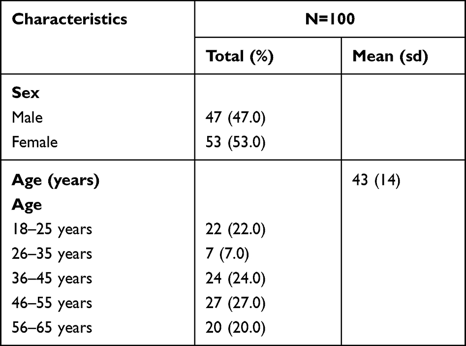 |
Table 1 Characteristics of Subjects |
The results of the seven auricular anthropometric parameters can be found in Table 2.
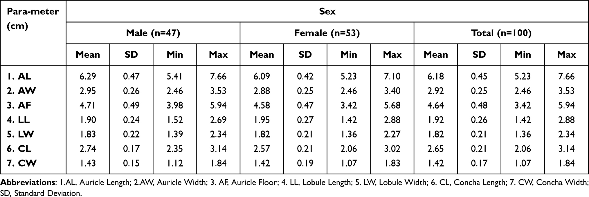 |
Table 2 Auricular Anthropometric Parameter Results |
Based on the age group, the average of AL, AW, AF, LL and Lw right-left is greatest at the age of 56–65 years. While the largest right-left CW is at the age of 36–45 years as presented in Table 3.
 |
Table 3 Comparison of Auricle Anthropometry by Age Group |
There have been various studies on ear size, one of which compared populations with different social and ethnic backgrounds and showed the differences in earlobe sizes (Table 4).
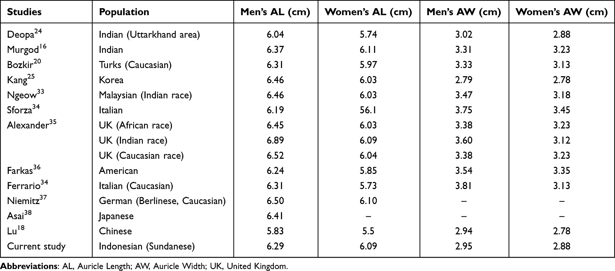 |
Table 4 Anthropometry of Auricle Length and Width from Various Populations |
Discussion
The auricle is an important face-forming component, both functionally and aesthetically. Every individual wants to have ears that look normal aesthetically, and the same expectation is felt particularly in those with congenital or acquired ear deformities. Achievement of functional rehabilitation and good auricular aesthetics increases not only self-confidence but also social acceptance. The external ear dimensions and its various components vary in different ethnic groups, and these are required by surgeons who base their reconstruction on data specifically collected from each ethnic group.17 Therefore, this study aims to provide data on the external ear for normal individuals in the Sundanese ethnic group.
Determine these dimension using auricular anthropometric analysis parameters, this technique is indirect measurement. Some of the advantages of indirect measurement: the subject does not move or move so it is easier to measure, error due to skin pressing and others when measurement does not occur, measurements can be repeated, using relatively shorter check times, images can be manipulated as rotated and enlarged for more detailed evaluation, data can be stored permanently so that it is possible to make comparisons at another day. Furthermore, some of the disadvantages of this method: the equipment (software and hardware) required is more expensive, installation use and maintenance of equipment requires higher technological capabilities, the inspection process that require stricter supervision and limited mobility, for pictures or scanning some areas of the body may be covered, making some points in these areas difficult to assess.8 Currently this method has been used scientifically to evaluate subjects qualitatively (anthropology) and quantitatively (anthropometry).6
In this study, the auricle length (AL) was 6.29 cm for men and 6.09 cm for women, and in all populations, men had a larger AL size than women. The AL size of Sundanese men was smaller when compared to almost all studies except those conducted by Sforza et al on the Caucasian race in Italy and Lu et al on the Chinese population. Conversely, the AL size of Sundanese women was not much different from the value reported in other studies. It was also larger compared to the population stated by Deopa et al in India, Bozkir et al and Sforza et al in Italy, Farkas et al in America, and Lu et al in China. The average AW was 2.95 cm in men and 2.88 cm in women compared to other studies in Table 4. Furthermore, the average size of AW tended to be smaller than other AW sizes discovered except in studies from Asian races, namely Korean and Chinese populations.16
Based on their AL and AW, the size of Sundanese auricles was closest to the value found in Koreans by Kang et al.25 According to Purkait, there is no standard shape known for the auricle. Even in groups with the same ethnic background, differences can occur in auricle shape and size.2 Differences in auricle dimensions of men and women have been reported in several studies, including Farkas et al,1992, Bozkir et al, 2006, Murgod et al, 2013, and Lee et al, 2018, stating the women’s auricle size is usually smaller compared to men.16,19–21,
In the examined Sundanese ethnic group, almost all auricle anthropometric parameters (AL, AW, AF, CL, and LW) were greater in males than females except for LL and LW sizes. Additionally, the average AL was found to be longer in men (6.29 cm) than in women (6.09 cm). This is in line with the study conducted by Eboh et al, 2013 on the Nigerian population.22 Ezon-Ebidor et al, 2018 discovered that in 217 samples with an age range of 18–65 years, the average AL and LL of men’s ears were longer than women’s, while women had a wider LW. This study concluded that at the same age, men’s auricle size was larger than women’s.23
In 2013, Deopa found the average LL for men to be 1.69 cm and 1.68 cm for women. A correlation was also reported between increasing age and LL. The LL elongation in women could be due to aging and the use of accessories such as earrings, while the aging process was the cause in men. Then, it was concluded that lobule length and gender had no statistically significant relationship. Meanwhile, in this current study, women’s LL was longer than men’s, but their LW tended to be the same.24
Recently, auricular anthropometric studies of various ethnic groups have been carried out, either directly or through photography. Despite ethnic variations concerning ear dimensions and position, the human auricle was found to continue growing even after reaching the cartilaginous skeleton maturation. With age, there was a gradual change in the microscopic structure of the ear cartilage, namely a decrease in the elastic fibers and density of cartilage cells. Purkait and Singh, as quoted by Japatti et al, mentioned the decline in skin elasticity and resilience with age. This may explain the increase in the macroscopic size of ear dimensions with age.17
Riedle et al found the elastic cartilage in the ear, larynx, and epiglottis to be similar to hyaline cartilage, except that the matrix contained elastic fibers for providing resilience and flexibility. A morphological study of age-related changes in human auricles showed that the elastic fibers in young individuals had a homogeneous diameter, but heterogeneous and fragmented diameter sizes were discovered in the elderly. Quantitative analysis of this study found that the auricular cartilage elastin content lost all its mechanical integrity of stress. Although further investigation is needed to elucidate the elastin constituent-function relationship, the age-related loss of elastic fibers is believed to produce less flexible and pliable auricular cartilage.26
In this current study on anthropometric comparisons of Sundanese auricles based on age groups, AL, AF, LL, and CL tended to increase with age. The elevation in auricle dimensions has not yet been proven to be correlated with age. Therefore, further investigation is needed with a more uniform sample size in all age groups.
The external ear consists of three main components, namely the antihelix and helix cluster, the concha cluster, and the lobules.27 According to Farkas, the auricle length and width could be useful in diagnosing syndromes associated with congenital external ear disorders, for example, microtia and disproportionately wide or narrow ears as in craniofacial syndromes.28, Lennon, 2018 found differences in the ear shape of patients with craniofacial syndromes, such as Crouzon syndrome. Ear width reached its maximum size at the age of seven years for boys and six years for girls.29
Deformities that could appear with age were the lengthening or descending of ear lobules. Earrings and other accessories used on the ear were known to affect the lobule length due to their additional pulling force. Additionally, in 2013, Eboh et al reported the average LL as equal to 1.58 cm. This value increased in both sexes once ear accessories such as earrings were used, and elevation in LL was found with increasing age. The auricle lobule enlargement with age in the vertical dimension was also caused by gravity, which initiated non-linear stretch changes deeply expressed in the lobules.22
Moreover, Livia et al stated that the ear shape is mainly determined by the proportions of its different dimensions, where CL and CW have a positive correlation with AL and AW. On the other hand, in Iraq, Farhan et al, 2019 reported a positive correlation of AW and CW with all auricle dimension parameters. This is suitable for planning reconstruction operations, specifically in estimating the parameters used.30,31
In a study conducted by Widiarni on microtia patients at Dr. Cipto Mangunkusumo Hospital Jakarta, ear reconstruction required a good anthropometric assessment before and after each surgery. The length and width, as well as details of the ear, were important parameters for postoperative evaluation.32
Study Limitation
This study was descriptive-oriented by displaying the range, mean, and standard deviation for samples’ age and gender. Therefore, further study is needed to determine the correlation of the mentioned characters. The samples used were not representative of the Sundanese population, leading to the need for another study with a larger number of samples to validate the stated results. The anthropometric data obtained can be useful for reconstructive ear surgery, anthropology, and forensics.
Conclusion
The length and width of the Sundanese normal auricle were closest to the size reported in the Korean population. In the Sundanese, the length, width, and base of the auricle, as well as the concha length in men tended to be larger than in women. On the other hand, women’s lobules seem to be longer compared to men’s, while their lobules and concha widths tended to be the same. From the anthropometric comparison of the by age group, it was discovered that auricle length, auricle floor, lobule length, and turbinate length had a propensity to increase with age. Besides, photogrammetry can be used to obtain auricular anthropometric data. This method can be used as a baseline for the normal auricle anthropometric characteristics of the Sundanese that is applicable in further anthropometric studies and planning for reconstructive surgery.
Abbreviation
AD, Anno Domini; CM, Centimeter; DR, Doctor; EOS, Electro-Optical System; MP, Mega Pixel; ORL-HNS, Otorhinolaryngology-Head and Neck Surgery; SD, Standard Deviation; SLR, Single Lens Reflex; UK, United Kingdom.
Acknowledgments
The authors are grateful to everyone that contributed to the writing of this manuscript.
Disclosure
The authors report no conflicts of interest in this work.
References
1. Widiarni D. Anthropometry of Ear in the congenital abnormalities of the auricle microtia. Dep Ilmu Penyakit THT FKUI RS Dr Cipto Mangunkusumo; 2012: 10.
2. Purkait R. Anthropometry of normal human auricle. In: Handbook of Anthropometry. Springer; 2012:903–917. doi:10.1007/978-1-4419-1788-1_53
3. Sharma N. Anthropometric measurement and cross-sectional surveying of ear pinna characteristics in Northern India. J Exp Clin Anat. 2016;15(2):102. doi:10.4103/1596-2393.200914
4. Deng K, Dai L, Yi L, Deng C, Li X, Zhu J. Epidemiologic characteristics and time trend in the prevalence of anotia and microtia in China. Birth Defects Res Part a Clin Mol Teratol. 2016;106(2):88–94. doi:10.1002/bdra.23462
5. Zhan G, Han L, Li Z, Liu Z, Fu J, Zhong K. Identification and documentation of auricle defects using three-dimensional optical measurements. Sci Rep. 2018;8(1):1–7. doi:10.1038/s41598-018-21289-x
6. Han K, Kwon HJ, Choi TH, Kim JH, Son D. Comparison of anthropometry with photogrammetry based on a standardized clinical photographic technique using a cephalostat and chair. J Cranio-Maxillofac Surg. 2010;38(2):96–107. doi:10.1016/j.jcms.2009.04.003
7. Ogawa Y, Wada B, Taniguchi K, Miyasaka S, Imaizumi K. Photo anthropometric variations in Japanese facial features: establishment of large-sample standard reference data for personal identification using a three-dimensional capture system. Forensic Sci Int. 2015;257:511.e1–511.e9. doi:10.1016/j.forsciint.2015.07.046
8. Liu B-S, Tseng H-Y, Chia T-C. Reliability of external ear measurements obtained by direct, photocopier scanning and photo anthropometry. Ind Eng Manag Syst. 2010;9(1):20–27.
9. Purkait R. Progression of growth in the external ear from birth to maturity: a 2-year follow-up study in India. Aesthetic Plast Surg. 2013;37(3):605–616. doi:10.1007/s00266-013-0097-1
10. Takari M, Deliana F, Fadlin NT, Netriroza A, Dewi H. Indonesian art community [Internet]. Pertama. Medan: Studia Kultura; 2008: 219.
11. Hidayah Z. Encyclopedia of Ethnicities in Indonesia [Internet].
12. Nurianingsih FR, Epsilawati L, Lubis N, Polii H. The height of the cortical bone mandible compared to height of the mandibular bone through panoramic radiography in Sunda Tribe. Bionatura. 2014;16(1):21–24.
13. Prasetyono TOH. Morphometry of deutero Malay female nose. Med J Indones. 2009;18(2):120–123. doi:10.13181/mji.v18i2.349
14. Widyanti A, Susanti L, Sutalaksana IZ, Muslim K. Ethnic differences in Indonesian anthropometry data: evidence from three different largest ethnics. Int J Ind Ergon. 2015;47:72–78. doi:10.1016/j.ergon.2015.02.008
15. Large DM. Sample in Medicine and Health Research.
16. Murgod V, Angadi P, Hallikerimath S, Kale A. Anthropometric study of the external ear and its applicability in sex identification: assessed in an Indian sample. Aust J Forensic Sci. 2013;45(4):431–444. doi:10.1080/00450618.2013.767374
17. Japatti SR, Engineer PJ, Reddy BM, Tiwari AU, Siddegowda CY, Hammannavar RB. Anthropometric assessment of the normal adult human ear. Ann Maxillofac Surg. 2018;8(1):121–123. doi:10.4103/ams.ams_183_17
18. Lu P, Tsao L, Yu C, Ma L. Survey of ear anthropometry for young college students in China and its implications for ear-related product design. Hum Factors Ergon Manuf. 2021;31(1):86–97. doi:10.1002/hfm.20871
19. Farkas LG, Posnick JC, Hreczko TM. Anthropometric Growth Study of the Ear. Cleft Palate-Craniofacial J. 1992;29(4):1–6. doi:10.1597/1545-1569_1992_029_0324_agsote_2.3.co_2
20. Bozkir MG, Karakaş P, Yavuz M, Dere F. Morphometry of the external ear in our adult population. Aesthetic Plast Surg. 2006;30(1):81–85. doi:10.1007/s00266-005-6095-1
21. Lee W, Yang X, Jung H, et al. Anthropometric analysis of 3D ear scans of Koreans and Caucasians for ear product design. Ergonomics. 2018;61(11):1480–1495. doi:10.1080/00140139.2018.1493150
22. Dennis E. Morphological changes of the human pinna in relation to age and gender of Urhobo people in Southern Nigeria. J Exp Clin Anat. 2013;12(2):68. doi:10.4103/1596-2393.127964
23. Ebidor E, Edibamode I, Paul JN, Hart JS, Okere AU. The anthropometric measurement of the auricle In Igbo and Yoruba ethnic groups of the Nigerian adult population. Sudan Med J. 2018;54(3):209–215.
24. Deopa D, Thakkar HK, Prakash C, Niranjan R, Barua MP. Anthropometric measurements of external ear of medical students in Uttarakhand region. J Anat Soc India. 2013;62(1):79–83. doi:10.1016/S0003-2778(13)80018-4
25. Kang H, Hu K, Song W, et al. Physical anthropologic characteristics of the auricle through the metric and non-metric analysis in Korean young adults. Korean J Phys Anthr. 2006;19(4):255–265. doi:10.11637/kjpa.2006.19.4.255
26. Riedler KL, Shokrani A, Markarian A, Fisher LM, Pepper JP. Age-related histologic and biochemical changes in auricular and septal cartilage. Laryngoscope. 2017;127(11):E399–407. doi:10.1002/lary.26807
27. Wang B, Dong Y, Zhao Y, Bai S, Wu G. Computed tomography measurement of the auricle in Han population of north China. J Plast Reconstr Aesthetic Surg. 2011;64(1):34–40. doi:10.1016/j.bjps.2010.03.053
28. Farkas LG, Katic MJ, Forrest CR, et al. International anthropometric study of facial morphology in various ethnic groups/races. J Craniofac Surg. 2005;16(4):615–646. doi:10.1097/01.scs.0000171847.58031.9e
29. Lennon C, Chinnadurai S. Nonsurgical Management of Congenital Auricular Anomalies. Facial Plast Surg Clin North Am. 2018;26(1):1–8. doi:10.1016/j.fsc.2017.09.001
30. Petrescu L, Catalin-Dumitru P. Anthropometric investigation of external ear morphology, as a pattern of uniqueness, useful in identifying the person. Publ House Rom Acad. 2018;20(2):95–104.
31. Farhan SS, Al-Jewari WM, Afar Al-Maathidy AQ, Al-Qtaitat A. Morphological assessment of Ear auricle in a group of Iraqi subjects and its possible role in personal identification. Ital J Anat Embryol. 2019;124(3):432–442.
32. Widiarni D, Trimartani WA. Ear Anthropometry as a Basis for Diagnosis and Planning for Reconstruction of Earlobe Abnormalities; 2011:19.
33. Ngeow WC, Aljunid ST. Craniofacial anthropometric norms of Malaysian Indians. Indian J Dent Res. 2009;20(3):313–319. doi:10.4103/0970-9290.57372
34. Sforza C, Grandi G, Binelli M, Tommasi DG, Rosati R, Ferrario VF. Age- and sex-related changes in the normal human ear. Forensic Sci Int. 2009;187(1–3):110.e1–110.e7. doi:10.1016/j.forsciint.2009.02.019
35. Alexander KS, Stott DJ, Sivakumar B, Kang N. A morphometric study of the human ear. J Plast Reconstr Aesthetic Surg. 2011;64(1):41–47. doi:10.1016/j.bjps.2010.04.005
36. Farkas L. Anthropometry of normal and anomalous ears. Clin Plast Surg. 1978;5(3):401–412. doi:10.1016/S0094-1298(20)32143-X
37. Niemitz C, Nibbrig M, Zacher V. Human ears grow throughout the entire lifetime according to complicated and sexually dimorphic patterns--conclusions from a cross-sectional analysis. Anthr Anz. 2007;65(4):391–413. doi:10.1127/anthranz/65/2007/391
38. Asai K, Yoshimura M, Yamada T. Why do old men have big ears? Correlation of ear length with age in Japan. BMJ. 1996;312(2):582. doi:10.1136/bmj.312.7030.582c
 © 2022 The Author(s). This work is published and licensed by Dove Medical Press Limited. The
full terms of this license are available at https://www.dovepress.com/terms
and incorporate the Creative Commons Attribution
- Non Commercial (unported, 3.0) License.
By accessing the work you hereby accept the Terms. Non-commercial uses of the work are permitted
without any further permission from Dove Medical Press Limited, provided the work is properly
attributed. For permission for commercial use of this work, please see paragraphs 4.2 and 5 of our Terms.
© 2022 The Author(s). This work is published and licensed by Dove Medical Press Limited. The
full terms of this license are available at https://www.dovepress.com/terms
and incorporate the Creative Commons Attribution
- Non Commercial (unported, 3.0) License.
By accessing the work you hereby accept the Terms. Non-commercial uses of the work are permitted
without any further permission from Dove Medical Press Limited, provided the work is properly
attributed. For permission for commercial use of this work, please see paragraphs 4.2 and 5 of our Terms.