
Back to Journals » Clinical, Cosmetic and Investigational Dermatology » Volume 19
Dermatitis Artefacta in a 38-Year-Old Female with Coexisting Anxiety Disorder: A Diagnostic Challenge and the Importance of Multidisciplinary Management
Authors Wang J  , Zhou X, Chen X
Received 6 December 2025
Accepted for publication 1 February 2026
Published 5 February 2026 Volume 2026:19 587040
DOI https://doi.org/10.2147/CCID.S587040
Checked for plagiarism Yes
Review by Single anonymous peer review
Peer reviewer comments 2
Editor who approved publication: Dr Jeffrey Weinberg
Jue Wang,1,* Xinyu Zhou,2,* Xinlong Chen1
1Department of Dermatology, Hospital of Chengdu University of Traditional Chinese Medicine, Chengdu, Sichuan, People’s Republic of China; 2Department of Dermatology, Nanbu People’s Hospital, Nanchong, Sichuan, People’s Republic of China
*These authors contributed equally to this work
Correspondence: Xinlong Chen, Department of Dermatology, Hospital of Chengdu University of Traditional Chinese Medicine, Chengdu, Sichuan, People’s Republic of China, Email [email protected]
Abstract: Dermatitis artefacta (DA) is a psychocutaneous disorder characterized by self-inflicted skin lesions and frequently associated with underlying psychological distress. We report a case of a 38-year-old woman who presented with recurrent erythema, papules, exudation, and severe pruritus involving the head and neck for over one year. The patient was initially misdiagnosed with atopic dermatitis and treated with immunosuppressive therapy, partly due to overlapping clinical features and the presence of peripheral eosinophilia, which was misinterpreted as evidence of allergic inflammation. Further evaluation revealed significant comorbid anxiety, and the diagnosis of dermatitis artefacta was established based on clinical morphology, disease course, behavioral features, and response to combined dermatological and psychiatric management. This case highlights how nonspecific laboratory abnormalities, such as eosinophilia, may contribute to diagnostic delay and inappropriate treatment escalation if psychocutaneous disorders are not considered. Early recognition of dermatitis artefacta and timely integration of psychological assessment are essential to prevent misdiagnosis and unnecessary immunosuppressive therapy in patients with atypical or treatment-refractory dermatoses.
Keywords: dermatitis, artefacta, anxiety, disorder
Introduction
Dermatitis artefacta (DA) is a psychocutaneous disorder characterized by deliberate, self-inflicted skin lesions that are typically denied or incompletely acknowledged by patients. It is most frequently observed in adult women and is often associated with underlying psychiatric conditions, including anxiety disorders, depression, and maladaptive coping mechanisms.1,2 Although DA shares overlapping features with neurotic excoriations and factitious disorder imposed on self,3,4 careful differentiation is essential for diagnostic accuracy. In the present case, the intentional production of skin lesions, denial of self-inflicted behavior, and the absence of secondary gaining behavior collectively support the diagnosis of DA. Laboratory abnormalities, including peripheral eosinophilia, may further complicate the diagnostic process. Although eosinophilia is commonly interpreted as a marker of allergic or immunologic disease, it is a nonspecific finding that may also reflect stress-related immune dysregulation or coexist with psychocutaneous disorders. Overreliance on eosinophil counts may therefore reinforce an inflammatory diagnostic framework and contribute to inappropriate escalation of immunosuppressive therapy, particularly when clinical features are atypical or treatment responses are inconsistent.
This case is presented to highlight a clinically relevant diagnostic pitfall: the convergence of anxiety-related psychocutaneous disease, nonspecific inflammatory laboratory findings, and lesion morphology resembling atopic dermatitis, which together led to prolonged misdiagnosis and unnecessary immunosuppressive treatment. By emphasizing key clinical and behavioral clues and the limitations of laboratory markers such as eosinophilia, this report aims to underscore the importance of early psychodermatological assessment in patients with refractory or atypical dermatoses.5,6
Case Presentation
A 38-year-old woman presented to our dermatology clinic with a 12-month history of recurrent erythema, papules, exudation, and severe pruritus predominantly involving the head and neck. The lesions were well-demarcated and localized to exposed areas, significantly impairing her quality of life. She denied photosensitivity, occupational exposure, or recent changes in cosmetics or skincare products.
On physical examination, erythematous patches and papules with exudation and crusting were observed on the face, scalp margin, and neck, accompanied by excoriations and areas of sharp demarcation (Figure 1). No similar lesions were noted on non–sun-exposed or less accessible areas. Initial laboratory investigations revealed peripheral eosinophilia, while other routine hematologic and biochemical parameters were within normal limits.
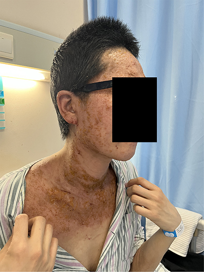 |
Figure 1 Clinical examination revealed widespread erythema, papules, and exudation involving the head and neck regions, with focal crusting and a mild increase in skin temperature. |
Prior to presentation at our institution, the patient had been treated at an outside facility. Given the chronic pruritic dermatitis, inflammatory appearance, and elevated eosinophil count, a provisional diagnosis of severe atopic dermatitis had been made. Before admission, she received systemic cyclosporine A for approximately a week; however, the treatment resulted in minimal clinical improvement.
Upon reevaluation at our clinic, the photodistributed pattern and chronic refractory course prompted reconsideration of alternative diagnoses, including allergic contact dermatitis (particularly airborne or cosmetic-related), photoallergic dermatitis, photocontact dermatitis, and chronic actinic dermatitis. Skin biopsy and patch testing were considered but not performed due to patient reluctance, representing a limitation of the diagnostic workup.
During follow-up visits, closer clinical observation revealed repetitive scratching and manipulation of lesions, predominantly involving easily reachable areas, often exacerbated during periods of emotional distress. The lesion morphology was inconsistent with classic inflammatory dermatoses, showing sharp borders, variable stages of healing, and a discrepancy between severe subjective pruritus and relatively limited objective inflammation. These features raised suspicion for a psychocutaneous disorder.
A formal psychiatric consultation was subsequently obtained. The patient met the diagnostic criteria for an anxiety disorder based on a structured clinical assessment performed by a psychiatrist, with a Hamilton Anxiety Rating Scale (HAM-A) score of 40. She reported longstanding anxiety symptoms and had not been receiving regular psychiatric treatment prior to referral. No generalized illness-seeking behavior or external incentives were identified. Taken together, the intentional lesion production, denial of self-inflicted behavior, absence of pervasive factitious behavior, and clinical course were most consistent with a diagnosis of dermatitis artefacta.
She reported longstanding anxiety symptoms and had not been receiving regular psychiatric treatment prior to referral. No generalized illness-seeking behavior or external incentives were identified. Taken together, the intentional lesion production, denial of self-inflicted behavior, absence of pervasive factitious behavior, and clinical course were most consistent with a diagnosis of dermatitis artefacta.
Cyclosporine was discontinued, and treatment was adjusted to address both dermatologic and psychiatric components. The patient was initiated on oral doxepin for its anxiolytic and antipruritic effects, along with topical fusidic acid to prevent secondary infection. Psychiatric follow-up was arranged. Over subsequent visits, gradual improvement of skin lesions was observed in parallel with stabilization of anxiety symptoms.
Discussion
Dermatitis artefacta (DA) is defined as a condition characterized by self-inflicted skin lesions in which the patient denies or is unwilling to acknowledge responsibility for the injury.7 This concealment distinguishes DA from other self-inflicted dermatoses, such as neurotic excoriations, and trichotillomania.8 Its diagnosis relies primarily on psychological factors rather than definitive histopathological or biochemical findings, often necessitating prolonged evaluation. Psychiatrists may miss opportunities for early intervention due to limited dermatologic awareness, whereas dermatologists may focus on common dermatoses and overlook psychiatric comorbidity, leading to inappropriate escalation of treatment. Characteristic lesions of dermatitis artefacta exhibit unusual shapes with irregular outlines and linear or geometric patterns that are sharply demarcated from the surrounding normal skin. The clinical spectrum may include erythematous patches, purpura, swelling, blisters, denuded areas, crusts, cuts, burns, and scars.9–11 Notably, these lesions usually do not evolve gradually but appear abruptly, often overnight, without preceding signs or symptoms. However, previously published reports tend to emphasize either strikingly bizarre lesion morphology or overt psychiatric comorbidity—features that may not be immediately apparent in routine clinical practice.
In contrast, the present case illustrates a more subtle and clinically challenging scenario, in which lesion morphology, distribution, and laboratory findings closely resembled inflammatory dermatitis, thereby delaying recognition of dermatitis artefacta. Importantly, several clinical “red flags” were retrospectively identified that could have prompted earlier diagnostic reconsideration. These included sharply demarcated lesions confined to easily accessible areas, fluctuating severity inconsistent with therapeutic escalation, a discrepancy between the extent of objective skin findings and the reported severity of symptoms, and behavioral patterns such as repetitive scratching observed during clinical encounters. Awareness of these features is critical, as they may provide early diagnostic clues even in the absence of overtly bizarre lesion patterns.8
A further clinically relevant aspect highlighted by this case is the risk associated with inappropriate immunosuppressive therapy in psychocutaneous disorders. In the absence of definitive diagnostic confirmation, systemic immunosuppression—such as cyclosporine—may reinforce an inflammatory disease framework, delay psychiatric assessment, and expose patients to unnecessary adverse effects without addressing the underlying driver of disease. This case underscores the importance of maintaining diagnostic flexibility and reconsidering the working diagnosis when treatment responses are inconsistent or paradoxical.
Several limitations of this case warrant explicit acknowledgment. Histopathological confirmation and patch testing were not performed, which limits the exclusion of alternative inflammatory or contact-related dermatoses. Furthermore, the diagnosis of dermatitis artefacta relied primarily on clinical pattern recognition and behavioral observation rather than objective diagnostic markers. While this reflects real-world clinical practice in many psychodermatological cases, it nonetheless represents an inherent limitation that should be considered when interpreting the findings.
Overall, this case contributes to existing literature by emphasizing that dermatitis artefacta may present without dramatic or classic features, and that its recognition often depends on identifying subtle behavioral and clinical inconsistencies rather than relying solely on morphology or laboratory results. Early consideration of psychocutaneous disorders in patients with refractory or atypical dermatitis may help prevent diagnostic delay, unnecessary immunosuppression, and prolonged patient morbidity.
Conclusion
This case illustrates the diagnostic challenges of dermatitis artefacta when accompanied by anxiety disorder and nonspecific inflammatory findings that mimic common dermatoses. Failure to recognize psychocutaneous features may lead to diagnostic delay and inappropriate immunosuppressive treatment. Early identification of behavioral clues, cautious interpretation of laboratory markers, and timely psychiatric assessment are essential for accurate diagnosis and optimal management.
Ethics Approval
The publication of case report does not require ethical approval. We confirm that no institutional approval was required for publishing the case details.
Informed Consent for Publication
The patient had signed informed consent and provided informed consent for the publication of the case details and any accompanying images.
Funding
There is no funding to report.
Disclosure
The authors report no conflicts of interest in this work.
References
1. Van Moffaert M, Vermander F, Kint A. Dermatitis artefacta. Int J Dermatol. 1985;24(4):236–4. doi:10.1111/j.1365-4362.1985.tb05769.x
2. Nayak S, Acharjya B, Debi B, Swain SP. Dermatitis artefacta. Ind J Psychiatry. 2013;55(2):189–191. doi:10.4103/0019-5545.111462
3. Cyr PR, K DG. Neurotic excoriations. Am Fam Phys. 2001;64(12):1981–1985.
4. Liu RK, Green J, Newton R. A scoping review of treatment for Factitious disorder (imposed on self). Psych Res Commun. 2025;5(2):100216. doi:10.1016/j.psycom.2025.100216
5. Brough MD. Dermatitis artefacta. Hand. 1977;9(3):283–286. doi:10.1016/S0072-968X(77)80117-4
6. Conde-Salazar L, Valks R, Ma P, et al. Dermatitis artefacta? Am J Contact Dermat. 2003;14(2):93–94. doi:10.1097/01634989-200306000-00009
7. Musalek M, Hobl B, Mossbacher U. Diagnóstico y clasificación en psicodermatología. In: Grimalt F, Cotterill JA, editors. Dermatología y Psiquiatría. Historias clínicas comentadas. Madrid: Grupo Aula Médica. 2002:
8. Torales J, Malvido K, Vázquez MA, et al. Dermatitis artefacta: a practical guide for diagnosis and management. Acta Dermatovenerol Croat. 2023;31(1):17–23.
9. Chen Y, Li L, Lu J. Purpura with regular shape in an adolescent: beware of dermatitis artefacta. Front Pediatr. 2022;10:959064. doi:10.3389/fped.2022.959064
10. Gupta M, Sharma A, Kumaran MS. An insight into adolescent dermatitis artefacta: a case report. Cureus. 2024;16(9):e68682. doi:10.7759/cureus.68682
11. Shah R, Jamgochian M, Haroon A, et al. 14-year-old with dermatitis artefacta secondary to aerosolized spray deodorant: a rare case with an important learning opportunity. JAAD Case Rep. 2021;19:71–73. doi:10.1016/j.jdcr.2021.11.014
 © 2026 The Author(s). This work is published and licensed by Dove Medical Press Limited. The
full terms of this license are available at https://www.dovepress.com/terms
and incorporate the Creative Commons Attribution
- Non Commercial (unported, 4.0) License.
By accessing the work you hereby accept the Terms. Non-commercial uses of the work are permitted
without any further permission from Dove Medical Press Limited, provided the work is properly
attributed. For permission for commercial use of this work, please see paragraphs 4.2 and 5 of our Terms.
© 2026 The Author(s). This work is published and licensed by Dove Medical Press Limited. The
full terms of this license are available at https://www.dovepress.com/terms
and incorporate the Creative Commons Attribution
- Non Commercial (unported, 4.0) License.
By accessing the work you hereby accept the Terms. Non-commercial uses of the work are permitted
without any further permission from Dove Medical Press Limited, provided the work is properly
attributed. For permission for commercial use of this work, please see paragraphs 4.2 and 5 of our Terms.