
Back to Journals » International Journal of Nanomedicine » Volume 21
Dendrocin ZM1 Nanoconjugates with Carbon Quantum Dots: A ROS-Generating Platform for Combating Multidrug-Resistant Bacteria
Authors Seyedjavadi SS, Zare-Zardini H  , Sharif Bakhtiar L , Razzaghi-Abyaneh M , Navidinia M, Rahmandoust M , Goudarzi M
Received 26 September 2025
Accepted for publication 27 January 2026
Published 2 February 2026 Volume 2026:21 565719
DOI https://doi.org/10.2147/IJN.S565719
Checked for plagiarism Yes
Review by Single anonymous peer review
Peer reviewer comments 4
Editor who approved publication: Dr Sachin Mali
Sima Sadat Seyedjavadi,1 Hadi Zare-Zardini,2 Leili Sharif Bakhtiar,3 Mehdi Razzaghi-Abyaneh,1 Masoumeh Navidinia,4 Moones Rahmandoust,3,5,* Mehdi Goudarzi6,7,*
1Department of Mycology, Pasteur Institute of Iran, Tehran, Iran; 2Department of Biomedical Engineering, Meybod University, Meybod, Iran; 3Protein Research Center, Shahid Beheshti University, Tehran, Iran; 4Department of Medical Laboratory Sciences, School of Allied Medical Sciences, Shahid Beheshti University of Medical Sciences, Tehran, Iran; 5Faculty of Life Sciences and Biotechnology, Shahid Beheshti University, Tehran, Iran; 6Infectious Diseases and Tropical Medicine Research Center, Shahid Beheshti University of Medical Sciences, Tehran, Iran; 7Department of Microbiology, School of Medicine, Shahid Beheshti University of Medical Sciences, Tehran, Iran
*These authors contributed equally to this work
Correspondence: Mehdi Goudarzi, Department of Microbiology, School of Medicine, Shahid Beheshti University of Medical Sciences, Koodak-Yar St., Daneshjoo Blvd, Velenjak, Chamran HWY, Tehran, Iran, Tel/Fax +98 21 23872556, Email [email protected] Moones Rahmandoust, Protein Research Center, Faculty of Life Sciences and Biotechnology, Shahid Beheshti University, Velenjak, Chamran HWY, Tehran, Iran, Tel +98 21 29905009, Email [email protected]
Introduction: The global increase in multidrug-resistant (MDR) bacterial infections highlights the urgent need for novel antimicrobial strategies. Antimicrobial peptides (AMPs), such as Dendrocin ZM1 derived from Zataria multiflora Boiss. offer promising therapeutic potential, but face limitations including poor stability and dose-dependent cytotoxicity. We developed a nanoconjugate of Dendrocin ZM1 with carbon quantum dots (CQDs) to enhance antimicrobial efficacy and reduced cytotoxicity.
Methods: The nanoconjugates were synthesized and characterized using FTIR, XPS, TEM, DLS, and fluorescence spectroscopy. Antibacterial activity against MDR Gram-positive and Gram-negative strains was evaluated using MIC determination, time–killing kinetics, ROS production, and ROS-scavenger rescue experiments. Hemolysis and MTT assays assessed biocompatibility on human red blood cells and HEK-293 cells.
Results: Dendrocin ZM1–CQDs showed a 2 to 4-fold reduction in MIC values compared with Dendrocin ZM1 alone, and its rapid bactericidal kinetics reached a reduction of ≥ 3-log10 CFU/mL within 30– 60 min at 2× MIC. ROS measurements showed a 3 to 4-fold increase in intracellular ROS levels, with scavenger treatments restoring bacterial viability, confirming ROS-mediated bacterial killing. Cytotoxicity assays showed > 90% cell viability and < 8% hemolysis even at high concentrations, indicating low toxicity to mammalian. Dendrocin ZM1–CQD nanoconjugates significantly enhance antibacterial efficacy while maintaining excellent biocompatibility.
Discussion: The synergistic combination of AMPs and CQDs offers a promising nanobiotechnology platform for next-generation antimicrobial therapeutics targeting MDR pathogens.
Keywords: dendrocin ZM1, carbon quantum dots, antimicrobial peptides, multidrug resistance, reactive oxygen species, nanoconjugates
Introduction
In recent decades, antibacterial resistance has been emerging globally, which will have a negative impact on health systems worldwide. Antibiotic resistance occurs due to incomplete antibiotic dosing and misuse,1 exposure to continuous stress, changes at the genomic level, and horizontal gene transfer. Combination therapy using different antibiotics can be used, but has some toxicity, limitations, and side effects.2,3 Therefore, there is an urgent need to develop a better treatment against drug-resistant infections.4,5 This highlights the emergency of developing alternatives to antibiotics that have high antimicrobial activity with less or negligible toxicity. Nanotechnology has emerged as a promising approach for antibacterial treatment, utilizing nanoparticles (NPs), which, due to their small size, can effectively penetrate and disrupt bacterial cell membranes.6 Commonly studied antibacterial nanomaterials include silver (Ag), titanium oxide (TiO2), copper oxide (CuO), and zinc oxide (ZnO).7 Although these NPs have shown significant antibacterial properties, their clinical use is often hampered by potential cytotoxicity and long-term persistence in the body.8 As a result, research has focused on developing materials that balance low toxicity and high biocompatibility with potent antibacterial efficacy, aiming to create safer and more effective options for antibacterial therapies.9,10 Carbon nanomaterials are very attractive for use in bioimaging, optoelectronics, and photocatalysis.11,12 Among these NPs, carbon quantum dots (CQDs), a carbon-based quantum material which is generally below 10-nm in diameter, offer several advantages, including easy modification, low cytotoxicity, excellent photostability, and high-water solubility.13 Due to these exceptional physicochemical properties, CQDs have shown promise in environmental applications and in biomedical fields, particularly in drug and gene delivery.14,15 Furthermore, CQDs show potential as antibacterial agents. The combined applications of CQDs in antibacterial treatment and bioimaging offer unique advantages compared to conventional antimicrobial agents.16
The photo-induced redox properties and intrinsic optical properties of CQDs can be enhanced, providing a promising platform for antimicrobial agents activated by natural or visible light.15,17 This allows CQDs to act effectively as antimicrobial agents and has the potential to modify the surface charge and functional group to optimize their performance.18 By interacting with bacterial cell surfaces, CQDs can cause cell wall damage and increased production of reactive oxygen species (ROS), leading to cytoplasmic leakage and bacterial cell death. Research has shown that certain CQDs are biocompatible in both in vivo and in vitro. Furthermore, CQDs can be modified and combined with peptides to achieve antimicrobial effects. Therefore, antimicrobial peptides and nanoparticles are a suitable alternative to traditional antibiotics.
The conjugation of peptides with NPs or other compounds allows for the creation of a diverse range of novel particles with potential medical applications, enabling simultaneous bioimaging for infection tracking, controlled drug release, and potent activity against biofilms or persister cells. Conjugating CQDs with antimicrobial peptides (AMPs), antibiotics, polymers, metals, or other functional moieties has emerged as a powerful strategy to amplify their antibacterial performance against multidrug-resistant pathogens, including MRSA, VRSA, carbapenem-resistant enterobacteriaceae, and intracellular bacteria such as Porphyromonas gingivalis, as demonstrated in peptide-CQD hybrids like HSER-CQDs or Dendrocin-like antimicrobial peptide.3,19,20 Dendrocin ZM1 (TTLRLNTLAYKVAWLVNVKAFWAA GRA LKKVGR) is a 33-amino acid residue peptide derived from Zataria multifora Boiss and exhibits a defense response against Gram-positive and Gram-negative bacteria.20 However, like many AMPs, its therapeutic application faces several limitations, including enzymatic degradation, poor stability under physiological conditions, and potential cytotoxicity at higher concentration.21 To overcome these challenges, nanotechnology-based approaches have attracted considerable attention. CQDs—a class of zero-dimensional, carbon-based nanomaterials- have emerged as attractive platforms for drug delivery due to their biocompatibility, chemical stability, ease of surface modification, and intrinsic fluorescence properties.18 CQDs can act as nanocarriers for AMPs, potentially increasing their solubility, protecting them from enzymatic degradation, and improving cellular uptake.17 Furthermore, CQDs themselves may contribute to antibacterial activity through mechanisms such as ROS generation or membrane disruption, thereby enhancing the overall antimicrobial effect through synergistic interactions.18 In this study, we report the synthesis and characterization of a novel Dendrocin ZM1–carbon quantum dot nanoconjugate designed to enhance antibacterial efficacy through synergistic ROS-mediated mechanisms. The novelty of this work lies in the integration of a plant-derived AMP with CQDs to simultaneously improve antimicrobial potency, stability, and biocompatibility. The main objectives of this study were (i) to evaluate the synergistic antibacterial activity of the Dendrocin ZM1–CQD nanoconjugate against representative bacterial strains, and (ii) to assess its biocompatibility and cytotoxicity. Through comprehensive physicochemical characterization and biological evaluation, this work provides new insights into AMP–CQD hybrid systems as next-generation antimicrobial platforms.
Materials and Methods
Materials and Instruments
Diammonium hydrogen citrate (≥99%, analytical grade) and urea (≥99%, analytical grade) were purchased from Merck (Darmstadt, Germany). Sodium hydroxide (NaOH, ≥98%, analytical grade) was obtained from Sigma-Aldrich (St. Louis, MO, USA). Carbon quantum dots (CQDs) were synthesized in-house as described in the Methods section. Mueller–Hinton broth and Mueller–Hinton agar (microbiological grade) were purchased from HiMedia Laboratories (Mumbai, India). Nutrient agar and nutrient broth (microbiological grade) were also obtained from HiMedia. Phosphate-buffered saline (PBS, pH 7.4, sterile) was purchased from Gibco (Thermo Fisher Scientific, USA). Triton X-100 (laboratory grade, ≥99%) was obtained from Sigma-Aldrich. Dimethyl sulfoxide (DMSO, ≥99.5%, cell culture grade) was purchased from Merck. Dulbecco’s modified Eagle’s medium (DMEM), fetal bovine serum (FBS, heat-inactivated), and trypsin-EDTA were purchased from Gibco (Thermo Fisher Scientific, USA). MTT reagent (≥98%, cell culture grade) and 2′,7′-dichlorodihydrofluorescein diacetate (DCFH-DA, ≥97%) were obtained from Sigma-Aldrich.
Fourier transform infrared spectroscopy (FTIR) (Thermo-Nicolet NEXUS 470, Illinois, USA) was employed. For XRD spectra in the 2θ range between 1 to 80 degrees, a DMAX-2500 diffractometer (Rigaku, Japan) was used using a Cu Kα1 X-ray radiation source. The zeta-potential and average hydrodynamic diameter of the particles were measured using a HeNe laser light source with a wavelength of 632.8 nm, with a maximum intensity of 10 mW, at the scattering angle of 90 degrees, using the dynamic light-scattering (DLS) method, (HORIBA model SZ-100, Kyoto, Japan), with three replicates, at room temperature. Ultraviolet-visible spectroscopy was used to characterize the optical properties of CQDs (PerkinElmer’s LAMBDA 950 UV/Vis/NIR Spectrophotometer, USA) in optical glass cuvettes with a path length 10 mm. A photoluminescence (PL) spectrophotometer (PerkinElmer LS45, Massachusetts, USA) was used. In the study of optical properties, the samples were diluted with different concentrations until the optical density of each reached about 0.1, to minimize the effect of re-absorbance.4 X-ray Photoelectron Spectroscopy (XPS) was performed using a PHI 5000 Versaprobe, Physical Electronics, Minnesota, USA. High-resolution, bright field transmission electron microscopy (TEM) was performed on selected CQDs samples to analyze their morphology, particle size, and aggregation behavior. Therefore, CQDs samples were highly diluted in distilled water. Then, the suspension was then homogenized in an ultrasonic bath for 5 min. Immediately after sonication, the micro-droplets were placed on 3 mm mesh carbon TEM grids (TED PELLA INC.) and left to dry for at least 30 min. Afterwards, the CQDs - containing grids were analyzed in a JEOL 2200FS TEM, Tokyo, Japan, operating at a voltage of 200 kV.
Peptide Synthesis
The peptide was synthesized by an external facility (Genscript USA Inc Piscataway, NJ, USA) using the solid phase method and Fmoc (9-fluorenyl-methoxycarbonyl) chemistry. The peptide was purified up to 95% using reverse phase-high performance liquid chromatography (RP-HPLC). The molecular weight of the peptide was also confirmed using the mass spectrometry method.
Molecular Dynamics (MD) Simulation
The structure was constructed directly from sequence (TTLRLNTLAYKVAWLVNVKAFWAA GRA LKKVGR) using the AlphaFold2 Protein Structure Database. The SAVES server and the PROCHECK module were used to evaluate the quality of the predicted 3D structure. Molecular dynamics simulations were performed using the open-source tool GROMAX version 2023.22 The peptide topology file was prepared using the charmm36 force field. The structures were placed in the center of the dodecahedron simulation box with periodic boundary conditions (PBC) and solvated in SPCE water molecules. Then, the box environment was neutralized in terms of charge by adding sodium and chlorine ions at a concentration of 0.15 M. In the next step, energy minimization was performed with 5000 steps and the steepest descent algorithm. After energy minimization, the systems were equilibrated for 5 nanoseconds using the Nose-Hoover algorithm at 310° Kelvin and then for 10 nanoseconds using the Parrinello-Rahman algorithm at 1 atmosphere pressure. Then, the simulation process was performed for each system for 1 microsecond. Every 10 picoseconds, the trajectory energy coordinates were saved. In this simulation, the cut-off of van der Waals and electrostatic interactions was set to 1.2 nm and the particle mesh Ewald (PME) method was used to measure the long-range electrostatic force. A Parrinello-Rahman barostat and a Nose-Hoover thermostat with a time constant of 0.2 picoseconds were used to set the temperature at 310° K and the pressure at 1 atmosphere, respectively. We also used the Linear Constraint Solver (LINCS) algorithm23 to keep the hydrogen bonds constant. Finally, the simulation results were analyzed using GROMACS internal modules. PyMol software was used to visualize the results of the 3D structure of proteins and XMGRACE and OriginPro software were used to prepare GROMACS graphs.24,25
Synthesis of N-Doped CQDs
In this method, 0.5 gr of diammonium hydrogen citrate and 0.5 gr of urea were mixed homogeneously in an agate mortar and placed in a glass beaker to be heated at 180°C for 60 min. The obtained dark synthesis product, containing N-doped CQDs, was diluted and neutralized using 1M NaOH solution to pH 7.0 and then centrifuged (20K rpm for 30 min) and filtered to remove the large particles.26
Peptide-CQD Conjugation
In this approach, 1 mg of Dendrocin ZM1 was added to 1 mL of CQDs solution and the obtained mixture was incubated for 24 h at 25°C under 650 rpm to complete the conjugation.19,27
Bacterial Strain
Bacterial strain, such as methicillin resistant S. aureus ATCC 43300, vancomycin-resistant S. aureus (VRSA), P. aeruginosa ATCC 27853, and E. coli ATCC 35218 (beta-lactam antibiotics resistant) were obtained from the Department of Microbiology, School of Medicine, Shahid Beheshti University of Medical Sciences, Tehran, Iran. Bacterial strains were cultured in Mueller Hinton (MH) media under sterile conditions to ensure the purity of the experimental setup. Initially, bacteria were inoculated onto nutrient agar plates and incubated at 37°C for 24 h to achieve colony formation. After incubation, a single colony was selected and transferred to sterile nutrient broth culture medium, and then incubated at 37°C with shaking at 150 rpm for optimal bacterial growth. After another 24 h, the bacterial culture reached mid-logarithmic phase, which was confirmed by measuring the optical density (OD) at 600 nm. In addition, dilution of the culture was performed using MH broth culture medium to obtain an absorbance of 0.1 (0.5 MacFarland standards) at OD600 nm and was used for subsequent experiments. All procedures were performed in a sterile environment to prevent contamination.
Minimum Inhibitory Concentration (MIC) Determination
The MIC of Dendrocin ZM1–CQDs was determined using the broth microdilution method, according to CLSI guidelines.28 A bacterial suspension was prepared from the overnight culture and adjusted to a concentration of 5×10⁵ CFU/mL. Next, 100 μL of the adjusted bacterial solution was added to each well of a 96-well polypropylene microtiter plate. After serially two-fold dilution of Dendrocin ZM1–CQDs, 100 μL was added to a 96-well plate to obtain concentrations from 1 to 128 μg/mL. The plates were incubated at 37°C for 24 h. The MIC was identified as the lowest concentration of antibacterial agent without visible bacterial growth, which was confirmed by comparison with the growth control. A positive control containing only bacterial solution without AMPs and a blank control containing only culture medium were used.
Time-Kill Assay
The time killing kinetics of Dendrocin ZM1–CQDs on S. aureus ATCC 43300 and E. coli ATCC 35218 were determined using a colony count-based method.29 Bacterial suspensions were prepared to a final concentration of 2×10⁵ CFU/mL and incubated with Dendrocin ZM1–CQDs at concentrations of 1× and 2× MIC. The solutions were incubated at 37°C with shaking at 120 rpm. Then, 50 µL of the bacterial solution was removed at specified time points (15, 45, 60, 90, and 120 min) and serially diluted in sterile saline. Aliquots (10 µL) were plated on Mueller-Hinton Agar medium and incubated at 37°C for 24 h. The number of colonies were recorded and the decrease in CFU/mL was used to assess the bactericidal effect of the antibacterial agent over time. A ≥3 log reduction in CFU/mL was considered bactericidal. A control of bacterial suspensions without peptide was performed under similar conditions. The assay was performed in triplicate independently.
Hemolytic Activity Assay
Fresh human red blood cells (RBCs) were collected, washed three times with phosphate-buffered saline (PBS), and diluted to a 2% RBC suspension. Dendrocin ZM1–CQDs at different concentrations (1×, 2×, and 4× MIC) was added to the RBC suspension and incubated at 37°C for 1 h. After incubation, the samples were centrifuged at 4500 rpm for 5 min and the supernatant was collected. Hemoglobin release was measured by measuring the absorption of the supernatant solution at a wavelength of 567 nm using an ELISA reader.20 PBS was used as a negative control (0% hemolysis), and Triton X-100 at a concentration of 0.1% was used as a positive control (100% hemolysis). The percentage of hemolysis was calculated relative to the positive control. Hemolysis (%) = [mean OD of test − mean OD of negative control)/([mean OD of test − mean OD of negative control mean OD of positive control – mean OD of negative control)] × 100. This study, which involved human blood cells, was approved by Shahid Beheshti University of Medical Sciences in Tehran, Iran (IR.SBMU.MSP.REC. 1402.445) and was conducted in accordance with the Declaration of Helsinki. Written informed consent was obtained from all donors before blood collection.30
Cytotoxicity Assay
To evaluate the toxicity Dendrocin ZM1–CQDs against the human embryonic kidney cell line 293 (HEK293), the 3-(4,5-dimethylthiazol-2-yl)-2,5-diphenyltetrazolium bromide (MTT) assay was used.20 HEK293 cells were obtained from the Microbiology Department of Shahid Beheshti University of Medical Sciences, and their use in this study was approved by Shahid Beheshti University of Medical Sciences in Tehran, Iran (IR.SBMU.MSP.REC. 1402.445). HEK293 cells were seeded in a 96-well plate with Dulbecco’s modified Eagle’s medium (DMEM) (supplemented with 10% fetal bovine serum) at a density of 1×10⁴ cells per well and allowed to adhere overnight at 37°C with 5% CO2. The cells were then treated with different concentrations of Dendrocin ZM1–CQDs (1×, 2×, and 4× MIC) for 24 h. Cytotoxicity was assessed using the MTT assay by adding MTT reagent (0.5 mg/mL) and incubating for 4 h under the same conditions. The resulting formazan crystals were dissolved in 100 μL DMSO (dimethyl sulfoxide) with gentle shaking, and the optical absorbance was measured using a microplate reader at a wavelength of 570 nm. Untreated cells were considered as control and the percentage of cell viability was calculated relative to the control. The findings were obtained from three separate experiments, each conducted in triplicate.
ROS Production Assay
Intracellular ROS was measured using a fluorometric method with 2′,7′-dichlorofluorescin diacetate (DCFH-DA).31 S. aureus ATCC 43300 and E. coli ATCC 35218 cells in mid-log phase were adjusted to 1×10⁶ CFU/mL and incubated with Dendrocin ZM1–CQDs at concentrations of 2× MIC for 1 h at 37°C with constant shaking (120 rpm). After treatment, cells were stained with a final concentration of 10 µM 2′,7′-dichlorodihydrofluorescein diacetate (DCFH-DA) and incubated for 30 min in the dark. ROS fluorescence intensity was measured on a FACS Calibur flow cytometer (BD Biosciences, San Jose, CA, United States). Untreated cells were considered as the control. Data from three independent experiments are reported in this study.
ROS Scavenger Rescue Assay
To confirm the contribution of oxidative stress to bactericidal activity induced by Dendrocin ZM1–CQDs, ROS scavenger rescue assays were performed according to established methods.32 Mid-logarithmic phase cultures of S. aureus ATCC 43300 and E. coli ATCC 35218 cultures were prepared as described above and incubated with N-acetylcysteine (NAC, 10 mM), thiourea (100 mM), or catalase (1000 U/mL) for 30 min at 37 °C before exposure to Dendrocin ZM1–CQDs. Cells were then treated with Dendrocin ZM1–CQDs (2× MIC) for 45 min, and intracellular ROS levels were determined using the DCFH-DA assay as described above. To assess bacterial viability, aliquots from each treatment group were serially diluted, plated on Mueller–Hinton agar, and incubated at 37 °C for 18–24 h. Colony-forming units (CFU) were counted and the results were expressed as log10 CFU/mL. The reduction of ROS signal along with CFU recovery relative to Dendrocin ZM1–CQDs alone was interpreted as evidence for a ROS-mediated mechanism of antibacterial activity.
Statistical Analysis
All experiments were performed in triplicate biological replicates with technical samples, and data were expressed as mean ± SD. ROS fluorescence intensities were normalized relative to untreated controls and logarithmically transformed when necessary. Comparisons between multiple groups (time points× treatments× species) were analyzed using two-way ANOVA with Tukey’s post-hoc test for multiple comparisons; scavenger assays at single time points were analyzed using one-way ANOVA followed by Tukey’s test. A significance threshold of p < 0.05 was applied, with p < 0.05 (*), p < 0.01 (**), and p < 0.001 (***) indicating statistical significance. Analyses were performed using GraphPad Prism v10 (GraphPad Software, USA).
Results
Peptide Structure
The 3D structure of Dendrocin ZM1 showed that the majority of Dendrocin ZM1 has an alpha helix structure and only the N-terminal and C-terminal subunits have a limited region with a random coil structure (Figure 1a). Ramachandran plot analysis showed that 100% of the residues were in the desired Region (Figure 1b).
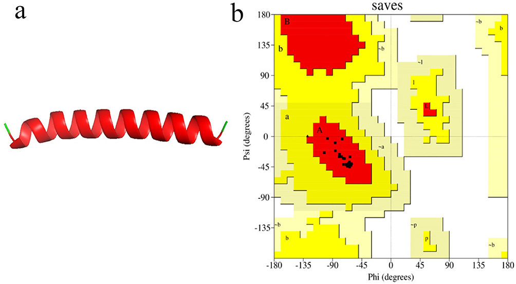 |
Figure 1 (a) 3D structure of Dendrocin ZM1, most of which has an alpha-helical structure and only the terminal parts are random coils. (b) Ramachandran plot of Dendrocin ZM1, with 100% of the residues in the most favored region. |
Analysis of the secondary structure of the residues during the simulation process showed that the middle residues (approximately residues 12 to 30) had an alpha helix structure most of the time during the simulation process, while residues 1, 33 and 34 had a coil structure. Residues 29 and 31 also had Turn and Bend structures, respectively, at most moments of the simulation (Figure 2a).
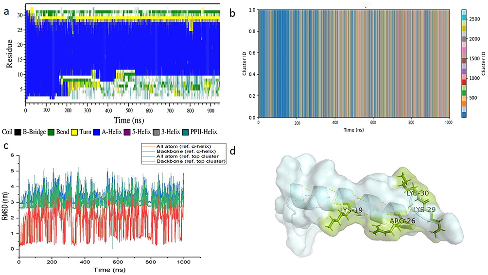 |
Figure 2 Molecular dynamics simulation of Dendrocin ZM1. (a) Secondary structure of residues during molecular dynamics simulation. The middle residues often had an alpha-helical structure during the simulation, but the initial and terminal regions had a coil structure. Also, the C-terminus of residue 31 often has a Bend; the alpha-helical, turn, and bend structures are shown in blue, yellow, and green, respectively. (b) Peptide clusters formed (peptide name) during the simulation. The peptide formed a wide range of clusters in the first 400 ns, but after that, this structural spectrum became narrower and the peptide acquired its main structure. (c) RMSD analysis for backbone and all atoms. The RMSD of the BACKBONE and all atoms of the alpha helix structure show more structural fluctuation during the simulation process compared to the top cluster structure. (d) Peptide structure at frame 72,837 corresponding to the top cluster state. The backbone and molecular surface structures are shown as cartoons in cyan, while the side chains of positively charged residues are shown in sticks style and in green. Intramolecular hydrogen bonds are also shown as yellow dashed lines. |
Analysis of different clusters during the simulation process based on the color spectrum of cluster types showed that the Dendrocin ZM1 had a wide structural diversity during the first 200 ns of simulation, but after about 400 ns, this structural diversity decreased (Figure 2b). Examination of Dendrocin ZM1 secondary structure in stable clusters at the corresponding simulation moments shows that the Dendrocin ZM1 often had a helical secondary structure at these moments.
Figure 2c is the structure of Dendrocin ZM1 in the top cluster state, which has numerous intrachain hydrogen bonds, and Table 1 shows the residues and atoms that donate and accept hydrogen bonds. This image shows that the middle parts of Dendrocin ZM1 consist of a helical secondary structure, while the initial and terminal regions have a coil structure. Also, as mentioned in the secondary structure analysis, the end of Dendrocin ZM1 has a bend, which can also be seen in the image shown.
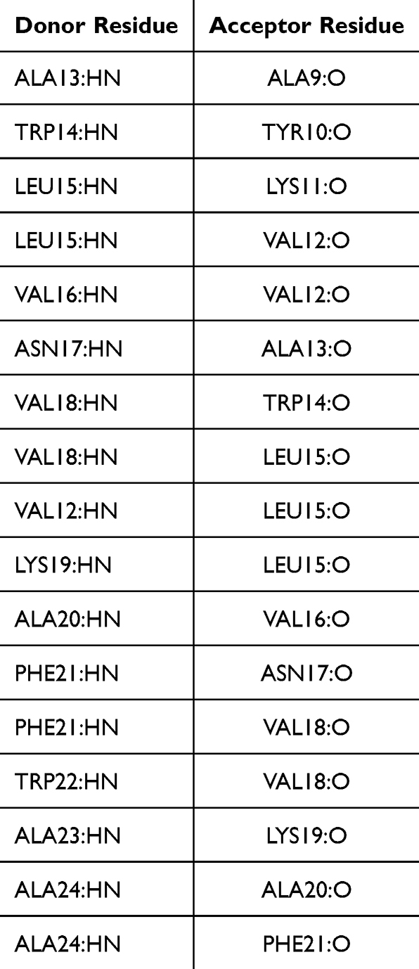 |
Table 1 Intrachain Hydrogen Bond Donor and Acceptor Residues |
Another analysis that was conducted was related to the stability of Dendrocin ZM1 structure in the primary structure (α-helix) as well as its conformation in the upper cluster state. Studying the RMSD of all atoms and Backbone of each of the mentioned conformations shows that the amount of Dendrocin ZM1 oscillation is greater in the α-helix state is higher compared to the upper cluster state, such that in the α-helix state, the oscillation range is about 3 angstroms, while in the upper cluster state it is about 2 angstroms (Figure 2d).
Characterization of CQDs
Morphology and Charge Distribution
The study of the average particle size and zeta potential of CQDs was performed in triplicate. As shown in Figure 3a and b, DLS and TEM results showed that the diameters of CQDs were 8.2±3.8 nm, with zeta potentials of −0.6±0.3 mV. It should be noted that the observed negative charge can be attributed to the presence of N-H amine groups, as confirmed by FTIR results (Figure 3c) shown below.
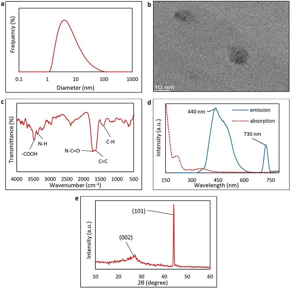 |
Figure 3 Characterization of the physicochemical properties of N-doped CQDs. (a) The DLS size distribution, and the (b) TEM results showed that the diameters of CQDs were 8.2±3.8 nm (c) the UV-visible spectrum showed a maximum absorption wavelength was around 355 nm, with two visible emission wavelengths at around 440 and 730 nm under UV excitation; the (d) FTIR, and (e) XRD patterns of the N-doped CQD confirmed remarkable existence of graphitic C=C sp2 bonds, with characteristic 2θ peaks around 25° for (002) and between 40° and 50° degrees for (101). |
FTIR of the CQDs
The characteristic chemical bond absorption patterns of CQDs, as shown in Figure 3c, were obtained using the FTIR spectra. It confirms that in N-doped CQD, the stretching and bending frequencies of the sp2 C=C bonds are significant at around 1560 and 842 cm−1. The C-H bending is also observed at 1377 cm−1, along with the amine N-H stretching and the amide carbonyl N-C=O stretching vibration, observed in the ranges of 3400–3300 cm−1 and 1800–1600 cm−1, respectively.
PL of the CQDs
In this study, UV-visible spectra of the synthesized CQDs were obtained, as shown in Figure 3d. The maximum absorption wavelength was around 355 nm. Under an excitation wavelength of 360 nm, the studied N-doped CQD exhibited two visible emission wavelengths at around 440 and 730 nm.
XRD of the CQDs
Figure 3e shows the XRD diagram of CQDs. As can be seen, in the range between 20 and 30 degrees, the broad peak (002) characteristic of carbon quantum dots is visible. This peak, which does not belong to graphite (ie around 25°), indicates that the crystal domains are not limited to graphite carbon, but rather that CQDs structures generally contain some amorphous polymer backbones with other crosslinks within them. Between 40 and 50 degrees, the (101) graphitic carbon crystalline phase is remarkable.
XPS
The elemental composition and chemical states of CQDs were further investigated using XPS analysis (Figure 4), including full and high-resolution spectra of C1s, O1s, and N1s, each of which accounts for 63.3%, 35.5% and 1.2% of CQDs atomic content, respectively (Figure 4a). As shown in Figures 4b–d the oxidation states in C and N are obtained by measuring the binding energy shift of core electrons experience due to changes in chemical environment. In Figure 4b, three binding energy peaks of 283.26, and 284.98 eV correspond to oxidation states for C-C sp2, and sp3, respectively, and the C=O bond at 286.78 eV [17]. As acknowledged by the FTIR results (Figure 3c), the O1s spectrum in Figure 4c, also shows the oxidation state of the N-C=O bond at 531.69 eV, and the C=O and –COOH, with peaks at 529.74 and 534.26 eV, respectively. The high-resolution N1s spectra reveal the existence of two types of C-N bond configurations, namely, pyrrolic-N and pyridinic-N oxidation states with binding energy peaks at 400.20 and 398.61 eV, respectively (Figure 4d).
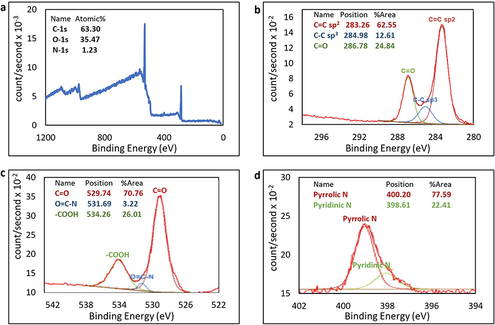 |
Figure 4 XPS spectrum of the CQDs. (a) The full, and high-resolution XPS spectrum of the N-doped CQD (b) C1s, showing the oxidation states of C-C sp2, and sp3, and the C=O bond at 283.26, 284.98, and 286.78 eV, respectively; (c) O1s with the oxidation states for N-C=O bond at 531.69 eV, and the C=O and –COOH, with peaks at 529.74 and 534.26 eV, respectively, and (d) N1s which confirms the existence pyridinic-N (398.61 eV), and pyrrolic-N (400.20 eV). |
Minimum Inhibitory Concentration (MIC)
The MIC values of Dendrocin ZM1–CQDs were determined using micro broth dilution against drug-resistant Gram-negative and Gram-positive strains. As shown in Table 2, the MIC values for carbon quantum dots alone are all >128 µg/mL, indicating minimal or no intrinsic antibacterial effect. Dendrocin ZM1 alone shows activity against E. coli strains (MIC 2–4 µg/mL). Activity against resistant strains of S. aureus (MRSA, VRSA) is weaker (MIC 16 µg/mL), indicating limitation against resistant Gram-positives.
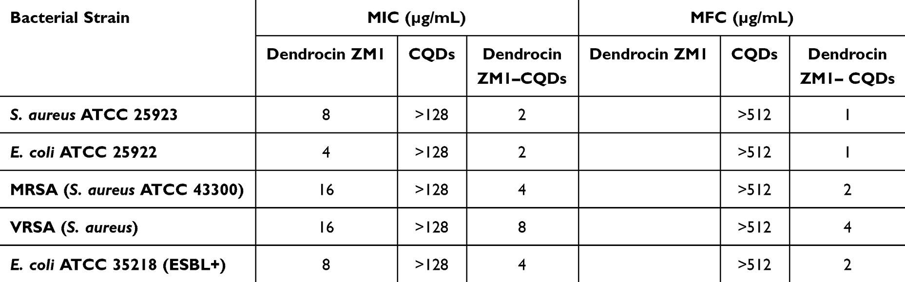 |
Table 2 Comparative MIC and MFC values (µg/mL) of Dendrocin ZM1 and Dendrocin ZM1–CQDs Against Standard and Resistant Bacterial Strains |
The MIC values decreased by 2 and 4 times in all strains compared to Dendrocin ZM1 alone. The strongest increase is observed in S. aureus ATCC 25923 and E. coli ATCC 25922 (MIC improved from 8 to 2 and 4 to 2 µg/mL, respectively). Even in resistant strains (MRSA, VRSA), MIC values decrease from 16 to 4 and 8 µg/mL, respectively. For every bacterial strain, the MBC values were two times higher than the MIC values.
Time-Kill Kinetics of Dendrocin ZM1–CQDs
To evaluate the bactericidal dynamics of the nanoconjugated peptide, a time–killing kinetic assay was performed against S. aureus ATCC 25923 and E. coli ATCC 25922. Dendrocin ZM1–CQD was tested at, 1×, 2× and 4× MIC concentrations, and bacterial viability was monitored over a 120 min period (Figure 5).
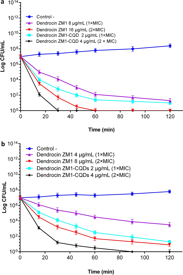 |
Figure 5 Time-kill kinetics of free and CQD-conjugated Dendrocin ZM1 (Dendrocin ZM1–CQDs). (a) against S. aureus ATCC 25923 and (b) against E. coli ATCC 25922. Bacterial suspensions were treated with 1×, and 2× MICs of either Dendrocin ZM1 alone or its conjugated form. Viable cell counts (log CFU/mL) were monitored over 120 min. Dendrocin ZM1 alone displayed dose-dependent bactericidal activity, achieving complete killing at higher concentrations. In contrast, the Dendrocin ZM1–CQDs exhibited significantly enhanced killing efficacy, especially at lower concentrations, and reduced the time required for bacterial eradication. Data are presented as mean ± SD from three independent experiments. |
As shown in Figure 5a, compared with Dendrocin ZM1 alone, which required 60 min at 2× MIC (16 µg/mL) to completely eradicate S. aureus, Dendrocin ZM1–CQDs exhibited significantly faster and more potent killing kinetics. At the concentration of 4 µg/mL (2× MIC), Dendrocin ZM1–CQDs completely killed bacteria within 30 min, which is twice as fast as Dendrocin ZM1 alone. At a concentration of 2 µg/mL (1× MIC), an approximately 3-log reduction was observed in 90 min, whereas Dendrocin ZM1 alone at its 1× MIC concentration required nearly 120 min to achieve similar bacterial suppression.
As shown in Figure 5b, time-killing studies showed that Dendrocin ZM1–CQDs also exerted greater bactericidal activity against E. coli ATCC 25922 compared to Dendrocin ZM1 alone. At a concentration of 2× MIC (8 µg/mL), Dendrocin ZM1–CQDs achieved a reduction of more than 3-log10 in CFU/mL within the first 60 min and achieved almost complete bacterial elimination within 90–120 min. This killing profile demonstrated a faster and more efficient bactericidal effect compared to Dendrocin ZM1 alone, which required longer exposure times to achieve similar results. At a concentration of 1× MIC (4 µg/mL), the Dendrocin ZM1–CQDs significantly reduced bacterial counts within 2 h and achieved faster bactericidal thresholds than Dendrocin ZM1 alone at its corresponding MIC concentration. These findings indicate that Dendrocin ZM1–CQDs not only reduces the MIC value for S. aureus and E. coli but also increases the kinetics of bacterial killing.
Hemolytic Activity of Dendrocin ZM1–CQDs
To evaluate the biosafety profile of the nanoconjugated peptide, hemolytic activity on human red blood cells (hRBCs) were investigated and compared with Dendrocin ZM1 alone. As shown in Figure 6a, Dendrocin ZM1–CQDs showed significantly reduced hemolytic activity at all concentrations tested (1×, 2×, and 4× MIC). At concentration of 4 µg/mL (1× MIC for E. coli) and 8 µg/mL (1× MIC for S. aureus), hemolysis remained less than 2%, which is considered biologically safe. Even at 2× and 4× MIC levels, hemolysis did not exceed 5%, indicating a favorable hemocompatibility profile.
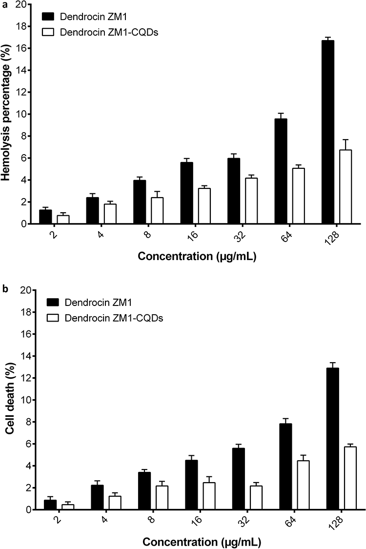 |
Figure 6 Hemolytic effect, cytotoxic effect, and stability of Dendrocin ZM1–CQDs. (a) Hemolytic activity of Dendrocin ZM1–CQDs on human red blood cells (RBCs) at increasing concentrations corresponding to 1×, 2×, and 4× MIC values. The conjugated form demonstrated significantly reduced hemolytic activity compared to Dendrocin ZM1 alone. (b) Cytotoxicity of Dendrocin ZM1–CQDs on HEK-293 cells at increasing concentrations (2–128 µg/mL). Cell death was determined after 24 h of exposure. The conjugated peptide exhibited lower toxicity compared to Dendrocin ZM1 alone, particularly at higher concentrations. Data represent mean ± SD of three independent replicates. |
In comparison, Dendrocin ZM1 alone showed more than 5% hemolysis at MIC concentrations (8–16 µg/mL), and hemolysis gradually increased with increasing concentration. At the highest dose tested (128 µg/mL), it did not exceed 7.8%, while at the same dose, 17% hemolysis occurred by Dendrocin ZM1 alone.
Cytotoxicity of Dendrocin ZM1–CQDs on HEK-293 Cells
The cytotoxic potential of Dendrocin ZM1–CQDs on human embryonic kidney (HEK-293) cells was assessed by serial peptide levels. As shown in Figure 6b, Dendrocin ZM1–CQDs showed less cytotoxicity on HEK293 cells compared to Dendrocin ZM1 alone. At concentrations corresponding to 1× and 2× MIC (4 and 8 µg/mL), cell viability remained above 97%, and even at a concentration of 128 µg/mL, Dendrocin ZM1–CQDs induced only 6.5% cell death, compared to 12.4% cell death induced by Dendrocin ZM1 alone.
ROS Generation and Scavenger Effects
As shown in Figure 7a and b, exposure of both S. aureus and E. coli strains to Dendrocin ZM1–CQDs at 2× MIC concentration caused a rapid and time-dependent increase in intracellular ROS levels, which peaked at 30–45 min after treatment (**p < 0.001 vs untreated control). The fold-change in ROS production at 45 min reached 3.3 ± 0.4 in S. aureus and 3.6 ± 0.4 in E. coli, whereas Dendrocin ZM1 and CQDs alone produced only slight increase (≤1.6-fold) over the same time period.
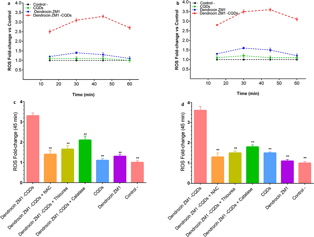 |
Figure 7 ROS generation kinetics and scavenger effects in S. aureus ATCC 25923 and E. coli ATCC 25922. (a and b) Time-dependent intracellular ROS levels in S. aureus and E. coli treated with Dendrocin ZM1–CQDs (2× MIC), Dendrocin ZM1, CQDs, or untreated controls, measured using DCFH-DA fluorescence. Dendrocin ZM1–CQDs induced a rapid and sustained ROS burst peaking at 30–45 min, with significantly higher fold-changes compared with all controls (**p < 0.001, two-way ANOVA with Tukey’s post-hoc test). (c and d) Effect of ROS scavengers (NAC, thiourea, catalase) on Dendrocin ZM1–CQDs induced ROS levels at 45 min. Pre-incubation with scavengers markedly reduced ROS production and restored bacterial viability in both S. aureus and E. coli (**p < 0.001 vs Dendrocin ZM1–CQDs), confirming the causal role of oxidative stress in antibacterial activity. Data are presented as mean ± SD from three independent experiments performed in triplicate. |
Importantly, pre-incubation with ROS scavengers including N-acetylcysteine (10 mM), thiourea (100 mM), and catalase (1000 U/mL) significantly reduced ROS production in both strains by 65–80% and restored bacterial viability by 1–2 log10 CFU (Figure 7c and d **p < 0.001 vs Dendrocin ZM1 alone). These findings confirm that ROS generation is the main mechanism of bactericidal activity of Dendrocin ZM1–CQDs against Gram-positive and Gram-negative bacteria.
Discussion
The present study demonstrates that nanoconjugation of the plant-derived antimicrobial peptide Dendrocin ZM1 with CQDs significantly enhances its antibacterial activity while maintaining biocompatibility. The nanoconjugate exhibited substantially reduced MIC values (approximately 2–4-fold) against both Gram-positive bacteria (including MRSA and VRSA) and Gram-negative strains (including ESBL-positive E. coli), together with accelerated bactericidal kinetics and rapid intracellular ROS generation. These findings are consistent with emerging evidence that nanoparticle-assisted delivery can potentiate the antimicrobial activity of AMPs by improving local concentration at bacterial membranes and facilitating intracellular access.16,19,33 Time-kill assays further revealed that the Dendrocin ZM1–CQDs nanoconjugate achieved ≥3 log10 CFU/mL reduction within 30–60 min at 2× MIC, whereas free Dendrocin ZM1 required longer exposure to reach comparable killing. This accelerated bactericidal action is likely attributable to enhanced membrane interaction and uptake mediated by CQDs, a mechanism reported for other AMP–nanomaterial platforms where nanocarriers promote efficient peptide–bacteria interactions and rapid onset of antimicrobial effects.18
One of the possible mechanisms of action of Dendrocin ZM1–CQD is to induce a strong robust intracellular ROS—including superoxide, hydrogen peroxide, and hydroxyl radicals—concurrent with membrane depolarization and cell death. As shown in Figure 7a–d, treatment with Dendrocin ZM1-CQDs caused a rapid and significant increase in intracellular ROS levels in both S. aureus and E. coli, peaking within 30–45 min. In contrast, CQDs alone and free Dendrocin ZM1 only resulted in a moderate (≤1.6-fold) increase in ROS, indicating that neither of these components alone is sufficient to induce strong oxidative stress. Carbon quantum dots have been reported to induce limited ROS production in bacterial cells, depending on the surface chemistry, heteroatom doping, and environmental conditions; However, the production of such ROS is generally modest and often insufficient to provide rapid bactericidal activity on its own.34,35
The significantly higher ROS levels observed for the Dendrocin ZM1-CQDs combination suggest a synergistic mechanism in which the CQDs act as a ROS-generating nanoplatform while the antimicrobial peptide enhances bacterial aggregation and membrane disruption. Antimicrobial peptides are known to disrupt bacterial membrane integrity and increase permeability, thereby facilitating intracellular access and enhancing oxidative damage caused by nanomaterials.36 A similar synergistic enhancement of ROS-induced antibacterial activity has been reported for peptide-nanomaterial combinations, where peptide-induced membrane destabilization sensitizes bacteria to nanoparticle-induced oxidative stress.37 This interpretation is further supported by ROS rescue assays, in which N-acetylcysteine, thiourea, and catalase significantly reduced intracellular ROS levels and partially restored bacterial viability. Such attenuation of bactericidal activity in the presence of ROS scavengers is consistent with previous reports on CQD-based antibacterial systems and nanoconjugates, confirming oxidative stress as a dominant killing mechanism.1,3,5 Collectively, these results indicate that although CQDs contribute little to the basal ROS production, the dominant and bactericidal oxidative stress response is primarily driven by the Dendrocin ZM1-CQDs conjugate, not solely by the nanocarrier itself.
While both S. aureus and E. coli showed significant ROS production, E. coli produced more ROS than S. aureus compared to its controls (Figure 7a and b). The results of ROS production are consistent with the fact that the thick cell wall of Gram-positive bacteria limits the entry of Dendrocin ZM1–CQDs compared to Gram-negative bacteria, hence producing less ROS. The ROS production leads to bacterial membrane distortion or bacterial cell lysis which ultimately leads to cell death.38
In addition to antibacterial potency, biocompatibility is a critical consideration for clinical applications. Dendrocin ZM1–CQD showed low hemolytic activity (<2% at the MIC; <5% at 4× MIC) and minimal cytotoxicity to human cell lines even at high concentrations. Hemolysis refers to the interaction and incompatibility of samples with RBCs. When the hemolysis rate of a material is less than 5%, it is considered biologically safe,39 and the results also showed that Dendrocin ZM1–CQDs has high potential as an antibacterial agent with low toxicity.40 These results are promising for the use of AMP– CQDs conjugates for systemic or topical therapeutic applications.
Beyond biological performance, physicochemical properties such as surface area and porosity can also influence the behavior of nanomaterial-based antimicrobials. In the present study, Brunauer–Emmett–Teller (BET) analysis was conducted for the Dendrocin ZM1–CQDs nanoconjugate to characterize its textural properties; however, BET measurements of the individual components were not performed. While a direct comparison between the surface areas of the individual materials and the composite would enable a more quantitative assessment of structural changes upon nanoconjugation, such analysis was beyond the scope of the current work. Previous studies such as those by Chen et al (2020)41 have confirmed that nanocomposite formation can substantially alter surface area and porosity due to interfacial interactions and nanoscale rearrangements, which in turn may affect biological interactions. Although direct comparison was not possible here, the observed BET characteristics of the nanoconjugate are consistent with these reported trends. Accordingly, the absence of comparative BET data for the individual components is acknowledged as a limitation of this study, and future investigations will include systematic BET analyses of both individual materials and the composite to further elucidate structure–activity relationships.
Taken together, these findings indicate that the enhanced antibacterial activity of the Dendrocin ZM1–CQDs nanoconjugate arises from a synergistic interplay between membrane disruption and ROS-mediated intracellular damage, while maintaining favorable biocompatibility. This dual mechanism underscores the promise of AMP–CQD nanoconjugates as next-generation broad-spectrum antimicrobial agents.
Conclusion
In this study, we developed and characterized a nanoconjugate of the antimicrobial peptide Dendrocin ZM1 with carbon quantum dots as a ROS-generating antibacterial system targeting multidrug-resistant (MDR) bacteria. By integrating a plant-derived AMP with CQDs, we achieved a 2–4-fold reduction in MIC values against both Gram-positive and Gram-negative MDR strains and demonstrated rapid bactericidal activity, with ≥3-log10 CFU/mL reduction within 30–60 minutes at 2× MIC. Mechanistic assays revealed a 3–4-fold increase in intracellular ROS levels, and the ability of ROS scavengers to rescue bacterial viability confirmed that oxidative stress plays a central role in the antibacterial activity of Dendrocin ZM1–CQDs.
Importantly, this enhancement in antibacterial efficacy was achieved without compromising safety toward mammalian cells, as evidenced by >90% viability in HEK293 cells and minimal hemolytic activity within the therapeutically relevant concentration range. These findings highlight CQD conjugation as an effective nanotechnological strategy to potentiate AMP activity while mitigating dose-dependent cytotoxicity, thereby addressing a critical challenge in AMP-based antimicrobial development.
Despite these advances, a clear research gap exists in the comprehensive evaluation of AMP–nanomaterial conjugates under physiologically relevant and in vivo conditions. The present study is limited to in vitro antibacterial and preliminary biosafety assessments and does not address long-term stability, pharmacokinetics, biodistribution, or host–pathogen interactions in complex biological systems. In addition, detailed cellular uptake pathways and downstream cytotoxic mechanisms in mammalian cells were not explored in this work.
Future studies will therefore focus on expanding the antibacterial evaluation to a broader range of clinically relevant MDR pathogens, assessing long-term and physiological stability (including serum stability), and conducting in vivo efficacy and safety studies. Furthermore, advanced imaging techniques and mechanistic analyses will be employed to elucidate cellular internalization pathways and host–nanoconjugate interactions. Collectively, these efforts will be essential to advance AMP–CQD nanoconjugates toward translational and therapeutic applications.
Abbreviations
CQDs, Carbon quantum dots; MDR, Multidrug-resistant; MIC, Minimum inhibitory concentration; ROS, Reactive oxygen species; AMPs, Antimicrobial peptides; ZnO, Zinc oxide; CuO, copper oxide; Ag, Silver; TiO2, Titanium oxide; NPs, Nanoparticles; FTIR, Fourier transform infrared spectroscopy; DLS, Dynamic light-scattering; PL, Photoluminescence; XPS, X-ray Photoelectron Spectroscopy; TEM, Transmission electron microscopy; RP-HPLC, Reverse phase-high performance liquid chromatography; MD, Molecular Dynamics; PBC, Periodic boundary conditions; LINCS, Linear Constraint Solver.
Data Sharing Statement
The data used to support the findings of this study are included within the article.
Ethics Statement
Ethical approval for conducting this study was obtained from the Shahid Beheshti University of Medical Sciences in Tehran, Iran (IR.SBMU.MSP.REC. 1402.445).
Author Contributions
All authors have made significant contributions to the reported work, whether in conceptualization, study design, execution, data collection, analysis, and interpretation, or in all of these areas; have participated in drafting, revising, or critically reviewing the article; have provided final approval of the version to be published; have agreed on the journal to which the article has been submitted; and agree to be accountable for all aspects of the work.
We confirm that this statement accurately reflects the contributions and responsibilities of all authors listed.
Funding
The authors disclosed receipt of the following financial support for the research, authorship, and/or publication of this article: The Infectious Diseases and Tropical Medicine Research Center, Shahid Beheshti University of Medical Sciences, Tehran, Iran (Grant No. 43006674).
Disclosure
The authors declare no conflicts of interest in this work.
References
1. Loffredo MR, Savini F, Bobone S. et al. Inoculum effect of antimicrobial peptides. Proc Natl Acad Sci. 2021;118(21):e2014364118. doi:10.1073/pnas.2014364118
2. Camlik G, Ozakca I, Bilakaya B, et al. Development of composite carbon quantum dots-insulin formulation for oral administration. J Drug Deliv Sci Technol. 2022;76:103833. doi:10.1016/j.jddst.2022.103833
3. de Souza AP, de Souza KR, Santos DL, et al. One-pot functionalization of carbon dots with ecPis-4s antimicrobial peptide. Mater Chem Phys. 2023;307:128101. doi:10.1016/j.matchemphys.2023.128101
4. Gao F, Ma S, Li J, et al. Rational design of high quality citric acid-derived carbon dots by selecting efficient chemical structure motifs. Carbon. 2017;112:131–18. doi:10.1016/j.carbon.2016.10.089
5. Mitchell MJ, Billingsley MM, Haley RM, Wechsler ME, Peppas NA, Langer R. Engineering precision nanoparticles for drug delivery. Nat Rev Drug Discov. 2021;20(2):101–124. doi:10.1038/s41573-020-0090-8
6. Hetta HF, Ramadan YN, Al-Harbi AI A, et al. Nanotechnology as a promising approach to combat multidrug resistant bacteria: a comprehensive review and future perspectives. Biomedicines. 2023;11(2):413. doi:10.3390/biomedicines11020413
7. Martínez-Corona Z, García-Zaleta DS, López-González R, et al. Antibacterial activity of TiO2, CuO, and CuO/TiO2 nanomaterials and their potential application on construction surfaces. J Mater Sci: Mater Eng. 2025;20(1):70. doi:10.1186/s40712-025-00288-7
8. Xuan L, Ju Z, Skonieczna M, Zhou PK, Huang R. Nanoparticles‐induced potential toxicity on human health: applications, toxicity mechanisms, and evaluation models. MedComm. 2023;4(4):e327. doi:10.1002/mco2.327
9. Ana ID, Vrana NE, Morita A, Satria GA, Hathroubi S. Antibacterial Surface Functionalization of Biomedical Scaffolds: a Transformation towards More Adaptive, Resilient Regenerative Therapy. Results Surf Interf. 2025;100481. doi:10.1016/j.rsurfi.2025.100481
10. Brindha B, Okla MK, Kokilavani S, et al. Dynamic Ag-mediated electron transfer confined ZnO nanorods for boosted photocatalytic bacterial disinfection. J Clean Prod. 2024;451:141908. doi:10.1016/j.jclepro.2024.141908
11. Harikumar B, Subhiksha V, Okla MK, et al. Magnetic Fe3O4 nanospheres supported N/S-SnO2 nanorod for highly effective visible light photocatalytic degradation of tetracycline. J Alloys Compd. 2024;986:174017. doi:10.1016/j.jallcom.2024.174017
12. Swetha S, Alahmadi TA, Ansari MJ, Khan SS. Strategically tailored double S-scheme heterojunction in h-MoO3 doped Bi7O9I3 decorated with Cr–CdS quantum dots for efficient photocatalytic degradation of phenolics. J Clean Prod. 2024;449:141656. doi:10.1016/j.jclepro.2024.141656
13. Qureshi ZA, Dabash H, Ponnamma D, Abbas MK. Carbon dots as versatile nanomaterials in sensing and imaging: efficiency and beyond. Heliyon. 2024;10(11). doi:10.1016/j.heliyon.2024.e31634
14. Kailasa SK, Bhamore JR, Koduru JR, Park TJ. Carbon dots as carriers for the development of controlled drug and gene delivery systems. J Biomed Nanotechnol. 2019;295–317. doi:10.1016/B978-0-12-816506-5.00006-1
15. Kokilavani S, AL-ghamdi AA, Okla MK, et al. Interfacial engineering of CQDs sensitized NiFe2O4 spheres anchored CoCr2O4/MoO3-x NSs for boosted visible light driven photodegradation of antibiotic, mechanistic insights, and its toxicity assessment. J Water Process Eng. 2023;51:103355. doi:10.1016/j.jwpe.2022.103355
16. Chu X, Wu F, Sun B, et al. Genipin cross-linked carbon dots for antimicrobial, bioimaging and bacterial discrimination. Colloids Surf B Biointerfaces. 2020;190:110930. doi:10.1016/j.colsurfb.2020.110930
17. Dong X, Liang W, Meziani MJ, Sun YP, Yang L. Carbon dots as potent antimicrobial agents. Theranostics. 2020;10(2):671. doi:10.7150/thno.39863
18. Pechnikova NA, Domvri K, Porpodis K, Istomina MS, Iaremenko AV, Yaremenko AV. Carbon quantum dots in biomedical applications: advances, challenges, and future prospects. Aggregate. 2025;6(3):e707. doi:10.1002/agt2.70028
19. Mazumdar A, Haddad Y, Milosavljevic V, et al. Peptide-carbon quantum dots conjugate, derived from human retinoic acid receptor responder protein 2, against antibiotic-resistant gram positive and gram negative pathogenic bacteria. J Nanomater. 2020;10(2):325. doi:10.3390/nano10020325
20. Seyedjavadi SS, Razzaghi-Abyaneh M, Nasiri MJ, et al. Isolation and chemical characterization of an alpha-helical peptide, dendrocin-ZM1, derived from Zataria multiflora boiss with potent antibacterial activity. Probiotics Antimicrob. 2022;14(2):326–336. doi:10.1007/s12602-022-09907-7
21. Min KH, Kim KH, Ki MR, Pack SP. Antimicrobial peptides and their biomedical applications: a review. Antibiotics. 2024;13(9):794. doi:10.3390/antibiotics13090794
22. Abraham M, Alekseenko A, Bergh C, et al. GROMACS 2023 1. Manual. GROMACS. The Netherlands: Groningen; 2023.
23. Hess B, Bekker H, Berendsen HJ, Fraaije JG. LINCS: a linear constraint solver for molecular simulations. J Comput Chem. 1997;18(12):1463–1472. doi:10.1002/(SICI)1096-987X(199709)18:12<1463::AID-JCC4>3.3.CO;2-L
24. Schrodinger LL. The PyMOL molecular graphics system. Version. 2015;1:8.
25. Vaught A. Graphing with Gnuplot and Xmgr: two graphing packages available under linux. Linux J. 1996;1996(28es):7.
26. Khan WU, Wang D, Zhang W, et al. High quantum yield green-emitting carbon dots for Fe (ІІІ) detection, biocompatible fluorescent ink and cellular imaging. Sci Rep. 2017;7(1):14866. doi:10.1038/s41598-017-15054-9
27. Li P, Han F, Cao W, et al. Carbon quantum dots derived from lysine and arginine simultaneously scavenge bacteria and promote tissue repair. Appl Mater Today. 2020;19:100601. doi:10.1016/j.apmt.2020.100601
28. Clinical and Laboratory Standards Institute (CLSI). Performance Standards for Antimicrobial Susceptibility Testing. In: CLSI Supplement M100.
29. Wang L, Zhao X, Xia X, et al. Antimicrobial peptide JH-3 effectively kills Salmonella enterica Serovar Typhimurium strain CVCC541 and reduces its pathogenicity in mice. Probiot Antimicrob Proteins. 2019;11:1379–1390. doi:10.1007/s12602-019-09533-w
30. General Assembly of the World Medical Association. World Medical Association Declaration of Helsinki: ethical principles for medical research involving human subjects. J Am Coll Dent. 2014;81(3):14–18. doi:10.1001/jama.2013.281053.
31. Sun CQ, Peng J, Yang LB, et al. A cecropin-4 derived peptide C18 inhibits Candida albicans by disturbing mitochondrial function. Front Microbiol. 2022;13:872322. doi:10.3389/fmicb.2022.872322
32. Hsieh HJ, Cheng CC, Wu ST, Chiu JJ, Wung BS, Wang DL. Increase of reactive oxygen species (ROS) in endothelial cells by shear flow and involvement of ROS in shear‐induced c‐fos expression. J Cell Physiol. 1998;175(2):156–162. doi:10.1002/(SICI)1097-4652(199805)175:2<156::AID-JCP5>3.3.CO;2-G
33. Ladavière C, Gref R. Toward an optimized treatment of intracellular bacterial infections: input of nanoparticulate drug delivery systems. Nanomed. 2015;10(19):3033–3055. doi:10.2217/nnm.15.128
34. Jelinek R. Carbon Quantum Dots: Synthesis, Properties and Applications. Springer; 2016; doi:10.1007/978-3-319-43911-2
35. Zhao WB, Liu KK, Wang Y, et al. Antibacterial carbon dots: mechanisms, design, and applications. Adv Healthc Mater. 2023;12(23):2300324. doi:10.1002/adhm.202300324
36. Mahlapuu M, Håkansson J, Ringstad L, Björn C. Antimicrobial peptides: an emerging category of therapeutic agents. Front Cell Infect Microbiol. 2016;6:194. doi:10.3389/fcimb.2016.00194
37. Liang J, Li W, Chen J, et al. Antibacterial activity and synergetic mechanism of carbon dots against gram-positive and-negative bacteria. ACS Appl Bio Mater. 2021;4(9):6937–6945. doi:10.1021/acsabm.1c00618
38. Qayyum S, Oves M, Khan AU. Obliteration of bacterial growth and biofilm through ROS generation by facilely synthesized green silver nanoparticles. PLoS One. 2017;12(8):e0181363. doi:10.1371/journal.pone.0181363
39. Yang Y, Ren X, Sun Z, et al. Toxicity and bio-distribution of carbon dots after single inhalation exposure in vivo. Chin Chem Lett. 2018;29(6):895–898. doi:10.1016/j.cclet.2018.04.018
40. Wu F, Sun B, Chu X, et al. Hyaluronic acid-modified porous carbon-coated Fe3O4 nanoparticles for magnetic resonance imaging-guided photothermal/chemotherapy of tumors. Langmuir. 2019;35(40):13135–13144. doi:10.1021/acs.langmuir.9b02300
41. Cheng L, He Y, Gong M, et al. MOF-derived synthesis of Co3O4 nanospheres with rich oxygen vacancies for long-term stable and highly selective n-butanol sensing performance. J Alloys Compd. 2021;857:158205. doi:10.1016/j.jallcom.2020.158205
 © 2026 The Author(s). This work is published and licensed by Dove Medical Press Limited. The
full terms of this license are available at https://www.dovepress.com/terms
and incorporate the Creative Commons Attribution
- Non Commercial (unported, 4.0) License.
By accessing the work you hereby accept the Terms. Non-commercial uses of the work are permitted
without any further permission from Dove Medical Press Limited, provided the work is properly
attributed. For permission for commercial use of this work, please see paragraphs 4.2 and 5 of our Terms.
© 2026 The Author(s). This work is published and licensed by Dove Medical Press Limited. The
full terms of this license are available at https://www.dovepress.com/terms
and incorporate the Creative Commons Attribution
- Non Commercial (unported, 4.0) License.
By accessing the work you hereby accept the Terms. Non-commercial uses of the work are permitted
without any further permission from Dove Medical Press Limited, provided the work is properly
attributed. For permission for commercial use of this work, please see paragraphs 4.2 and 5 of our Terms.