
Back to Journals » Journal of Blood Medicine » Volume 15
Deep Vein Thrombosis as a Complication of Gemcitabine-Capecitabine Chemotherapy in Adenocarcinoma of Gallbladder
Authors Widiasi ED  , Romadhon PZ , Ashariati A, Bintoro SUY , Diansyah MN, Amrita PNA, Savitri M
Received 8 August 2024
Accepted for publication 2 December 2024
Published 19 December 2024 Volume 2024:15 Pages 523—531
DOI https://doi.org/10.2147/JBM.S482753
Checked for plagiarism Yes
Review by Single anonymous peer review
Peer reviewer comments 2
Editor who approved publication: Dr Martin H Bluth
Etha Dini Widiasi,1 Pradana Zaky Romadhon,1– 3 Ami Ashariati,1,3 Siprianus Ugroseno Yudho Bintoro,1,3 Muhammad Noor Diansyah,1– 3 Putu Niken Ayu Amrita,1,3 Merlyna Savitri1,3
1Department of Internal Medicine, Faculty of Medicine, Universitas Airlangga, Surabaya, Indonesia; 2Department of Internal Medicine, Universitas Airlangga Hospital, Faculty of Medicine, Universitas Airlangga, Surabaya, Indonesia; 3Division of Hematology and Medical Oncology, Department of Internal Medicine, Dr. Soetomo General Academic Hospital, Surabaya, Indonesia
Correspondence: Pradana Zaky Romadhon, Department of Internal Medicine, Universitas Airlangga Hospital, Faculty of Medicine, Universitas Airlangga, Jalan Dharmahusada Permai, Mulyorejo, Surabaya, Jawa Timur, Indonesia, 60115, Email [email protected]
Abstract: Gallbladder adenocarcinoma has a high mortality rate, with approximately 1.7% cancer-related deaths worldwide. Cancer-associated thrombosis (CAT), including deep vein thrombosis (DVT), can significantly increase the risk of mortality within cancer patients, especially in pancreatic, brain, and intra-abdominal cancers, as well as in advanced and metastatic cancers. In this case report, there was a 45-year-old male patient diagnosed with advanced gallbladder adenocarcinoma UICC stage IVB with a TNM stage of T2b, N0, M1 with liver metastases who experienced pain and swelling in both lower limbs after undergoing a VI-A cycle of chemotherapy with gemcitabine capecitabine. The risk of thrombosis was calculated using the modified Khorana-Vienna CAT scores, which increased during every chemotherapy session. In this case, the Khorana-Vienna CAT score was calculated during two latest cycle of chemotherapy that somewhat considered delayed as the patient had already shown hypercoagulopathy symptoms and developed a poorer prognosis. Early CAT scoring, ideally before starting chemotherapy session, potentially improves thrombosis prognosis. The patient’s condition improved after administration of antithrombotic agents. Chemotherapy agents and other factors, including the cancer site and presence of metastatic cancer, influence the risk of CAT. Risk predictor scores are required to assess the risk of CAT and benefits of prophylactic treatment. Prophylactic therapy can be initiated in patients with high-risk CAT, calculated using the modified Khorana and Vienna CAT scores, to prevent thrombosis and improve patient outcomes.
Keywords: gallbladder cancer, deep vein thrombosis, cancer-associated thrombosis, Khorana-Vienna CAT score, chemotherapy, thromboprophylaxis
Introduction
Gallbladder adenocarcinoma is a highly lethal and rare cancer that emerges in the epithelial layer of the gallbladder.1 Moreover, the gallbladder is a sac located beneath the hepatic lobe responsible for storing bile fluid produced in the liver, which is then transported to the small intestine. Bile fluid is deposited in the duodenum and contributes positively to digestion, particularly in the lipid metabolism. The accurate incidence of gallbladder cancer is uncertain owing to challenging diagnostics and rapid disease progression, especially in developing countries with limited facilities.2 Gallbladder adenocarcinoma has a high mortality rate, accounting for approximately 1.7% of cancer-related deaths and a 5-years survival rate of <5% worldwide. Clinically, it often occurs without any symptoms at the early stage of gallbladder adenocarcinoma. The symptoms might only appear in the late stage of the cancer; thus, the prognosis is poor.2,3 The clinical presentation could differ depending on the stage of the disease at the time of assessment. Intense paroxysmal throbbing pain in the right hypochondriac region that radiates to the right shoulder tip with nausea-vomiting is the most common symptoms of gallbladder cancer. A palpable gallbladder mass and jaundice can be found in later stages of gallbladder cancer. Jaundice may result in a worse clinical presentation of gallbladder cancer because it appears following tumor invasion, obstruction of the bile duct due to lymphadenopathy or tumor compression, and liver metastases.4 Surgical resection of the gallbladder is the main therapy for gallbladder carcinoma, although the recurrence rate after radical resection of gallbladder cancer is still high.5
Chemotherapy should be administered as an adjuvant therapy after gallbladder resection surgery to decrease the recurrence rate. The National Comprehensive Cancer Network (NCCN) recommends administering systemic chemotherapy for unresectable or metastatic gallbladder carcinoma.6 The suggested systemic therapy for biliary tract cancer is currently divided into two categories, one is designated for patients with superior performance status and the other is specialized for patients with poor performance status. Single-agent therapies, such as gemcitabine, capecitabine, and single-agent fluorouracil, are also beneficial for patients with poor performance status, elderly patients, and patients with concurrent medical conditions as symptom control, compared to supportive therapy alone. Combination therapy can be used to treat biliary tract cancer, gemcitabine with 5-fluorouracil (5-FU) or gemcitabine with capecitabine. Patients with a good performance status will establish a good response to combination therapy that can be administered in the form of gemcitabine/platinum-based regimens (cisplatin or oxaliplatin if cisplatin is contraindicated), 5-FU/platinum-based regimens, gemcitabine/capecitabine, or capecitabine/platinum-based regimens. One study recommends a gemcitabine/cisplatin combination as a first-line systemic therapy; however, other studies suggest that other combinations, such as gemcitabine/oxaliplatin and gemcitabine/capecitabine, have similar efficacy to gemcitabine/cisplatin. The gemcitabine/capecitabine regimen can be administered with a capecitabine dose of 1500 mg/m2 divided into two doses from days 1 to 14, and gemcitabine 1000 mg/m2 on days 1 and 8 every 3 weeks.1,6,7 If systemic therapy does not yield significant outcomes, clinical trial medications, like immunotherapy and targeted therapy, may be considered. Palliative chemotherapy and best supportive care may be an option of last resort for advanced or metastatic gallbladder carcinoma that does not respond to treatment and has a poor prognosis.6 Some guidelines categorize palliative chemotherapy into first-line, second-line, and third-line regimens, which their administration are determined based on performance score (PS). Patients with a PS of 0–1 may receive palliative gemcitabine-cisplatin chemotherapy, while those with a PS ≥2 may be offered gemcitabine monotherapy. Beyond PS, additional factors such as advanced age at diagnosis, lymph node and distant metastases, poor tumor cell differentiation, gallbladder involvement, and albumin and bilirubin levels significantly impact the outcome of palliative chemotherapy in improving patient quality of life and slowing cancer progression.8 Gemcitabine and capecitabine combination therapy was applied in this case.
Cancer-associated thrombosis (CAT) is more likely to occur since cancer is a predisposing factor for thrombosis. Thromboembolism risk in cancer patients is seven-fold higher compared with the normal population. Chemotherapy increased the possibility of CAT by 5–10%.9 Deep vein thrombosis (DVT), a manifestation of CAT, significantly increases the risk of mortality among patients with cancer, especially in pancreatic, brain, and intra-abdominal cancers, as well as in advanced and metastatic cancer.10,11 Administration of thromboprophylaxis can reduce the risk of a venous thromboembolism (VTE) by up to 50% in high-risk patients. Risk predictor scores are required to assess the risk of CAT and the benefits of thromboprophylaxis in cancer patients, especially those who have undergone chemotherapy. Studies have been conducted to develop a predictive risk model for CAT. Several risk score has already examined for validity and compared with other scores for best outcome, such as the Khorana score, Vienna-CAT score, PROTECHT score, and CONKO score. These scores are required to assess the eligibility of potential cancer patient for thromboprophylaxis therapy.9,12 The National Comprehensive Cancer Network (NCCN) and the American Society of Hematology (ASH) recommend the Khorana score as a valid and reliable CAT predictive score. The Khorana score alone seems to encounter several limitations, thus Khorana score application together with Vienna-CAT score (Table 1) appeared to have more superior outcome by discriminate better between low and high CAT-risk patients.
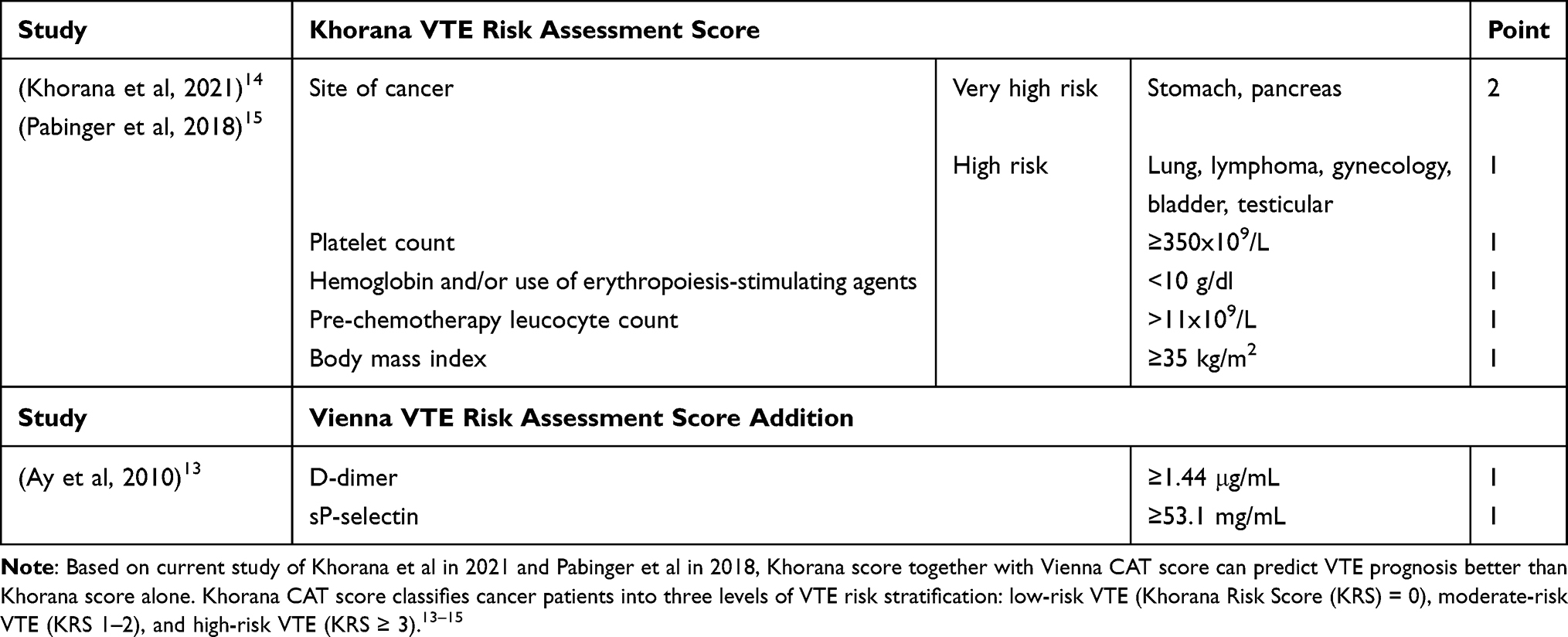 |
Table 1 The Modified Vienna CAT Score Integrates the Criteria from the Khorana Score and the Addition of Two Biochemical Parameters.13–15 |
Case Presentation
A 45-year-old male diagnosed with advanced gallbladder adenocarcinoma UICC stage IVB with a TNM stage of T2b, N0, and M1 with liver metastases experienced throbbing pain and swelling in both lower limbs after undergoing a VI-A cycle of gemcitabine and capecitabine chemotherapy. The pain was exaggerated with movement but improved when his legs were in a straight position. The color of the legs did not change. Both legs felt warm, and the left leg was warmer than the other. There were no complaints of cough, shortness of breath, pain while swallowing, fever, nausea, or vomiting. Based on previous medical history, the patient complained of intermittent pain in the right upper abdomen that had been experienced for nine months prior to the first admission to the hospital. Abdominal pain increased, especially during meals, accompanied by nausea without vomiting, decreased appetite, and perceived weight loss of approximately two kilograms per month. The abdominal pain was felt to be increasingly heavy and interfered with activities. Abdominal ultrasonography revealed the presence of multiple gallstones. Therefore, gallstone removal and biliary bypass were performed. During the surgery, a lump was observed in the gallbladder. Subsequently, the operator performed a biopsy to obtain a tissue sample for the anatomical pathological examination of the gallbladder. An anatomical pathological examination revealed a malignant gallbladder tumor.
The histological differentiation could provide the extent or size of the tumor and how far the cancer has grown into the wall of the gallbladder that indicated the tumor (T) grade from UICC (Union for International Cancer Control) TNM staging.16,17 In this case, microscopic histopathologic examination of the gallbladder biopsy sample showed malignant tumor tissue fragments that formed an irregular glandular arrangement lined with anaplastic epithelium, columnar shape, round oval nucleus, pleomorphic, coarse chromatin, prominent nucleoli, and sufficient cytoplasm as shown in Figure 1. The tumor cells growth invaded perimuscular connective tissue on the side of the liver without extension beyond the serosa or into the liver (T2b). The conclusion of the gallbladder biopsy material was adenocarcinoma NOS (not otherwise specified). There were no molecular characteristics examinations performed in this case due to the lack of fund and facilities. MRI-MRCP showed a malignant mass in the gallbladder with metastasis to the liver (M), without enlargement or signs of metastasis to the lymph nodes (N). Based on histopathology and radiology examination results, the patient had suffered from gallbladder adenocarcinoma UICC stage IVB with a TNM stage of T2b, N0, and M1 liver metastasis as guided by the UICC staging system.16,17 Biliary tract cancer, including gallbladder carcinoma and cholangiocarcinoma, has been known to be associated with genetic predisposition that can be detected through molecular testing from germline mutation. The most abnormality findings were BRCA2 mutation followed with BRCA1 mutation, and at least MLH1, MSH2, PALB2, RAD51D, BAP1 and ATM mutation. Recommended molecular testing for gallbladder carcinoma includes ERBB2, ERBB3, ELF3, BRAF V600E mutation, NTRK gene fusion, MSI-H/dMMR, RET gene fusion, and also HER2 mutation.18,19 In this case, we did not perform molecular testing due to lack of facilities and cost-related issues.
 |
Figure 1 The patient’s microscopic histopathologic examination of the gallbladder biopsy sample using total magnification lenses 100x (A) and 400x (B). |
The patient was then advised to undergo chemotherapy that was planned for a total of six cycles every 28 days (with divided doses every 14 days) using a gemcitabine-capecitabine chemotherapy regimen. After the first chemotherapy session (I-A cycle), the patient experienced side effects, such as nausea, vomiting, intermittent right abdominal pain, occasional back pain and right-sided chest pain. After the VI-A cycle of chemotherapy, the patient experienced pain and swelling in both the legs. This patient was diagnosed with hypertension 5 years prior with routine treatment of once daily 10 mg of amlodipine and once daily 20 mg of atorvastatin every night, previous liver disease or jaundice was denied, history of tuberculosis was denied, history of diabetes mellitus was denied, history of kidney disease was denied. There was no history of malignancy with other family members. This patient was a non-smoker, and alcohol consumption was excluded. The patient underwent the I-A cycle until VI-B chemotherapy with 1200 mg of gemcitabine was consumed on day-1 and day-8, accompanied by 1000 mg capecitabine twice daily for seven days.
On physical examination after the VI-A cycle of chemotherapy, hemodynamics were stable: blood pressure was 128/73 mmHg, heart rate was 89 beats per minute, respiratory rate was 18 breaths/min, temperature was 36.8 °C, oxygen saturation was 97% on room air, and oxygen saturation of the lower extremities was 97–98% on room air. A physical examination revealed a distended abdomen, visible collateral veins, liver enlargement (three fingers below the costae arc), and tenderness in the right upper quadrant. The patient showed a positive Homan’s sign in both lower limbs. Laboratory examinations were performed after VI-A cycle of chemotherapy to determine the patient’s condition. The following values were obtained: hemoglobin, 12.2 g/dl; hematocrit, 37.4%; MCV, 101.6 fl; MCH, 33.2 pg; MCHC, 32.6 g/dl; leukocytes 8200/μL with a dominant neutrophil count of 71.9%; lymphocytes, 15.6%; monocytes, 11.2%; eosinophils, 0.9%; basophils, 0.4%; platelets, 269,000/μL; SGOT, 248 U/L; SGPT, 97 U/L; non-reactive HBsAg; non-reactive Anti-HCV. In this case, signs of hypercoagulopathy were examined using D-dimer serum levels and Doppler ultrasonography examination of lower extremities to evaluate thrombus development, while the predictive risk score for VTE was determined using the modified Khorana-Vienna CAT scores. The Khorana-Vienna CAT score in this case was calculated twice: during VI-A chemotherapy cycle and 14 days after VI-B chemotherapy cycle. These calculations were considered pretty late, because the patient has already developed hypercoagulopathy symptoms and a poorer prognosis. The progression of D-dimer and predictive risk score of DVT increased. During VI-A chemotherapy cycle, D-dimer level was 0.49 ug/mL with a modified CAT score 9.2% and the Khorana-Vienna CAT score 2 or intermediate score. Meanwhile, in 14 days after VI-B chemotherapy cycle, D-dimer level increased to 2.4 ug/mL with progression of modified CAT score to 12.7% and the Khorana-Vienna CAT score 3 or high-risk score (the risk score explanation can be seen in Table 1).
Doppler ultrasound examination of the patient’s lower limb vessels revealed evidence of DVT (left: common femoral and popliteal veins; right: common femoral vein). The occurrence of DVT in this patient was suspected to be a complication of gemcitabine because the symptoms of DVT presented before the last cycle of chemotherapy, although disease progression itself might exaggerate the probability of CAT. The patient was administered a subcutaneous injection of fondaparinux sodium 2.5 mg once daily, followed by a subcutaneous injection of 40 mg of enoxaparin sodium once daily for one week (VTE prophylaxis therapy can be seen in Table 2). The patient’s condition improved. Swelling and pain in both legs decreased. The patient was advised to continue treatment as an outpatient. Rivaroxaban 10 mg once daily was administered as an ambulatory therapy. After the last cycle of chemotherapy, the patient underwent an MRI chemotherapy response examination which showed that the malignant mass of the gallbladder was still prominent, liver metastasis still existed, and hepatomegaly. Because the latest abdominal MRI results showed that the mass in the gallbladder was still present, the patient was then planned for palliative chemotherapy using Gemcitabine monotherapy.
Discussion
Adenocarcinoma of the gallbladder is one of the rarest cancers of the abdominal organ. It has a poor prognosis due to its highly malignant character and is often discovered incidentally during laparoscopic cholecystectomy with a delayed presentation.20 The International Agency for Research on Cancer (IARC) Globocan in 2018 estimated that gallbladder cancer accounts for 1.7% of cancer-related deaths worldwide, with 220,000 new cases diagnosed each year and a female predominance 3–6 times higher than in males. The highest incidence rates per 100,000 persons were found in Latin America, East Asia, and Eastern Europe, with the highest incidence recorded in Chile (27), followed by Northern India (21.5), Poland (14), Pakistan (11.3), Japan (7), and Israel (5). Incidence in the United States varies significantly by ethnicity, with Native Americans showing a rate of 3.3 compared to non-Native Americans (0.4–1.5), where the incidence is higher in Caucasians than in Black Americans but lower than in Hispanics.3 More than two-thirds of people diagnosed with gallbladder cancer are over the age of 65, with an average age of 72 years.2 The early symptoms of gallbladder adenocarcinomas vary. Symptoms experienced by patients manifest as abdominal pain, nausea-vomiting, jaundice, weight loss, and abdominal masses that can be found in physical examination or radiologic imaging.1 In this case, the patient complained of intermittent upper right abdominal pain, nausea, and weight loss, and cancer was incidentally found during gallstone surgery. Surgery is the gold standard curative therapy for gallbladder adenocarcinoma, with a 5-year survival rate of 63.2%; however, only 10% of patients with early-stage disease are eligible for surgery.7 The overall median survival rate for unresectable or metastatic biliary tract cancer is less than one year, with gallbladder malignancy becoming the most progressive biliary tract cancer with less than 5% of a 5-year survival rate.7 The recurrence rate of post-resection cancer is still high, therefore systemic chemotherapy is still recommended following surgery, either as adjuvant or palliative treatments.21
Patients with malignancies are prone to either arterial or venous thrombosis, also known as cancer-associated thrombosis (CAT).22 The predominant presentation of CAT typically involves VTE, including DVT, which occurs in 5–10% of patients after a year of chemotherapy.9,12 The increased risk of VTE is influenced by various factors, including the type and stage of cancer, patient characteristics, and the chemotherapy protocol. Patients with pancreatic, brain, and digestive system cancers, advanced-stage cancer, and metastatic cancer have a tendency to develop VTEs. Patients diagnosed with breast, prostate, and early-stage cancers have a lower risk of developing VTE. Meanwhile, patients with brain, lung, uterine, bladder, pancreatic, gastric, and kidney cancers have an incidence of VTE in a year after diagnosis. Furthermore, the risk of VTE increases significantly in patients with metastasis, ranging from 4 to 13 times higher.10,11 Ultrasonography and contrast-enhanced computed tomography (CT) are recommended for early performance after chemotherapy initiation. Asymptomatic VTE is more threatening because it is more difficult to detect; thus, biomarker examinations should be performed following radiological imaging. Patients with VTE often exhibit increased D-dimer levels, degradation of fibrin products, and interleukin (IL)-6 levels, all of which can be used as potential VTE biomarkers. Monitoring D-dimer levels during and after chemotherapy initiation could serve as a sensitive and straightforward approach for promptly detecting asymptomatic VTE and prevent potential complications.23 The patient in this case was reported with an increased D-dimer serum level from 0.49 ug/mL after the VI-A-cycle of chemotherapy with 2.4 ug/mL at 14 days after the VI-B-cycle of chemotherapy, whilst the VTE was confirmed by Doppler ultrasound examination of lower extremity that supported the image of DVT by the discovery of thrombus from the level of the common femoral vein and popliteal vein of the left lower extremity, and up to the level of the common femoral vein of the right lower extremity (Figure 2).
 |
Figure 2 Doppler ultrasound examination of the patient supported the Deep Vein Thrombosis (DVT) image. Thrombus was found from the level of the common femoral vein of the right lower extremity (A) and from the common femoral vein and popliteal vein of the left lower extremity (B). |
The role of gemcitabine in activating the coagulation and hemostasis cascade is still poorly understood. Studies on cancer-associated thrombosis (CAT) in gallbladder carcinoma explained one possible mechanism of that cancer complication involving a protein called sciellin (SCEL), which is highly expressed in epithelial tissues and played important role in stress resilience and barrier functions. SCEL overexpression is highly associated with various cancer progression, including gallbladder carcinoma. SCEL induces interleukin (IL) 8 secretion that lead to the formation of neutrophil extracellular traps (NETs) which attract platelets and contribute to thrombus formation. NET formation is driven by neutrophil releasing chromatin and granular proteins, forming a fibrillar matrix that leads to extensive clotting in the affected area. An animal study found that SCEL-overexpressed group showed higher IL-8 level, increase myeloperoxidase DNA or MPO-DNA (a NETs marker), and larger clot formations compared to normal subjects. Tumour growth factor, tumour-educated platelet, and cytokines, such as granulocyte colony-stimulating factor (G-CSF) and IL-8, increased NETs formation in cancer and worsened progression of the cancer by developing CAT.24 However, both thrombocytopenia and thrombocytosis are well-known side effects of gemcitabine, one of which may have an important role in thrombophilia-related regulation and balance. Gemcitabine-induced thrombocytopenia follows when this agent causes vascular endothelial changes.25 Other studies have shown that administering gemcitabine was not associated with a significant increase in VTE. The combined data showed that gemcitabine has a tendency to enhance the risk of VTE in cancer patients, although the exact mechanism is still unclear. Statistically, gemcitabine chemotherapy did not increase the incidence of VTE in cancer patients compared to other chemotherapy regimens.26 Gemcitabine may cause microvascular thrombosis through the development of thrombotic microangiopathies (TMA). Gemcitabine-related TMA has been previously reported; however, these cases are very rare.10,27 Previous research using univariate analysis confirmed the relative increased risk of VTE with gemcitabine and platinum-based therapies, both regimens apparently increased vascular toxicity. In the latest multivariate studies, VTE risk was adjusted with the CAT predictive score that categorized the tumor site and D-dimer level between gemcitabine therapy and platinum-based therapy. These findings suggest that most cases of VTE in cancer patients treated with gemcitabine or platinum-based therapy may be related to the underlying thrombotic risk (such as the cancer site) rather than the chemotherapy agent itself.28
Table 1 shows that the VTE predictive score recommended by NCCN includes the modified Vienna CAT score and the Khorana score with two additional biomarker parameters from the Vienna CAT score. The National Comprehensive Cancer Network (NCCN) and the American Society of Hematology (ASH) recommend the Khorana score,29 that developed by Khorana et al in 2008, as a tool to predict and identify VTE risk in patients with cancer. As time goes by, the Khorana score was discovered to have some limitations. The Khorana score itself cannot be applied to all types of malignancies, particularly lymphoid malignancies, since leukocytosis criteria cannot serve as benchmark. In lung and pancreatic cancers, the Khorana score does not accurately represent the risk of VTE, as these malignancies have a huge incidence rate of VTE. Additionally, the Khorana score itself is considered less effective in predicting VTE events in low and moderate risk groups with a KRS of 0–1.30 The modified Vienna CAT score that was created by Ay et al in 2010 is based on the Khorana VTE Risk Assessment Score (that consists of site of cancer, platelet count, hemoglobin and/or use of erythropoiesis-stimulating agents, leucocyte count before administering chemotherapy, and body mass index) by adding two VTE biomarker parameters, D-dimer and soluble P-selectin.13 Based on the current study of Khorana et al in 2021 and Pabinger et al in 2018, Khorana score together with Vienna CAT score can depict hemostatic system activation better and can be independent prognostic factors for VTE in cancer (Table 1).14,15 Based on the current study of Khorana et al in 2021 and Pabinger et al in 2018, Khorana score together with Vienna CAT score can predict VTE prognosis better than Khorana score alone.14,15 Prognosis of the thrombosis might be improved if the CAT score was performed earlier and advised to be performed before the chemotherapy session started. The risk of CAT increases following chemotherapy administration, thus the Khorana-Vienna CAT score should ideally be re-evaluated before each chemotherapy cycle begins.31
The cancer site was the most influential substance in this predictive tool. Modified predictor scores, such as the Vienna CAT modified scores, were established to support the limitations of the Khorana score. The specificity and sensitivity of the scoring system were considered to be low when a single biomarker was used. The specificity of the Khorana scoring system increases with modification of the combination of clinical symptoms and dual biomarkers.30 Khorana CAT score classifies cancer patients into three levels of VTE risk stratification: low-risk VTE (Khorana Risk Score (KRS) = 0), moderate-risk VTE (KRS 1–2), and high-risk VTE (KRS ≥ 3).14,15
Table 2 depicts the drug of choices for VTE prophylaxis. Streiff et al in 2021 contributed to establish recommendation for VTE prophylaxis that has been recommended by the NCCN Cancer-Associated Venous Thromboembolic Disease Guideline.32 This guideline is divided into two groups: prophylaxis for ambulatory patient and for hospitalized patient. In ambulatory setting with high risk thrombosis patient, the drug of choices are apixaban, rivaroxaban, dalteparin, or enoxaparin. Whilst in a hospitalized setting with critically ill, long immobilization and high-risk thrombosis could be given with Dalteparin, Enoxaparin, Fondaparinux, or UFH (Table 2).32 The administration of anticoagulant therapy can also be assessed using the Khorana score, with guidelines stating that anticoagulant therapy can be initiated in patients with a Khorana score of ≥2.9,30 The higher the risk score, the more beneficial is the thromboprophylaxis therapy. Prophylactic therapy can be initiated to prevent thrombosis in patients with high-risk CAT to prevent thrombosis.11,28,33 Increased CAT predictive scores necessitated the administration of thromboprophylactic therapy in this patient. The patient received a subcutaneous injection of fondaparinux sodium followed by a subcutaneous injection of enoxaparin sodium for seven days, followed by oral administration of rivaroxaban for ambulatory therapy.
 |
Table 2 NCCN Cancer-Associated Venous Thromboembolic Disease Guideline Recommendation for Venous Thromboembolism (VTE) Prophylaxis Options.32 |
Conclusion
A 45-year-old male with advanced gallbladder adenocarcinoma UICC stage IVB with a TNM stage of T2b, N0, M1 with liver metastases and DVT complications after a VI-A chemotherapy cycle was reported. He had a high risk of cancer-associated thrombosis (CAT) based on the calculation of the Khorana-Vienna CAT scores and was administered thromboprophylactic therapy to improve the patient’s condition. Early evaluation of CAT predictive score will prevent thrombosis event in cancer patient. Ideally, the Khorana-Vienna CAT score should be evaluated before every cycle of chemotherapy regardless of the regiments to improve patient’s prognosis. Chemotherapy agents alone cannot increase the risk of CAT; other factors, including the cancer site and presence of metastatic cancer, also influence the risk of CAT. Some regiments, like Gemcitabine, have a tendency to induce venous thromboembolism, but the mechanism is still unclear. Clinicians can use predictor risk scoring, such as the Khorana-Vienna score, to calculate the risk of CAT and decide whether to initiate thromboprophylaxis in cancer patients before chemotherapy, which can aid in improving patient outcomes.
Ethics and Consent
Written informed consent was obtained from the patient during admission to unveil the case details, including the examination results and other accompanying images, for publication and educational purposes. There was no institutional approval that required for publication.
Acknowledgments
We would like to thank Universitas Airlangga Hospital for providing the essential data and access to the patient and facilities required to this study. We are especially thankful to the patient for granting permission to publish this case. We also wish to acknowledge Prof. Mei-Feng Lin and Mrs. Rismia Agustina from Department of Nursing, College of Medicine, National Cheng Kung University, Tainan, Taiwan for their valuable input and suggestions regarding the possible mechanism of venous thromboembolism on gallbladder carcinoma and on the palliative management strategies. We are deeply grateful to all those who have supported us throughout the writing of this manuscript.
Disclosure
The authors report no conflicts of interest in this work.
References
1. Khleif SN, Rixe O, Skeel RT. Skeel’s Handbook of Cancer Chemotherapy.
2. Schmidt MA, Marcano-Bonilla L, Roberts LR. Gallbladder cancer: epidemiology and genetic risk associations. Chin Clin Oncol. 2019;8(4):31. doi:10.21037/cco.2019.08.13
3. Rawla P, Sunkara T, Thandra KC, Barsouk A. Epidemiology of gallbladder cancer. Clin Exp Hepatol. 2019;5(2):93–102. doi:10.5114/ceh.2019.85166
4. Adam KM, Abdelrahim EY, Doush WM, Abdelaziz MS. Clinical presentation and management modalities of gallbladder cancer in Sudan: a single-center study. JGH Open. 2023;7(5):365–371. doi:10.1002/jgh3.12906
5. Zhou Y, Yuan K, Yang Y, et al. Gallbladder cancer: current and future treatment options. Front Pharmacol. 2023;14:1183619. doi:10.3389/fphar.2023.1183619
6. Benson AI, D’Angelica MI, Abbott DE, Ahmed A, Anaya DA. 2022. NCCN clinical practice guidelines in oncology (NCCN Guidelines®) hepatobiliary cancers. NCCN. Ver 4. 2022.
7. Javle M, Zhao H, Abou-Alfa GK. Systemic therapy for gallbladder cancer. Chin Clin Oncol. 2019;8(4):44. doi:10.21037/cco.2019.08.14
8. Thol F, Gairing SJ, Czauderna C, et al. Outcomes in patients receiving palliative chemotherapy for advanced biliary tract cancer. JHEP Rep. 2022;4(3):100417. doi:10.1016/j.jhepr.2021.100417
9. Bao Y, Wan X, Fu J, Wu B. The risk of venous thromboembolism in cancer patients receiving chemotherapy: a meta-analysis with systematic review. Ann Transl Med. 2021;9(4):277. doi:10.21037/atm-20-3292
10. Grover SP, Hisada YM, Kasthuri RS, Reeves BN, Mackman N. Cancer therapy-associated thrombosis. Arterioscler Thromb Vasc Biol. 2021;41(4):1291–1305. doi:10.1161/ATVBAHA.120.314378
11. Sheth RA, Niekamp A, Quencer KB, et al. Thrombosis in cancer patients: etiology, incidence, and management. Cardiovasc Diagn Ther. 2017;7(Suppl 3):S178–S85. doi:10.21037/cdt.2017.11.02
12. Mosaad M, Elnaem MH, Cheema E, et al. Cancer-associated thrombosis: a clinical scoping review of the risk assessment models across solid tumours and haematological malignancies. Int J Gen Med. 2021;14:3881–3897. doi:10.2147/IJGM.S320492
13. Ay C, Dunkler D, Marosi C, et al. Prediction of venous thromboembolism in cancer patients. Blood. 2010;116(24):5377–5382. doi:10.1182/blood-2010-02-270116
14. Khorana AA, DeSancho MT, Liebman H, Rosovsky R, Connors JM, Zwicker J. Prediction and prevention of cancer-associated thromboembolism. Oncologist. 2021;26(1):e2–e7. doi:10.1002/onco.13569
15. Pabinger I, van Es N, Heinze G, et al. A clinical prediction model for cancer-associated venous thromboembolism: a development and validation study in two independent prospective cohorts. Lancet Haematol. 2018;5(7):e289–e98. doi:10.1016/S2352-3026(18)30063-2
16. Rathanaswamy S, Misra S, Kumar V, et al. Incidentally detected gallbladder cancer- The controversies and algorithmic approach to management. Indian J Surg. 2012;74(3):248–254. doi:10.1007/s12262-012-0592-7
17. Sobin LH, Gospodarowicz MK, W C. Digestive system: gallbladder. TNM classification of malignant tumours, seventh edition-Wiley-Blackwell publication; digestive system tumours. Gallbladder Cancer. 2009;118–121.
18. Benson AB, D’Angelica MI, Abrams T, et al. NCCN guidelines(R) insights: biliary tract cancers, Version 2.2023. J Natl Compr Canc Netw. 2023;21(7):694–704. doi:10.6004/jnccn.2023.0035
19. Giraldo NA, Drill E, Satravada BA, et al. Comprehensive molecular characterization of gallbladder carcinoma and potential targets for intervention. Clin Cancer Res. 2022;28(24):5359–5367. doi:10.1158/1078-0432.CCR-22-1954
20. Halaseh SA, Halaseh S, Shakman R. A review of the etiology and epidemiology of gallbladder cancer: what you need to know. Cureus. 2022;14(8):e28260. doi:10.7759/cureus.28260
21. Yuan Z, Shui Y, Liu L, Guo Y, Wei Q. Postoperative recurrent patterns of gallbladder cancer: possible implications for adjuvant therapy. Radiat Oncol. 2022;17(1):118. doi:10.1186/s13014-022-02091-6
22. Xiong W. Current status of treatment of cancer-associated venous thromboembolism. Thromb J. 2021;19(1):21. doi:10.1186/s12959-021-00274-x
23. Yamai T, Ikezawa K, Hiraga E, et al. Early detection of venous thromboembolism after the initiation of chemotherapy predicts a poor prognosis in patients with unresectable metastatic pancreatic cancer who underwent first-line chemotherapy with gemcitabine plus nab-paclitaxel. PLoS One. 2022;17(3):e0264653. doi:10.1371/journal.pone.0264653
24. Li Y, Yuan R, Ren T, et al. Role of Sciellin in gallbladder cancer proliferation and formation of neutrophil extracellular traps. Cell Death Dis. 2021;12(1):30. doi:10.1038/s41419-020-03286-z
25. Huang PL. A comprehensive definition for metabolic syndrome. Dis Model Mech. 2009;2(5–6):231–237. doi:10.1242/dmm.001180
26. Qi WX, Lin F, Sun YJ, Tang LN, Shen Z, Yao Y. Risk of venous and arterial thromboembolic events in cancer patients treated with gemcitabine: a systematic review and meta-analysis. Br J Clin Pharmacol. 2013;76(3):338–347. doi:10.1111/bcp.12203
27. Efe O, Goyal L, Galway A, Zhu AX, Niles JL, Zonozi R. Treatment of gemcitabine-induced thrombotic microangiopathy followed by gemcitabine rechallenge with eculizumab. Kidney Int Rep. 2021;6(5):1464–1468. doi:10.1016/j.ekir.2021.03.001
28. Moik F, van Es N, Posch F, et al. Gemcitabine and platinum-based agents for the prediction of cancer-associated venous thromboembolism: results from the Vienna cancer and thrombosis study. Cancers. 2020;12(9):2493. doi:10.3390/cancers12092493
29. Khorana AA, Kuderer NM, Culakova E, Lyman GH, Francis CW. Development and validation of a predictive model for chemotherapy-associated thrombosis. Blood. 2008;111(10):4902–4907. doi:10.1182/blood-2007-10-116327
30. Overvad TF, Nielsen PB, Nielsen PB, et al. Validation of the Khorana score for predicting venous thromboembolism in 40 218 patients with cancer initiating chemotherapy. Blood Adv. 2022;6(10):2967–2976. doi:10.1182/bloodadvances.2021006484
31. Verzeroli C, Giaccherini C, Russo L, et al. Utility of the Khorana and the new-Vienna CATS prediction scores in cancer patients of the HYPERCAN cohort. J Thromb Haemost. 2023;21(7):1869–1881. doi:10.1016/j.jtha.2023.03.037
32. Streiff MB, Holmstrom B, Angelini D, et al. Cancer-associated venous thromboembolic disease, Version 2.2021, NCCN clinical practice guidelines in oncology. J Natl Compr Canc Netw. 2021;19(10):1181–1201. doi:10.6004/jnccn.2021.0047
33. Lai-Tiong F, Duval Y, Krabansky F. Gemcitabine-associated thrombotic microangiopathy in a patient with lung cancer: a case report. Oncol Lett. 2017;13(3):1201–1203. doi:10.3892/ol.2017.5576
 © 2024 The Author(s). This work is published and licensed by Dove Medical Press Limited. The
full terms of this license are available at https://www.dovepress.com/terms
and incorporate the Creative Commons Attribution
- Non Commercial (unported, 3.0) License.
By accessing the work you hereby accept the Terms. Non-commercial uses of the work are permitted
without any further permission from Dove Medical Press Limited, provided the work is properly
attributed. For permission for commercial use of this work, please see paragraphs 4.2 and 5 of our Terms.
© 2024 The Author(s). This work is published and licensed by Dove Medical Press Limited. The
full terms of this license are available at https://www.dovepress.com/terms
and incorporate the Creative Commons Attribution
- Non Commercial (unported, 3.0) License.
By accessing the work you hereby accept the Terms. Non-commercial uses of the work are permitted
without any further permission from Dove Medical Press Limited, provided the work is properly
attributed. For permission for commercial use of this work, please see paragraphs 4.2 and 5 of our Terms.