Back to Journals » Journal of Experimental Pharmacology » Volume 12
Curcumin Supplementation Alleviates Polymyxin E-Induced Nephrotoxicity
Authors Vazin A ![]() , Heidari R
, Heidari R ![]() , Khoddami Z
, Khoddami Z ![]()
Received 3 April 2020
Accepted for publication 15 May 2020
Published 4 June 2020 Volume 2020:12 Pages 129—136
DOI https://doi.org/10.2147/JEP.S255861
Checked for plagiarism Yes
Review by Single anonymous peer review
Peer reviewer comments 4
Editor who approved publication: Professor Bal Lokeshwar
Afsaneh Vazin,1 Reza Heidari,2 Zahra Khodami1
1Department of Clinical Pharmacy, School of Pharmacy, Shiraz University of Medical Sciences, Shiraz, Iran; 2Pharmaceutical Sciences Research Center, Shiraz University of Medical Sciences, Shiraz, Iran
Correspondence: Afsaneh Vazin; Reza Heidari
Department of Clinical Pharmacy, School of Pharmacy, Shiraz University of Medical Sciences and Pharmaceutical Sciences Research Center, Shiraz University of Medical Sciences, PO Box 1583, Karafarin St. Roknabad, Shiraz, Fars 71345, Iran
Tel +98 7132424127-238
Fax +98 7132424126
Email [email protected]; [email protected]
Background: The last-line agent for gram-negative bacteria that have developed resistance towards commonly used antibiotics is polymyxin E (PolyE). The renal toxicity attributed to this agent limits its use, proper dosing, and eventually its clinical efficacy. Although the exact mechanism of PolyE-induced nephrotoxicity is not obvious, some investigations suggest the role of oxidative stress and its associated events in this complication. Curcumin (CUR) is a potent antioxidant molecule. The aim of the current investigation was the evaluation of the potential nephroprotective properties of CUR in PolyE-treated mice.
Materials and Methods: Mice were randomly allocated into five groups (n = 8 per group). PolyE (15 mg/kg/day, i.v, for 7 days) alone or in combination with CUR (10, 100 and 200 mg/kg, i.p) were administered to mice. Renal injury biomarkers, in addition to markers of oxidative stress and kidney histopathological alterations, were evaluated.
Results: Plasma creatinine (Cr) and blood urine nitrogen (BUN) significantly raised in PolyE group. Oxidative stress biomarkers consisting of reactive oxygen species (ROS) and lipid peroxidation (LPO) also increased, and concomitantly GSH and antioxidant capacity of renal cells significantly decreased following the use of PolyE. Interstitial nephritis, tissue necrosis, and glomerular atrophy were all induced by the use of PolyE in the mice kidney. CUR (10, 100, and 200 mg/kg, i.p) treatment alleviated PolyE-induced oxidative stress and histopathological alterations in the kidney tissue significantly.
Conclusion: According to the results of this study, CUR has a protective role against renal toxicity induced by PolyE. Hence, more research is necessary until this compound could be clinically applicable to alleviate PolyE-induced renal injury.
Keywords: antibiotics, mitochondrial impairment, nephrotoxicity, oxidative stress
Introduction
Increasing resistance patterns to antibiotic therapy limit the use of many previously developed antibiotics available and necessitates the selection of newer agents.1 Therefore, new drugs have been developed to overcome bacterial drug resistance. The last-line agent for gram-negative bacteria that have developed resistance towards commonly used antibiotics is polymyxin E (PolyE).2,3 Although yet effective, adverse reactions such as nephrotoxicity related to PolyE limits its use and thereafter its efficacy.4–7 Therefore, finding supportive treatments to alleviate PolyE-induced renal injury could be of great worth.
Some studies mentioned the oxidative stress mechanism in PolyE-induced nephrotoxicity.8,9 It has been well-documented that oxidative stress biomarkers, including lipid peroxidation, protein carbonylation, and disturbance in cellular antioxidant mechanisms, are evident in the kidney of PolyE-treated animals.8,9 Hence, the administration of protective agents with antioxidant properties could alleviate PolyE-induced renal injury.
Curcumin (CUR) is a potent radical scavenger and an antioxidant molecule that has been vastly studied for its cytoprotective characteristics.10,12 It has been stated in previous studies that CUR could have antioxidant qualities, and this statement has been supported by showing improvements in cellular antioxidant capacity, reducing oxidative stress, and alleviation of xenobiotics-induced cytotoxicity following the use of CUR13,14 at the same time showing renoprotective effects.12
In the current investigation, the potential nephroprotective properties of CUR supplementation have been evaluated in an in vivo model of PolyE-induced renal injury. Data collected from this study could propose the use of agents that can help towards the better efficacy of antibiotic treatment while reducing their adverse reactions.
Materials and Methods
Chemicals
Reduced glutathione (GSH), 2′,7′-Dichlorofluorescein diacetate (DCFH-DA), oxidized glutathione (GSSG), trichloroacetic acid (TCA), Malondialdehyde (MDA), 2,4-dinitrofluorobenzene (DNFB), Coomassie brilliant blue, 2, 4, 6-tripyridyl-s-triazine (TPTZ), Dithiothreitol (DTT), Ferric chloride hexahydrate (FeCl3.6H2O), curcumin, rhodamine123 (Rh 123), 6-hydroxy-2,5,7,8-tetramethylchroman-2-carboxylic acid (Trolox), thiobarbituric acid (TBA), and iodoacetic acid, were purchased from Sigma (Sigma-Aldrich, St. Louis, MO). Commercial kits from Pars Azmoon® (Tehran, Iran) were used to evaluate plasma biomarkers of renal injury. Colistin was purchased from Beacon Pharmaceuticals. Meta-Phosphoric acid, Ethylenediaminetetraacetic acid (EDTA), 2-amino-2-hydroxymethyl-propane-1, 3-diol-hydrochloride (Tris-HCl), and n-butanol were bought from Merck (Darmstadt, Germany).
Animals
This study was approved by ethics committee of Shiraz University of Medical Sciences,(IR.sums.REC.1396.S365) and a local ethical guideline approved by Shiraz University of Medical Sciences followed for the welfare of the laboratory animals. This study was performed on BALB/c mice weighing 20–25 g (n = 40, 7–8 weeks’ age). Animals were kept in an ambient temperature of 23 ± 2ºC, 12 h light/dark cycle with ≈40% relative humidity. Mice had free access to a commercial animal diet (Behparvar®, Tehran, Iran) and tap water. All procedures were approved by an ethics committee at Shiraz University of Medical Sciences, Shiraz, Iran (1396–01-36-14397). Ethical approval was obtained from the ethics committee of Shiraz University of Medical Sciences (1396–01-36-14397).
Experimental Setup
Mice were randomly allocated into 5 groups (n = 8 per group): 1) serve as control (Vehicle-treated group, received normal saline for 7 consecutive days), 2) received PolyE (15 mg/kg/day, i.v) for 7 consecutive days; 3) received PolyE (15 mg/kg/day, i.v) + CUR (10 mg/kg/day, gavage) for 7 consecutive days; 4) received PolyE (15 mg/kg/day, i.v) + CUR (100 mg/kg/day, gavage) for 7 consecutive days; and 5) received PolyE (15 mg/kg/day, i.v) + CUR (200 mg/kg/day, gavage) for 7 consecutive days. Normal saline (Sodium chloride 0.9% w: v) was used as PolyE and CUR vehicle (2.5 mL/kg). Intravenous administration of PolyE (15 mg/kg/day of COL for 7 consecutive days) induced noticeable kidney injury in mice.15
Kidney Samples and Plasma Biochemistry
Mice were anesthetized (Thiopental 80 mg/kg, i.p), twenty-four hours after the final dose of PolyE. Kidney and blood and specimens were collected. Serum urea and creatinine levels were assessed using a Mindray BS-200® auto analyzer and commercial kits (Pars Azmoon®, Tehran, Iran).16
Histopathology of the Kidney Tissue
Kidney samples were fixed in 10% neutral buffered formalin solution (0.6% w: v sodium phosphate dibasic, 0.4% w: v sodium phosphate monobasic, 10% v: v formaldehyde, and sodium phosphate mono-basic, in double-distilled water), and embedded in paraffin. Paraffin-embedded samples were cut using a microtome (5 μm) and stained with hematoxylin-eosin (H&E). A quantitative method was employed to grade kidney injury. Tissue histopathological changes have reported as: 0 (no change), +1 (mild change), +2 (mild to moderate change), +3 (moderate change), +4 (moderate to severe change) and +5 (severe change). Tissue histopathological changes were evaluated blindly.
Assessment of Kidney Tissue Reactive Oxygen Species (ROS) Formation
The kidney tissue samples (200 mg) were homogenized in an ice-cooled Tris-HCl buffer (2 mL; 40 mM, pH = 7.4, 4ºC). Afterward, 100 µL of tissue homogenate was mixed with 1 mL of Tris-HCl buffer (40 mM, pH = 7.4) and 2′, 7′-dichlorofluorescein diacetate (10 µL; final concentration 10 µM). The mixture was incubated in the dark in (15 min, 37ºC). Finally, the fluorescence intensity of the samples was measured using a FLUOstar Omega® (BMG Labtech, Germany) multifunctional microplate reader (λ excitation = 485 nm and λ emission = 525 nm).16,17
Renal Reduced and Oxidized Glutathione Content
A high-performance liquid chromatography (HPLC) method based on samples derivatization with iodoacetic acid and fluoro-2,4-dinitrobenzene was used to assess the reduced (GSH) and oxidized (GSSG) glutathione levels in the kidney tissue. The HPLC system consisted of the stationary phase of an NH2 column (Bischoff chromatography, Leonberg, Germany, 25 cm, 5 µm) was used.18 The mobile phases (Flow rate 1 mL/min) consist of buffer A (Acetate buffer: Water; 1:4 v/v) and buffer B (Methanol: Water; 4:1 v/v) and a gradient method (a steady increase of buffer B to 95% in 25 minutes). The UV detector was set at λ = 252 nm.18 Samples of kidney tissue (200 mg) were homogenized (Tris-HCl buffer 250 mM; pH = 7.4; 4ºC). Then, 500 µL of meta-phosphoric acid (50% w: v) was added, and samples were incubated on ice (5 min). Then, samples were centrifuged (17,000 g, 20 min, 4ºC). Afterward, 1 mL of the supernatant was added to a 5 mL tube, and ≈200 µL of the NaOH: NaHCO3 (2 M: 2 M) was added. Afterward, iodoacetic acid (100 µL of 1.5% w: v solution in water) was added, and samples were incubated in the dark (1 h, 4ºC). After the incubation time, 500 µL of 2, 4-dinitrofluorobenzene (1.5% v: v in absolute ethanol) was added, and samples were incubated for 24 hours at 25ºC. Finally, the homogenates were centrifuged (17,000 g, 20 min), and 25 µL of the supernatant was injected into the described HPLC system.18,19
Lipid Peroxidation in the Renal Tissue
The thiobarbituric acid reactive substances (TBARS) was used as an index of lipid peroxidation in the renal tissue. Briefly, one mL of tissue homogenate (10% w: v, 4ºC) was added to 3 mL of a reaction mixture consisted of thiobarbituric acid (0.375% w: v in double-distilled water), trichloroacetic acid (15% w: v in double-distilled water), and 1 mL of hydrochloric acid (12 N). Samples were vortexed (5 min) and heated in a water bath (100 °C, 45 min).20,21 Subsequently, n-butanol (2 mL) was added, and vortexed (5 min). Then, the reaction mixture was centrifuged (10,000 g, 10 min) and the absorbance of the developed color in the upper phase was determined (λ = 532 nm), using an Epoch® plate reader (Bio-Tek® Instruments, Highland Park, USA).22,23
Ferric Reducing Antioxidant Power (FRAP) of the Kidney
The total antioxidant capacity of the renal tissue was determined by FRAP assay.24 The FRAP reagent consisted of 10 volumes of the acetate buffer (300 mmol/L, pH = 3.6), 1 volume of ferric chloride (FeCl3, 20 mmol/L in deionized water), and 1 volume of TPTZ (10 mmol/L in 40 mmol/L hydrochloric acid). Kidney tissue homogenate was prepared in 250 mM Tris-HCl buffer (containing 200 mM sucrose, and 5 mM DTT, pH = 7.4, 4ºC). Then, 100 µL of the tissue homogenate was added to 900 µL of the FRAP working solution.25 The reaction mixture was then incubated for five minutes in the dark (37ºC). Finally, samples were centrifuged (17,000 g, 2 min,4ºC), and the absorbance of developed color was read (λ = 595 nm, Epoch® plate reader, Bio-Tek® Instruments, Highland Park, USA).25,26
Statistical Analysis
Data are given as the mean ± SD. Data comparison by the one-way analysis of variance (ANOVA) with Tukey’s multiple comparisons as the post hoc test was performed. Histopathological changes scores are given as median and quartiles for eight random pictures per group. The statistical analysis of histopathological scores was performed by the Kruskal–Wallis tests, followed by the Mann Whitney U-test. We considered P < 0.05 as statistically significant.
Results
Biomarkers of Renal Injury in Polymixin E-Treated Animals
Renal injury, according to the rise in serum creatinine (Cr) and blood urine nitrogen (BUN) was evident in PolyE-treated animals (Table 1). Furthermore, CUR treatment (10, 100, and 200 mg/kg/day) significantly decreased biomarkers of renal injury in PolyE-treated animals (Table 1).
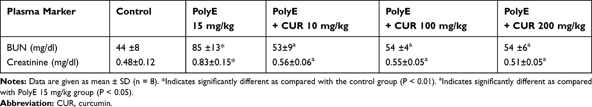 |
Table 1 Plasma Biochemical Measurements in Polymyxin E (PolyE)-Treated Mice |
Effects of Curcumin on Polymyxin E-Induced Oxidative Stress in the Renal Tissue
Oxidative stress-induced by PolyE was seen in the renal tissue of the study population showing ROS and tissue oxidized glutathione (GSSG) formation with higher levels of lipid peroxidation. (Figure 1). With intravenous PolyE injection, the level of reduced glutathione (GSH) stores and tissue antioxidant capacity was significantly decreased (Figure 1). CUR administration (10, 100, and 200 mg/kg/day) significantly alleviated oxidative stress biomarkers in the kidney of PolyE-treated mice (Figure 1).
 |
Figure 1 Kidney tissue markers of oxidative stress in polymyxin E (PolyE)-treated mice and the effect of curcumin (CUR) treatment. Data are given as mean ± SD (n = 8). ***Indicates significantly different as compared with the control group (P < 0.01). aIndicates significantly different as compared with the PolyE group (P < 0.05). |
Curcumin Alleviates Polymyxin-Induced Renal Histopathological Alterations
Renal tubular changes consisting of acute necrosis, casts, and dilation evaluated histopathologically were obvious in PolyE-treated animals (Figure 2 and Table 2). CUR treatment (10, 100, and 200 mg/kg/day) mitigated renal histopathological alterations due to PolyE injections (Figure 2 and Table 2).
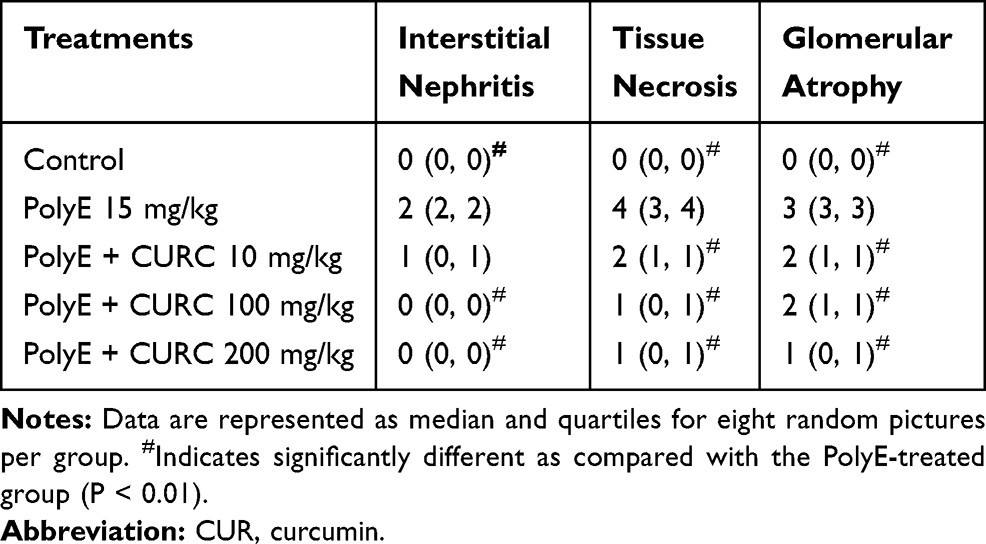 |
Table 2 Grade of Kidney Tissue Histopathological Alterations in Polymyxin E (PolyE)-Treated Mice |
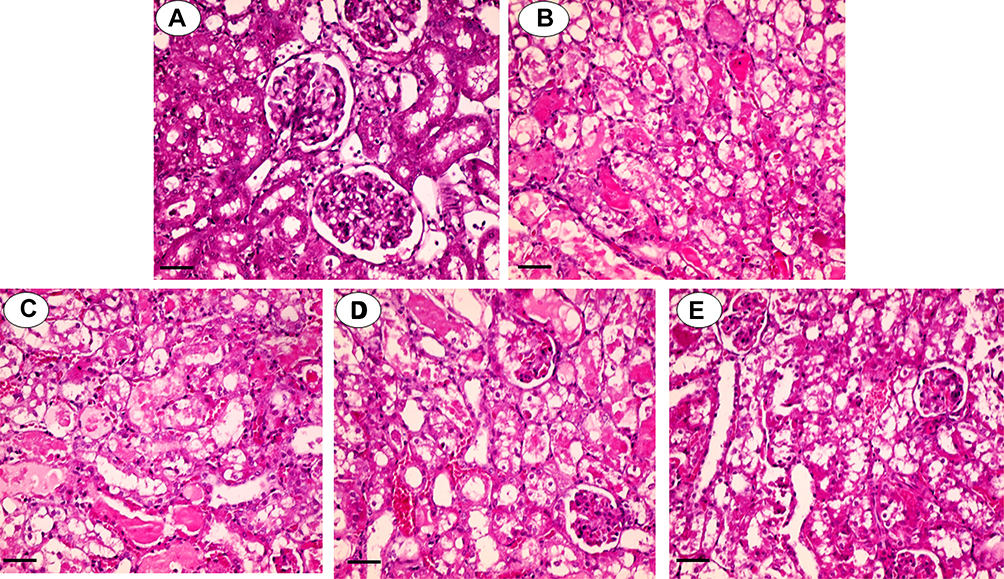 |
Figure 2 Kidney histopathological alterations in polymyxin E (PolyE)-treated animals. Typical kidney tissue histopathology (A). Tubular necrosis, interstitial inflammation, and atrophy were evident in PolyE-treated (15 mg/kg/day, i.v, seven consecutive days) animals (B). It was found that curcumin administration significantly decreased PolyE-induced renal injury (PolyE + curcumin; (C–E) for 10, 100, and 200 mg/kg of curcumin, respectively). The grades of kidney histopathological alterations are given in Table 2. |
Discussion
In this study, we demonstrated that co-administration of CUR with PolyE in a vivo model could ameliorate PolyE-induced renal injury. Bacterial resistance to antibiotics is a severe clinical challenge.9,27 Several strategies have been developed to overcome this complication.9,27 The last-line agent for gram-negative bacteria that have developed resistance towards commonly used antibiotics is PolyE.6,27 However, renal injury is a severe adverse effect due to PolyE administration.28,29 Although minimizing PolyE dose might limit its nephrotoxicity, but drug efficacy is also significantly influenced. Hence, finding ancillary treatments could mitigate PolyE-induced nephrotoxicity and enhance its efficacy.
Different studies mentioned the pivotal role of oxidative stress in the mechanism of PolyE-induced nephrotoxicity.30–35 The increase of reactive oxygen species (ROS) level, deterioration of biological membrane lipids performance, increased cellular protein injury, and alteration in cellular antioxidant systems have been documented in the kidney of PolyE-treated animals.8,9 It was found that CUR treatment significantly decreased PolyE-induced oxidative stress in the mice kidney.
CUR is a phenolic compound widely studied for its pharmacological properties. The antioxidant properties of CUR as an active ingredient of polyphenolic curcuminoids have been repeatedly investigated.10,12,36-41 The direct radical scavenging properties of CUR play a crucial role in the antioxidant properties of this compound.11,38 On the other hand, it has been found that CUR significantly upregulates cellular enzymatic and non-enzymatic antioxidant systems.10,40,42,43 It has been found that CUR effectively protects the kidney against oxidative stress induced by diseases or several nephrotoxic chemicals.12,36,39 Pivotal mechanisms of curcumin renoprotective activity include: master regulator of antioxidant response nuclear factor erythroid-derived 2 (Nrf2) induction, mitochondrial dysfunction inhibition, inflammatory response amelioration, antioxidant enzymes preservation, and oxidative stress prevention.44 Curcumin was effective in the prevention of an increase in serum levels of urea, creatinine, and MDA, compared with the control group. In a study by Mercantepe et al, the effects of curcumin in cisplatin-induced nephrotoxicity in rats was evaluated. The results showed a protective role of curcumin in cisplatin-induced nephropathy. Its renoprotective effects can lead to the maintenance of the renal function and redox balance of mitochondria.45 Several clinical trials mentioned the therapeutic role of curcumin in a variety of human diseases.1 The effects of curcumin against Alzheimer’s disease, acute coronary syndrome, atherosclerosis, diabetic nephropathy, diabetic microangiopathy, gallbladder, alcohol intoxication, has been undergoing clinical trials.1 A wide range of curcumin doses has been applied for clinical purposes (eg, 10–180 mg/day for two months in coronary artery diseases or 1–4 g/day for Alzheimer’s disease).1 Hence, this compound could be considered as a safe agent for clinical applications.
In another study, evaluating the protective effects of curcumin in gentamicin-induced nephrotoxicity, it was shown that the positive effects of curcumin are due to the reduction of oxidative stress. Curcumin could reduce the biomarkers of renal injury and renal tubule necrosis in AKI.46 Based on the data obtained from this study, the antioxidative effects of CUR play a significant role in its renoprotective properties against PolyE nephrotoxicity. Many clinical trials showed the protective effects of CUR in human diseases.47,48 Hence, this compound could be clinically applicable to drug-induced adverse effects such as PolyE-induced renal injury.
Our study results support the protective quality of CUR towards renal toxicity induced by PolyE. However, more research is necessary to ascertain the probable beneficiary effects of CUR on PolyE induced toxicity regarding the exclusion of possible drug interactions.
Acknowledgments
This investigation was financially supported by the Vice-Chancellor of Research Affairs of Shiraz University of Medical Sciences (Grant 14397/14823). Authors thank the Pharmaceutical Sciences Research Center (PSRC) of Shiraz University of Medical Sciences for providing technical facilities to carry out this study.
Author Contributions
AV and RH conceived and planned the experiments. RH and ZK performed the measurements and contributed to sample preparation. AV and RH were involved in the interpretation of the results. All authors contributed toward drafting and revising the paper, gave final approval of the version to be published, and agree to be accountable for all aspects of the work.
Disclosure
The authors declare that there is no conflict of interest.
References
1. Gupta SC, Patchva S, Aggarwal BB. Therapeutic roles of curcumin: lessons learned from clinical trials. AAPS J. 2013;15(1):195–218. doi:10.1208/s12248-012-9432-8
2. Vazin A, Karimzadeh I, Zand A, Hatami-Mazinani N, Firouzabadi D. Evaluating Adherence of Health-Care Team to Standard Guideline of Colistin Use at Intensive Care Units of a Referral Hospital in Shiraz, Southwest of Iran. Adv Pharm Bull. 2017;7(3):391–397. doi:10.15171/apb.2017.047
3. Li J, Nation RL, Turnidge JD, et al. Colistin: the re-emerging antibiotic for multidrug-resistant Gram-negative bacterial infections. Lancet Infect Dis. 2006;6(9):589–601. doi:10.1016/S1473-3099(06)70580-1
4. Kim J, Lee K-H, Yoo S, Pai H. Clinical characteristics and risk factors of colistin-induced nephrotoxicity. Int J Antimicrob Agents. 2009;34(5):434–438. doi:10.1016/j.ijantimicag.2009.06.028
5. Binh NG, Hayakawa K, Co DX, et al. The efficacy and nephrotoxicity associated with colistin use in an intensive care unit in Vietnam: use of colistin in a population of lower body weight. Int J Infect Dis. 2015;35:18–23. doi:10.1016/j.ijid.2015.03.020
6. Desai K, Kazi M, Ajbani K, et al. Clinical outcomes and safety of colistin in treatment of gram negative infections: a prospective observational study. Egypt J Crit Care Med. 2016;4(2):67–72. doi:10.1016/j.ejccm.2016.07.001
7. Vazin A, Japoni A, Shahbazi S, Davarpanah MA. Vancomycin utilization evaluation at hematology-oncology ward of a teaching hospital in iran. Iran J Pharm Res. 2012;11(1):163–170.
8. Heybeli C, Oktan MA, Çavdar Z. Rat models of colistin nephrotoxicity: previous experimental researches and future perspectives. Eur J Clin Microbiol Infect Dis. 2019;38(8):1387–1393. doi:10.1007/s10096-019-03546-7
9. Gai Z, Samodelov SL, Kullak-Ublick GA, Visentin M. Molecular mechanisms of colistin-induced nephrotoxicity. Molecules. 2019;24(3):653. doi:10.3390/molecules24030653
10. Balogun E, Hoque M, Gong P, et al. Curcumin activates the haem oxygenase-1 gene via regulation of Nrf2 and the antioxidant-responsive element. Biochem J. 2003;371(3):887–895. doi:10.1042/bj20021619
11. Ak T, Gülçin İ. Antioxidant and radical scavenging properties of curcumin. Chem Biol Interact. 2008;174(1):27–37. doi:10.1016/j.cbi.2008.05.003
12. Trujillo J, Chirino YI, Molina-Jijón E, Andérica-Romero AC, Tapia E, Pedraza-Chaverrí J. Renoprotective effect of the antioxidant curcumin: recent findings. Redox Biol. 2013;1(1):448–456. doi:10.1016/j.redox.2013.09.003
13. Motterlini R, Foresti R, Bassi R, Green CJ. Curcumin, an antioxidant and anti-inflammatory agent, induces heme oxygenase-1 and protects endothelial cells against oxidative stress. Free Radical Biol Med. 2000;28(8):1303–1312. doi:10.1016/S0891-5849(00)00294-X
14. Cao J, Liu Y, Jia L, et al. Curcumin attenuates acrylamide-induced cytotoxicity and genotoxicity in HepG2 cells by ROS scavenging. J Agric Food Chem. 2008;56(24):12059–12063. doi:10.1021/jf8026827
15. Dai C, Li J, Tang S, Li J, Xiao X. Colistin-induced nephrotoxicity in mice involves the mitochondrial, death receptor, and endoplasmic reticulum pathways. Antimicrob Agents Chemother. 2014;58(7):4075–4085. doi:10.1128/AAC.00070-14
16. Jamshidzadeh A, Heidari R, Mohammadi-Samani S, et al. A comparison between the nephrotoxic profile of gentamicin and gentamicin nanoparticles in mice. J Biochem Mol Toxicol. 2015;29(2):57–62. doi:10.1002/jbt.21667
17. Socci DJ, Bjugstad KB, Jones HC, Pattisapu JV, Arendash GW. Evidence that oxidative stress is associated with the pathophysiology of inherited hydrocephalus in the H-Tx rat model. Exp Neurol. 1999;155(1):109–117. doi:10.1006/exnr.1998.6969
18. Meeks RG, Harrison S. Hepatotoxicology. New York: CRC Press; 1991.
19. Truong DH, Eghbal MA, Hindmarsh W, Roth SH, O’Brien PJ. Molecular mechanisms of hydrogen sulfide toxicity. Drug Metab Rev. 2006;38(4):733–744. doi:10.1080/03602530600959607
20. Heidari R, Taheri V, Rahimi HR, Yeganeh BS, Niknahad H, Najibi A. Sulfasalazine-induced renal injury in rats and the protective role of thiol-reductants. Ren Fail. 2016;38(1):137–141. doi:10.3109/0886022X.2015.1096731
21. Shafiekhani M, Ommati MM, Azarpira N, Heidari R, Salarian AA. Glycine supplementation mitigates lead-induced renal injury in mice. J Exp Pharmacol. 2019;11:15–22. doi:10.2147/JEP.S190846
22. Heidari R, Babaei H, Roshangar L, Eghbal MA. Effects of enzyme induction and/or glutathione depletion on methimazole-induced hepatotoxicity in mice and the protective role of N-Acetylcysteine. Adv Pharm Bull. 2014;4(1):21–28. doi:10.5681/apb.2014.004
23. Heidari R, Behnamrad S, Khodami Z, Ommati MM, Azarpira N, Vazin A. The nephroprotective properties of taurine in colistin-treated mice is mediated through the regulation of mitochondrial function and mitigation of oxidative stress. Biomed Pharmacother. 2019;109:103–111. doi:10.1016/j.biopha.2018.10.093
24. Katalinic V, Modun D, Music I, Boban M. Gender differences in antioxidant capacity of rat tissues determined by 2,2ʹ-azinobis (3-ethylbenzothiazoline 6-sulfonate; ABTS) and ferric reducing antioxidant power (FRAP) assays. Comp Biochem Physiol. 2005;140(1):47–52.
25. Heidari R, Niknahad H. The role and study of mitochondrial impairment and oxidative stress in cholestasis. In: Vinken M, editor. Experimental Cholestasis Research. New York, NY: Springer; 2019:117–132.
26. Alía M, Horcajo C, Bravo L, Goya L. Effect of grape antioxidant dietary fiber on the total antioxidant capacity and the activity of liver antioxidant enzymes in rats. Nutr Res. 2003;23(9):1251–1267. doi:10.1016/S0271-5317(03)00131-3
27. Velkov T, Roberts KD, Nation RL, Thompson PE, Li J. Pharmacology of polymyxins: new insights into an ‘old’ class of antibiotics. Future Microbiol. 2013;8(6):6. doi:10.2217/fmb.13.39
28. Falagas ME, Fragoulis KN, Kasiakou SK, Sermaidis GJ, Michalopoulos A. Nephrotoxicity of intravenous colistin: a prospective evaluation. Int J Antimicrob Agents. 2005;26(6):504–507. doi:10.1016/j.ijantimicag.2005.09.004
29. Pogue JM, Lee J, Marchaim D, et al. Incidence of and risk factors for colistin-associated nephrotoxicity in a large academic health system. Clin Infect Dis. 2011;53(9):879–884. doi:10.1093/cid/cir611
30. Ateşşahin A, Çeribaşı AO, Yılmaz S. Lycopene, a carotenoid, attenuates cyclosporine-induced renal dysfunction and oxidative stress in rats. Basic Clin Pharmacol Toxicol. 2007;100(6):372–376. doi:10.1111/j.1742-7843.2007.00060.x
31. Ozyilmaz E, Ebinc FA, Derici U, et al. Could nephrotoxicity due to colistin be ameliorated with the use of N-acetylcysteine? Intensive Care Med. 2011;37(1):141–146. doi:10.1007/s00134-010-2038-7
32. Yousef JM, Chen G, Hill PA, Nation RL, Li J. Melatonin attenuates colistin-induced nephrotoxicity in rats. Antimicrob Agents Chemother. 2011;55(9):4044–4049. doi:10.1128/AAC.00328-11
33. Ozkan G, Ulusoy S, Orem A, et al. How does colistin-induced nephropathy develop and can it be treated? Antimicrob Agents Chemother. 2013;57(8):3463–3469. doi:10.1128/AAC.00343-13
34. Ghlissi Z, Hakim A, Sila A, et al. Evaluation of efficacy of natural astaxanthin and vitamin E in prevention of colistin-induced nephrotoxicity in the rat model. Environ Toxicol Pharmacol. 2014;37(3):960–966. doi:10.1016/j.etap.2014.03.004
35. Azad MAK, Sivanesan S, Wang J, et al. Methionine ameliorates polymyxin-induced nephrotoxicity by attenuating cellular oxidative stress. Antimicrob Agents Chemother. 2018;62(1):e01254–01217. doi:10.1128/AAC.01254-17
36. Farombi EO, Ekor M. Curcumin attenuates gentamicin-induced renal oxidative damage in rats. Food Chem Toxicol. 2006;44(9):1443–1448. doi:10.1016/j.fct.2006.05.005
37. Cekmen M, Ilbey YO, Ozbek E, Simsek A, Somay A, Ersoz C. Curcumin prevents oxidative renal damage induced by acetaminophen in rats. Food Chem Toxicol. 2009;47(7):1480–1484. doi:10.1016/j.fct.2009.03.034
38. Sevgiler Y, Karaytug S, Karayakar F. Antioxidative effects of N-acetylcysteine, lipoic acid, taurine, and curcumin in the muscle of Cyprinus carpio L. exposed to cadmium. Arh Hig Rada Toksikol. 2011;62(1):1–9. doi:10.2478/10004-1254-62-2011-2082
39. Ueki M, Ueno M, Morishita J, Maekawa N. Curcumin ameliorates cisplatin-induced nephrotoxicity by inhibiting renal inflammation in mice. J Biosci Bioeng. 2013;115(5):547–551. doi:10.1016/j.jbiosc.2012.11.007
40. García-Niño WR, Pedraza-Chaverrí J. Protective effect of curcumin against heavy metals-induced liver damage. Food Chem Toxicol. 2014;69:182–201. doi:10.1016/j.fct.2014.04.016
41. Ghosh S, Bhattacharyya S, Rashid K, Sil PC. Curcumin protects rat liver from streptozotocin-induced diabetic pathophysiology by counteracting reactive oxygen species and inhibiting the activation of p53 and MAPKs mediated stress response pathways. Toxicol Rep. 2015;2:365–376. doi:10.1016/j.toxrep.2014.12.017
42. Mishra P, Paital B, Jena S, et al. Possible activation of NRF2 by Vitamin E/Curcumin against altered thyroid hormone induced oxidative stress via NFĸB/AKT/mTOR/KEAP1 signalling in rat heart. Sci Rep. 2019;9(1):7408. doi:10.1038/s41598-019-43320-5
43. Turpaev KT. Keap1-Nrf2 signaling pathway: mechanisms of regulation and role in protection of cells against toxicity caused by xenobiotics and electrophiles. Biochem Moscow. 2013;78(2):111–126. doi:10.1134/S0006297913020016
44. Motaharinia J, Panahi Y, Barreto GE. Efficacy of curcumin on prevention of drug-induced nephrotoxicity: a review of animal studies. Biofactors. 2019;45(5):690–702. doi:10.1002/biof.1538
45. Mercantepe T, Unal D, Tumkaya L, Yazici ZA. Protective effects of amifostine, curcumin and caffeic acid phenethyl ester against cisplatin-induced testis tissue damage in rats. Exp Ther Med. 2018;15(4):3404–3412. doi:10.3892/etm.2018.5819
46. Al-Kuraishy H, Al-Gareeb A, Rasheed H. Antioxidant and anti-inflammatory effects of curcumin contribute into attenuation of acute gentamicin-induced nephrotoxicity in rats. Asian J Pharm Clin Res. 2019;466–468. doi:10.22159/ajpcr.2019.v12i3.30875
47. Hsu C-H, Cheng A-L. Clinical studies with curcumin. Adv Exp Med Biol. 2007;595:471–480.
48. Gupta SC, Patchva S, Aggarwal BB. Therapeutic roles of curcumin: lessons learned from clinical trials. AAPS J. 2012;15(1):195–218.
 © 2020 The Author(s). This work is published and licensed by Dove Medical Press Limited. The
full terms of this license are available at https://www.dovepress.com/terms
and incorporate the Creative Commons Attribution
- Non Commercial (unported, 3.0) License.
By accessing the work you hereby accept the Terms. Non-commercial uses of the work are permitted
without any further permission from Dove Medical Press Limited, provided the work is properly
attributed. For permission for commercial use of this work, please see paragraphs 4.2 and 5 of our Terms.
© 2020 The Author(s). This work is published and licensed by Dove Medical Press Limited. The
full terms of this license are available at https://www.dovepress.com/terms
and incorporate the Creative Commons Attribution
- Non Commercial (unported, 3.0) License.
By accessing the work you hereby accept the Terms. Non-commercial uses of the work are permitted
without any further permission from Dove Medical Press Limited, provided the work is properly
attributed. For permission for commercial use of this work, please see paragraphs 4.2 and 5 of our Terms.