Back to Journals » International Journal of Chronic Obstructive Pulmonary Disease » Volume 10 » Issue 1
COPD: it is time to change!
Authors Tantucci C, Pini L
Received 30 April 2015
Accepted for publication 23 June 2015
Published 11 November 2015 Volume 2015:10(1) Pages 2451—2457
DOI https://doi.org/10.2147/COPD.S87696
Checked for plagiarism Yes
Review by Single anonymous peer review
Peer reviewer comments 3
Editor who approved publication: Prof. Dr. Richard Russell
Claudio Tantucci, Laura Pini
Respiratory Medicine Unit, Department of Experimental and Clinical Sciences, University of Brescia, Brescia, Italy
Abstract: COPD is a common cause of disability, morbidity and mortality worldwide and a major global health problem with enormous direct and indirect health care costs. Different reasons can be advanced to explain it, but among them the possibility that the recommended diagnostic and therapeutic approaches to COPD were less effective than they could be, should be also considered. The pharmacological baseline treatment of stable COPD has been widely based on the severity of airflow obstruction and recently, of chronic symptoms and on the annual number of previous exacerbations. These recommendations do not take into account the underlying prevalent disease that should be treated and the future risk. Our suggestion is that the therapy must be firstly tailored on the prevalent disease leading to COPD, independently from the degree of FEV1 reduction and chronic dyspnea and only after that, according to the severity of the disorder (and age of patient), to establish the level of the treatment in order to freeze, when possible, and not to follow the underlying pathological process, running after it. Moreover, given the relevance of exacerbations in the natural history of COPD, greater effort should be placed on recognition of their prevalent type in frequent exacerbators and to prevent them using more tailored and specific treatment.
Keywords: COPD, therapeutic approach of COPD, prevention of COPD exacerbations
Introduction
COPD is a common cause of disability, morbidity, and mortality worldwide and a major global health problem with enormous direct and indirect health care costs.1,2 The worst notice, however, is that the mortality rate (number of deaths per 100,000 persons) for COPD in the last 50 years (at least in USA) has progressively increased in both men and women older than 60 years in every 5-year period.3 Moreover, among the 20 leading causes of deaths in USA, the only one that has substantially increased the age-standardized relative rank of death in the last 20 years is COPD.4 In Europe, the situation seems better, and in many European countries, the mortality rate (number of deaths for 100,000 persons) for COPD is decreasing, particularly in men.5 However, for 100,000 deaths, the percentage of mortality due to COPD is increasing, suggesting that among chronic noncommunicable diseases COPD is less effectively treated.5
Therefore, according to this trend, it is estimated that COPD will become the third worldwide cause of mortality in 2030.6 Of course, there are many causes for this, but among them the possibility that the recommended diagnostic and therapeutic approaches to COPD were less effective than what they could be should be seriously considered.
Definition
Let us start with definition which is a fundamental point. The most disseminated definition says that COPD is a disease.1 In fact, D in the COPD acronym stands for disease. It must be acknowledged, however, that COPD is recognized in the definition as an obstructive functional defect associated with a chronic immune inflammatory process, occurring in the lungs of susceptible individuals with known risk factors, in the absence of other causes. Thus, strictly speaking, D in the COPD acronym should stand for disorder. Since the 1960s, based on pathological findings, it has been clear that similar functional disorder could arise from different diseases, essentially two, one originating from small airways and the other from lung parenchyma.7
Today, we know that chronic bronchiolitis, which is a progressive, proliferative, and fibrosing pathology of the membranous and terminal bronchioli due to persistent inflammatory, immune, and remodeling process, is by far the most common disease leading to COPD.8 In contrast, panlobular emphysema, which is a destructive and hyporegenerative pathology of the alveolar walls with some autoimmune features, is a rare disease that may originate among individuals with severe alpha-1-antitrypsin deficiency and heavy smokers and also leads to COPD.9,10
Moreover, chronic bronchiolitis can be frequently associated with centrilobular emphysema, when the inflammatory process involves the respiratory bronchioli with subsequent destruction of the secondary lobule, from the center to periphery. This may configure the presence of mild (<5% of the lung volume with low attenuation area [LAA] <−950 HU, showing round- or oval-shaped LAA with well-defined border), moderate (>5% of the lung volume with LAA <−950 HU, showing polygonal- or irregular-shaped LAA with ill-defined border), or confluent (showing irregular-shaped LAA with ill-defined border coalescent with each other) centrilobular emphysema.11 Actually, centrilobular emphysema does not occur without chronic bronchiolitis, and there is growing evidence that injury of terminal bronchioli would precede the enlargement of centrilobular spaces.12
In a cross-sectional high-resolution computed tomography analysis, it is often impossible to distinguish between panlobular emphysema and confluent centrilobular emphysema, the only distinctive clue being the prevalence of destructive lesions in the lower lobes in the former.
At the stage of severe-to-very severe airflow obstruction, fibrosing chronic bronchiolitis with (same degree of) centrilobular emphysema is the pathological condition most frequently encountered. Although not definitely proved, isolated fibrosing chronic bronchiolitis should be the most common form of COPD at earlier stages of airflow obstruction.
In all circumstances, however, a progressive reduction in maximal expiratory and inspiratory flow rates may develop because of an increase in airway resistance and/or a decrease in pulmonary static elastance. Diminution of the elastic recoil pressure can augment the expiratory flow resistance by distortion and collapse of the small airways because of reduction in tethering forces. This phenomenon is amplified by the loss of airway–parenchyma interdependence due to progressive disappearance of alveolar attachments.
Diagnostic and severity assessment
With the above-mentioned background, in a patient with COPD, the priority should be to define the prevalent underlying disease. Generally, clinical, radiological (chest X-ray), and adequate functional assessments are sufficient to achieve this (Tables 1 and 2), but sometimes this task requires a more extensive work-up including high-resolution computed tomography lung scan.
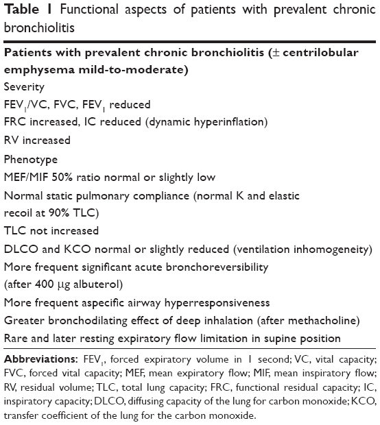 | Table 1 Functional aspects of patients with prevalent chronic bronchiolitis |
 | Table 2 Functional aspects of patients with prevalent emphysema |
Is it useful? Many lines of evidence suggest yes. Based on different underlying disease, namely prevalent chronic bronchiolitis versus prevalent pulmonary emphysema, in patients with COPD the effect of anti-inflammatory treatments (inhaled corticosteroids and/or phosphodiesterase 4 inhibitors, for instance) on top of long-acting bronchodilators is different in terms of improvement in function, symptoms, quality of life,13,14 and prevention of exacerbations.15–17 In addition, the functional decline is differently affected by treatment,18,19 the all-cause and respiratory mortality is different in long-term follow-up,20 and finally, the clinical phenotypes and some comorbidities tend to cluster differently.21,22
After that it is mandatory to stage the COPD severity, which cannot rely only on forced expiratory volume in 1 second (FEV1) and, as recently suggested, on symptoms, requiring instead a multidimensional approach not only to have a prognostic evaluation but also to establish the level of initial treatment and timing of monitoring. Presently, the updated BODE (body mass index, airflow obstruction, dyspnea and exercise capacity) index appears the most suitable tool for such purpose,23 taking into account also the distribution of the symptoms during the day and the gas exchange abnormalities. Prospectively, determination of some plasma biomarkers, such as C-protein and fibrinogen, might be useful.24 All of this information must be weighted for the age of the patient.
Subsequently, an annual history of COPD exacerbations, as much as is possible to detail, must be performed because a high frequency of COPD exacerbations is an undisputable marker of disease severity that needs to be aggressively controlled. It is of limited utility to simply count the previous COPD exacerbations, and every effort should be made to recognize the prevalent type, and so choose the best available strategy to prevent them.25
The various comorbidities should finally be investigated and graduated by severity in order to treat adequately those known to have a major impact on mortality in patients with COPD.26
Therapeutic approach
The pharmacological baseline treatment of stable COPD has been widely based on the severity of airflow obstruction and recently also of chronic symptoms and on the number of exacerbations in the previous year.1
With the interesting but somehow confusing exception of the Spanish guidelines,27 these recommendations do not take into account the underlying prevalent disease that should be treated and the future risk.
Moreover, the exacerbations are just enumerated with no attempt to define their etiology, always suggesting the stereotyped use of inhaled corticosteroids (ICS) on top of bronchodilators when they are two or more in the previous year (or one severe, requiring hospitalization).1
In contrast, the therapy must be first tailored on the prevalent disease underlying COPD, independent from the degree of FEV1 reduction and chronic dyspnea score, and only after that, according to the severity of the disorder (and age of the patient), to establish the level of the treatment in order to freeze, when possible, and not to follow the underlying pathological process, running after it (Figure 1).
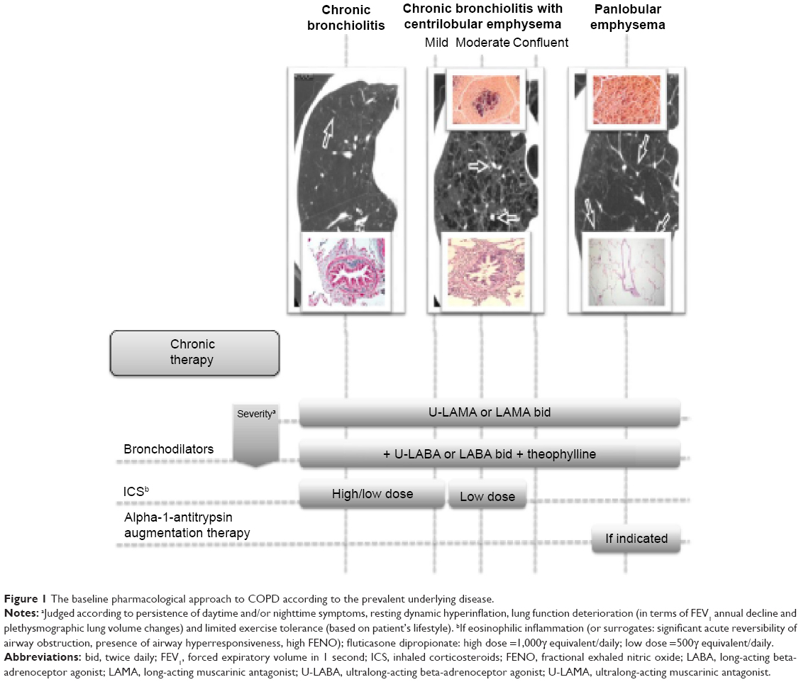 | Figure 1 The baseline pharmacological approach to COPD according to the prevalent underlying disease. |
We do not need expensive randomized controlled trials (RCTs) anymore where thousands of patients with COPD having different underlying diseases and having different severity of airflow obstruction are enrolled all together, trying to understand the effect of a same treatment. On the contrary, more valuable information about any given targeted intervention might be collected, studying small numbers of well-selected patients with COPD, with same underlying disease, similar clinical phenotypes, same degree of airflow obstruction and/or BODE index, and similar age range, for an adequate span of time.
It can be speculated, but it must be proved with well-designed prospective studies that this approach will be more effective in terms of lung function decline and patient-related outcomes, particularly if applied in the initial phases of COPD, implying an early diagnosis of chronic airflow obstruction and a better characterization of the underlying disease.
Perhaps, we would learn that more aggressive treatments have to be implemented in the earlier stages of COPD, instead of using them in the more advanced ones as recommended today, to obtain the best possible outcomes. This has been suggested by the post hoc subgroups analysis in the previous interventional large studies on COPD where improvement in symptoms, exacerbation frequency, FEV1 annual decline, and all-cause mortality was demonstrated only for patients with COPD stages II and III (Global Initiative for Chronic Obstructive Lung Disease).28,29
The goal should be to have patients dying with COPD (when allowed by the underlying disease, essentially chronic bronchiolitis) and not because of COPD.
Exacerbations
The prevention of COPD exacerbations is a point of paramount importance in the management of COPD that needs a completely different approach because it cannot be addressed simply with the baseline pharmacological treatment. We know a lot about COPD exacerbations, even if their diagnosis, essentially based on worsening of chronic symptoms reported as relevant by the patients, is presently still based on the exclusion of other diseases. To suffer from two or more COPD exacerbations or from one severe COPD exacerbation leading to hospitalization in the previous year is without doubt a marker of COPD severity, independent from the underlying disease, degree of airflow obstruction, and entity of symptoms or BODE index, even if lower FEV1 is associated with higher risk of frequent and more severe exacerbations. Although the probability of having a new COPD exacerbation is greater in those patients with COPD who previously experienced COPD exacerbations (so-called frequent exacerbators),30 such status, with few exceptions,31–33 cannot be identified as a distinct phenotype, rather as a condition requiring more strict social and medical attention. In fact, it is quite easy to shift from a frequent exacerbator to a nonfrequent exacerbator and vice versa,34 sometimes without an obvious reason, but often clearly because of a more adequate and comprehensive treatment.35
Given the prognostic importance of COPD exacerbations,36 however, we cannot be limited to simply counting exacerbations; we must learn how to consistently recognize the prevalent type in a single patient. Such an approach is crucial to prevent more effectively the COPD exacerbations using more tailored and specific therapies (Figure 2).
 | Figure 2 The different preventive approach to acute exacerbation in COPD. |
Some plasma, blood, or sputum biomarkers have been shown to be associated with high sensitivity and specificity to a prevalent clinical type of COPD exacerbation: eosinophilic, infectious either virus or bacteria associated, or pauci-inflammatory, due to several possible causes that have to be identified.37 More interestingly, these specific biomarkers tend to be detectable also in stable conditions, at least in eosinophilia- and bacteria-associated exacerbations, allowing an easier identification of the most likely future pattern of COPD exacerbations.37
Thus, when possible, each prevalent type of COPD exacerbation in a given patient who is a frequent exacerbator should be appropriately prevented by specific treatments that cannot be invariably the use of high-dose ICS, as presently recommended.1,27
Long-acting bronchodilators and mainly ultralong-acting bronchodilators (both beta-2 agonist and antimuscarinic drugs) are able to prevent up to 30% of COPD exacerbations when given alone38 and possibly more when given in combination.39
ICS are very useful to prevent the eosinophilic exacerbations,15–17 but it is difficult to see how ICS can prevent infectious exacerbations, or even noneosinophilic and noninfectious exacerbations, apart from strengthening the action of bronchodilators such as long-acting beta-2 agonists (LABA) in some circumstances.38
In patients with COPD having bronchiectasis and chronic bronchitis, COPD exacerbations are often infectious and bacterial in nature and must be prevented with antibiotic prophylaxis, at least in the winter season. There is strong evidence that macrolides may significantly reduce COPD exacerbations,40–42 suggesting that this kind of exacerbation can be prevented in some clinical phenotypes of patients with COPD having high risk of bacterial colonization or chronic infections.
Presently, only adequate immunization against influenza virus can be offered for viral exacerbations in patients with COPD, and we urgently need the same effective tools for rhinovirus infection, the most common cause of virus-associated COPD exacerbation.
All other types of COPD exacerbations (noninfectious and noneosinophilic) deserve accurate work-up and specific preventative treatment should be applied when the cause is recognized (Figure 2). Many of these exacerbations, in fact, can be largely avoided with targeted approaches.38,43–45 For instance, in the presence of patients with COPD having presumable high oxidative stress (those who continue smoking, those with chronic bronchitis, and those with high exposure to environment pollution), an adequate antioxidant support might significantly reduce COPD exacerbations.46,47
Finally, a strict adherence to baseline chronic therapy should be assured throughout a strong relationship between patients with COPD and their physicians and caregivers because this aspect is crucial to obtain improved control of COPD exacerbations and their consequences.48
Conclusion
In summary, in patients with COPD, first treat as soon as possible the underlying disease, aiming to freeze the disease and halt progression, ready to implement treatment (essentially with different bronchodilators, including theophylline) to control symptoms and improve lung function, exercise tolerance, and quality of life, if necessary. Second, look at the exacerbations of COPD, if they are frequent (more than one in a year), define their prevalent phenotype (eosinophilic, bacterial, or due to other causes) and treat to prevent accordingly.
Disclosure
The authors report no conflicts of interest in this work.
References
Global Initiative for Chronic Obstructive Pulmonary Disease. Global strategy for the Diagnosis, Management and Prevention of Chronic Obstructive Pulmonary Disease. Global Initiative for Chronic Obstructive Pulmonary Disease: 2014. Available from: http://www.goldcopd.org/uploads/users/files/GOLD_Report_2014_Jun 11.pdf. Accessed December 15, 2014. | ||
Menn P, Heinrich J, Huber RM, KORA Study Group, et al. Direct medical costs of COPD – an excess cost approach based on two population-based studies. Respir Med. 2012;106:540–548. | ||
Thun MJ, Carter BD, Feskanich D, et al. 50-year trends in smoking-related mortality in the United Sates. N Engl J Med. 2013;368:351–364. | ||
Murray CJL, Phil D, Lopez AD. Measuring the global burden of disease. N Engl J Med. 2013;369:448–457. | ||
ERS White Book. The Burden of Lung Disease. Sheffield: ERS White Book; 2013. [Chapt 1:1–15 and COPD Chapt 13:1–12]. | ||
World Health Organization. Burden of COPD. Geneva: World Health Organization; 2014. | ||
Burrows B, Fletcher CM, Heard BE, Jones NL, Wootliff JS. The emphysematous and bronchial types of chronic airways obstruction. A clinicopathological study of patients in London and Chicago. Lancet. 1966;1:830–835. | ||
Hogg JC, Chu F, Utokaparch S, et al. The nature of small-airway obstruction in chronic obstructive pulmoinary disease. N Engl J Med. 2004;350:2645–2653. | ||
Eriksson S. Pulmonary emphysema and alpha-1-antitrypsin deficiency. Acta Med Scand. 1964;175:197–205. | ||
Finkelstein R, Fraser RS, Ghezzo H, Cosio M. Alveolar inflammation and its relation to emphysema in smokers. Am J Respir Crit Care Med. 1995;152:1666–1672. | ||
Kim WD, Eidelman DH, Izquierdo JL, Ghezzo H, Saetta MP, Cosio M. Centrilobular and panlobular emphysema in smokers. Two distinct morphologic and functional entities. Am Rev Respir Dis. 1991;144:1385–1390. | ||
McDonough JE, Yuan R, Suzuki M, et al. Small-airway obstruction and emphysema in chronic obstructive pulmonary disease. N Engl J Med. 2011;365:1567–1575. | ||
Brightling CE, McKenna S, Hargadon B, et al. Sputum eosinophilia and the short term response to inhaled mometasone in chronic obstructive pulmonary disease. Thorax. 2005;60:193–198. | ||
Lee J-H, Lee YK, Kim E-K, Kim T-H. Response to inhaled long-acting beta-agonist and corticosteroid according to COPD subtype. Respir Med. 2010;104:542–551. | ||
Siva R, Green RH, Brightling CE, et al. Eosinophilic airway inflammation and exacerbations of COPD: a randomized controlled trial. Eur Respir J. 2007;29:906–913. | ||
Di Santostefano RL, Li H, Rubin DB, Stempel DA. Which patients with chronic obstructive pulmonary disease benefit from the addition of an inhaled corticosteroid to their bronchodilator? A cluster analysis. BMJ Open. 2013;3(4):3e001838. | ||
Liesker JJW, Bathoorn E, Postma DS, Vonk JM, Timens W, Kerstjens AM. Sputum inflammation predicts exacerbations after cessation of inhaled corticosteroids in COPD. Respir Med. 2011;105:1853–1860. | ||
Lapperre TS, Snoeck-Stroband JB, Gosman MM, Groningen Leiden Universities Corticosteroids in Obstructive Lung Disease Study Group, et al. Effect of fluticasone with and without salmeterol on pulmonary outcomes in chronic obstructive pulmonary disease. Ann Intern Med. 2009;151:517–527. | ||
Nishimura M, Makita H, Nagai K, Hokkaido COPD Cohort Study Investigators, et al. Annual Change in pulmonary function and clinical phenotype in chronic obstructive pulmonary disease. Am J Respir Crit Care Med. 2012;185:44–52. | ||
Johannessen A, Skorge TD, Bottai M, et al. Mortality by level of emphysema and airway wall thickness. Am J Respir Crit Care Med. 2013;187:602–608. | ||
Hersh CP, Make BJ, Lynch DA, COPDGene and ECLIPSE Investigators, et al. Non-emphysematous chronic obstructive pulmonary disease is associated with diabetes mellitus. BMC Pulm Med. 2014;14:164. | ||
Han MK, Kazerooni EA, Lynch DA, COPDGene Investigators, et al. Chronic obstructive pulmonary disease exacerbations in the COPDGene study: associated radiologic phenotypes. Radiology. 2011;261:274–282. | ||
Puhan MA, Garcia-Aymerich J, Frey M, et al. Expansion of the prognostic assessment of patients with chronic obstructive pulmonary disease: the updated BODE index and the ADOindex. Lancet. 2009;374:704–711. | ||
Agustí A, Edwards LD, Rennard SI, Evaluation of COPD Longitudinally to Identify Predictive Surrogate Endpoints (ECLIPSE) Investigators, et al. Persistent systemic inflammation is associated with poor clinical outcomes in COPD: a novel phenotype. PLoS One. 2012;7(5):e37483. | ||
Hurst JR. Exacerbation phenotyping in chronic obstructive pulmonary disease. Am J Respir Crit Care Med. 2011;184:625–626. | ||
Divo M, Cote C, de Torres JP, BODE Collaborative Group, et al. Comorbidities and risk of mortality in patients with chronic obstructive pulmonary disease. Am J Respir Crit Care Med. 2012;186:155–161. | ||
Miravittles M, Soler-Cataluna JJ, Calle M, et al. Spanish guideline for COPD (gesEPOC). Update 2014. Arch Bronconeumol. 2014;48(suppl 1):2–58. | ||
Jenkins CR, Jones PW, Calverley PM, et al. Efficacy of salmeterol/fluticasone propionate by GOLD stage of chronic obstructive pulmonary disease: analysis from the randomized placebo-controlled TORCH study. Respir Res. 2009;10:59–68. | ||
Decramer M, Celli B, Kesten S, et al; UPLIFT Investigators. Effect of tiotropium on outcomes in patients with moderate chronic obstructive pulmonary disease (UPLIFT): a prespecified subgroup analysis of a randomized controlled trial. Lancet. 2009;374:1171–1178. | ||
Hurst JR, Vestbo J, Anzueto A, et al. Susceptibility to exacerbation in chronic obtructive pulomary disease. New Engl J Med. 2010;63:1128–1138. | ||
Lin CL, Siu LK, Lin JC, et al. Mannose-binding lectine gene polymorphism contributes to the recurrence of exacerbation in patients with COPD. Chest. 2011;139:43–51. | ||
Foreman MG, DeMeo DL, Hersh CP, et al. Polymorphic variation in surfactant protein B is associated with COPD exacerbations. Eur Respir J. 2008;32:938–944. | ||
Mallia P, Message SD, Gielen V, et al. Experimental rhinovirus infection as a human model of chronic obstructive pulmonary disease exacerbation. Am J Respir Crirt Care Med. 2011;183:734–742. | ||
Donaldson GC, Müllerova H, Locantore N, et al. Factors associated with change in exacerbation frequency in COPD. Respir Res. 2013;14:79–88. | ||
Wedzicha JA, Rabe KF, Martinez FJ, et al. Efficacy of roflumilast in the COPD frequent exacerbator phenotype. Chest. 2013;143:1302–1311. | ||
Soler-Cataluna JJ, Martìnez-Garcìa MA, Romàn Sanchez P, Salcedo E, Navarro M, Ochado R. Severe acute exacerbation and mortality in patients with chronic obstructive pulmonary disease. Thorax. 2005;60:925–931. | ||
Bafadhel M, McKenna S, Terry S, et al. Acute exacerbations of chronic obstructive pulmonary disease. Identification of biollogic clusters and their biomarkers. Am J Respir Crirt Care Med. 2011;184:662–671. | ||
Wedzicha JA, Decramer M, Seemungal AR. The role of bronchodilators in the prevention of exacerbation of COPD. Eur Respir J. 2012;40:1545–1554. | ||
Wedzicha JA, Decramer M, Ficker JH, et al. Analysis of chronic obstructive pulmonary disease exacerbations with the dual bronchodilator QVA149 compared with glycopironium and tiotropium (SPARK): a randomized, double-blind, parallel-group study. Lancet Respir Med. 2013;1:199–209. | ||
Seemungal TAR, Wilkinson TMA, Hurst JR, Perera WR, Sapsford RJ, Wedzicha JA. Long-term erytromycin therapy is associated with dcreased chronic obstructive pulmonary disease exacerbations. Am J Respir Crti Care Med. 2008;178:1139–1147. | ||
Albert RK, Connett J, Bailey WC, COPD Clinical Research Network, et al. Azithromicin for prevention of exacerbations of COPD. New Engl J Med. 2011;365:689–698. | ||
Uzun S, Djamin RS, Kluytmans JA, et al. Azithromycin maintenance treatment in patients with frequent exacerbations of chronic obstructive pulmonary disease (COLUMBUS): a randomized, double-blind, placebo-controlled trial. Lancet Respir Med. 2014;2:361–368. | ||
Washko GR, Fan VS, Ramsey SD, National Emphysema Treatment Trial Research Group, et al. The effect of lung volume reduction surgery on chronic obstructive pulmonary disease exacerbations. Am J Respir Crirt Care Med. 2008;177:164–169. | ||
Marin JM, Soriano JB, Carrizo SJ, Boldova A, Celli BR. Outcomes in patients with chronic obstructive pulmonary disease and obstructive sleep apnea. The overlap sindrome. Am J Respir Crirt Care Med. 2010;182:325–331. | ||
Lehouck A, Mathieu C, Carremans C, et al. High does of Vitamin D to reduce exacerbations in chronic obstructive pulmonary disease. Ann Intern Med. 2012;156:105–114. | ||
Zheng JP, Kang J, Huang SG, et al. Effect of carbocistein on acute exacerbation of chronic obstructive pulmonary disease (PEACE study): a randomized placebo-controlled study. Lancet. 2008;371:2013–2018. | ||
Zheng JP, Wen FQ, Bai CX, On behalf of the PANTHEON study group, et al. Twice daily N-acetylcysteine 600 mg for exacerbations of chronic obstructive pulmonary disease (PANTHEON): a randomised, double-blind placebo-controlled trial. Lancet Respir Med. 2014;2:187–194. | ||
Vestbo J, Anderson JA, Calverley PM, et al. Adherence to inhaled therapy, mortality and hospital admission in COPD. Thorax. 2009;64:939–943. |
 © 2015 The Author(s). This work is published and licensed by Dove Medical Press Limited. The
full terms of this license are available at https://www.dovepress.com/terms
and incorporate the Creative Commons Attribution
- Non Commercial (unported, 3.0) License.
By accessing the work you hereby accept the Terms. Non-commercial uses of the work are permitted
without any further permission from Dove Medical Press Limited, provided the work is properly
attributed. For permission for commercial use of this work, please see paragraphs 4.2 and 5 of our Terms.
© 2015 The Author(s). This work is published and licensed by Dove Medical Press Limited. The
full terms of this license are available at https://www.dovepress.com/terms
and incorporate the Creative Commons Attribution
- Non Commercial (unported, 3.0) License.
By accessing the work you hereby accept the Terms. Non-commercial uses of the work are permitted
without any further permission from Dove Medical Press Limited, provided the work is properly
attributed. For permission for commercial use of this work, please see paragraphs 4.2 and 5 of our Terms.