
Back to Journals » International Medical Case Reports Journal » Volume 19
Conservative Methotrexate Management of Ovarian Ectopic Pregnancy in a Resource-Constrained Setting: A Rare Case Report
Authors Adawe MMM, Ibrahim FH  , Mudei NM , Elmi AH
Received 27 February 2026
Accepted for publication 8 May 2026
Published 15 May 2026 Volume 2026:19 605583
DOI https://doi.org/10.2147/IMCRJ.S605583
Checked for plagiarism Yes
Review by Single anonymous peer review
Peer reviewer comments 3
Editor who approved publication: Professor Thomas E Hutson
Mariam Mohamed Mohamud Adawe,1,2 Fahmo Hussein Ibrahim,1,2 Nasteho Mohamud Mudei,1 Abdullahi Hassan Elmi2,3
1Department of Obstetrics and Gynecology, Dr Sumait Hospital, SIMAD University, Mogadishu, Somalia; 2Faculty of Medicine and Health Sciences, SIMAD University, Mogadishu, Somalia; 3Department of Nursing and Midwifery, Dr Sumait Hospital, SIMAD University, Mogadishu, Somalia
Correspondence: Mariam Mohamed Mohamud Adawe, Email [email protected]
Introduction: Ovarian ectopic pregnancy is a rare form of ectopic implantation and can be difficult to distinguish from common ovarian findings such as a corpus luteum cyst, particularly in settings with limited diagnostic and surgical resources. Early recognition using transvaginal ultrasonography and appropriate patient selection are essential to reduce the risk of rupture and preserve fertility.
Case Presentation: A 27-year-old woman (gravida 7, para 4+2) with five weeks of amenorrhea and a previous vaginal birth after cesarean section presented with five days of nausea and right-sided pelvic pain, without vaginal bleeding. She was hemodynamically stable, with marked right adnexal tenderness. Serum β-hCG was 3233.83 IU/L. Transvaginal ultrasound demonstrated an empty anteverted uterus with an 11.27 mm endometrial thickness and a gestational sac located within the right ovarian tissue, without a yolk sac or embryo, consistent with ovarian ectopic pregnancy. After counseling, conservative medical management with methotrexate was initiated. The patient remained clinically stable, and serial β-hCG levels declined appropriately (2095.41 IU/L on day 4 and 1318.43 IU/L on day 7), with sonographic regression of the ovarian gestational sac. She was discharged with weekly follow-up, and by the third week her β-hCG level fell to < 5 IU/L. Follow-up ultrasound confirmed complete resolution.
Conclusion: Ovarian ectopic pregnancy is a rare but important diagnosis in early pregnancy and may be difficult to distinguish from other adnexal lesions. In this patient, transvaginal ultrasound and serum β-hCG findings supported the diagnosis of presumed ovarian ectopic pregnancy. Conservative management with methotrexate was chosen because the patient was hemodynamically stable and could undergo close follow-up. Serial β-hCG monitoring and follow-up ultrasound demonstrated progressive regression and complete resolution without surgery. This case highlights that carefully selected patients may be managed successfully with fertility-preserving medical treatment, even in a resource-constrained setting.
Keywords: ovarian ectopic pregnancy, methotrexate, medical management, transvaginal ultrasound, β-hCG monitoring, early pregnancy, fertility preservation, resource-limited setting
Introduction
Ectopic pregnancy remains a significant cause of morbidity in early pregnancy and should always be considered in reproductive-aged women who present with amenorrhea, pelvic pain, and/or vaginal bleeding. Although most ectopic pregnancies occur in the fallopian tube, non-tubal ectopic implantations are uncommon and often more difficult to diagnose, especially when symptoms are mild and diagnostic resources are limited. Delayed recognition can lead to rupture, hemorrhage, and increased maternal risk, making early clinical suspicion and focused ultrasonographic assessment essential for timely and safe management.1–4
Ovarian ectopic pregnancy is a particularly rare form of extrauterine implantation and may closely resemble more common ovarian conditions, especially corpus luteum cysts, on both clinical evaluation and imaging. This overlap can make diagnosis challenging and may contribute to delays in treatment. Recent evidence indicates that transvaginal ultrasonography plays a central role in diagnosis, particularly when it demonstrates an empty uterus together with a gestational sac located within or inseparable from ovarian tissue. Recognizing these features is important for distinguishing ovarian ectopic pregnancy from other adnexal findings and for guiding appropriate management without delay.1,4,5
Management of ovarian ectopic pregnancy depends on the patient’s clinical stability, ultrasound findings, serum β-hCG level, and the availability of close follow-up. While surgery has traditionally been used in many cases, methotrexate offers a fertility-preserving alternative in carefully selected, hemodynamically stable patients. This approach is especially valuable in resource-constrained settings, where access to operative services, blood products, and advanced monitoring may be limited. In such contexts, successful conservative treatment can reduce surgical risk while preserving ovarian tissue and future reproductive potential.2,3,6,7
In this case report, we describe the successful methotrexate management of a rare ovarian ectopic pregnancy in a resource-limited setting. We highlight the practical diagnostic value of transvaginal ultrasound, the importance of careful patient selection, and the role of structured β-hCG follow-up in achieving complete resolution without surgical intervention.
Case Presentation
A 27-year-old woman (gravida 7, para 4+2) with a five-week history of amenorrhea and a prior vaginal birth after cesarean section (VBAC) presented to the outpatient department with five days of nausea and right-sided pelvic pain. She denied any per-vaginal bleeding. Her menstrual cycles had previously been regular, and she had no notable past medical history.
On assessment, she was hemodynamically stable Abdominal examination revealed localized tenderness in the right iliac fossa. Speculum examination showed no vaginal bleeding, and the cervix was closed. On bimanual examination, the uterus was consistent with an estimated gestational age of approximately 5–7 weeks, with marked tenderness over the right adnexa.
Initial laboratory testing demonstrated a serum β-human chorionic gonadotropin (β-hCG) level of 3233.83 IU/L. Given the strong clinical concern for ectopic pregnancy, a transvaginal ultrasound (TVS) was performed. This showed an anteverted uterus with no intrauterine gestational sac and a thickened endometrium measuring 11.27 mm (Figure 1A). A gestational sac was identified within the right ovarian tissue, without a visible yolk sac or embryo (Figure 1B).
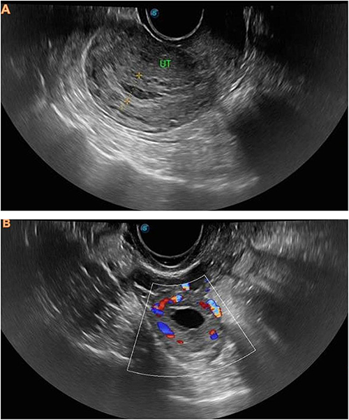 |
Figure 1 (A) Transvaginal ultrasound of the uterus in longitudinal (sagittal) view showing an empty uterine cavity with no visible intrauterine gestational sac. The endometrial thickness is measured at 11.27 mm. (B) Transvaginal ultrasound with color Doppler demonstrating a gestational sac located within or inseparable from the right ovarian tissue, with surrounding peripheral vascularity, supporting the sonographic diagnosis of presumed ovarian ectopic pregnancy. Notes: Letters denote the labeled structures or key findings, and yellow lines highlight the area of interest. |
Based on the clinical presentation and transvaginal ultrasound findings, a sonographically diagnosed presumed ovarian ectopic pregnancy was made. Because the patient was managed conservatively without surgical intervention, histopathologic confirmation was not available. The diagnosis was supported by the presence of an empty uterine cavity together with a gestational sac located within or inseparable from the right ovarian tissue on transvaginal ultrasound. The lesion was distinct from a simple adnexal cyst and was associated with right adnexal tenderness, making ovarian ectopic pregnancy more likely in the clinical context of early pregnancy and elevated serum β-hCG. The patient and her family were counseled regarding the diagnosis, available management options, and expected outcomes. Considering her hemodynamic stability, the absence of signs of rupture, the feasibility of close follow-up, and the practical limitations in immediate access to more resource-intensive surgical care, conservative medical management with methotrexate was selected.
Before methotrexate administration, the patient underwent baseline clinical and laboratory assessment to confirm suitability for medical treatment, including evaluation of hematologic, renal, and hepatic status. She was counseled about the expected treatment course, the importance of serial follow-up, and possible adverse effects of methotrexate, including abdominal pain, gastrointestinal upset, oral ulceration, and symptoms suggestive of hepatic intolerance. During follow-up, she was monitored clinically for treatment-related toxicity in addition to serial β-hCG measurement and repeat ultrasonography. She remained clinically stable throughout treatment, and no methotrexate-related adverse effects were observed. Serial β-hCG levels declined appropriately, from 2095.41 IU/L on day 4 to 1318.43 IU/L on day 7. Repeat ultrasonography also demonstrated clear regression of the right ovarian gestational sac.
She was discharged with instructions for weekly β-hCG follow-up. By the third week after methotrexate administration, her β-hCG level had decreased to <5 IU/L, and follow-up ultrasound confirmed complete resolution of the ovarian gestational sac (Figure 2).
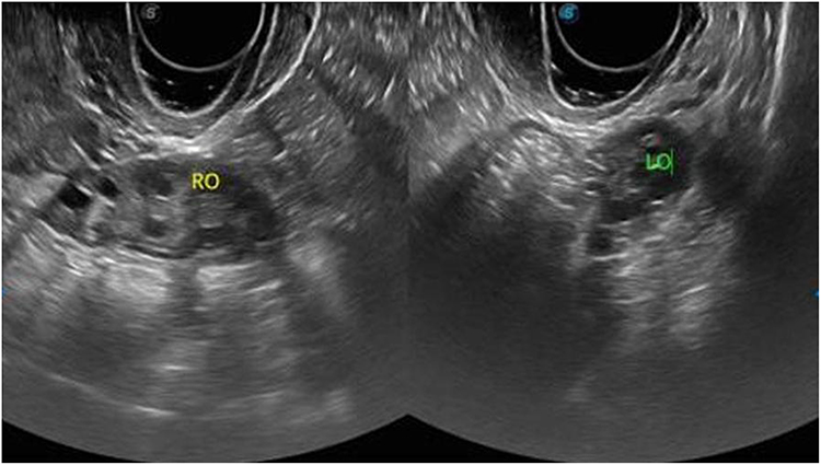 |
Figure 2 Follow-up transvaginal ultrasound of the right ovary after methotrexate treatment showing regression and complete resolution of the previously identified ovarian gestational sac, with no persistent gestational structure seen on follow-up imaging. Notes: Letters denote the labeled structures or key findings, and yellow lines highlight the relevant boundary or area of interest. |
Discussion
Ovarian ectopic pregnancy is an uncommon but clinically important subtype of ectopic gestation because it can be easily mistaken for more frequent ovarian conditions, particularly a hemorrhagic corpus luteum. This diagnostic overlap is a major reason ovarian ectopic pregnancies may be detected late, increasing the risk of rupture and significant hemorrhage. In our patient, the combination of early pregnancy symptoms, focal right-sided pelvic pain, and an empty uterus on transvaginal ultrasound prompted urgent evaluation for ectopic pregnancy.1 The key imaging feature supporting the diagnosis was the identification of a gestational sac located within the right ovarian tissue in the presence of an empty uterine cavity, an ultrasound pattern emphasized as highly suggestive of ovarian implantation in contemporary reviews.1,7
Recent data on ovarian ectopic pregnancy highlight that a timely transvaginal ultrasound is central to diagnosis and triage, particularly when clinical findings are non-specific. Solangon et al describe the typical clinical characteristics and ultrasound appearances of ovarian ectopic pregnancy and underscore that clear visualization of a sac within or inseparable from ovarian tissue, rather than simply “an adnexal mass,” helps distinguish ovarian implantation from tubal ectopic pregnancy and corpus luteum-related findings.1 In addition, atypical presentations of ectopic pregnancy are well described in the literature, reminding clinicians that absence of vaginal bleeding does not exclude ectopic gestation and that reliance on classic “textbook” triads may delay care.4 Our case aligns with these observations: the patient had no per-vaginal bleeding yet had localized adnexal tenderness and ultrasound findings consistent with ovarian ectopic pregnancy.
Once ovarian ectopic pregnancy is suspected, management must balance immediate maternal safety with fertility preservation and local resource availability. Historically, many ovarian ectopic pregnancies were managed surgically, often because diagnosis occurred after rupture or because the ovarian mass was presumed to require operative confirmation. However, when patients are hemodynamically stable, have reliable follow-up, and meet appropriate selection criteria, conservative management, either ovarian-sparing surgery or medical therapy, may be feasible.1 In our setting, preserving ovarian tissue was a priority, and access to immediate operative resources can be constrained. These practical realities made a carefully monitored medical approach particularly valuable.
Methotrexate is an established treatment for ectopic pregnancy and is recommended in major guidance documents for appropriately selected, clinically stable patients.2,3 Although ACOG guidance focuses primarily on tubal ectopic pregnancy, the same treatment principles, hemodynamic stability, absence of contraindications to methotrexate, and the capacity for close follow-up, are commonly applied when considering medical treatment for other ectopic locations on an individualized basis.2,3 In this case, the patient remained stable and had a demonstrable biochemical response, with a progressive decline in β-hCG from baseline to day 4 and day 7. This pattern is consistent with treatment success criteria used in standard methotrexate protocols, and supports continued conservative follow-up rather than escalation to surgery.2,3 Importantly, ultrasonography also demonstrated regression of the ovarian gestational sac, providing supportive anatomic correlation alongside biochemical improvement.
This case is especially relevant to resource-constrained environments, where the risks of delayed surgery, limited blood products, and constrained anesthesia and theater capacity can be substantial. In such settings, successful methotrexate treatment, when clinically appropriate, may reduce the need for emergency surgery and help preserve ovarian tissue, which is particularly meaningful for future fertility. The clinical course in our patient illustrates that, even without advanced diagnostic modalities, a structured pathway using careful clinical assessment, targeted transvaginal ultrasound, and serial β-hCG monitoring can safely support conservative management when strict criteria are met.1,3,8,9
Although methotrexate is an established option for selected ectopic pregnancies, its success depends on appropriate patient selection and close follow-up rather than on treatment administration alone. In this patient, progressive β-hCG decline and follow-up sonographic regression confirmed treatment success without the need for surgical intervention. Accordingly, the key clinical message is not pharmacogenetic variability, which was not assessed in this case, but the practical value of early diagnosis, conservative management, and structured monitoring in a stable patient.5–12 Our patient’s favorable outcome likely reflects several supportive factors: early diagnosis before rupture, hemodynamic stability, absence of embryo/yolk sac on ultrasound, a moderate baseline β-hCG level, and reliable follow-up with serial β-hCG monitoring until complete resolution. These features align with general principles used to select patients for methotrexate therapy and to ensure safety during outpatient follow-up.2,3 The complete biochemical resolution by week three and confirmatory ultrasound findings demonstrate that medical therapy can be effective for ovarian ectopic pregnancy when applied in a carefully selected patient and supported by close monitoring.1–3
Future reproductive risk should also be considered after ectopic pregnancy. A history of ectopic pregnancy may increase the likelihood of recurrent ectopic implantation and other adverse outcomes in subsequent pregnancies, underscoring the importance of fertility-preserving treatment whenever safely feasible. In this case, successful conservative management avoided surgery and preserved ovarian tissue, which is particularly relevant in a young patient with future reproductive potential. This further supports the value of careful follow-up and counseling regarding early assessment in subsequent pregnancies.7,8,10
An important limitation in this case is that the diagnosis was not confirmed histopathologically because the patient was treated successfully without surgery. The diagnosis should therefore be understood as a presumed ovarian ectopic pregnancy based on sonographic and clinical findings. The main differential diagnoses included a corpus luteum cyst and a hemorrhagic ovarian cyst, both of which may mimic ovarian ectopic pregnancy on ultrasound. However, in this patient, the finding of an empty uterus together with a gestational sac located within or inseparable from the right ovarian tissue favored ovarian ectopic implantation. In addition, the clinical picture of amenorrhea, pelvic pain, adnexal tenderness, and a positive β-hCG level further supported the diagnosis. These findings, together with serial follow-up showing regression after methotrexate treatment, strengthened the working diagnosis despite the absence of tissue confirmation.
This case adds to the growing evidence that ovarian ectopic pregnancy, although rare, can be diagnosed confidently with transvaginal ultrasound and managed conservatively in selected hemodynamically stable patients.1–4,9 In resource-limited contexts, methotrexate offers a fertility-preserving alternative to surgery when strict follow-up is feasible, while recognizing that variable drug response and toxicity risk make ongoing clinical and biochemical surveillance indispensable.
Conclusion
Ovarian ectopic pregnancy is a rare and diagnostically challenging form of ectopic gestation that may closely resemble more common ovarian conditions, particularly in the early stages of presentation. This case highlights the importance of maintaining a high index of suspicion in women presenting with early pregnancy symptoms and using careful transvaginal ultrasound assessment together with serial serum β-hCG monitoring to support diagnosis and guide management. In carefully selected hemodynamically stable patients, conservative treatment with methotrexate can be a safe and fertility-preserving alternative to surgery when close follow-up is feasible. In our patient, this approach resulted in progressive biochemical decline and complete sonographic resolution without surgical intervention. This case adds to the growing evidence that presumed ovarian ectopic pregnancy can be successfully managed without surgery in selected patients, even in resource-constrained settings, provided that diagnosis is timely, monitoring is structured, and follow-up is reliable.
Abbreviations
β-Hcg, Beta–human chorionic gonadotropin; MTX, Methotrexate; OEP, Ovarian ectopic pregnancy; TVS, Transvaginal ultrasound; VBAC, Vaginal birth after cesarean section.
Ethics and Consent
Written informed consent was obtained from the patient for publication of this case report and the accompanying images. In line with institutional policy, ethics committee approval was not required for single-patient case reports.
Acknowledgments
This research was supported by SIMAD University, Mogadishu, Somalia. We sincerely thank the Center for Research and Development at SIMAD University for their helpful guidance and insightful, constructive recommendations.
Author Contributions
All authors made substantial contributions to the conception and design of the study, data acquisition, analysis, and interpretation of the findings. They were all involved in drafting the manuscript or critically revising it for important intellectual content, approved the final version to be published, agreed on the target journal for submission, and accept accountability for all aspects of the work.
Disclosure
The authors declare no conflicts of interest in this work.
References
1. Solangon SA, Naftalin J, Jurkovic D. Ovarian ectopic pregnancy: clinical characteristics, ultrasound diagnosis and management. Ultrasound Obstetrics Gynecol. 2024;63(6):815–6. doi:10.1002/uog.27549
2. American College of Obstetricians and Gynecologists’ Committee on Practice Bulletins—Gynecology. ACOG Practice Bulletin No. 193: tubal Ectopic Pregnancy. Obstetrics Gynecol. 2018;131(3):e91–e103. 10.1097/AOG.0000000000002560.
3. Ren F, Liu G, Wang T, Li M, Guo Z. Unruptured ovarian ectopic pregnancy: two case reports and literature review. Front Physiol. 2022;13:1036365. doi:10.3389/fphys.2022.1036365. PMID: 36388130; PMCID: PMC9641050.
4. Birch EM, Torres Molina M, Oliver JJ. Not Like the Textbook: an Atypical Case of Ectopic Pregnancy. Cureus. 2022;14(10):e29881. doi:10.7759/cureus.29881
5. Deng J, Chen L, Xue H, Zeng FX, Niu PG, Shi DH. Contribution of genetic polymorphism of methylene tetrahydrofolate reductase on the effect of methotrexate in ectopic pregnancy patients. J Clin Lab Analysis. 2020;34(1):e23030. doi:10.1002/jcla.23030
6. Papageorgiou D, Sapantzoglou I, Prokopakis I, Zachariou E. Tubal Ectopic Pregnancy: from Diagnosis to Treatment. Biomedicines. 2025;13(6):1465. doi:10.3390/biomedicines13061465. PMID: 40564186; PMCID: PMC12191334.
7. Wang S, Zuo S, Liu Z, Ji X, Yao Z, Wang X. Association of MTHFR and RFC1 gene polymorphisms with methotrexate efficacy and toxicity in Chinese Han patients with rheumatoid arthritis. J Int Med Res. 2020;48(2):300060519879588. doi:10.1177/0300060519879588
8. Wessels JA, de Vries-Bouwstra JK, Heijmans BT, et al. Efficacy and toxicity of methotrexate in early rheumatoid arthritis are associated with single-nucleotide polymorphisms in genes coding for folate pathway enzymes. Arthritis Rheum. 2006;54(4):1087–1095. doi:10.1002/art.21726
9. Saleh MM, Irshaid YM, Mustafa KN. Methylene tetrahydrofolate reductase genotypes frequencies: association with toxicity and response to methotrexate in rheumatoid arthritis patients. Int J Clin Pharmacol Therap. 2015;53(2):154–162. doi:10.5414/CP202242
10. Scheuern A, Fischer N, McDonald J, Brunner HI, Haas JP, Hügle B. Mutations in the MTHFR gene are not associated with Methotrexate intolerance in patients with juvenile idiopathic arthritis. Pediatric Rheumatol J. 2016;14(1):11. doi:10.1186/s12969-016-0071-y
11. van Dijkhuizen EH, Wulffraat NM. Prediction of methotrexate efficacy and adverse events in patients with juvenile idiopathic arthritis: a systematic literature review. Pediatric Rheumatol J. 2014;12:51. doi:10.1186/1546-0096-12-51
12. Kyvsgaard N, Mikkelsen TS, Als TD, Christensen AE, Corydon TJ, Herlin T. Single nucleotide polymorphisms associated with methotrexate-induced nausea in juvenile idiopathic arthritis. Pediatric Rheumatol J. 2021;19(1):51. doi:10.1186/s12969-021-00539-9
 © 2026 The Author(s). This work is published and licensed by Dove Medical Press Limited. The
full terms of this license are available at https://www.dovepress.com/terms
and incorporate the Creative Commons Attribution
- Non Commercial (unported, 4.0) License.
By accessing the work you hereby accept the Terms. Non-commercial uses of the work are permitted
without any further permission from Dove Medical Press Limited, provided the work is properly
attributed. For permission for commercial use of this work, please see paragraphs 4.2 and 5 of our Terms.
© 2026 The Author(s). This work is published and licensed by Dove Medical Press Limited. The
full terms of this license are available at https://www.dovepress.com/terms
and incorporate the Creative Commons Attribution
- Non Commercial (unported, 4.0) License.
By accessing the work you hereby accept the Terms. Non-commercial uses of the work are permitted
without any further permission from Dove Medical Press Limited, provided the work is properly
attributed. For permission for commercial use of this work, please see paragraphs 4.2 and 5 of our Terms.