
Back to Journals » Clinical Ophthalmology » Volume 20
Congenital Hereditary Endothelial Dystrophy: A Review of the Molecular Pathogenesis, Genetic Basis, and Emerging Treatments
Authors Jomar DE  , Khayyat W, Almontashiri NAM, Ahmad K , Hameed S , Al-Swailem SA
Received 15 July 2025
Accepted for publication 15 February 2026
Published 5 March 2026 Volume 2026:20 553795
DOI https://doi.org/10.2147/OPTH.S553795
Checked for plagiarism Yes
Review by Single anonymous peer review
Peer reviewer comments 2
Editor who approved publication: Dr Scott Fraser
Deema E Jomar,1 Waleed Khayyat,2 Naif AM Almontashiri,2,3 Khabir Ahmad,2 Syed Hameed,2 Samar A Al-Swailem1
1Anterior Segment Division, King Khaled Eye Specialist Hospital, Riyadh, Saudi Arabia; 2Research Department, King Khaled Eye Specialist Hospital, Riyadh, Saudi Arabia; 3Center for Genetics and Inherited Diseases (CGID) Taibah University, Madinah, Saudi Arabia
Correspondence: Samar A Al-Swailem, Anterior Segment Division, King Khaled Eye Specialist Hospital, 2775 AlUrubah Road, Umm AlHammam AlGharbi, Unit 2, P.O. Box 7191, Riyadh, 11462, Saudi Arabia, Tel +966-1-482-1234 ext. 1152, Fax +966114829311, Email [email protected]; [email protected]
Abstract: Congenital hereditary endothelial dystrophy (CHED) is a rare autosomal recessive disease more common in populations with high consanguinity. This review aims to provide a comprehensive overview of CHED focusing on the underlying genetic and molecular bases, listing identified mutations, and evaluating current and future therapeutic strategies. We performed a comprehensive literature review using PubMed with the keywords “Congenital hereditary endothelial dystrophy (CHED); SLC4A11 gene; Endothelial keratoplasty (EK) and pediatric corneal disease; Genetic therapy and corneal endothelial disease” from 1988 to 2025. Of the reviewed articles (n=494), English-language studies published in the last decade were prioritized and analyzed (n=107). CHED is commonly caused by biallelic SLC4A11 mutations, causing dysfunction of the corneal endothelium, progressive stromal edema, and visual loss. It typically manifests at birth or infancy as bilateral, asymmetric corneal opacification with variable severity. Although penetrating keratoplasty and EK remain the gold standards for the recovery of corneal transparency, novel therapeutics, including gene therapies and mitochondrial-targeted antioxidants, have shown promising results in preclinical studies. Emerging therapeutic strategies are likely to markedly change the current treatment of CHED with less invasive and more effective therapeutic options on the horizon.
Keywords: SLC4A11 gene, keratoplasty, gene therapy, oxidative stress, primary congenital glaucoma, pediatric corneal diseases
Introduction and Objectives
Congenital hereditary endothelial corneal dystrophy (CHED) is an inherited non-inflammatory corneal endothelial dystrophy that follows an autosomal recessive inheritance pattern.1 Owing to stromal edema and Descemet’s membrane (DM) thickening caused by endothelial dysfunction, CHED typically manifests at birth or infancy as bilateral, asymmetric, non-progressive corneal opacification with variable severity. Reliable global estimates for CHED prevalence are lacking; however, regional data suggest its significance. Underdiagnosing or misclassifying CHED as other congenital corneal diseases is common.2 Historically, CHED was classified into two variants: type 1, an autosomal dominant disease that presents after the first year of life and is now considered a variant of posterior polymorphous corneal dystrophy (PPCD), and type 2, an autosomal recessive disease that presents immediately after birth and is caused by mutations in the SLC4A11 gene.1 Today, it is widely accepted by ophthalmologists that the term CHED only refers to the autosomal recessive form (formerly CHED2).
SLC4A11 is an 891–amino acid membrane transporter specifically linked to corneal endothelial dystrophies. In the cornea, it localizes to the basolateral surface of endothelial cells facing Descemet’s membrane and functions as an electrogenic H⁺/OH− transporter that supports water movement and endothelial adhesion to Descemet’s membrane.3,4
SLC4A11 dysfunction disrupts corneal endothelial pump activity by impairing water and proton transport and by increasing intracellular oxidative stress, ultimately promoting endothelial cell damage and apoptosis.5
CHED typically presents with diffuse, bilateral corneal clouding noticed at birth or in early infancy, in the absence of epiphora and photophobia. Clinically, slit lamp examinations will show thick corneas with a mosaic ground glass pattern of corneal opacity involving the entire cornea with the absence of buphthalmos, markedly increased central corneal thickness, and a normal-for-age corneal diameter.1
As the disorder manifests early in the development of the visual system, the corneal opacification may result in disabling amblyopia if not treated in a timely manner.6 Current treatment strategies are surgical in the form of corneal transplantation, either using penetrating keratoplasty (PKP) or endothelial keratoplasty (EK).6 Ongoing research into new nonsurgical therapies based on the known genetic and molecular mechanisms, holds significant promise for transforming the treatment basis of CHED. Advances in gene-editing technologies, such as gene editing or replacement therapies through targeted delivery methods, offer the potential to correct or compensate for the underlying genetic defects. Additionally, mitochondrial-targeted antioxidants suggest potential therapeutic effectiveness as a nonsurgical therapeutic option. This review aimed to provide an overview of the disease with a dedicated focus on its current understanding, genetic landscape, management strategies, and future therapeutic directions, being areas not fully addressed in previous reviews.
Literature Search Strategy
A structured literature search was conducted on PubMed by two reviewers (DJ and WK) between January 1988 and January 2025. The search strategy used the keywords “Congenital hereditary endothelial dystrophy (CHED),” “SLC4A11 gene,” “Endothelial keratoplasty (EK) and pediatric corneal disease,” and “Genetic therapy and corneal endothelial disease.”
Titles and abstracts of all retrieved records were screened to remove duplicate publications, non-English articles, and studies unrelated to CHED or the previously mentioned keywords. Full-text review was then performed for all potentially eligible studies. Data extraction and eligibility decisions were independently made by both reviewers, and disagreements were resolved by discussion and consensus. Although PubMed was selected as the primary database because it captures the majority of peer-reviewed biomedical and genetic literature relevant to CHED, the reference lists of included studies and review articles were additionally screened to identify any missed publications.
Inclusion criteria included relevant studies addressing CHED, SLC4A11, or corneal endothelial disorders; English-language publications; and articles published within the targeted timeframe, with particular emphasis on the past 10 years to capture contemporary management and molecular insights. Exclusion criteria included non-English publications and abstracts without full-text availability.
The initial PubMed search retrieved 494 articles. After title/abstract screening and full-text eligibility review, 107 studies met the inclusion criteria and were included into the final analysis. Figure 1 shows the flow diagram of the search strategy and study selection.
 |
Figure 1 Workflow of the literature search strategy. |
Original Endothelial Dystrophy Classification and Its Modification
Historically, CHED referred to two distinct entities: the autosomal dominant (CHED1) and autosomal recessive (CHED2) forms. CHED1 differs from CHED2 as cases of the former typically present with epiphora and photophobia, rarely have nystagmus, and have a later onset in the first two years of life with a progressive course. CHED1 was reported in only five families; some of the cases were confirmed to have genetic mutations in the genetic locus 20p.11.2.-q.11.2., similar to cases of posterior polymorphous corneal dystrophy (PPCD) type 1.1 Due to overlapping clinical, histopathological, and genetic features between PPCD and CHED1, the International Classification of Corneal Dystrophies (IC3D, 2nd Edition) reclassified CHED1 under PPCD, recognizing only autosomal recessive cases (previously CHED2) as CHED.7
Epidemiology
Reliable global estimates for CHED prevalence are lacking; however, regional data suggest its significance. In India, the rate of consanguineous marriages was reported to be 9.9%,8 and CHED was recognized among the ocular disorders with the highest overall prevalence of diseases with a family history of consanguinity.9 The disease burden can be estimated from its representation among the pool of patients receiving corneal transplants in published studies. In Saudi Arabia, a review of data from King Khaled Eye Specialist Hospital (KKESH) over 20 years (1983–2002) showed that among 8318 cases of corneal transplantations, 0.92% were due to CHED.10 In a study of all PKP cases at KKESH in children younger than 13 years, CHED was present in 21.2% of the recipients.11
A report from Iran’s Central Eye Bank on indications of corneal transplantations across all age groups over 27 years (1991–2017) showed that CHED represented the indication for 0.44% of all corneal transplants (n=95,057) and 7.6% of all corneal dystrophies needing transplantation (n=4943).12 Another report from a tertiary care center in Turkey showed that of 815 corneal transplantations performed over 11 years, CHED or PPCD was present in 0.9% of all cases.13 In a report from a different tertiary center in Turkey of 5016 transplantations performed over 15 years, CHED was present in 0.1% of all cases and in 3.8% of all endothelial dystrophy cases needing transplantation.14 In a review at a tertiary center in South India of 144 dystrophy cases who underwent keratoplasty, CHED was the most common indication (34%).15 A center in the United States of America has published a report of 60 cases of keratoplasty performed in children 14 years and younger, among which, CHED was the indication in 11.7%.16 A study in New Zealand over 13 years of pediatric keratoplasties found that CHED represented 22.2% of all congenital indications.17 A recent systematic review of the English literature on EK performed in children was published in 2022 and found that the most common indication overall was CHED (70%).18
Etiology and Pathophysiology
The corneal endothelium is a critical barrier between the aqueous humor and the stroma, maintaining corneal transparency. During fetal development (3rd to 8th month of gestation), endothelial cells synthesize proteins that form the anterior banded zone of DM, whereas postnatally, they contribute to the posterior non-banded zone.19 Nutrients diffuse through the endothelial cell monolayer, but maintenance of corneal clarity requires active ion transport mechanisms that pump fluid from the stroma into the aqueous humor, preserving stromal hydration at approximately 78%.20,21 As endothelial cells (ECs) possess minimal regenerative capacity, their progressive loss—especially below a threshold of 500–700 cells/mm2—results in stromal edema and loss of transparency.22 Mutations that cause CHED affect the gene that encodes the SLC4A11 protein. Figure 2 shows a mutation map of the SLC4A11 gene, and Table 1 lists the mutations in the SLC4A11 gene reported so far. Mutational heterogeneity exists for CHED, whereby variable mutations in the same gene can cause the disease.23 SLC4A11 is a large (891 amino acids) membrane protein that belongs to the SLC4 bicarbonate transporter gene family.3 SLC4A11, expressed in most body tissues, has been implicated only in the pathogenesis of corneal endothelial dystrophies.3 In the cornea, it localizes to the basolateral side of endothelial cells, facing DM.4 In mice, SLC4A11 is expressed on day 18 of gestation, corresponding to the 5th month of gestation in humans when CHED-associated changes are believed to start.24 SLC4A11 is thought to function as an electrogenic H+ (OH−) transporter and assists in the movement of water across the endothelium.4,25 This transporter also enhances the attachment of ECs to DM.26
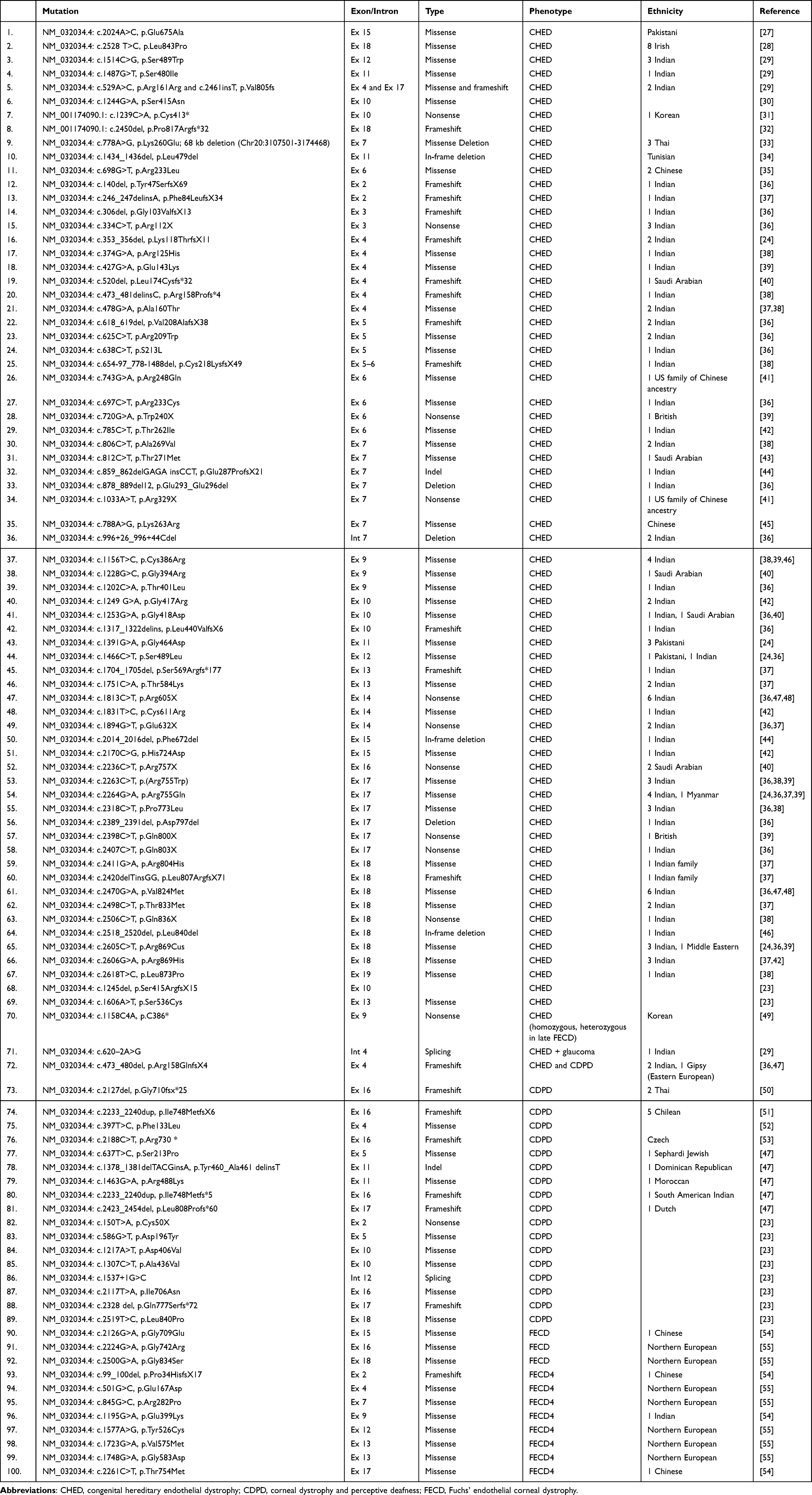 |
Table 1 Literature Review of All Reported CHED/CDPD Mutations |
 |
Figure 2 Mutation map of the SLC4A11 gene. |
SLC4A11 impairment causes loss of its water and H+ transport function, thereby directly affecting the “pump” function of the EC. Indirectly, SLC4A11 mutations cause a loss of EC function through increased intracellular production of reactive oxygen species (ROS). This upregulated ROS production is caused by the loss of the proposed SLC4A11 function in transporting H+ into the mitochondrial matrix and NH3 out of the mitochondrial matrix, as a build-up of NH3 increases the production of ROS.56,57 Moreover, SLC4A11 seems to play a role in regulating antioxidant signaling pathways. Nuclear factor erythroid-2 related factor 2 (NRF2), a trigger for antioxidant signaling, is downregulated in the absence of SLC4A11.58 Therefore, SLC4A11 dysfunction may cause both increased ROS production and impairment of the antioxidant pathway, hence increasing oxidative stress in ECs and eventually leading to EC apoptosis.5
An intact SLC4A11 function is also important for maintaining EC adhesion to DM.26 Defects in the attachment of epithelial cells trigger apoptosis.59 Thus, the loss of EC–DM adhesion during the early development of the cornea might be responsible for reduced EC density. Another possible pathophysiological mechanism is that mutations in SLC4A11 cause misfolding of the expressed protein in the endoplasmic reticulum (ER), and accumulation of these misfolded proteins in the ER may trigger apoptosis.60,61
Although SLC4A11 is the only well-established causative gene for CHED so far, the failure to identify SLC4A11 mutations in a number of patients suggests that other pathogenic genetic variants may contribute to this disease. One study reported the gene encoding multiple PDZ domain protein (MPDZ) as a different disease-causing gene, where a homozygous mutation in MPDZ was identified in a patient with a confirmed CHED diagnosis without a mutation in SLC4A11.23 Mutations in MPDZ have been implicated in the pathogenesis of some cases of hydrocephalus through the loss of connections between choroid plexus cells, leading to increased leakage of cerebrospinal fluid. Corneal endothelial cells (CECs) harbor embryological resemblance to choroid plexus cells; thus, it is speculated that mutations in MPDZ may cause loss of cell–cell connection integrity, leading to overhydration of the corneal stroma.62,63
A pathogenic homozygous mutation in the FAM149A gene (NM_015398.3: c.991A>G, p.R331G) was also suggested as a plausible causative etiology for a CHED phenotype in a patient with detected but nonpathogenic variants of SLC4A11 (identified by an in-house bioinformatics pipeline and characterized by having poor quality or low coverage). The FAM149A protein is highly expressed in the murine corneal endothelium and plays a role in protecting CECs against oxidative stress, maintaining the corneal endothelium’s health and stability. Zhang et al reported that the expression of FAM149A was increased in response to oxidative stress, and similar to a mutated SLC4A11 protein,58 the suppression of the FAM149A gene resulted in increased intracellular ROS levels and impaired NRF2-driven antioxidant signaling pathways64 The above studies imply that the current knowledge about the genetic predisposition of CHED remains limited, and the use of whole-exome sequencing methods for further exploration of other mutant pathogenic variants is required.
Corneal Dystrophy and Perceptive Deafness (CDPD)
Some patients with CHED also develop progressive sensorineural hearing loss (SNHL), a syndrome known as Harboyan syndrome or CDPD.65 In previous reports, all patients diagnosed with CHED whose auditory function was tested had SNHL at varying degrees.50,51 Homozygous or compound heterozygous mutations causing this syndrome are not distinct from those found in patients with CHED.47 Similarly, SLC4A11−/− mice have poor auditory and vestibular function.66 Besides the corneal endothelium, SLC4A11 is also expressed within the inner ear cells, namely the fibrocytes of the stria vascularis, and helps in OH− transport and endolymph maintenance.67 All patients with CDPD have high-frequency SNHL, suggesting a pathology at the basal part of the cochlea.68 The age of onset and severity of SNHL vary even among patients with the same mutation.50 Tissue-specific regulation of gene splicing may contribute to this variability in presentation.69 Owing to the variable age of onset, undertesting of SNHL among patients with CHED, and SNHL in some cases being only mild or asymptomatic, CDPD might be underreported in the literature.
Association with Fuchs’ Endothelial Corneal Dystrophy (FECD)
FECD is another type of corneal endothelial dystrophy with a late age of onset (4th to 5th decade of life) and variable degrees of severity. FECD is marked by impaired endothelial cell function which is confirmed clinically by the presence of corneal guttae in slit lamp examinations. As the disease progresses, the cornea thickens and loses its transparency, causing reduced vision and, in severe instances, a painful bullous keratopathy. In some countries, FECD represents about a quarter of all indications for corneal transplantation.70,71 Similar to CHED, it shows an abnormal posterior non-banded zone of DM on histopathological assessment, implying EC dysfunction.72 This dystrophy is typically sporadic, but an autosomal dominant inheritance pattern is recognized in just less than half of the patients.73 Different genetic mutations mapped at different loci, including SLC4A11 and MPDZ, were identified as causative of FECD.23,54 Some parents of children with CHED are affected by FECD.49,52,74 These data suggest that homozygous or compound heterozygous allelic mutations may cause CHED, whereas mutations of a single allele may cause FECD through autosomal dominant inheritance.
Clinical Presentation
CHED typically presents with diffuse, bilateral corneal clouding noticed at birth or in early infancy, in the absence of epiphora and photophobia. Nystagmus may also be present, depending on the severity of the disease and visual impairment. Clinically, slit lamp examinations will show thick corneas with a mosaic ground glass pattern of corneal opacity involving the entire cornea with the absence of buphthalmos, markedly increased central corneal thickness, and a normal-for-age corneal diameter.1
A Common Diagnostic Dilemma in Consanguineous Populations
A child presenting with a cloudy cornea and high intraocular pressure (IOP) is a common dilemma that can result in unnecessary glaucoma surgeries in the early stages of CHED. Similar to CHED, primary congenital glaucoma (PCG) has an autosomal recessive pattern of inheritance, and in areas of the world with high consanguinity rates, such as Saudi Arabia (reaching up to 66.7%),75 relatively high incidence rates of both diseases can occur. Mutations in CYP1B1, MYOC, LTBP2, FOXC1, NTF4, and WDR36 have been reported in Arab countries, with the CYP1B1 gene, a member of the cytochrome P450 gene family, being the major contributor to PCG.76
Few observations point towards the diagnosis of CHED rather than PCG, including a failure of corneal clearance despite adequate IOP control with topical or surgical management, a normal-for-age corneal diameter, and the absence of buphthalmos. A thick cornea with a mosaic ground glass pattern of corneal opacity, as well as the absence of DM breaks known as Haab’s striae, are classic features of CHED.77 By contrast, patients with PCG have variable corneal scarring, noted mostly centrally, an enlarged corneal diameter, buphthalmos, and Haab’s striae.78 Table 2 highlights some points that facilitate the clinical differentiation of CHED from PCG.
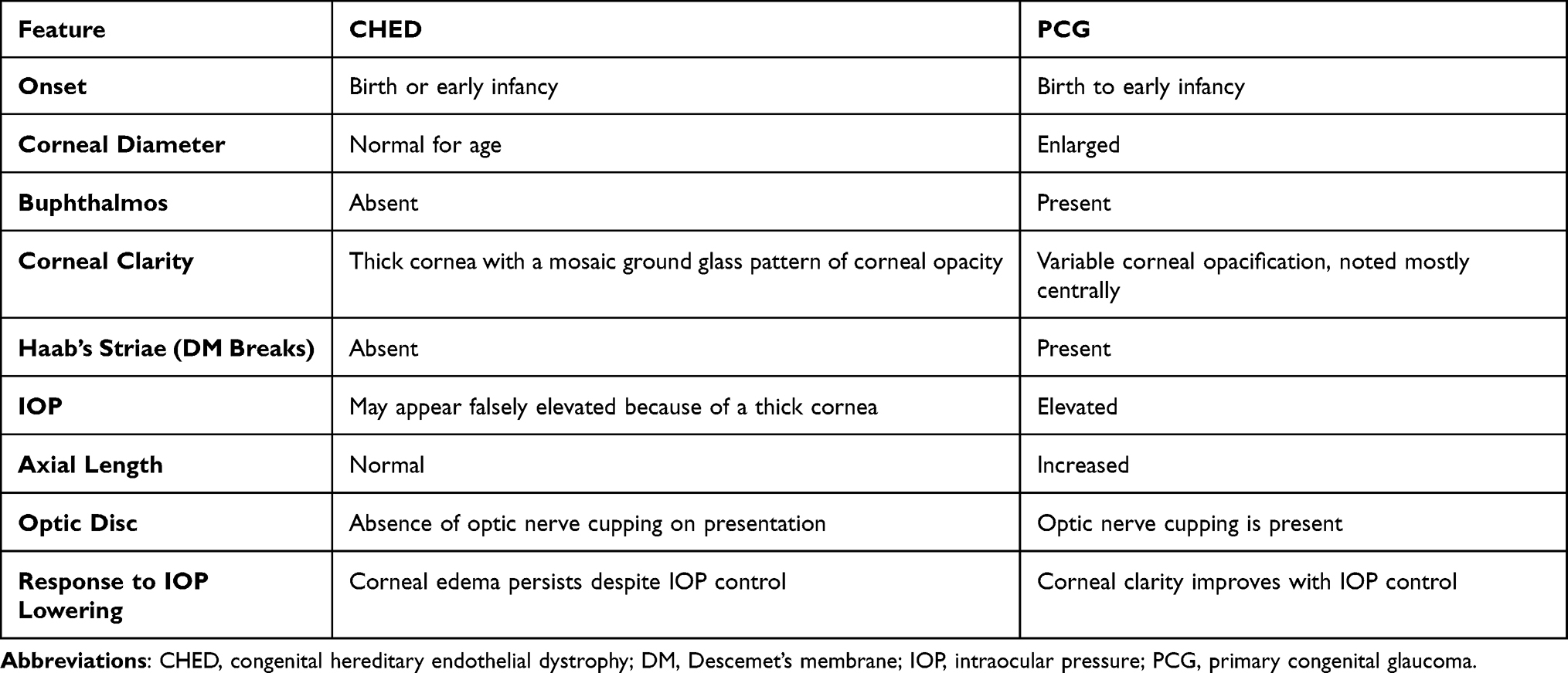 |
Table 2 Clinical Differentiation Between CHED and PCG |
Various reports describe individuals with CHED misdiagnosed as PCG.2,79,80 One of the earliest publications described three children who had elevated IOP with corneal opacification and partial aniridia. Multiple glaucoma procedures were required to control the IOP, and a corneal transplant was planned to clear the view because of persistent corneal edema despite a normalized IOP. Although genetic analysis was not performed, the histopathological evaluation of corneal buttons revealed thick corneas with a markedly abnormal DM and absent endothelial cell layer, suggesting a diagnosis of CHED.79
Khan et al described a child with an isolated clinical finding of ocular coloboma, treated with topical anti-glaucoma drugs since infancy for a suspected glaucoma Sequencing of SLC4A11 proved the presence of a known homozygous mutation (c.1228G>C, p.Gly394Arg), confirming a diagnosis of CHED. The child maintained a normal IOP (20 mmHg) after cessation of all anti-glaucoma drops for 3 years of follow-up.2 More recently, Yousaf et al identified a Pakistani family that underwent multiple surgical interventions over 9 years for an initially misdiagnosed PCG The absence of CYP1B1 gene variants in the family’s genetic analysis questioned the initial diagnosis, and whole-exome sequencing analysis identified a homozygous missense variant (Glu675Ala) in the SLC4A11 gene.80 While a negative genetic result for CHED does not rule out the diagnosis, mutation analysis of SLC4A11 can be confirmatory.
Identifying a positive family history of PCG can be of help; however, it should be kept in mind that other family members with similar phenotypes might have been misdiagnosed and undergone unnecessary glaucoma surgeries. This necessitates the need to obtain a careful past medical and surgical history and to examine all other affected family members as applicable.
An association between PCG and CHED has been proposed;79,81 however, insufficient proof to support this hypothesis exists up to now, as IOP measurements can vary and be deceptively high in abnormally thick corneas. Corneal thickness may not be the only deceptive factor, since corneal hysteresis and corneal resistance factor values have a more significant effect on the measured IOP in patients with PCG than in healthy individuals.82 Future research, including studies of corneal biomechanics in eyes with dual phenotype, is needed to help a more accurate interpretation of the IOP and prove whether the two entities can co-exist.
As cases of faulty PCG diagnosis continue to be reported, this scenario clearly remains a therapeutic dilemma for many treating physicians. For atypical cases of PCG, revisiting the diagnosis with serial assessments and genetic testing when available, can help in minimizing unnecessary glaucoma interventions and can also allow an earlier visual rehabilitation of patients with CHED (Figure 3).
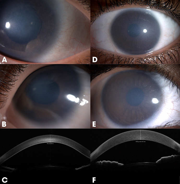 |
Figure 3 (A–C) Primary congenital glaucoma: More central dense scarring (A), Haab’s striae (B), and a relatively thinner cornea in AS-OCT images (C). (D–F) Congenital hereditary endothelial dystrophy: Diffuse haze from limbus to limbus with a ground-glass pattern (D and E) and a thicker cornea in AS-OCT images (F). Abbreviation: AS-OCT, anterior segment optical coherence tomography. |
Management
Surgical Management
Prior to the introduction of EK, conventional PKP used to be the mainstay surgical treatment for CHED.83 Despite offering good visual outcomes, PKP carries the intraoperative risk of suprachoroidal hemorrhage, in addition to the risk of postoperative suture-related complications and wound dehiscence. Following the development of many posterior lamellar keratoplasty surgical techniques in the last decade, EK in the form of Descemet stripping automated endothelial keratoplasty (DSAEK) was used as a safe alternative with good graft survival rates, fast visual rehabilitation84 and clinical outcomes equivalent to those of standard PKP.85,86
A recently published study evaluated the outcomes of PKP versus DSAEK in 111 eyes of 63 patients with CHED in Saudi Arabia. The authors reported equivalent graft survival rates and best-corrected visual acuity (BCVA) at 2 years postoperatively for both procedures; however, the DSAEK group had a better postoperative safety profile.85 DM stripping causes intraoperative difficulties in patients with CHED due to poor visibility and tighter adhesion of DM in pediatric eyes. Whether to strip or leave the DM in place is a subject of debate, with a growing literature that supports a non-Descemet stripping approach of EK in patients with CHED.87–89
Descemet’s membrane endothelial keratoplasty (DMEK) has the advantages of a faster visual recovery and a lower rejection rate (1%) compared to that of DSAEK (10%),90 because a very thin layer of DM and endothelium is transplanted, closely replicating the natural anatomy of the cornea. However, it is technically more challenging, especially among pediatric age groups with poor corneal visibility.90 The challenges of performing EK for CHED arise from the difficulty in maneuvering within the small anterior chamber of a child with a clear lens, compounded by poor visualization caused by the overlying corneal edema. A small case series examined the appearance of DM preoperatively in 12 eyes with CHED using anterior segment optical coherence tomography (AS-OCT), then selectively sorted the patients into either DMEK or non-Descemet’s membrane endothelial keratoplasty (n-DMEK) based on their preoperative DM appearance.88 An increased DM thickness correlating with hyperreflectivity on AS-OCT was noticed in older patients (13–39 years) who had their DM removed intraoperatively, whereas younger patients (3–8 years) with preoperatively normal-appearing DM (absence of hyperreflectivity on AS-OCT), denoting a normal thickness of DM, underwent n-DMEK. Apart from one patient in the n-DMEK group who required a repeated air injection, all patients had a successful postoperative attachment with comparable visual outcomes (notably, the authors used 20% SF6 for tamponade in the n-DMEK group vs air in the DMEK group). This report highlights an interesting role for AS-OCT as a diagnostic tool in the preoperative assessment of DM and intraoperative planning (Figure 4). Although the initial results of DMEK in CHED appear promising,89,91 the current literature remains insufficient, and additional comparative studies are warranted.
 |
Figure 4 AS-OCT images and slit lamp photographs of two patients with CHED. A hyperreflective Descemet’s membrane in a patient is shown in (A). Absence of Descemet’s membrane hyperreflectivity in another patient is shown in (B). Abbreviations: AS-OCT, anterior segment optical coherence tomography; CHED, congenital hereditary endothelial dystrophy. |
Descemet stripping only (DSO) with or without a tissue-engineering approach is another regenerative therapeutic modality that is suggested to be effective in mild-to-moderate stages of FECD,92 but its efficacy is yet to be established in patients with CHED. Notably, a good density of healthy peripheral endothelial cells would be a prerequisite to increase its likelihood of success. This may be the case in the early stages of FECD, where only central guttae are present while retaining a peripheral rim of healthy CECs, unlike in CHED, which is characterized by non-progressive generalized endothelial cell atrophy in early stages.93
Injection of cultured CECs into the anterior chamber, either following in vitro expansion or de novo generation from pluripotent stem cells, is another less invasive procedure that can provide therapeutic endothelial cells for more patients in the era of donor tissue scarcity. Currently, three ongoing clinical trials (NCT04191629, UMIN000034334, and UMIN000012534) assess the results of CED injection in patients with FECD and pseudophakic bullous keratopathy. Owing to the rarity of the disease, similar studies have still not been conducted in patients with CHED.
Mehta et al proposed an algorithm for CHED management based on the anterior segment visibility (Ramappa grading of corneal haze)94 and OCT findings Conservative medical management (nonsteroidal anti-inflammatory drugs [NSAIDs]) in addition to optical correction and amblyopia treatment was suggested for mild cases with an acceptable corneal clarity, allowing the discrimination of anterior segment details. In moderate cases where only the pupillary silhouette can be seen, early-stage surgical intervention in the form of DSAEK is recommended, in opposition to severe cases with obscured view and anterior or posterior corneal scarring on OCT, which may benefit from undergoing PKP (Figure 5).95
 |
Figure 5 Subjective grading of CHED based on the visibility of iris structures. (A) Mild: Iris details are visible. (B) Moderate: Iris details are obscured, but the pupillary silhouette is visible. (C) Severe: Corneal cloudiness obscuring both iris and pupillary details. Abbreviation: CHED, congenital hereditary endothelial dystrophy. |
Nonsurgical Management
Although the most efficient treatment for CHED remains to be corneal transplantation, the worldwide shortage of donor tissue is a great challenge with only 1 cornea available for 70 needed, as per a global survey.96 In a shift away from surgical treatment, attempts have been made to decrease the stress related to misfolding defects, using topical NSAIDs. Most SLC4A11 mutations hinder the transport of the protein into the plasma membrane and lead to its accumulation in the ER, resulting in high levels of ROS, which in turn predispose the cell to apoptosis.97 Alka et al studied the therapeutic potential of five ophthalmic NSAIDs on HEK293 cells with CHED and FECD caused by SLC4A11 mutation and their ability to correct the mutant cell-surface trafficking Nepafenac and diclofenac significantly increased the surface abundance of SLC4A11 mutants in 20 out of 30 test cells, reaching up to 20–30% of the wild-type levels and restoring its water flux activity to a level similar to that of wild-type SLC4A11.97 These findings have further encouraged the testing of ophthalmic NSAIDs as a treatment for CHED. An ongoing clinical trial (NCT04843839) in India started in 202198 and is currently evaluating the effectiveness of nepafenac 0.1% in patients with CHED aged 9 years or younger. Another palliative medical treatment uses hypertonic saline drops or ointment to temporarily draw osmotically the water out of the edematous cornea.
Future Therapeutic Directions
Gene Therapy
The future role of genetic therapy holds significant promise for transforming the treatment basis of CHED. Advances in gene-editing technologies, such as Clustered regularly interspaced short palindromic repeats-Cas protein (CRISPR-Cas9), and gene replacement therapies offer the potential to correct or compensate for the underlying genetic defects. Researchers are exploring targeted delivery methods, including viral vectors and nanoparticle-based systems, to introduce functional copies of the defective gene or repair mutations directly within CECs.99,100
It was previously reported that intracameral injections of adeno-associated virus (AAV)-9 serotype can successfully transduce mouse corneal endothelium.101 This has led Shyam et al to explore the potential of AAV-mediated gene therapy to rescue the disease phenotype in SLC4A11 knockout mice After delivering a functional SLC4A11 gene tagged with hemagglutinin (HA) into the corneal endothelium of young (5 weeks old) and old (11 weeks old) mice, AAV9-HA-SLC4A11 injection showed a reversal of corneal edema, reduction in stromal lactate levels, and restoration of endothelial cell density, mitochondrial function, and lactate transporter expression to near wild-type levels in young animals. By contrast, the structural damage to the cornea was more severe in older mice, limiting the therapeutic potential.99 This study provides a foundation for developing gene therapy approaches for CHED and highlights the importance of early intervention before cell loss commences and structural corneal damage becomes irreversible. Future research should improve the transduction efficiency to enhance therapeutic outcomes, especially in older animals with advanced disease.
CRISPR-Cas9 is a genetic engineering tool that allows scientists to edit genes with high precision. A study investigated its potential role in the treatment of a murine model of FECD secondary to collagen type VIII alpha 2 chain (COL8A2) gene mutation, using an adenovirus delivery system into CECs. With a single injection, the designed gene editing tool was efficient in knocking down the expression of the mutant COL8A1, which prevented the loss of CECs and restored the pumping function of the corneal endothelium.100
While challenges such as ensuring precise targeting, minimizing off-target effects, and achieving long-term efficacy remain, ongoing preclinical studies and clinical trials will pave the way for more efficient treatments. If successful, genetic therapy could provide a curative approach, reducing the reliance on corneal transplants and significantly improving the quality of life for individuals with CHED.
Mitochondria-Targeted Antioxidants
The resulting mitochondrial oxidative stress in individuals with corneal endothelial dystrophy is a primary cause of endothelial cell dysfunction, which drives the disease phenotype.102,103 The SLC4A11 protein facilitates the suppression of mitochondrial ROS production in endothelial cells.57 In SLC4A11 knockout mouse models, trials attempted to reduce mitochondrial ROS production by instilling dimethyl-α-ketoglutarate eye drops57 or by injecting intraperitoneally the mitochondria-targeted antioxidant mitoquinone.103 These procedures resulted in decreased endothelial cell loss and a reduction in corneal edema.
More recently, Peshkar-Kulkarni et al reported increased cell viability in vitro human CECs with CHED-associated SLC4A11 mutations, comparing mitoquinone-treated with untreated cells104 Additionally, in vitro experiments using immortalized wild-type human CECs showed under stress enhanced cell viability and mitochondrial membrane potential in FECD and wild-type cells pretreated with mitoquinone or idebenone (a non-targeted antioxidant).105 This implies that mitochondria-targeted antioxidants can be viable nonsurgical therapeutic options for the treatment of CHED and other oxidative stress-related disorders. Further in vivo studies are required to evaluate the efficacy of this approach in reversing corneal edema and disease phenotype.
Visual Rehabilitation
Amblyopia management is of paramount importance in the visual rehabilitation of patients with CHED. Despite having an optically clear cornea, the failure to start a timely amblyopia treatment and optical correction by pediatric ophthalmologists in the immediate postoperative period can negatively influence surgical outcomes. Although no consensus on the appropriate timing for surgical intervention in pediatric patients with CHED exists, it has been proven that the child’s age is a major factor in achieving a better surgical outcome, with earlier intervention carrying a better prognosis.106 Comparing the results of Descemet stripping endothelial keratoplasty in 16 patients with CHED between age groups (infants vs children), patients in the infant group had a statistically better postoperative BCVA, which can be attributed to the fact that the infant group was subjected to an earlier initiation of amblyopia treatment.106
Role of Genetic Counselling
Genetic counseling is a vital resource for individuals and families affected by CHED. As an autosomal recessive condition, two copies of the pathogenic variant have to be inherited for a child to be affected (one from each parent). Genetic counseling helps identify couples at risk through carrier screening for the identified mutations in affected relatives. Besides providing education about the inheritance pattern, guidance on reproductive choices can be offered, such as prenatal diagnosis or preimplantation genetic testing. Emotional and psychosocial concerns should be addressed adequately, helping families understand the condition and its implications.
Knowledge Gaps and Future Directions
Despite identification of SLC4A11 as the principal gene associated with CHED, the genetic basis of the disease remains incompletely understood. Emerging evidence from isolated reports has suggested the potential involvement of other genes, including MPDZ and FAM149A; however, these associations require further validation and replication in independent cohorts. The limited scope of existing genetic studies and the rarity of CHED have hindered comprehensive elucidation of its genetic heterogeneity. Future research should prioritize systematic, large-scale genetic screening studies using approaches such as whole-exome or whole-genome sequencing to identify novel causative genes associated with CHED. Moreover, integrating genetic findings with detailed clinical phenotyping may facilitate improved genotype–phenotype correlations and help refine diagnostic criteria.
Another significant knowledge gap lies in the absence of a standardized, objective grading system for corneal haze severity in CHED. Current clinical decision-making regarding the timing of surgical intervention relies largely on subjective slit-lamp assessment and visual behavior, which may vary considerably across examiners. The lack of an objective, reproducible grading framework limits the ability to accurately stratify disease severity, monitor progression, and determine the optimal timing for intervention. In this context, further studies incorporating multimodal imaging techniques, such as AS-OCT and Scheimpflug-based imaging (eg., Pentacam), are needed to define the strengths and limitations of each modality. Establishing standardized imaging protocols and objective metrics may enable more precise assessment of corneal opacity and support evidence-based clinical decision-making in CHED. Addressing these gaps will be critical for advancing understanding of CHED pathogenesis and for laying the groundwork for precision-based diagnostic and therapeutic strategies.
Limitations
This study has some limitations. Being a narrative review carries the potential for selection bias, limiting its comprehensiveness. Additionally, some of the reported CHED-related SLC4A11 variants were initially identified; however, these publications lacked important data regarding variant descriptions and the exact locations of the mutation, which hindered their presentation in a standardized format. Thus, they were excluded from this review.
Conclusion
CHED, a rare but impactful corneal endothelial dystrophy, demands early diagnosis, multidisciplinary management, and ongoing research to develop less invasive and curative therapies. Advances in genetics and disease modelling have expanded the understanding of its pathophysiology, identifying oxidative stress, impaired water and proton transport, and endothelial apoptosis as key pathogenic pathways. Although emerging gene-based and antioxidant modulation therapies have produced promising preclinical results, substantial barriers, particularly pertaining to safe and targeted delivery, long-term efficacy, and regulatory approval, still limit their clinical translation. Continued collaboration across laboratory research and clinical trials will be crucial to move these experimental approaches toward practical alternatives to corneal transplantation. Ongoing genetic studies aimed at identifying additional causative mutations may further clarify the disease etiology and inform future therapeutic targets.
Data Sharing Statement
The data supporting the findings of this study are available from the corresponding author upon reasonable request. All relevant anonymized data can be provided to qualified researchers for academic purposes following institutional and ethical guidelines.
Ethics Approval
This study was approved by the Institutional Review Board of King Khaled Eye Specialist Hospital (KKESH, approval number RP 22117) and adhered to the tenets of the Declaration of Helsinki.
Funding
There is no funding to report.
Disclosure
The authors report no conflicts of interest related to this work.
References
1. Aldave AJ, Han J, Frausto RF. Genetics of the corneal endothelial dystrophies: an evidence-based review. Clin Genet. 2013;84(2):109–19. doi:10.1111/cge.12191
2. Khan AO, Aldahmesh MA, Alkuraya F. Congenital hereditary endothelial dystrophy, not glaucoma, in a child with iris colobomas. J AAPOS. 2016;20(4):370–372. doi:10.1016/j.jaapos.2016.03.017
3. Parker MD, Ourmozdi EP, Tanner MJ. Human BTR1, a new bicarbonate transporter superfamily member and human AE4 from kidney. Biochem Biophys Res Commun. 2001;282(5):1103–1109. doi:10.1006/bbrc.2001.4692
4. Vilas GL, Loganathan SK, Liu J, et al. Transmembrane water-flux through SLC4A11: a route defective in genetic corneal diseases. Hum Mol Genet. 2013;22(22):4579–4590. doi:10.1093/hmg/ddt307
5. Roy S, Praneetha DC, Vendra VPR. Mutations in the corneal endothelial dystrophy–associated gene SLC4A11 render the cells more vulnerable to oxidative insults. Cornea. 2015;34(6):668–674. doi:10.1097/ICO.0000000000000421
6. Vanathi M, Raj N, Kusumesh R, Aron N, Gupta N, Tandon R. Update on pediatric corneal diseases and keratoplasty. Surv Ophthalmol. 2022;67(6):1647–1684. doi:10.1016/j.survophthal.2022.07.010
7. Weiss JS, Møller HU, Aldave AJ, et al. IC3D classification of corneal dystrophies—edition 2. Cornea. 2015;34(2):117–159. doi:10.1097/ICO.0000000000000307
8. Sharma SK, Kalam MA, Ghosh S, Roy S. Prevalence and determinants of consanguineous marriage and its types in India: evidence from the National Family Health Survey, 2015–2016. J Biosoc Sci. 2021;53(4):566–576. doi:10.1017/S0021932020000383
9. Rauniyar D, Das AV. Consanguinity and ocular disorders in India: electronic medical records driven big data analytics. Indian J Ophthalmol. 2022;70(7):2401–2407. doi:10.4103/ijo.IJO_1553_21
10. Al-Towerki AE, Gonnah ES, Al-Rajhi A, Wagoner MD. Changing indications for corneal transplantation at the King Khaled Eye Specialist Hospital (1983–2002). Cornea. 2004;23(6):584–588. doi:10.1097/01.ico.0000121708.58571.5b
11. Al-Ghamdi A, Al-Rajhi A, Wagoner MD. Primary pediatric keratoplasty: indications, graft survival, and visual outcome. J AAPOS. 2007;11(1):41–47. doi:10.1016/j.jaapos.2006.09.012
12. Ali Javadi MA, Kanavi MR, Safi S. A 27-year report from the Central Eye Bank of Iran. J Ophthalmic Vis Res. 2020;15(2):149–159. doi:10.18502/jovr.v15i2.6731
13. Bozkurt TK, Acar B, Kilavuzoglu AE, et al. An 11-year review of keratoplasty in a tertiary referral center in Turkey: changing surgical techniques for similar indications. Eye Contact Lens. 2017;43(6):364–370. doi:10.1097/ICL.0000000000000274
14. Tanyildiz B, Oklar M, Günaydın NT, Kandemir B. Changing trends in the corneal transplantation and the impact of the COVID-19 pandemic on corneal transplant recipient selection. Saudi J Ophthalmol. 2022;36(1):95–101. doi:10.4103/sjopt.sjopt_251_21
15. Pandrowala H, Bansal A, Vemuganti GK, Rao GN. Frequency, distribution, and outcome of keratoplasty for corneal dystrophies at a tertiary eye care center in South India. Cornea. 2004;23(6):541–546. doi:10.1097/01.ico.0000126324.58884.b9
16. Huang C, O’Hara MO, Mannis MJ. Primary pediatric keratoplasty: indications and outcomes. Cornea. 2009;28(9):1003–1008. doi:10.1097/ICO.0b013e3181a186c0
17. Patel HY, Ormonde S, Brookes NH, Moffatt LS, McGhee CNJ. The indications and outcome of paediatric corneal transplantation in New Zealand: 1991–2003. Br J Ophthalmol. 2005;89(4):404–408. doi:10.1136/bjo.2004.053116
18. Mohebbi M, Mehrpour M, Sanij AD, Mohammadi N, Mirghorbani M. Pediatric endothelial keratoplasty: a systematic review and individual participant data meta-analysis. Graefes Arch Clin Exp Ophthalmol. 2022;260(4):1069–1082. doi:10.1007/s00417-021-05459-8
19. Wulle KG. Electron microscopy of the fetal development of the corneal endothelium and descemet’s membrane of the human eye. Invest Ophthalmol. 1972;11(11):897–904.
20. Bonanno JA. Molecular mechanisms underlying the corneal endothelial pump. Exp Eye Res. 2012;95(1):2–7. doi:10.1016/j.exer.2011.06.004
21. Willoughby CE, Ponzin D, Ferrari S, Lobo A, Landau K, Omidi Y. Anatomy and physiology of the human eye: effects of mucopolysaccharidoses disease on structure and function—a review. Clin Exp Ophthalmol. 2010;38:2–11. doi:10.1111/j.1442-9071.2010.02363.x
22. DelMonte DW, Kim T. Anatomy and physiology of the cornea. J Cataract Refract Surg. 2011;37(3):588–598. doi:10.1016/j.jcrs.2010.12.037
23. Moazzeni H, Javadi MA, Asgari D, et al. Observation of nine previously reported and 10 non-reported SLC4A11 mutations among 20 Iranian CHED probands and identification of an MPDZ mutation as possible cause of CHED and FECD in one family. Br J Ophthalmol. 2020;104(11):1621–1628. doi:10.1136/bjophthalmol-2019-314377
24. Vithana EN, Morgan P, Sundaresan P, et al. Mutations in sodium-borate cotransporter SLC4A11 cause recessive congenital hereditary endothelial dystrophy (CHED2). Nat Genet. 2006;38(7):755–757. doi:10.1038/ng1824
25. Ogando DG, Jalimarada SS, Zhang W, Vithana EN, Bonanno JA. SLC4A11 is an EIPA-sensitive Na+ permeable pHi regulator. Am J Physiol Cell Physiol. 2013;305(7):C716–C727. doi:10.1152/ajpcell.00056.2013
26. Malhotra D, Jung M, Fecher-Trost C, et al. Defective cell adhesion function of solute transporter, SLC4A11, in endothelial corneal dystrophies. Hum Mol Genet. 2020;29(1):97–116. doi:10.1093/hmg/ddz259
27. Kaul H, Suman M, Khan Z, Ullah MI, Ashfaq UA, Idrees S. Missense mutation in SLC4A11 in two Pakistani families affected with congenital hereditary endothelial dystrophy (CHED2). Clin Exp Optom. 2016;99(1):73–77. doi:10.1111/cxo.12276
28. Hand CK, McGuire M, Parfrey NA, Murphy CC. Homozygous SLC4A11 mutation in a large Irish CHED2 pedigree. Ophthalmic Genet. 2017;38(2):148–151. doi:10.3109/13816810.2016.1151901
29. Salman M, Verma A, Chaurasia S, et al. Identification and in silico analysis of a spectrum of SLC4A11 variations in Indian familial and sporadic cases of congenital hereditary endothelial dystrophy. Orphanet J Rare Dis. 2022;17(1):361. doi:10.1186/s13023-022-02521-4
30. Kumawat BL, Gupta R, Sharma A, Sen S, Gupta S, Tandon R. Delayed onset of congenital hereditary endothelial dystrophy due to compound heterozygous SLC4A11 mutations. Indian J Ophthalmol. 2016;64(7):492–495. doi:10.4103/0301-4738.190100
31. Park SH, Jeong HJ, Kim M, Kim MS. A novel nonsense mutation of the SLC4A11 gene in a Korean patient with autosomal recessive congenital hereditary endothelial dystrophy. Cornea. 2013;32(7):e181–e182. doi:10.1097/ICO.0b013e31828d9ffd
32. Cunnusamy K, Bowman CB, Beebe W, Gong X, Hogan RN, Mootha VV. Congenital corneal endothelial dystrophies resulting from novel de novo mutations. Cornea. 2016;35(2):281–285. doi:10.1097/ICO.0000000000000670
33. Puangsricharern V, Yeetong P, Charumalai C, Suphapeetiporn K, Shotelersuk V. Two novel mutations including a large deletion of the SLC4A11 gene causing autosomal recessive hereditary endothelial dystrophy. Br J Ophthalmol. 2014;98(10):1460–1462. doi:10.1136/bjophthalmol-2014-305584
34. Chibani Z, Abid IZ, Söderkvist P, Feki J, Aifa MH. Autosomal recessive congenital hereditary corneal dystrophy associated with a novel SLC4A11 mutation in two consanguineous Tunisian families. Br J Ophthalmol. 2022;106(2):281–287. doi:10.1136/bjophthalmol-2020-318204
35. Liu M, Xia JL, Yang H, Yu L. Compound heterozygous mutations in the SLC4A11 gene associated with congenital hereditary endothelial dystrophy in a Chinese family. Ophthalmic Genet. 2022;43(4):538–542. doi:10.1080/13816810.2022.2051192
36. Sultana A, Garg P, Ramamurthy B, Vemuganti GK, Kannabiran C. Mutational spectrum of the SLC4A11 gene in autosomal recessive congenital hereditary endothelial dystrophy. Mol Vis. 2007;13:1327–1332.
37. Jiao X, Sultana A, Garg P, et al. Autosomal recessive corneal endothelial dystrophy (CHED2) is associated with mutations in SLC4A11. J Med Genet. 2007;44(1):64–68. doi:10.1136/jmg.2006.044644
38. Hemadevi B, Veitia RA, Srinivasan M, et al. Identification of mutations in the SLC4A11 gene in patients with recessive congenital hereditary endothelial dystrophy. Arch Ophthalmol. 2008;126(5):700–708. doi:10.1001/archopht.126.5.700
39. Ramprasad VL, Ebenezer ND, Aung T, et al. Novel SLC4A11 mutations in patients with recessive congenital hereditary endothelial dystrophy (CHED2). Hum Mutat. 2007;28(5):522–523. doi:10.1002/humu.9487
40. Aldahmesh MA, Khan AO, Meyer BF, Alkuraya FS. Mutational spectrum of SLC4A11 in autosomal recessive CHED in Saudi Arabia. Invest Ophthalmol Vis Sci. 2009;50(9):4142–4145. doi:10.1167/iovs.08-3006
41. Aldave AJ, Yellore VS, Bourla N, et al. Autosomal recessive CHED associated with novel compound heterozygous mutations in SLC4A11. Cornea. 2007;26(7):896–900. doi:10.1097/ICO.0b013e318074bb01
42. Kodaganur SG, Kapoor S, Veerappa AM, et al. Mutation analysis of the SLC4A11 gene in Indian families with congenital hereditary endothelial dystrophy 2 and a review of the literature. Mol Vis. 2013;19:1694–1706.
43. Shah SS, Al-Rajhi A, Brandt JD, et al. Mutation in the SLC4A11 gene associated with autosomal recessive congenital hereditary endothelial dystrophy in a large Saudi family. Ophthalmic Genet. 2008;29(1):41–45. doi:10.1080/13816810701850033
44. Kumar A, Bhattacharjee S, Prakash DR, Sadanand CS. Genetic analysis of two Indian families affected with congenital hereditary endothelial dystrophy: two novel mutations in SLC4A11. Mol Vis. 2007;13:39–46.
45. Zhen T, Li Y, Guo Q, Yao S, You Y, Lei B. Pathogenicity and function analysis of two novel SLC4A11 variants in patients with congenital hereditary endothelial dystrophy. Transl Vis Sci Technol. 2023;12(10):1. doi:10.1167/tvst.12.10.1
46. Paliwal P, Sharma A, Tandon R, et al. Congenital hereditary endothelial dystrophy—mutation analysis of SLC4A11 and genotype-phenotype correlation in a north Indian patient cohort. Mol Vis. 2010;16:2955–2963.
47. Desir J, Moya G, Reish O, et al. Borate transporter SLC4A11 mutations cause both Harboyan syndrome and non-syndromic corneal endothelial dystrophy. J Med Genet. 2007;44(5):322–326. doi:10.1136/jmg.2006.046904
48. Hemadevi B, Srinivasan M, Arunkumar J, Prajna NV, Sundaresan P. Genetic analysis of patients with Fuchs endothelial corneal dystrophy in India. BMC Ophthalmol. 2010;10:3. doi:10.1186/1471-2415-10-3
49. Kim JH, Ko JM, Tchah H. Fuchs endothelial corneal dystrophy in a heterozygous carrier of congenital hereditary endothelial dystrophy type 2 with a novel mutation in SLC4A11. Ophthalmic Genet. 2015;36(3):284–286. doi:10.3109/13816810.2014.881510
50. Tananuvat N, Tananuvat R, Chartapisak W, et al. Harboyan syndrome: novel SLC4A11 mutation, clinical manifestations, and outcome of corneal transplantation. J Hum Genet. 2021;66(2):193–203. doi:10.1038/s10038-020-00834-5
51. Romero PT, Donoso R, López P, et al. Clinical features and possible founder mutation of the 8bp duplication mutation in the SLC4A11 gene causing corneal dystrophy and perceptive deafness in three South American families. Ophthalmic Genet. 2019;40(2):91–98. doi:10.1080/13816810.2019.1571615
52. Siddiqui S, Zenteno JC, Rice A, et al. Congenital hereditary endothelial dystrophy caused by SLC4A11 mutations progresses to Harboyan syndrome. Cornea. 2014;33(3):247–251. doi:10.1097/ICO.0000000000000041
53. Liskova P, Dudakova L, Tesar V, et al. Detailed assessment of renal function in a proband with Harboyan syndrome caused by a novel homozygous SLC4A11 nonsense mutation. Ophthalmic Res. 2015;53(1):30–35. doi:10.1159/000365109
54. Vithana EN, Morgan PE, Ramprasad V, et al. SLC4A11 mutations in Fuchs endothelial corneal dystrophy. Hum Mol Genet. 2008;17(5):656–666. doi:10.1093/hmg/ddm337
55. Riazuddin SA, Vithana EN, Seet LF, et al. Missense mutations in the sodium borate cotransporter SLC4A11 cause late-onset Fuchs corneal dystrophy. Hum Mutat. 2010;31(11):1261–1268. doi:10.1002/humu.21356
56. Murthy CRK, Rama Rao KV, Bai G, Norenberg MD. Ammonia-induced production of free radicals in primary cultures of rat astrocytes. J Neurosci Res. 2001;66(2):282–288. doi:10.1002/jnr.1222
57. Ogando DG, Choi M, Shyam R, Li S, Bonanno JA. Ammonia sensitive SLC4A11 mitochondrial uncoupling reduces glutamine induced oxidative stress. Redox Biol. 2019;26:101260. doi:10.1016/j.redox.2019.101260
58. Guha S, Chaurasia S, Ramachandran C, Roy S. SLC4A11 depletion impairs NRF2 mediated antioxidant signaling and increases reactive oxygen species in human corneal endothelial cells during oxidative stress. Sci Rep. 2017;7(1):4074. doi:10.1038/s41598-017-03654-4
59. Gilmore AP. Anoikis. Cell Death Differ. 2005;12(Suppl 2):1473–1477. doi:10.1038/sj.cdd.4401723
60. Alka K, Casey JR. Molecular phenotype of SLC4A11 missense mutants: setting the stage for personalized medicine in corneal dystrophies. Hum Mutat. 2018;39(5):676–690. doi:10.1002/humu.23401
61. Almanza A, Carlesso A, Chintha C, et al. Endoplasmic reticulum stress signalling—from basic mechanisms to clinical applications. FEBS J. 2019;286(2):241–278. doi:10.1111/febs.14608
62. Yang J, Simonneau C, Kilker R, et al. Murine MPDZ‐linked hydrocephalus is caused by hyperpermeability of the choroid plexus. EMBO Mol Med. 2019;11(1):e9540. doi:10.15252/emmm.201809540
63. Feldner A, Adam MG, Tetzlaff F, et al. Loss of Mpdz impairs ependymal cell integrity leading to perinatal‐onset hydrocephalus in mice. EMBO Mol Med. 2017;9(7):890–905. doi:10.15252/emmm.201606430
64. Zhang J, Dai Y, Wu D, Li Y, Xu J. Whole exome sequencing identified FAM149A as a plausible causative gene for congenital hereditary endothelial dystrophy, affecting Nrf2-Antioxidant signaling upon oxidative stress. Free Radic Biol Med. 2021;173:117–124. doi:10.1016/j.freeradbiomed.2021.07.029
65. Desir J, Abramowicz M. Congenital hereditary endothelial dystrophy with progressive sensorineural deafness (Harboyan syndrome). Orphanet J Rare Dis. 2008;3:28. doi:10.1186/1750-1172-3-28
66. Lopez IA, Rosenblatt MI, Kim C, et al. Slc4a11 gene disruption in mice: cellular targets of sensorineuronal abnormalities. J Biol Chem. 2009;284(39):26882–26896. doi:10.1074/jbc.M109.008102
67. Morris KA, Snir E, Pompeia C, et al. Differential expression of genes within the cochlea as defined by a custom mouse inner ear microarray. J Assoc Res Otolaryngol. 2005;6(1):75–89. doi:10.1007/s10162-004-5046-x
68. Damkier HH, Nielsen S, Praetorius J. Molecular expression of SLC4-derived Na+-dependent anion transporters in selected human tissues. Am J Physiol Regul Integr Comp Physiol. 2007;293(5):R2136–R2146. doi:10.1152/ajpregu.00356.2007
69. Cartegni L, Chew SL, Krainer AR. Listening to silence and understanding nonsense: exonic mutations that affect splicing. Nat Rev Genet. 2002;3(4):285–298. doi:10.1038/nrg775
70. Keenan TDL, Jones MNA, Rushton S, Carley FM; National Health Service Blood and Transplant Ocular Tissue Advisory Group and Contributing Ophthalmologists (Ocular Tissue Advisory Group Audit Study 8). Trends in the indications for corneal graft surgery in the United Kingdom: 1999 through 2009. Arch Ophthalmol. 2012;130(5):621–628. doi:10.1001/archophthalmol.2011.2585
71. Park CY, Lee JK, Gore PK, Lim CY, Chuck RS. Keratoplasty in the United States: a 10-year review from 2005 through 2014. Ophthalmology. 2015;122(12):2432–2442. doi:10.1016/j.ophtha.2015.08.017
72. Klintworth GK. Corneal dystrophies. Orphanet J Rare Dis. 2009;4:7. doi:10.1186/1750-1172-4-7
73. Krachmer JH, Purcell JJ, Young CW, Bucher KD. Corneal endothelial dystrophy: a study of 64 families. Arch Ophthalmol. 1978;96(11):2036–2039. doi:10.1001/archopht.1978.03910060424004
74. Chaurasia S, Ramappa M, Kannabiran C. Parents of patients with congenital hereditary endothelial dystrophy should be evaluated for Fuchs endothelial corneal dystrophy. Cornea. 2017;36(12):e34–e35. doi:10.1097/ICO.0000000000001397
75. Tadmouri GO, Nair P, Obeid T, Al Ali MT, Al Khaja N, Hamamy HA. Consanguinity and reproductive health among Arabs. Reprod Health. 2009;6:17. doi:10.1186/1742-4755-6-17
76. Jemmeih S, Malik S, Okashah S, Zayed H. Genetic epidemiology of primary congenital glaucoma in the 22 Arab countries: a systematic review. Ophthalmic Epidemiol. 2022;29(1):1–12. doi:10.1080/09286586.2021.1883676
77. Khan AO. Conditions that can be mistaken as early childhood glaucoma. Ophthalmic Genet. 2011;32(3):129–137. doi:10.3109/13816810.2010.544363
78. Badawi AH, Al-Muhaylib AA, Al Owaifeer AM, Al-Essa RS, Al-Shahwan SA. Primary congenital glaucoma: an updated review. Saudi J Ophthalmol. 2019;33(4):382–388. doi:10.1016/j.sjopt.2019.10.002
79. Mullaney PB, Risco JM, Teichmann K, Millar L. Congenital hereditary endothelial dystrophy associated with glaucoma. Ophthalmology. 1995;102(2):186–192. doi:10.1016/S0161-6420(95)31037-8
80. Yousaf K, Naz S, Mushtaq A, et al. Exome sequencing reveals SLC4A11 variant underlying congenital hereditary endothelial dystrophy (CHED2) misdiagnosed as congenital glaucoma. Genes. 2023;14(2):310. doi:10.3390/genes14020310
81. Ramamurthy B, Sachdeva V, Mandal AK, Vemuganti GK, Garg P, Sangwan VS. Coexistent congenital hereditary endothelial dystrophy and congenital glaucoma. Cornea. 2007;26(6):647–649. doi:10.1097/ICO.0b013e31804e4579
82. Morales-Fernandez L, Saenz-Frances F, Pérez-García P, et al. Effects of corneal biomechanical properties on rebound tonometry (Icare200) and applanation tonometry (Perkins) readings in patients with primary congenital glaucoma. J Glaucoma. 2022;31(3):183–190. doi:10.1097/IJG.0000000000001913
83. Al-Rajhi AA, Wagoner MD. Penetrating keratoplasty in congenital hereditary endothelial dystrophy. Ophthalmology. 1997;104(6):956–961.
84. Mohebbi M, Nabavi A, Fadakar K, Hashemi H. Outcomes of Descemet-stripping automated endothelial keratoplasty in congenital hereditary endothelial dystrophy. Eye Contact Lens. 2020;46(1):57–62. doi:10.1097/ICL.0000000000000604
85. Al-Dahan D, AlRajhi A, AlHazzani A, Alabdulwahid R, Alqarni A, Ahad MA. Penetrating keratoplasty versus descemet stripping automated endothelial keratoplasty in children with congenital hereditary endothelial dystrophy: long-term results. Eye Contact Lens. 2022;48(12):521–526. doi:10.1097/ICL.0000000000000942
86. Ashar JN, Ramappa M, Vaddavalli PK. Paired-eye comparison of descemet’s stripping endothelial keratoplasty and penetrating keratoplasty in children with congenital hereditary endothelial dystrophy. Br J Ophthalmol. 2013;97(10):1247–1249. doi:10.1136/bjophthalmol-2012-302602
87. Bellucci C, Mora P, Tedesco SA, Gandolfi S, Chierego C, Bellucci R. 12-year follow-up of the first endothelial keratoplasty without descemet stripping in a 3-month newborn with congenital hereditary endothelial dystrophy (CHED). BMC Ophthalmol. 2023;23(1):433. doi:10.1186/s12886-023-03180-0
88. Fogla R. Role of anterior segment OCT for descemet membrane stripping during descemet membrane endothelial keratoplasty in eyes with congenital hereditary endothelial dystrophy. Cornea. 2021;40(4):458–461. doi:10.1097/ICO.0000000000002428
89. Saad A, Ghazal W, Keaik M, Indumathy TR, Fogla R. Outcomes of descemet’s membrane endothelial keratoplasty for congenital hereditary endothelial dystrophy. J AAPOS. 2020;24(6):
90. Deng SX, Lee WB, Hammersmith KM, et al. Descemet membrane endothelial keratoplasty: safety and outcomes: a report by the American Academy of Ophthalmology. Ophthalmology. 2018;125(2):295–310. doi:10.1016/j.ophtha.2017.08.015
91. Mittal V, Sehdev N, Mittal R. Descemet membrane endothelial keratoplasty in congenital hereditary endothelial dystrophy: initial experiences. Cornea. 2021;40(8):972–976. doi:10.1097/ICO.0000000000002701
92. Artaechevarria Artieda JA, Wells M, Devasahayam RN, Moloney G. 5-year outcomes of descemet stripping only in Fuchs dystrophy. Cornea. 2020;39(8):1048–1051. doi:10.1097/ICO.0000000000002270
93. Cockerham GC, Laver NV, Hidayat AA, McCoy DL. An immunohistochemical analysis and comparison of posterior polymorphous dystrophy with congenital hereditary endothelial dystrophy. Cornea. 2002;21(8):787–791. doi:10.1097/00003226-200211000-00012
94. Ramappa M, Mohamed A, Achanta DSR, Tumati CSK, Chaurasia S, Edward DP. Descemet stripping automated endothelial keratoplasty in pediatric age group: a decade of our experience. Cornea. 2021;40(12):1571–1580. doi:10.1097/ICO.0000000000002811
95. Mehta N, Ramappa M. Novel proposed algorithm in congenital hereditary endothelial dystrophy. Semin Ophthalmol. 2023;38(2):108–115. doi:10.1080/08820538.2022.2094713
96. Gain P, Jullienne R, He Z, et al. Global survey of corneal transplantation and eye banking. JAMA Ophthalmol. 2016;134(2):167–173. doi:10.1001/jamaophthalmol.2015.4776
97. Alka K, Casey JR. Ophthalmic nonsteroidal anti-inflammatory drugs as a therapy for corneal dystrophies caused by SLC4A11 mutation. Invest Ophthalmol Vis Sci. 2018;59(10):4258–4267. doi:10.1167/iovs.18-24301
98. Ched MNY. Congenital hereditary endothelial dystrophy: new paradigm shift in therapy using topical eye drops. Available from: https://clinicaltrials.gov/study/NCT04843839.
99. Shyam R, Ogando DG, Kim ET, Murugan S, Choi M, Bonanno JA. Rescue of the congenital hereditary endothelial dystrophy mouse model by adeno-associated virus–mediated Slc4a11 replacement. Ophthalmol Sci. 2022;2(1):100084. doi:10.1016/j.xops.2021.100084
100. Uehara H, Zhang X, Pereira F, et al. Start codon disruption with CRISPR/Cas9 prevents murine Fuchs’ endothelial corneal dystrophy. eLife. 2021;10:e55637. doi:10.7554/eLife.55637
101. O’Callaghan J, Crosbie DE, Cassidy PS, et al. Therapeutic potential of AAV-mediated MMP-3 secretion from corneal endothelium in treating glaucoma. Hum Mol Genet. 2017;26(7):1230–1246. doi:10.1093/hmg/ddx028
102. Jurkunas UV. Fuchs endothelial corneal dystrophy through the prism of oxidative stress. Cornea. 2018;37(Suppl 1):S50–S54. doi:10.1097/ICO.0000000000001775
103. Shyam R, Ogando DG, Choi M, Liton PB, Bonanno JA. Mitochondrial ROS induced lysosomal dysfunction and autophagy impairment in an animal model of congenital hereditary endothelial dystrophy. Invest Ophthalmol Vis Sci. 2021;62(12):15. doi:10.1167/iovs.62.12.15
104. Peshkar-Kulkarni S, Chung DD, Aldave AJ. Antioxidant MitoQ increases viability of human corneal endothelial cells with congenital hereditary endothelial dystrophy-associated SLC4A11 mutations. Ophthalmic Genet. 2025;46(2):166–173. doi:10.1080/13816810.2025.2450455
105. Böhm M, Parekh M, Deshpande N, et al. Mitochondria-targeted antioxidant (MitoQ) and nontargeted antioxidant (idebenone) mitigate mitochondrial dysfunction in corneal endothelial cells. Cornea. 2025;44(4):492–503. doi:10.1097/ICO.0000000000003801
106. Yang F, Hong J, Xiao G, et al. Descemet stripping endothelial keratoplasty in pediatric patients with congenital hereditary endothelial dystrophy. Am J Ophthalmol. 2020;209:132–140. doi:10.1016/j.ajo.2019.08.010
 © 2026 The Author(s). This work is published and licensed by Dove Medical Press Limited. The
full terms of this license are available at https://www.dovepress.com/terms
and incorporate the Creative Commons Attribution
- Non Commercial (unported, 4.0) License.
By accessing the work you hereby accept the Terms. Non-commercial uses of the work are permitted
without any further permission from Dove Medical Press Limited, provided the work is properly
attributed. For permission for commercial use of this work, please see paragraphs 4.2 and 5 of our Terms.
© 2026 The Author(s). This work is published and licensed by Dove Medical Press Limited. The
full terms of this license are available at https://www.dovepress.com/terms
and incorporate the Creative Commons Attribution
- Non Commercial (unported, 4.0) License.
By accessing the work you hereby accept the Terms. Non-commercial uses of the work are permitted
without any further permission from Dove Medical Press Limited, provided the work is properly
attributed. For permission for commercial use of this work, please see paragraphs 4.2 and 5 of our Terms.