
Back to Journals » Clinical, Cosmetic and Investigational Dermatology » Volume 19
Conceptual Approaches to Facial Lifting Using Hyaluronic Acid Fillers: An Anatomical and Biomechanical Perspective
Authors Kwon BS, Hong GW, Lim Y
Received 10 April 2026
Accepted for publication 27 June 2026
Published 14 July 2026 Volume 2026:19 616026
DOI https://doi.org/10.2147/CCID.S616026
Checked for plagiarism Yes
Review by Single anonymous peer review
Peer reviewer comments 3
Editor who approved publication: Dr Monica K. Li
Byung-So Kwon,1 Gi-Woong Hong,2 Youngbin Lim3
1NVU Clinic, Seoul, South Korea; 2Samskin Plastic Surgery Clinic, Seoul, South Korea; 3The Walts Clinic, Seoul, South Korea
Correspondence: Byung-So Kwon, NVU Clinic, 4F, Wonil Building, 5-2, Apgujeong-ro 46-gil, Gangnam-gu, Seoul, South Korea, Tel +821062330759, Email [email protected]
Abstract: This Perspective reviews contemporary approaches to hyaluronic acid (HA) filler-based facial lifting, focusing on how different strategies define anatomical treatment targets and propose underlying biomechanical mechanisms. A key contribution is the shift toward interpreting the continuous facial fibrous supporting network as a unified biomechanical unit—an aspect not emphasized in previous frameworks. HA fillers have become central to nonsurgical facial rejuvenation, with clinical applications expanding beyond focal volume correction toward structural and multidimensional interpretations of facial aging. Representative frameworks include myomodulation, which targets functional muscle balance; depth-oriented dual-plane strategies, which differentiate structural support by injection depth; and retaining ligament–targeted approaches, which define focal anchoring structures as anatomical treatment targets. Although these frameworks share a recognition of structural elements as key therapeutic targets, they differ substantially in their mechanistic interpretations and each addresses discrete layers, functional units, or focal structures—leaving underexplored the anatomical continuity and collective biomechanical role of the facial fibrous supporting network as a unified system. These approaches are summarized from anatomical and biomechanical perspectives, and an integrative interpretive framework is proposed in which the continuous fibrous supporting network is reconsidered as a unified therapeutic unit, offering a structural basis for reinterpreting facial lifting with HA fillers and for future clinical application and research design. This review aims to provide a structural and functional basis for reinterpreting facial lifting with HA fillers and to support future clinical application and research design. The fibrous network-oriented framework discussed herein is intended as a hypothesis-generating model that requires prospective validation.
Keywords: facial aging, fibroseptal network, retaining ligaments, injection depth, biomechanics, fibrous augmentation lifting technique
Introduction
Hyaluronic acid (HA) fillers are among the most widely performed non-surgical aesthetic procedures worldwide. Owing to their ability to provide immediate volume restoration, predictable outcomes, and a favorable safety profile with reversibility, HA fillers have become a cornerstone in the non-surgical management of facial aging.1–3 According to the 2024 global survey by the International Society of Aesthetic Plastic Surgery (ISAPS), HA filler procedures accounted for approximately 30.9% of all non-surgical aesthetic treatments, representing a 56.4% increase compared with 2020.4 This sustained growth reflects an expanding clinical demand not only for wrinkle correction, but also for non-surgical approaches aimed at addressing age-related changes in facial contour and structural sagging.
Facial aging has traditionally been explained primarily in terms of superficial or volumetric changes, such as dermal collagen loss, reduced skin elasticity, and deep fat atrophy.5,6 However, accumulating anatomical and clinical evidence suggests that facial aging is not confined to these processes. Instead, it is increasingly understood as a multifactorial phenomenon involving inferior displacement of subcutaneous fat, elongation and weakening of retaining ligaments, and structural alterations of the supporting framework, including fibrous septa.7,8 These structural changes manifest clinically as characteristic hollows and contour deformities, such as the mid-cheek groove, nasolabial fold, and marionette line. Such observations have led some authors to suggest9 that facial aging may be more closely related to weakening of support structures and consequent structural descent than to simple tissue loss, although this interpretation remains an area of active investigation.
In response to this evolving understanding, multiple HA filler-based approaches have been proposed to address facial lifting beyond simple volume replacement. Earlier approaches were primarily directed toward focal volume restoration, with lifting effects attributed largely to mechanical support of adjacent structures—an interpretation that has since been revisited as anatomical understanding has evolved. More recent frameworks have expanded this view and include myomodulation, which seeks to influence lifting by modulating facial muscle function; dual-plane approaches that differentiate the biomechanical roles of deep and superficial filler placement; and ligament-targeted anchoring strategies that establish focal support points through injection adjacent to retaining ligaments.9–12 Nevertheless, the precise biomechanical mechanisms underlying filler-induced lifting effects remain incompletely understood, and the extent to which structural reinforcement, volumetric displacement, or tissue tension redistribution each contributes to observed clinical outcomes continues to be debated.
Although these approaches share a common recognition of structural elements as therapeutic targets, they differ substantially in the anatomical units emphasized and in their underlying mechanistic interpretations. Accordingly, this Perspective reviews major HA filler lifting approaches from anatomical and biomechanical viewpoints, focusing on how each defines its therapeutic unit and explains lifting effects. This article represents a narrative conceptual Perspective rather than a systematic review; literature was selected based on relevance to the anatomical and biomechanical interpretation of HA filler–based facial lifting, and the three frameworks reviewed—myomodulation, dual-plane approaches, and ligament-targeted anchoring—were selected as representative examples that collectively cover functional, depth-oriented, and structural-anchoring interpretive paradigms. In addition, we propose an integrative interpretive framework that reconceptualizes the continuous facial fibrous supporting network as a unified biomechanical therapeutic unit. Unlike previous reviews of HA filler biomechanics, which have largely addressed discrete anatomical layers, individual structures, or focal functional units, this Perspective is the first to explicitly position the fibrous network continuum—spanning from the dermis through the SMAS to the periosteum—as a unifying interpretive basis for reexamining existing filler-based lifting strategies.
Anatomical and Biomechanical Overview of the Facial Fibrous Support Network
In conventional anatomical descriptions, facial anatomy has been understood according to a horizontal lamellar structure, progressing from the skin and subcutaneous fat to the superficial musculoaponeurotic system (SMAS), deep fat and musculature, and ultimately the periosteum. This layered model has provided a useful framework for procedural planning; however, it is limited in its ability to fully explain the dynamic mechanical behavior of facial tissues. Anatomical observations indicate that the face is not simply composed of independently stacked layers, but rather is organized as a complex fibrous supporting network, in which numerous collagen fiber bundles and septal structures are interwoven both vertically and horizontally throughout the face.13–17
Morphological stability of the face is therefore not maintained by individual soft tissue layers acting independently. Instead, it is sustained by a continuous fibroseptal network extending from the dermis through the SMAS and deep fascia to the periosteum, forming a tightly interconnected fibrous supporting system (Figure 1).18 A key component of this network is the retinacula cutis, which consist of collagenous septal pillars originating in the dermis and traversing the subcutaneous tissue to insert into the SMAS or deep fascia. These structures subdivide the superficial fat into compartments and mechanically couple the skin to deeper supporting tissues. While their precise contribution to age-related sagging remains under investigation, a reduced density of the retinacula cutis in subcutaneous tissue has been observed to correlate negatively with sagging scores.8
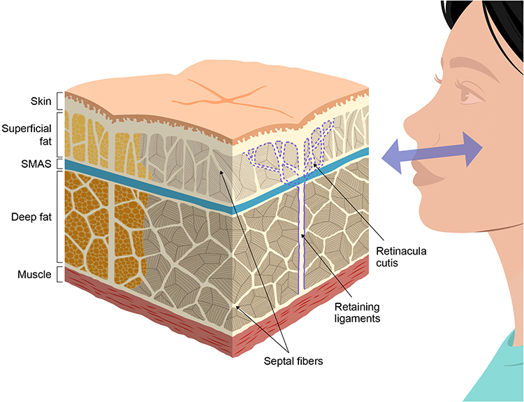 |
Figure 1 Schematic illustration of the three-dimensional facial fibrous supporting network. This figure is a schematic conceptual illustration based on established anatomical descriptions; it does not represent empirically validated tissue architecture. |
From a biomechanical perspective, this fibrous network—including the retinacula cutis—exhibits substantial tensile strength, enabling resistance to external mechanical forces and maintaining tissue position. Age-related tissue sagging should therefore not be attributed solely to volumetric loss within specific layers. Rather, it reflects a progressive reduction in the density and tensile integrity of the fibrous supporting network, leading to increased interlayer shearing and gradual failure of structural support (Figure 2).
 |
Figure 2 Schematic illustration of age-related changes in retaining ligaments and fibrous supporting tissues. This figure is a schematic conceptual illustration; the depicted changes are based on anatomical and histological observations but the mechanistic interpretations remain hypothetical. ↑ indicates structural improvement or direction of force; ↓ indicates sagging or structural weakening. |
Accordingly, in establishing therapeutic strategies, it is insufficient to approach individual layers or localized regions in isolation. Instead, the fibrous supporting network that spans the entire face should be recognized as a single integrated biomechanical unit. This structural continuum provides a theoretical basis for understanding HA fillers not merely as volumizing agents, but as mechanical mediators capable of modulating structural support by increasing local tissue density and redistributing mechanical forces within the fibrous network. On this basis, the therapeutic targets and strategies proposed by existing filler-based approaches can be reinterpreted within a broader biomechanical framework.
Myomodulation: A Functional Muscle Based Paradigm
Myomodulation represents a theoretical expansion of HA fillers from simple soft-tissue volumizing agents to tools capable of modulating the functional balance of facial mimetic muscles. This approach was first introduced in 2018 by the Brazilian doctor Maurício de Maio and was further refined through subsequent publications, including an updated theoretical framework published in 2020.9,19 By incorporating fillers into a domain that has been largely managed with neuromodulators, myomodulation proposes a paradigm shift in facial treatment strategies.
Conventionally, fillers have been used primarily to correct static wrinkles, depressions, and localized volume deficits, whereas neuromodulators have been employed to attenuate excessive muscle contraction in order to improve dynamic wrinkles and facial asymmetry.20–23 Clinically, however, it has been observed that in the presence of structural support deficiency, filler injection alone may alter muscle movement patterns. Myomodulation was proposed as a mechanistic explanation for these observations.
The core mechanism of myomodulation lies in the premise that filler placement can influence the muscle length–tension relationship and functional vectors of contraction by altering the biomechanical support environment. By positioning fillers adjacent to a muscle’s origin or insertion, or within the superior or inferior planes relative to the muscle, fillers may indirectly modify the mechanical leverage and excursion pathways during muscle contraction. Importantly, this effect should not be interpreted as a direct action on the muscle fibers themselves. Rather, myomodulation represents a form of mechanical intervention in which functional outcomes are altered through reconfiguration of the anatomical support environment in which muscles operate.
This concept is schematically illustrated in Figure 3. In youthful facial anatomy, the underlying support structures formed by skin, fat, and skeletal elements provide appropriate curvature and mechanical fulcrums for muscle fibers, allowing a balanced interaction between levator and depressor muscles (Figure 3A). With aging, soft tissue descent and loss of structural support diminish mechanical advantage at anchoring points, resulting in relative dominance of depressor activity and acceleration of facial sagging (Figure 3B). From a myomodulation perspective, filler injection is hypothesized to compensate for these structural changes by restoring mechanical advantage for levator muscles and improving contraction efficiency, thereby rebalancing opposing muscle groups (Figure 3C).
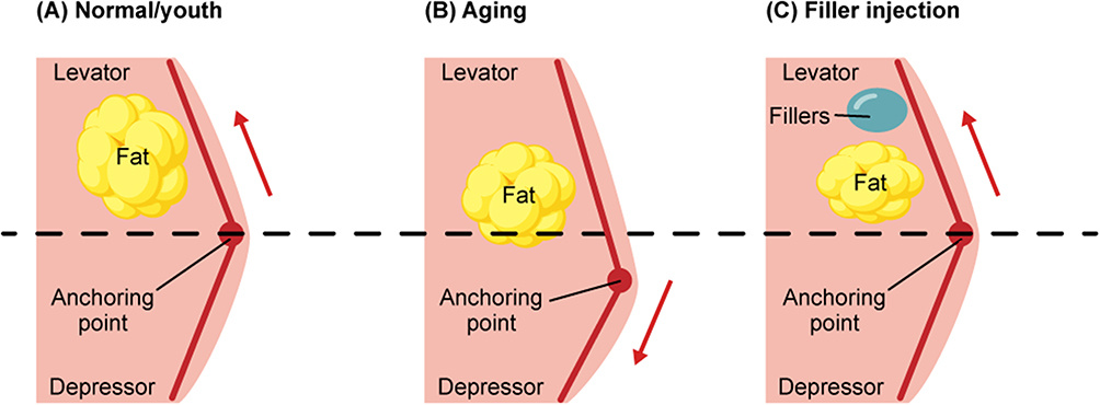 |
Figure 3 Schematic illustration of the myomodulation framework and its hypothesized biomechanical effects on facial muscle balance. The diagram compares youthful facial anatomy (A), age-related structural changes (B), and the proposed biomechanical effects of filler-based myomodulation (C). In youthful anatomy, adequate underlying structural support provides appropriate curvature and mechanical fulcrums, allowing balanced interaction between levator and depressor muscles. With aging, loss of structural support and soft tissue descent reduce mechanical advantage at anchoring points, leading to relative dominance of depressor activity and facial sagging. Filler injection is hypothesized to restore structural support at these anchoring points, re-establish mechanical advantage, and improve functional balance between opposing muscle groups without direct pharmacologic muscle inhibition. ↑ indicates improved mechanical advantage or levator activity; ↓ indicates reduced mechanical advantage or depressor dominance. This figure is a conceptual illustration and should not be interpreted as implying proven biomechanical causation. Notes: Adapted from de Maio M. Myomodulation with injectable fillers: an innovative approach to addressing facial muscle movement. Aesthetic Plastic Surgery. 2018;42(3):798–814. Distributed under the Creative Commons Attribution 4.0 International License (CC BY 4.0). |
In the existing literature, discussions of muscle modulation have largely focused on the upper face, particularly the balance between levator and depressor muscles in the forehead.24 The myomodulation framework extends this concept to the entire face. Facial expression is not the result of isolated muscle action, but rather emerges from coordinated interactions among synergistic and antagonistic muscle groups. Structural support deficiencies disrupt this balance and contribute to aged expression vectors. By indirectly modulating such structural–functional imbalances through filler placement, myomodulation suggests that improvements in facial expression and contour may be achieved without excessive volumetric augmentation.
Clinical application of this framework has been reported in subsequent large-scale retrospective analyses. In one retrospective case series including 1,352 patients treated with HA fillers between 2016 and 2021, strategic filler placement adjacent to mimetic muscles was associated with clinical changes consistent with altered muscle function.25 Specifically, in the lateral eyebrow region, filler injection near structures associated with brow depressor muscles was associated with changes in eyebrow position and expression. In the nasolabial fold and midface regions, contour improvement was reported in association with altered functional balance between upper and lower muscle groups. In the oral commissure and lower face, changes in expression vectors were observed following filler placement near depressor-associated structures. These findings support the notion that fillers may influence functional outcomes by modifying the anatomical support environment rather than exerting direct neurologic effects on muscle tissue.
However, because myomodulation primarily interprets facial aging through the lens of muscular functional imbalance and changes in action vectors, it has inherent limitations in explaining the structural support environment within which muscles operate. Although this approach suggests that filler injection may be associated with observable changes in muscle function, its interpretive framework is limited in distinguishing whether such changes arise from intrinsic alterations in muscle properties or are indirectly mediated by weakening and redistribution of the fibrous structures that support and connect muscles.
Furthermore, by defining the therapeutic unit mainly as individual muscles or interacting muscle groups, myomodulation does not explicitly address the structural changes and tension distribution of the continuous fibrous supporting network extending from the skin through the SMAS to the deep fascia as an integrated unit. As a result, while myomodulation provides a useful interpretive framework for interpreting changes in expression vectors and dynamic contour, it offers a limited explanation of how structural sagging and hollowing are linked to the progressive, global weakening of the fibrous supporting network.
Dual-Plane Approaches: A Depth-Oriented Structural Strategy
The dual-plane approach is a depth-oriented strategic framework for distributing HA fillers across multiple anatomical depths, each characterized by distinct biomechanical roles. This approach was systematically described in 2015 by the Italian surgeon Giovanni Salti and is based on the recognition that facial aging does not arise from isolated alterations within a single layer, but rather from coordinated changes involving weakening of deep supporting structures and positional displacement of superficial soft tissues.10 Conventional single-plane injection techniques may be effective for correcting localized volume deficits or improving superficial contours; however, they are limited in their ability to simultaneously address deep structural support loss and superficial contour deformation. The dual-plane approach was developed in response to these clinical limitations and proposes a layer-based framework in which the role and objective of filler placement are functionally differentiated according to injection depth.
Within this framework, the therapeutic unit is defined as anatomically distinct tissue planes determined by depth, with filler placement guided by the functional role of each layer—structural support and deep volumization in the supraperiosteal or deep fat plane, and contour refinement and surface modulation in the subdermal or superficial fat plane. Facial soft tissue is not interpreted as a single continuous volume; instead, the deep plane is regarded as a load-bearing layer responsible for positional stabilization, whereas the superficial plane functions primarily as a shape-modulating layer involved in contour formation and surface harmonization. This depth-oriented interpretation provides a mechanistic basis for separating the supportive and contour-modifying effects transmitted by fillers. As illustrated schematically in Figure 4, the dual-plane strategy conceptualizes filler placement as a coordinated interaction between deep structural support and superficial contour refinement.
 |
Figure 4 Schematic illustration of the dual-plane filler strategy based on injection depth. This figure is a schematic conceptual illustration; the depicted depth-based functional differentiation represents a theoretical framework and has not been experimentally validated. ↑indicates superficial plane effects; → indicates superficial contour refinement. |
The core biomechanical principles underlying the dual-plane strategy include functional compartmentalization by depth and rheological differentiation of filler materials. In the deep plane, which typically corresponds to the supraperiosteal level or the sub-SMAS space, high-elasticity (high G′; firm) fillers resist deformation, provide relatively greater structural support to the overlying tissues, and counteract gravitational descent. In contrast, low-elasticity, highly cohesive fillers applied to the superficial plane, such as the superficial fat layer or subdermal plane, transmit the lifting effect generated in the deep layer toward the surface while absorbing shear stress associated with facial expression, thereby achieving natural contour refinement.26 These depth-specific functional roles assume that the deep and superficial layers do not operate independently, but rather as interrelated components of a unified structural system. Structural support established in the deep plane provides positional stability for overlying soft tissues, upon which superficial filler placement fine-tunes contour and surface morphology. By functionally separating deep structural support from superficial contour modulation, this strategy aims to minimize excessive surface fullness while achieving more natural clinical outcomes.
In 2024, Korean plastic surgeon Gi-Woong Hong introduced the Skin and SMAS layer remodeling technique (SSRT), which can be understood as a conceptual development of the dual-plane concept by integrating not only anatomical layer-based injection strategies but also structural support elements, including retaining ligaments.27 In SSRT, fillers intended for structural support are placed in the sub-SMAS or deep subcutaneous layers, whereas fillers targeting surface uniformity and wrinkle attenuation are applied to more superficial planes. In this regard, SSRT is not confined to a purely layer-based approach, but may be interpreted as a transitional framework that bridges layer-oriented strategies with ligament-targeted concepts.
Alongside this development, alternative approaches have been proposed that focus on specific structural support elements, such as retaining ligaments, rather than anatomical layers. Techniques such as True Lift target specific retaining ligaments, aiming to induce lifting effects by increasing the density beneath these structures, thereby enhancing structural support.
Retaining Ligament–Targeted Anchoring Approaches: Focal Structural Support
Among the structural changes associated with facial aging, elongation and loss of tensile support of the retaining ligaments have long been recognized as key pathological factors.11,14,28,29 Retaining ligaments are critical fibrous supporting elements that anchor the skin, subcutaneous fat, and the superficial musculoaponeurotic system (SMAS) to the deep facial skeleton or deep fascial structures. A reduction in their tensile integrity directly contributes to inferior displacement of soft tissues and subsequent contour breakdown. Based on this anatomical understanding, certain filler injection strategies have proposed defining retaining ligaments as focal therapeutic units for localized structural stabilization, with the aim of indirectly improving soft tissue sagging.30
In these approaches, fillers are applied to reinforce the structural support surrounding ligament bases, thereby promoting relative positional stabilization of the SMAS–soft tissue complex. By increasing the density and mechanical stability of tissues adjacent to ligament attachment sites, fillers function as localized anchoring mediators that enhance the supportive role of these regions within the facial support system.
This approach was presented in 2022 by Huang et al in a report describing the True Lift® technique, in which the structural role of retaining ligaments was applied as a therapeutic target based on existing anatomical understanding.11 The True Lift® technique defines the bases of major true retaining ligaments, including the orbital retaining ligament, zygomatic retaining ligament, and mandibular retaining ligament, as focal treatment targets and proposes the distributed injection of limited volumes of high-elasticity (high G′) HA fillers. The central premise of this strategy is that creating localized structural support adjacent to ligament attachment sites can restrain inferior displacement of the associated SMAS and soft tissues, thereby restoring a relatively stable tissue configuration (Figure 5).
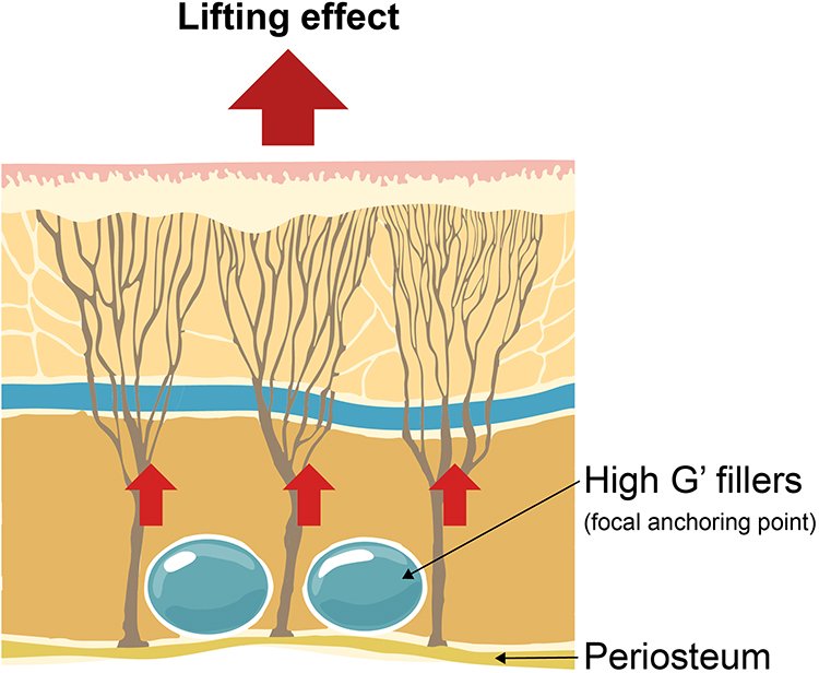 |
Figure 5 Schematic illustration of a focal structural anchoring strategy targeting the retaining ligaments for facial lifting. This figure is a schematic conceptual representation and does not constitute an exact procedural map. ↑ indicates direction of proposed lifting effect. |
In the reported case series, improvements in contour and changes in soft tissue position in the midface and lower face regions were observed following the True Lift procedure using three-dimensional measurements, suggesting that visible structural changes may be achieved with relatively small filler volumes. However, these findings are based on observational data derived from a limited number of cases and single-operator experience. Quantitative analyses evaluating how periligamentous filler injection influences ligament tension distribution, SMAS displacement, or the biomechanical properties of the fibrous supporting system as a whole remain insufficient.
These limitations indicate that approaches defining retaining ligaments as discrete and focal anchoring structures are constrained in their ability to explain integrated structural changes and tension redistribution across the continuous fibrous supporting system of the face. This recognition provides the conceptual background for subsequent approaches that reinterpret facial support not as isolated anatomical structures, but as a continuous fibrous network functioning as a unified therapeutic unit.
Conceptual Extension: Fibrous Augmentation Lifting Technique
Fibrous augmentation lifting technique (FALT) is proposed as an interpretive framework that builds upon anatomical insights derived from retaining ligament-based injection approaches, while extending beyond focal support restoration limited to individual ligaments to emphasize the anatomical continuity and network characteristics of the facial fibrous architecture, including the retinacula cutis fibers. Within this framework, facial aging is interpreted not simply as a consequence of soft tissue volume loss, but rather as the result of progressive weakening and elongation of the fibrous supporting network, leading to loss of structural support and imbalance in tensile force distribution. The anatomical space in which these changes are most prominently manifested, namely the micro-spaces between fibrous bundles, is defined as the treatment unit and referred to as the inter-fibrous space.15,31
From this perspective, HA fillers are interpreted not merely as soft tissue volumizers, but as materials capable of reinforcing mechanical continuity and local stability within the fibrous supporting framework. FALT hypothesizes that when fillers are distributed within inter-fibrous spaces, localized stiffness may be enhanced and network-level tension distribution may be modulated without inducing excessive morphological alteration. Accordingly, high-elasticity HA fillers capable of maintaining structural stability and resisting external mechanical forces are considered theoretically suitable for this approach.
FALT differs from existing HA filler-based lifting strategies primarily in its interpretive focus, as summarized in Table 1. While myomodulation explains changes in facial appearance through modulation of muscular functional balance and resulting expression vectors, and dual-plane approaches interpret contour improvement through harmonized volume distribution between deep and superficial layers, FALT emphasizes micro-distributed placement of highly cohesive HA filler within inter-fibrous spaces. Rather than targeting localized volumization, this approach proposes that lifting effects may be interpreted through hypothesized alterations in overall network stiffness and redistribution of tensile forces across the fibrous supporting system.
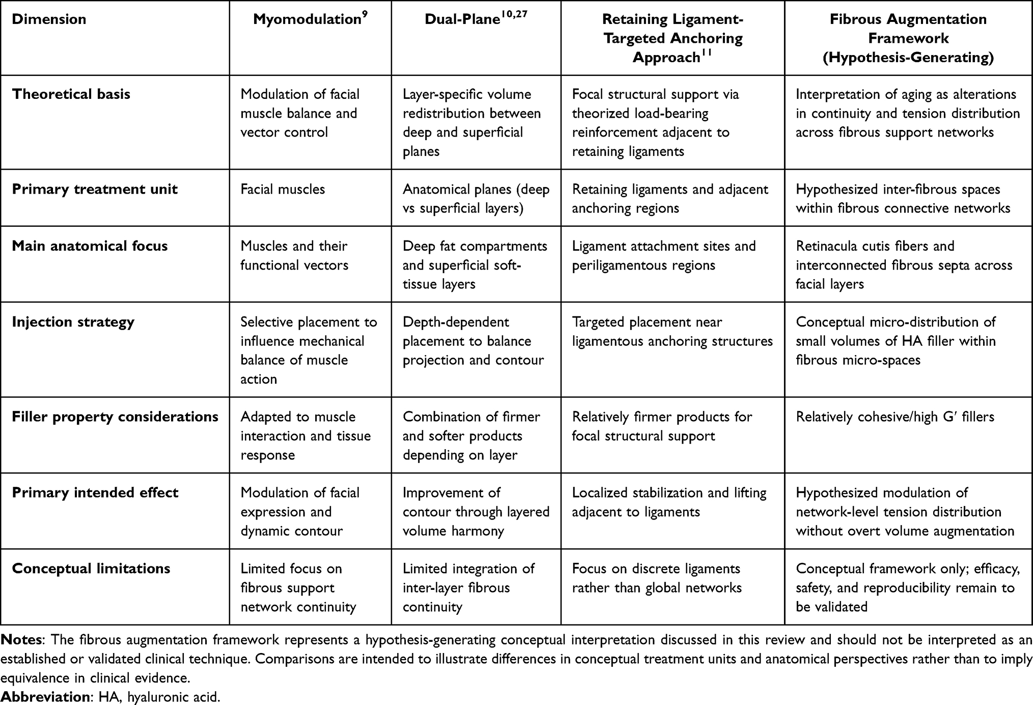 |
Table 1 Conceptual Comparison of HA Filler-Based Lifting Frameworks |
Within this framework, age-related hollowing is interpreted not solely as quantitative soft tissue atrophy, but as the combined result of fibrous elongation and localized tethering forces exerted by retaining ligaments and related fibrous structures. When HA filler is micro-distributed between weakened fibrous bundles, partial restoration of the structural support environment may occur through modulation of tensile force distribution across the network, without inducing excessive volume augmentation. This interpretation offers a possible explanatory basis for certain clinical observations, such as localized lifting effects adjacent to ligamentous structures, which are difficult to fully account for using volume-centric strategies alone.
In applying this framework, careful pre-procedural assessment of regions exhibiting age-related hollowing and the anatomical course of retaining ligaments involved in these changes is important. Such evaluation allows for the definition of injection zones centered on areas of suspected structural support weakening. From a technical standpoint, differences in tissue interaction and maneuverability between needles and cannulas may be considered, with device selection discussed in relation to target depth and tissue resistance.
FALT may be described using the principles of a micro-multiple injection technique, which emphasizes precise depth recognition and repeated micro-deposition rather than bulk volumization. After confirming anatomical reference points such as the periosteum using a cannula, fillers may be distributed in small, repeated aliquots, typically less than 0.05 mL per injection point, targeting inter-fibrous spaces defined as regions between bundles of fibrous septa, while advancing sequentially from deeper to more superficial planes. This approach differs fundamentally from traditional bolus injection or simple linear threading by prioritizing multiple discrete reinforcement points distributed throughout the fibrous supporting network, rather than continuous volumetric filling along a single plane. In the authors’ clinical experience, this micro-distributed strategy may potentially allow a reduction in total filler volume compared with conventional volumetric techniques; however, further systematic investigation is required to validate this observation.
As illustrated schematically in Figure 6, the proposed distinction between conventional volumetric filler augmentation and FALT may be understood at the level of network mechanics rather than surface appearance alone. Figure 6A depicts the baseline-aged state, in which the facial fibrous supporting network shows reduced tensile integrity, elongation of fibrous septa, and directional deformation associated with structural weakening. Figure 6B represents the conventional volumetric filler approach, where filler is placed as a localized space-occupying material to improve contour in a focal manner; although this may correct surface hollowing or localized depression, it does not substantially alter the overall configuration or mechanical state of the fibrous supporting network. In contrast, Figure 6C illustrates the conceptual basis of FALT, in which cohesive HA filler is micro-distributed within inter-fibrous spaces between weakened fibrous bundles. In this framework, the filler is hypothesized not merely to restore lost volume, but to reinforce local stiffness, improve load-bearing continuity, and promote redistribution of tensile forces across the fibrous supporting system without bulk volumetric expansion.
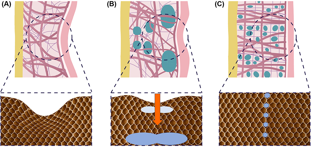 |
Figure 6 Conceptual comparison of volumetric filler augmentation and fibrous augmentation lifting technique (FALT). All panels are schematic conceptual illustrations and do not represent histologically validated tissue architecture. (A) Baseline state showing reduced tensile integrity and directional deformation of the facial fibrous supporting network, with elongation and weakening of fibrous septa. (B) Conventional volumetric filler approach, in which localized space-occupying augmentation improves surface contour; however, as indicated by ↓, strong vertical resistance forces may displace the filler, potentially resulting in insufficient correction of the central area and peripheral bulging of the surrounding tissue, without substantially modifying the global configuration or mechanical properties of the fibrous supporting network. (C) Fibrous augmentation lifting technique (FALT), illustrating micro-distributed placement of cohesive HA filler within inter-fibrous spaces. This approach is hypothesized to reinforce local network stiffness and redistribute tensile forces across the fibrous supporting system, promoting structural reorganization and lifting effects without bulk volumetric expansion. |
From a biomechanical perspective, the hypothesized mechanism of FALT is based on micro-distributed reinforcement of a pre-existing fibrous framework. By placing cohesive filler in small aliquots within inter-fibrous spaces, the local stiffness and load-bearing capacity of the network may be enhanced without substantial overall volume expansion. As a conceptual analogy, this mechanism may be compared to the principle by which functional rigidity arises from microstructural filling of enclosed spaces and subsequent tensioning of a surrounding fibrous envelope, rather than from gross tissue enlargement alone.
Accordingly, FALT may be understood as a strategy that emphasizes modulation of structural tension at the level of points and lines within the fibrous network, rather than surface contour correction driven primarily by volumetric replacement. While this framework offers a biomechanical interpretation that may account for certain clinical lifting effects, its implications remain largely theoretical.
At present, FALT should be understood not as an established clinical technique supported by quantitative outcome data, but as an anatomical and interpretive framework. Prospective clinical studies evaluating efficacy, safety, and long-term durability remain limited, and objective evidence demonstrating biomechanical effects of micro-distributed filler within inter-fibrous spaces through imaging, histologic, or mechanical analyses has yet to be sufficiently accumulated. Accordingly, future prospective clinical studies and experimental or biomechanical investigations will be required to determine whether modulation of fibrous network tension can yield reproducible and clinically meaningful outcomes.
Discussion
HA filler-based treatment strategies have continuously evolved over recent decades alongside advances in anatomical knowledge and the accumulation of clinical experience. Early applications primarily focused on correcting localized volume deficits. However, increasing recognition that facial aging cannot be sufficiently explained by volumetric loss alone has led to the development of diverse theoretical and strategic approaches addressing structural contributors. Within this context, the approaches reviewed in this article, namely myomodulation, dual-plane strategies, and retaining ligament-based injection techniques, can be regarded as representative interpretive frameworks that interpret facial aging and guide treatment strategies according to functional units, anatomical depth, and structural anchoring points, respectively.
Myomodulation broadened the role of fillers beyond soft tissue augmentation by interpreting them as biomechanical mediators capable of influencing muscular functional balance. Rather than focusing solely on volumetric correction, this approach introduced a functional perspective in which filler placement relative to facial mimetic muscles may modify contraction efficiency and functional vectors through alterations in the surrounding support environment. In this way, myomodulation expanded conventional interpretations of filler-based treatment that were primarily centered on static volumization. However, because this model primarily defines muscle function as the central therapeutic unit, comparatively less emphasis is placed on the continuous fibrous supporting structures that stabilize and interconnect the muscular system. As a result, while myomodulation provides a useful interpretive framework for interpreting changes in expression dynamics and functional contour, it offers a more limited explanation of how progressive weakening of the fibrous supporting network contributes to global structural sagging and hollowing with aging.
Dual-plane approaches proposed a depth-oriented strategy in which structural support restoration and surface contour refinement are achieved in a stepwise manner through functional differentiation by injection depth. By recognizing that deep and superficial layers operate within distinct mechanical environments and serve different clinical objectives, this approach offers a practical treatment paradigm for procedural planning. Nevertheless, because anatomical layer distinction constitutes its primary organizing principle, dual-plane strategies have limited capacity to integrate the continuity and progressive weakening of fibrous supporting structures that span across layers as a single therapeutic unit.
Retaining ligament-based injection strategies interpret facial aging in relation to laxity and reduced supportive capacity of retaining ligaments and aim to stabilize adjacent soft tissues through focal reinforcement at specific anchoring sites. This approach has clinical relevance in emphasizing structural fixation at ligament attachment regions. However, because targeting is centered on individual ligaments, comparatively less attention is directed toward the broader fibrous supporting network, including fibrous septa and retinacula cutis fibers that interconnect multiple retaining structures.
Anatomical observations indicate that facial form and stability are maintained through an integrated fibrous supporting system that extends continuously from the skin to the superficial musculoaponeurotic system, deep fascia, and periosteum. Accordingly, age-related sagging and hollowing should be understood not as isolated changes affecting individual structures, but as manifestations of cumulative alterations in tensile force distribution and structural continuity across the fibrous supporting network. From this perspective, the existing approaches reviewed here need not be viewed as mutually exclusive. Rather, they highlight the need for an additional interpretive perspective capable of interpreting network-level structural changes beyond the functional units, layers, or anchoring points emphasized by prior strategies.
FALT may be understood as a theoretical extension arising from the present discussion. While preserving the structural insights introduced by retaining ligament-based approaches, FALT proposes a shift from ligament-specific targeting toward an integrated view of the facial fibrous supporting network, including retinacula cutis fibers, as a continuous anatomical system.
The micro-multiple injection strategy associated with FALT describes the placement of highly cohesive HA fillers in very small aliquots within inter-fibrous spaces, with the objective of locally reinforcing the fibrous support environment rather than producing discrete volumetric augmentation. Conceptually, this approach emphasizes proposed enhancement of internal structural rigidity through micro-distributed reinforcement of a pre-existing fibrous framework, whereby hypothesized increases in stiffness and load-bearing capacity may be achieved without substantial changes in overall tissue volume. This strategy may also offer potential advantages in minimizing complications associated with excessive volumization, such as edema, overfilled appearance, and unnatural contour formation. By limiting filler diffusion into surrounding tissues and enabling more precise, localized structural support, this approach can be distinguished from conventional volume-centric techniques. Furthermore, such micro-distributed placement may be associated with a reduction in the total volume of filler required, although this observation remains primarily based on clinical experience rather than controlled comparative studies.
With respect to vascular safety, prior anatomical studies have reported that the intravascular volume required for retrograde embolization from the supraorbital or supratrochlear arteries to the central retinal artery is approximately 0.083 mL. Based on this anatomical consideration, the authors hypothesize that injection strategies employing less than 0.05 mL per injection point, as typically applied in FALT, may theoretically limit the likelihood of delivering a sufficient intravascular bolus to reach the retinal circulation.32 This interpretation, however, represents a mechanistic hypothesis rather than an evidence-based conclusion, and no direct clinical data currently demonstrate a reduced incidence of vascular complications with FALT. In addition, the suggestion that FALT may reduce overall filler consumption by approximately 20% reflects the author’s experiential observation in clinical practice rather than results derived from controlled comparative studies. This proposed reduction is discussed here as a potential consequence of point-based, micro-distributed structural reinforcement that avoids excessive volumetric deposition, but it has not yet been systematically evaluated or formally reported in the literature.
Taken together, FALT should be regarded nowadays as an anatomical and biomechanical interpretive framework rather than an established clinical technique. Assertions regarding its potential advantages—including modulation of structural stiffness, redistribution of tensile forces, vascular safety, and filler efficiency—remain speculative and require validation through prospective clinical studies and quantitative analyses. Some observed lifting effects may not be solely attributable to a distinct fibrous network mechanism; rather, conventional volumetric support, compartmental projection changes, skin tension redistribution, and viscoelastic tissue interactions may collectively contribute to the clinical outcomes observed following HA filler injection.
At present, these mechanisms and effects remain theoretical in nature. Future studies aimed at validating the FALT framework may employ a range of methodological approaches, including high-resolution ultrasound, MRI, or elastography for in vivo assessment of fibrous network architecture and filler distribution;33,34 finite element biomechanical modeling to simulate hypothesized tensile force redistribution within the fibrous network; cadaveric mechanical testing to evaluate structural changes at periligamentous injection sites; and prospective comparative clinical studies with standardized outcome measures, including three-dimensional surface imaging and patient-reported outcomes.
Conclusion
Concepts guiding HA filler-based approaches to understanding and treating facial aging have been structured around functional units, injection depth, and structural anchoring points, with each strategy proposing distinct therapeutic units and mechanistic interpretations. This review comparatively examined these existing approaches from anatomical and biomechanical perspectives and discussed a complementary conceptual framework that interprets facial aging through the continuity of the fibrous supporting network and alterations in tensile force distribution. A key conceptual contribution of this perspective is the proposal to reconceptualize the continuous facial fibrous supporting network as a unified biomechanical therapeutic unit, offering a complementary interpretive basis for reexamining existing filler-based lifting strategies. FALT, as introduced herein, represents a hypothesis-generating framework rather than an established clinical technique, and its validation will require future prospective clinical and biomechanical investigations. Such efforts may ultimately allow facial lifting strategies to be reconsidered within a more structurally integrated framework.
Abbreviations
HA, Hyaluronic Acid; SMAS, Superficial Musculoaponeurotic System; ISAPS, International Society of Aesthetic Plastic Surgery; SSRT, Skin and SMAS Layer Remodeling Technique; FALT, Fibrous Augmentation Lifting Technique.
Acknowledgments
The authors would like to thank those who provided administrative assistance during the preparation of this manuscript. The authors also express their gratitude to Editage for technical support in the preparation and refinement of the figures used in this review.
Author Contributions
All authors made a significant contribution to the work reported, whether that is in the conception, study design, execution, acquisition of data, analysis and interpretation, or in all these areas; took part in drafting, revising or critically reviewing the article; gave final approval of the version to be published; have agreed on the journal to which the article has been submitted; and agreed to be accountable for all aspects of the work.
Funding
This research received no external funding.
Disclosure
The authors report no conflicts of interest in this work.
References
1. Narins RS, Brandt F, Leyden J, Lorenc ZP, Rubin M, Smith S. A randomized, double-blind, multicenter comparison of the efficacy and tolerability of Restylane versus Zyplast for the correction of nasolabial folds. Dermatol Surg. 2003;29(6):588–14. doi:10.1046/j.1524-4725.2003.29150.x
2. Monheit GD, Coleman KM. Hyaluronic acid fillers. Dermatol Ther. 2006;19(3):141–150. doi:10.1111/j.1529-8019.2006.00065.x
3. Brandt FS, Cazzaniga A, Brandt F. Hyaluronic acid gel fillers in the management of facial aging. Clin Interv Aging. 2008;3(1):153–159. doi:10.2147/CIA.S2135
4. International Society of Aesthetic Plastic Surgery. ISAPS international survey on aesthetic/cosmetic procedures performed in 2023. Available from: https://www.isaps.org/media/oogpzodr/isaps-global-survey_2024.pdf.
5. Samizadeh S. Anatomy and pathophysiology of facial ageing. In: Samizadeh S, editor. Thread Lifting Techniques for Facial Rejuvenation and Recontouring. Cham: Springer International Publishing; 2024:61–89. doi:10.1007/978-3-031-47954-0_3
6. Bar O, Valiukevičienė S. Skin aging and type I collagen: a systematic review of interventions with potential collagen-related effects. Cosmetics. 2025;12(4):129. doi:10.3390/cosmetics12040129
7. Swift A, Liew S, Weinkle S, Garcia JK, Silberberg MB. The facial aging process from the “inside out”. Aesthet Surg J. 2021;41(10):1107–1119. doi:10.1093/asj/sjaa339
8. Sakata A, Abe K, Mizukoshi K, Gomi T, Okuda I. Relationship between the retinacula cutis and sagging facial skin. Skin Res Technol. 2018;24(1):93–98. doi:10.1111/srt.12395
9. de Maio M. Myomodulation with injectable fillers: an innovative approach to addressing facial muscle movement. Aesthetic Plast Surg. 2018;42(3):798–814. doi:10.1007/s00266-018-1116-z
10. Salti G, Rauso R. Facial rejuvenation with fillers: the dual plane technique. J Cutan Aesthet Surg. 2015;8(3):127–133. doi:10.4103/0974-2077.167264
11. Huang P, Li CW, Yan YQ. Efficacy evaluation of True Lift®, a nonsurgical facial ligament retightening injection technique: two case reports. World J Clin Cases. 2022;10(27):9783–9792. doi:10.12998/wjcc.v10.i27.9783
12. Carruthers JDA, Carruthers A. Facial sculpting and tissue augmentation. Dermatol Surg. 2005;31(Suppl 4):1604–1612. doi:10.2310/6350.2005.31248
13. Bryan M, Wong CH. Anatomy of the aging face. In: Neligan PC, editor. Plastic Surgery.
14. Furnas DW. The retaining ligaments of the cheek. Plast Reconstr Surg. 1989;83(1):11–16. doi:10.1097/00006534-198901000-00003
15. Rohrich RJ, Pessa JE. The retaining system of the face: histologic evaluation of the septal boundaries of the subcutaneous fat compartments. Plast Reconstr Surg. 2008;121(5):1804–1809. doi:10.1097/PRS.0b013e31816c3c1a
16. Wong CH, Mendelson B. Facial soft-tissue spaces and retaining ligaments of the midcheek: defining the premaxillary space. Plast Reconstr Surg. 2013;132(1):49–56. doi:10.1097/PRS.0b013e3182910a57
17. Mendelson B, Wong CH. Changes in the facial skeleton with aging: implications and clinical applications in facial rejuvenation. Aesthetic Plast Surg. 2012;36(4):753–760. doi:10.1007/s00266-012-9904-3
18. Minelli L, Brown CP, van der Lei B, Mendelson B. Anatomy of the facial glideplanes, deep plane spaces, and ligaments: implications for surgical and nonsurgical lifting procedures. Plast Reconstr Surg. 2024;154(1):95–110. doi:10.1097/PRS.0000000000011078
19. de Maio M. Myomodulation with injectable fillers: an update. Aesthetic Plast Surg. 2020;44(4):1317–1319. doi:10.1007/s00266-020-01768-1
20. Carruthers J, Carruthers A, Tezel A, Kraemer J, Craik L. Volumizing with a 20-mg/mL smooth, highly cohesive, viscous hyaluronic acid filler and its role in facial rejuvenation therapy. Dermatol Surg. 2010;36(Suppl 3):1886–1892. doi:10.1111/j.1524-4725.2010.01778.x
21. Callan P, Goodman GJ, Carlisle I, et al. Efficacy and safety of a hyaluronic acid filler in subjects treated for correction of midface volume deficiency: a 24 month study. Clin Cosmet Invest Dermatol. 2013;6:81–89. doi:10.2147/CCID.S40581
22. Ascher B, Talarico S, Cassuto D, et al. International consensus recommendations on the aesthetic usage of botulinum toxin type A (Speywood Unit)--Part I: upper facial wrinkles. J Eur Acad Dermatol Venereol. 2010;24(11):1278–1284. doi:10.1111/j.1468-3083.2010.03631.x
23. Goldberg RA, Fiaschetti D. Filling the periorbital hollows with hyaluronic acid gel: initial experience with 244 injections. Ophthal Plast Reconstr Surg. 2006;22(5):335–341. doi:10.1097/01.iop.0000235820.00633.61
24. Olson JJ. Balanced botox chemodenervation of the upper face: symmetry in motion. Semin Plast Surg. 2007;21(1):47–53. doi:10.1055/s-2007-967748
25. Coimbra DD, Stefanello B. Myomodulation with facial fillers: a comprehensive technical guide and retrospective case series. Aesthetic Plast Surg. 2023;47(3):1162–1174. doi:10.1007/s00266-022-03193-y
26. Pierre S, Liew S, Bernardin A. Basics of dermal filler rheology. Dermatol Surg. 2015;41(Suppl 1):S120–S126. doi:10.1097/DSS.0000000000000334
27. Hong GW, Yi KH. Skin and SMAS layer remodeling technique (SSRT): achieving both volume and lifting effects with filler treatments. Skin Res Technol. 2024;30(6):e13809. doi:10.1111/srt.13809
28. Kang MS, Kang HG, Nam YS, Kim IB. Detailed anatomy of the retaining ligaments of the mandible for facial rejuvenation. J Craniomaxillofac Surg. 2016;44(9):1126–1130. doi:10.1016/j.jcms.2016.06.018
29. Alghoul MS, Codner MA. Retaining ligaments of the face: review of anatomy and clinical applications. Aesthet Surg J. 2013;33(6):769–782. doi:10.1177/1090820X13495405
30. de Maio M, Wu WTL, Goodman GJ, Monheit G. Alliance for the future of aesthetics consensus committee. Facial assessment and injection guide for botulinum toxin and injectable hyaluronic acid fillers: focus on the lower face. Plast Reconstr Surg. 2017;140(3):393e–404e. doi:10.1097/PRS.0000000000003646
31. Herlin C, Chica-Rosa A, Subsol G, et al. Three-dimensional study of the skin/subcutaneous complex using in vivo whole body 3T MRI: review of the literature and confirmation of a generic pattern of organization. Surg Radiol Anat. 2015;37(7):731–741. doi:10.1007/s00276-014-1409-0
32. Zhang L, Pan L, Xu H, et al. Clinical observations and the anatomical basis of blindness after facial hyaluronic acid injection. Aesthetic Plast Surg. 2019;43(4):1054–1060. doi:10.1007/s00266-019-01374-w
33. Drelichman VS, Eandi JA, Deligia R, et al. Hyaluronic acid filler in HIV-associated facial lipoatrophy: evaluation of tissue distribution and morphology with MRI. Dermatol Surg. 2014;40(4):420–428. doi:10.1111/dsu.12451
34. Ronzatti M, Toniolo C, Ribero S, et al. Ultra-high-frequency ultrasound as an innovative imaging evaluation of hyaluronic acid filler in nasolabial folds. Skin Res Technol. 2023;29(11):e13495. doi:10.1111/srt.13495
 © 2026 The Author(s). This work is published and licensed by Dove Medical Press Limited. The
full terms of this license are available at https://www.dovepress.com/terms
and incorporate the Creative Commons Attribution
- Non Commercial (unported, 4.0) License.
By accessing the work you hereby accept the Terms. Non-commercial uses of the work are permitted
without any further permission from Dove Medical Press Limited, provided the work is properly
attributed. For permission for commercial use of this work, please see paragraphs 4.2 and 5 of our Terms.
© 2026 The Author(s). This work is published and licensed by Dove Medical Press Limited. The
full terms of this license are available at https://www.dovepress.com/terms
and incorporate the Creative Commons Attribution
- Non Commercial (unported, 4.0) License.
By accessing the work you hereby accept the Terms. Non-commercial uses of the work are permitted
without any further permission from Dove Medical Press Limited, provided the work is properly
attributed. For permission for commercial use of this work, please see paragraphs 4.2 and 5 of our Terms.