
Back to Journals » Drug Design, Development and Therapy » Volume 20
Comprehensive Review on the Toxicity of Five Main AQ Constituents from Rhubarb: Mechanisms, Challenges and Future Perspectives
Authors Yu L, Jiang Y, Li P, Wang J, Tang P, Zhao Y 
Received 3 February 2026
Accepted for publication 25 April 2026
Published 15 May 2026 Volume 2026:20 600863
DOI https://doi.org/10.2147/DDDT.S600863
Checked for plagiarism Yes
Review by Single anonymous peer review
Peer reviewer comments 3
Editor who approved publication: Professor Anastasios Lymperopoulos
Linyuan Yu,1,2 Yongxian Jiang,2 Ping Li,1 Jun Wang,1 Peng Tang,1 Yongli Zhao1
1Department of Pharmacy, Chengdu Integrated TCM & Western Medicine Hospital, Chengdu, Sichuan, 610095, People’s Republic of China; 2Department of Pharmacy, Sichuan Provincial Maternity and Child Health Care Hospital, Chengdu, Sichuan, 610041, People’s Republic of China
Correspondence: Yongli Zhao, Chengdu Integrated TCM & Western Medicine Hospital, No.18, Wanxiang North Road, Gaoxin District, Chengdu, 610095, People’s Republic of China, Email [email protected]
Background: Emodin, rhein, aloe-emodin, physcion, and chrysophanol are five representative anthraquinones (AQs) in rhubarb. They exhibit diverse pharmacological activities, including antitumor, anti-inflammatory, antibacterial, and antioxidant effects, and are widely used in traditional Chinese medicines (TCMs), dietary supplements, and functional foods. However, with their increasing application, the potential toxicity of AQs has become increasingly prominent. Moreover, their toxic mechanisms and metabolism-related toxicities remain incompletely elucidated, which has limited their clinical development and safe application to a certain extent.
Objective: This paper systematically reviews the toxicological characteristics, toxic mechanisms, and metabolism-related toxicity of the five major AQs in rhubarb, and explores research strategies for AQs toxicity based on advanced technologies, aiming to provide new insights and references for their safe application and further studies.
Methods: A comprehensive search was conducted in PubMed, Google Scholar, Web of Science, and CNKI for peer-reviewed research articles and reviews published in the past 15 years. The research progress of the five major rhubarb AQs and the applications of cutting-edge technologies in their toxicity studies were summarized.
Results: The toxicities of AQs mainly target the liver, kidney, heart, reproductive system, and nervous system. Their toxic mechanisms involve multiple pathways, including mitochondrial apoptosis, oxidative stress, death receptor pathway, endoplasmic reticulum (ER)-related apoptosis, caspase-dependent apoptosis, autophagy, inflammation, DNA damage, and bilirubin metabolism. The in vivo metabolism of AQs is complicated, and both Phase I and Phase II metabolites are linked to toxicity. The diverse metabolites can interconvert and undergo various reactions in vivo, posing great challenges for toxicity research. In the future, the application of advanced technologies, such as multi-omics and single-cell sequencing, artificial intelligence (AI) and computer simulation, microfluidics, and mass spectrometry imaging (MSI) will provide strong support for accurately elucidating toxic mechanisms, enabling toxicity prediction, and optimizing detoxification strategies, and will become effective approaches to advance toxicity studies of rhubarb AQs.
Conclusion: The intrinsic toxicity and complex metabolic processes of rhubarb AQs restrict their clinical application. Current studies on the toxic mechanisms, metabolism–toxicity relationships, and multifactorial regulation of AQs still require further in-depth investigation. Future research should focus on the metabolism-toxicity relationship, key toxic targets, and detoxification strategies of AQs, and establish an integrated toxicity research system combined with advanced technologies, so as to provide robust support for the safe application and clinical development of rhubarb AQs.
Keywords: rhubarb AQs, toxicity, toxicity mechanism, metabolism, perspectives
Introduction
Rhubarb is the dried root and rhizome of plants of the genus Rheum in the family Polygonaceae. It has been widely used in clinical practice since its use for constipation, jaundice and other conditions was first documented in Shennong’s Classic of Materia Medica.1,2 The Pharmacopoeia of the People’s Republic of China (2020 Edition) specifies emodin, rhein, aloe‑emodin, physcion, and chrysophanol as the five indicator components for the quality control of rhubarb. As the core constituents of anthraquinones (AQs), these five components are abundant in rhubarb and possess the most potent biological activities.3,4 Modern pharmacological studies have demonstrated that these five AQs, as representative active components of rhubarb, exhibit a broad spectrum of biological activities, including improving gastrointestinal function, exerting antitumor effects, regulating gut microbiota, displaying antibacterial activity, protecting the liver, and producing anti‑inflammatory effects.5,10 In addition, our previous research revealed that these five AQs possess anti‑hepatocellular carcinoma activity.11
However, the clinical development and application of these five AQs are limited by their potential toxicities, including hepatotoxicity, nephrotoxicity, reproductive toxicity and cardiotoxicity.12 Furthermore, key issues such as their toxic doses, toxic mechanisms, and in vivo metabolic profiles remain unclear, posing major challenges to the toxicological research of rhubarb AQs. Therefore, this review comprehensively discusses toxicity‑related studies on AQs published before 2025 by searching PubMed, Web of Science, and CNKI databases. It aims to clarify their toxic mechanisms, explore toxicity‑attenuating strategies from the perspective of drug metabolism and other aspects, and propose future directions in the toxicological research of AQs combined with cutting‑edge technologies, so as to provide guidance and support for the clinical research and development of rhubarb AQs.
The Content of AQS
The 2020 edition of the Chinese Pharmacopoeia stipulates that the total content of five compounds (emodin, rhein, chrysophanol, aloe-emodin, and physcion) in qualified rhubarb should not be less than 1.5%. The molecular structures and relative content ratios of five AQs in rhubarb are presented in Table 1. Specifically, the content ranges for emodin, rhein, chrysophanol, aloe-emodin, and physcion are as follows: 0.018–1.15%, 0.009–1.072%, 0.007–1.465%, 0.102–2.864%, and 0.065–3.385%, respectively.
 |
Table 1 Basic Information of Five AQs |
Pharmacological Effects of AQS
The modern pharmacological effects of AQs have been widely studied, mainly including anti-tumor, anti-inflammatory, anti-bacterial, and antioxidant.6,16,18 The pharmacological effects and mechanisms involved are shown in Table 2 and Figure 1.
 |
Table 2 Pharmacological Effects and Mechanisms of Five AQs |
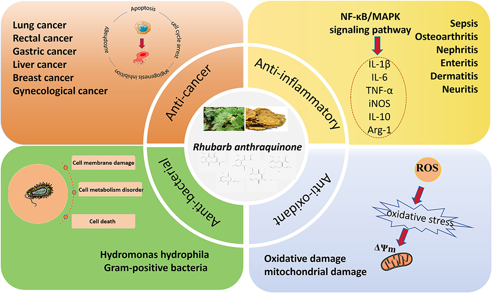 |
Figure 1 Pharmacological effects and mechanisms of AQs. Note: Red arrows indicate the gradual relationship between processes. |
Anti-Tumor
AQs exhibit efficacy in combating a diverse range of cancers, including gynecological cancer, liver cancer, colorectal cancer, gastric cancer, lung cancer, breast cancer, etc. Anti-cancer activity is associated with promoting tumor cell apoptosis and cell cycle arrest, and inhibiting tumor cell growth, migration and invasion. The related mechanisms involve apoptosis, autophagy, inflammation and energy metabolism pathways.
Research findings indicated that emodin possessed the capability to trigger apoptotic processes in gynecological cancer cells and significantly reduced mitochondrial membrane potential (MMP) and adenosine triphosphate (ATP) release, and also induced cell cycle arrest in the G0/G1 phase. Further mechanism studies have shown that emodin may regulate multiple targets to induce apoptosis (caspase-9 and cleaved-caspase-3) and autophagy (MAP LC3, Beclin-1 and Atg12-Atg5), cell cycle arrest (Cyclin D and Cyclin E) and angiogenesis inhibition (VEGF, VEGFR-2).19 The apoptosis mechanism involving the cysteinyl aspartate specific proteinase(caspase) family might be the main anti-cancer pathway of emodin. Xiong Simin et al20 found that emodin caused the accumulation of reactive oxygen species (ROS) in HepG2 cells, which subsequently triggered the opening of mitochondrial permeability transition pores, and resulted in the outflow of calcium ions and cytochrome C, thereby activating caspase and leading to cell apoptosis. In addition, the inflammatory mechanism involving Mitogen-activated protein kinase (MAPK) is also an important anti-cancer pathway of emodin. Emodin significantly inhibited the migration and invasion of MHCC-97H human hepatocellular carcinoma cells, which might be related to the activation of the p38 MAPK signaling pathway and the inhibition of the extracellular signal-regulated protein kinases (ERK)/MAPK and phosphatidylinositol 3-kinase/protein kinase B(PI3K/AKT) signaling pathways.21
Rhein and chrysophanol can also participate in anti-tumor effects by mediating apoptosis and autophagy pathways. Yang et al discovered that rhein was able to promote the expression of the apoptotic protein Bcl-2-associated X protein (Bax) by inhibiting the Signal transducer and activator of transcription 3 (STAT3) pathway. Additionally, it downregulated the expression of the anti-apoptotic protein B-cell lymphoma/Leukemia-2 (Bcl-2), thereby inducing apoptosis in non-small cell lung cancer cells.22 In addition, rhein also promoted apoptosis in human HepG2 cells by regulating the tumor protein p53 (P53) and CD95/CD95L apoptotic systems.23 The mammalian target of rapamycin (mTOR) is a major regulator of cell growth and metabolism.41 Rhein inhibited the growth of colorectal cancer cells by inhibiting the mTOR pathway.24 Similarly, with regard to inducing tumor cell apoptosis, chrysophanol primarily induced apoptosis in colorectal cancer cell line HCT-116 by upregulating the expression of Bax and p53 and downregulating the expression of Bcl-2.25 In terms of inhibiting tumor cell proliferation, chrysophanol also induced ferroptosis in tumor cells by inhibiting the solute carrier family 7 member 11/glutathione peroxidase 4 (SLC7A11/GPX4) signaling pathway, thereby inhibiting cell proliferation, migration, and invasion. This also might be related to the ability of chrysophanol to regulate the mTOR signaling pathway.26
Aloe-emodin participates in anti-tumor effects mainly by mediating inflammatory and energy metabolism signaling pathways. Shen et al27 found that aloe-emodin induced autophagy in non-small cell lung cancer cells by activating the MAPK signaling pathway and inhibiting the Akt/mTOR pathway. Furthermore, aloe-emodin exerted an inhibitory effect on glucose metabolism through downregulating the expression level of glucose transporter-1, thereby promoting apoptosis of cervical cancer cells and reducing the expression of human papillomavirus-related proteins E6 and E7.28 Modern research has found that glucose transporter-1 may play an important role in the progression of cancer by mediating glycolysis and proliferation.42
The anticancer effect of physcion is mainly related to promoting tumor cell apoptosis. Physcion promoted apoptosis of cervical cancer cells by activating caspases 3/7 and reducing the expression of Bcl-2.29 AMPK and Nuclear factor erythroidderived 2-like 2/nuclear factor kappa-B (Nrf2/NF-κB) signaling pathways were also involved in the apoptosis pathway mediated by physcion. For example, physcion stimulated endoplasmic reticulum stress (ERS) by activating the AMPK signaling pathway, thereby causing mitochondrial dysfunction and inducing apoptosis of human HCC cells.30 In addition, physcion regulated oxidative stress-mediated mitochondrial apoptosis and immune response by affecting the transduction of Nrf2/NF-κB signaling, ultimately causing apoptosis of breast cancer cells.31
Anti-Inflammatory
AQs exhibit significant anti-inflammatory effects. Studies have found that AQs exert anti-inflammatory effects by regulating inflammatory factors such as IL-1β, TNF-α, IL-6, iNOS, IL-10 and Arg-1. The signaling pathway involved is mainly NF-κB/MAPK, which has a therapeutic effect on a variety of inflammatory diseases such as sepsis, osteoarthritis, kidney inflammation, brain inflammation, dermatitis, intestinal inflammation and neuroinflammation by regulating inflammatory factors.35,43,46 NF-κB is a recognized classic molecular target for the treatment of inflammatory diseases and is particularly important in the anti-inflammatory effects of AQs.47 Lipopolysaccharide (LPS), recognized as a classic activator of the NF-κB signaling pathway, elicits a pronounced upregulation in the expression of pro-inflammatory cytokines, thereby exacerbating systemic inflammatory responses and ultimately precipitating tissue damage in the host organism.47 Rhein inhibited neuroinflammation in LPS-stimulated mouse microglia by inhibiting the PI3K/Akt/mTOR and p38 signaling pathways, activating the expression of ERK1/2 and inhibiting the Toll-like receptor 4 (TLR4)/NF-κB signaling pathways.33 Emodin alleviated acute radiation proctitis in mice by inhibiting the AKT/ERK/NF-κB/VEGF signaling pathway and inducing c-Jun N-terminal kinase (JNK) and p38-mediated apoptosis.32 Aloe-emodin reduced the secretion of inflammatory cytokines and alleviated intestinal immune cell infiltration through the IL-4/IL-13 pathway, thereby treating inflammatory bowel disease.34 Dong X et al found that physcion attenuated the oxygen glucose deprivation/reoxygenation (OGD/R)-induced inflammatory response in rat neurons by inhibiting the TLR4/NF-κB pathway and significantly reduced the expression of proinflammatory factors TNF-α, IL-1β, IL-6, and IL-10.35 The study on the anti-inflammatory effect of chrysophanol on the lung injury mouse model found that it reduced the levels of malondialdehyde (MDA), myeloperoxidase and inflammatory cytokines, and increased the activity of superoxide dismutase (SOD). Moreover, chrysophanol increased the expression of Peroxisome proliferator-activated receptor gamma (PPARγ) and inhibited the activation of NF-κB pathway after lung injury inflammation.36 In addition, emodin, rhein, and aloe-emodin exerted at least dual-target (NF-κB, iNOS) inhibitory effects on the inflammatory response of LPS-induced RAW264.7 macrophages.48 However, compared with rhein and emodin, aloe-emodin exhibits a significantly stronger anti-inflammatory effect, with its inhibitory potency on iNOS protein expression being approximately twice that of NF-κB phosphorylation.35
Other
In addition to the anti-tumor and anti-inflammatory pharmacological effects mentioned above, AQs also has multiple pharmacological effects such as anti-bacterial and antioxidant. Studies have demonstrated that emodin permeates bacterial cell membranes and inhibits cellular functions of Aeromonas hydrophila through DNA binding.37 Aloe-emodin exhibits anti-bacterial activity against clinically prevalent Gram-positive pathogens, including multidrug-resistant strains. The anti-bacterial mechanism might involve binding to the peptidoglycan layer of the cell outer membrane and the destruction of cell membrane permeability, which led to cell metabolic disorders and ultimately cell death.38 Under normal physiological conditions, the oxidative and antioxidant systems of the organism is in balance. When the body is stimulated, the balance between ROS and antioxidant enzymes is broken, and antioxidant enzymes cannot remove excess reactive oxygen and free radicals in time, leading to oxidative damage.49 AQs have the effect of resisting free radical damage, which is beneficial to cell anti-aging and body repair.50 Emodin significantly protected against mitochondrial fission and apoptosis in PC12 neuron-like cells from oxidative damage by activating the Pyruvate kinase M2 (PKM2)/Nrf2/antioxidant response element (ARE) pathway.39 Rhein significantly reduced mitochondrial ROS levels, reversed the depletion of MMP, and protected neurons from oxidative stress-related apoptosis, all of which were attributed to its excellent antioxidant activity. Rhein regulated mitochondrial biogenesis by activating the Sirtuin1/Peroxisome proliferator-activated receptor-γ coactivator-1α (SIRT1/PGC-1α) pathway. PGC-1α in the nucleus activates the downstream transcription factor NRF1, which produces healthy mitochondria by promoting mitochondrial biogenesis, thereby counteracting the oxidative stress induced by Aβ1-42 oligomers.40 Physcion reduced OGD/R-induced oxidative stress in SH-SY5Y cells, inhibited ROS/MDA production, and increased glutathione (GSH)/SOD activity, suggesting that physcion has a protective effect on neuronal oxidative damage.35
In summary, AQs exhibits multiple physiological effects and demonstrates potential for therapeutic development. However, its inherent toxicity significantly restricts clinical development and application.
The Main Toxicity and Toxicity Mechanism of AQs
Although AQs exhibit a wide range of pharmacological activities, recent studies have revealed that AQs exhibits discernible toxic effects, with toxicity predominantly localized to target organs including the gastrointestinal tract, liver, heart, and kidneys.51,53 The toxicity mechanism of AQs involves multiple pathways, including mitochondrial apoptosis, oxidative stress, death receptors, ER-associated apoptosis, caspase-dependent apoptosis, autophagy, inflammation, DNA toxicity, and bilirubin metabolism. Its toxicity and related mechanism are shown in Table 3.
 |
Table 3 Toxicity and Related Mechanisms of AQs |
Toxicity and Mechanism of Emodin
The toxicity profile of emodin primarily encompasses hepatotoxicity, nephrotoxicity, and reproductive toxicity, as evidenced by extensive in vivo and in vitro investigations. The toxicity of emodin is the most extensively studied of the AQs.
Hepatotoxicity of Emodin
According to some experimental and clinical studies, high-dose and long-term use of emodin or herbal medicine containing emodin was associated with liver cell damage and abnormal liver function.92,95,98 Apoptosis is the main pathway of emodin-induced hepatotoxicity. For example, Lin et al demonstrated that L02 cells exhibited apoptosis following 48-hour incubation with emodin (50 μM), and emodin suppressed the activity of all respiratory chain complexes in hepatic mitochondrial, thereby disrupting oxidative phosphorylation, which subsequently reduced MMP, elevated ROS levels, impaired ATP synthesis, and ultimately induced mitochondrial dysfunction and hepatocellular apoptosis. These effects may be associated with caspase pathway activation.54 Similarly, emodin (20–80 μM) arrested HepaRG cells at the S and G2/M phases and increased ROS production in HepaRG cells through the mitochondrial apoptosis pathway, thereby inducing apoptosis. The pathways involved included the mitochondrial apoptosis pathway, and molecular regulation was demonstrated by up-regulating the expression of Bax, cleaved caspase-3, 8, and 9, and cleaved poly (ADP-ribose) polymerase (PARP).55 Yang et al also demonstrated that oral administration of emodin (150 mg/kg) to SD rats for 4 weeks could result in certain damage to the rat liver. Emodin inhibited flavin adenine dinucleotide/nicotinamide adenine dinucleotide phosphate (FADH/NADPH) proton transport, activated the mitochondrial apoptosis pathway, and induced the expression of caspase-9 and caspase-3.56 In addition, oxidative stress is also associated with the apoptosis pathway implicated in the effects of emodin. When emodin was administered at a dose of 150 mg/kg, it induced oxidative stress damage in rat hepatocytes. This damage might be related to the binding of emodin to very long-chain acyl-CoA dehydrogenase and the consequent inhibition of fatty acid β-oxidation, the citric acid cycle, and oxidative phosphorylation in mitochondria, thereby triggering hepatic oxidative stress.57 Cui et al incubated grass carp hepatocytes with emodin (1–25 μg/mL) for 24 h and found that emodin showed severe hepatotoxicity, and destroyed ΔΨm and generated ROS in grass carp hepatocytes, and significantly reduced the SOD activity and total antioxidant capacity, thereby inducing hepatocyte apoptosis.58 In addition, p38-activated mitochondrial or death receptor pathways are also worthy of attention. Emodin (20–80 μM for 24 h) induced apoptosis in HepG2 cells via mitochondrial and death receptor-mediated pathways. The emodin-mediated death pathway was manifested by the simultaneous activation of the cell-surface fas receptor (Fas), Fas-ligand (Fas-L), caspase 8, and tBid. In addition, emodin also induced apoptosis in HepG2 cells by inhibiting the PI3K/Akt and ERK pathways and activating the p38 protein.59 ERS-induced apoptosis served as the primary pathway of hepatotoxicity caused by emodin, and the signaling pathway involved was mainly Inositol-requiring enzyme type 1-X-box binding protein 1 (IRE1α-XBP1). ERS induces the formation of large, microscopically visible IRE1 clusters or foci in IRE1 during its activation.99 Emodin (30 μM, 24 h) induced ERS by activating the IRE1α-XBP1 pathway and reduced the metabolic activity of HepG2 cells. The molecular mechanism was reflected in the increased expression of ERS marker proteins (such as BiP, IRE1α, and CHOP). In addition, HepG2 cells treated with emodin also increased the Bax/Bcl-2 ratio and the relative protein expression of cleaved caspase-3 in a dose-dependent manner.60 Qiu et al used emodin (25–50 μM) to induce hepatotoxicity in L02 cells and found that emodin (25–50 μM, 24h) promoted excessive ROS generation and redox imbalance in human hepatocytes, thereby triggering BiP/IRE1α/CHOP signaling-mediated ERS and unfolded protein response and intracellular Ca2+ overload, leading to ER-related cell apoptosis rather than mitochondria-dependent cell apoptosis.61 ERS induced by emodin metabolites is also one of the factors that induce hepatotoxicity. Jiang L L et al found that when human primary hepatocytes were incubated with emodin (10–100 μM), the metabolic enzyme CYP3A was activated and GSH was depleted, leading to toxicity in hepatocytes.62 Studies have found that emodin (360 mg/kg in mice, 50 μM in L02 cells) can activate the aryl hydrocarbon receptor (AHR), inducing CYP1A1 expression, leading to a decrease in GSH and an increase in ROS in hepatocytes, thereby triggering ERS and producing hepatotoxicity.63 The hepatotoxicity resulting from combined medication should not be overlooked. When emodin is combined with hepatotoxic drugs, an increase in hepatotoxicity will ensue. For example, Wu et al found that the combined use of emodin (150 mg/kg) and probenecid exacerbated the hepatotoxicity of emodin in rats. Therefore, it is suggested that gout patients should avoid long-term use of emodin-containing preparations combined with probenecid.64 Additionally, Tu et al demonstrated that neither emodin (20, 40, 80 mg/kg) nor LPS (2.8 mg/kg) alone could induce liver damage in rats. However, when rats were fed a non-toxic dose of LPS in combination with emodin for 10 hours, acute liver damage occurred in the rats.64
Nephrotoxicity of Emodin
Emodin has the potential to induce nephrotoxicity when administered in large doses over an extended period. The renal tubules represent the primary site where emodin-induced nephrotoxicity occurs.100 Ferroptosis has been confirmed as a pathway through which emodin induces renal injury. Xing et al reported that severe and extensive renal tubular lesions were observed in mice following intraperitoneal injection of emodin (0.4–0.8 mg/kg for 14days). Moreover, in vitro studies showed that the activity of renal tubular epithelial cells in rat treated with emodin (40–160 μM) was greatly reduced, which might be related to emodin inhibiting the Nrf2/GPX4 antioxidant system and inducing renal damage through ROS-mediated iron allergy.65 In addition, apoptosis is also an important pathway of renal injury induced by emodin. When administered at a concentration of 40 μM for a duration of 24 h, emodin was observed to induce a reduction in the viability of human renal proximal tubular epithelial cells, caspase-3 cleavage and activation, loss of ΔΨm, and release of Cyt-C from mitochondria to the cytosol, leading to HK-2 cell apoptosis. This complex mechanism might be related to the activation of proliferator-activated receptor γ (PPARγ).66 Furthermore, emodin (40 μM, 24 h) increased the expression of cathepsin B protein, thereby activating caspase-3, leading to the decreased viability of HK-2 cell.67 In addition, mesangial cells are also regarded as the main target of renal toxicity induced by emodin,101 which is associated with emodin-induced cell cycle arrest at the G1 phase.102
Reproductive Toxicity of Emodin
It has been reported that the toxicity of emodin also manifests in the reproductive system. Emodin (50–400 μM) has been shown to reduce the overall motility, forward-progressive motility, and linear velocity parameters of human sperm. It alters the sperm’s penetration ability in viscous media, along with progesterone-initiated capacitation and the acrosome reaction. The underlying mechanism primarily involves reducing sperm intracellular calcium ion concentration ([Ca(2+)]i) and inhibiting tyrosine phosphorylation.68 Emodin has been identified as an inhibitor of casein kinase 2. Male mice with the casein kinase 2 gene knocked out were found to be infertile. It was found that mice fed with emodin (1000 mg/kg) could cause decreased sperm production, increased eosinophils and germ cell apoptosis. The reproductive toxicity of emodin might be related to the insulin-like growth factor 1 (IGF-1) receptor signaling pathway, and the differentially expressed genes indicated that emodin affected the expression of casein kinase 2 and spermatogenesis.68 In addition, Mouse oocytes treated with emodin (20–40 μM for 24h) showed reduced maturation and fertilization rates, accompanied by impairment of early embryonic development. Pretreatment with a caspase-3 specific inhibitor effectively reduced the series of effects triggered by emodin, suggesting that the reproductive toxicity of emodin might occur through a caspase-dependent apoptotic process.70 Treatment of mouse blastocysts with emodin (25–75 μM for 24 hours) resulted in inhibition of early embryonic development to the blastocyst stage. Emodin triggered damage in mouse blastocysts via the activation of intrinsic apoptotic signaling pathways, but the specific mechanism was unclear.71 Emodin (0.25 μg/mL 7–72 hpf) caused edema, trunk bending, and morphological abnormalities in zebrafish embryos, which reduced the survival and hatching success rate of zebrafish embryos. The main mechanism was related to increased expression of CYP3A and multidrug resistance 1 (MDR1) proteins.72 Generally, the reproductive toxicity of emodin is evident in sperm, oocytes, and embryos. However, the specific toxicity mechanism remains to be further investigated.
In summary, hepatotoxicity has been studied relatively extensively, and its mechanisms mainly involve mitochondrial apoptosis, ERS and enzyme activity regulation. Nephrotoxicity primarily occurs in the renal tubules, involving ferroptosis and mitochondrial apoptosis pathways. The mechanisms of reproductive toxicity require further investigation. Indeed, current research on the nephrotoxicity and reproductive toxicity of emodin is limited, suggesting that the liver may be the primary site of toxicity. The toxic mechanism of emodin is shown in Figure 2.
 |
Figure 2 The toxic mechanism of emodin. Note: |


Toxicity and Mechanism of Rhein
Rhein serves as a crucial component of AQs. The toxicities of rhein primarily encompass hepatotoxicity, nephrotoxicity, reproductive toxicity, and cardiotoxicity. It is widely acknowledged that its toxicity is associated with both dose and duration.103
Hepatotoxicity of Rhein
Mitochondrial-mediated apoptosis and oxidative stress are the main pathways of rhein-induced hepatotoxicity. For example, studies have found that rhein (75–100 μM for 24 h) promoted the production of ROS, the loss of MMP and S phase cell cycle arrest, thereby inhibiting the viability of HepaRG cells and inducing cell death. The apoptosis mechanism involved was mainly the mitochondrial-mediated apoptosis pathway, which was reflected in the up-regulation of the expression of p53, p21, Bax, caspases-3 and PARP, and the down-regulation of the expression of Bcl-2, cyclin A and cyclin-dependent kinase 2 proteins.73 Similarly, Yang et al also found that rhein (4g/kg for 24 h) caused mitochondrial dysfunction through the oxidative stress pathway, activated the mitochondrial apoptosis pathway, and caused apoptosis of mouse liver cells, showing increased levels of ROS, NRF-2 and MDA, and decreased levels of SOD.74 In addition, Guy-Armel Bounda et al found that the hepatotoxicity caused by rhein was related to mitochondrial apoptosis pathway, oxidative stress, and lipid imbalance.The study revealed that rhein (50–100 μM for 12h) mediated the excessive production of ROS, decreased MMP, and increased synthesis of triglycerides (TG) and total cholesterol (TC). Rhein significantly up-regulated the expression of caspase-3, caspase-9, caspase-8, p53, p53-up-regulated modulator of apoptosis (PUMA), and Apaf-1, and participated in cell apoptosis. In addition, it also increased the expression of three key enzymes involved in lipid metabolism: 3-hydroxy-3-methyl glutaryl coenzyme A reductase (HGM-CoAR), glycerol-3-phosphate acyltransferase (GPAT), and Acetyl-CoA carboxylase (ACoAC). Immunoblotting showed that cyt-c was released from mitochondria into the cytosol in large quantities.75 Studies have also shown that hepatotoxicity caused by rhein is a very complex process involving multiple signaling pathways. Gati Krushna Panigrahi et al discovered that rhein (50 μM for 24 hours) caused apoptosis in rat primary hepatocytes by generating ROS, increasing intracellular Ca(2+), reducing MMP, and consuming intracellular GSH content. This was mainly related to the DNA damage, thereby increasing the expression of p53 and p21, and ultimately activating the endogenous apoptosis pathway mediated by Bax, Bcl2, cyt-c, caspase-3, caspase-9, and PARP, which involved multiple pathways such as p53, MAPK, and mitochondrial.76 In addition, ERS and increased intracellular calcium ion levels were the main reasons for rhein-induced hepatocyte apoptosis. The ERS pathway involved in rhein was reflected in the up-regulation of the expression of glucose-regulated protein 78 (GRP 78), protein kinase receptor-like ER kinase (PERK), JNK, and CCAAT/enhancer binding protein homologous protein (CHOP). At the same time, the ERS marker caspase-4 was also activated, followed by caspase-3. In addition, rhein could also promote the increase of intracellular calcium ion concentration, thereby inducing cell apoptosis, which might be related to JNK activation.77 Kågedal et al incubated rat hepatocytes with rhein (50μM for 16 hours) and also found that rhein increased the Ca2+ concentration in hepatocytes, thereby inducing hepatocyte apoptosis.78 Studies have shown that treatment with rhein can increase intracellular free Ca2+ concentration by nearly 10 times, which is related to mitochondrial dysfunction.104 TNFα-mediated apoptosis and autophagy have also been shown to be pathways for rhein-induced hepatotoxicity. Rhein (50–100 μM for 24 h)-induced hepatocyte apoptosis might be related to the increase of intracellular ROS, and induced hepatocyte L02 apoptosis through the TNF-α apoptosis pathway and the mitochondrial pathway, which was manifested by up-regulating the expression of TNF-α, Tumor Necrosis Factor Receptor 1 (TNFR1), tumor necrosis factor receptor-associated death domain protein (TRADD) and cleaved caspase-3, and down-regulating the expression of procaspase-8. At the same time, rhein up-regulated the expression of Bax and down-regulated the expression of procaspase-9 and procaspase-3, indicating that the mitochondrial pathway was activated. In addition, after treatment with rhein, the expression of autophagy-related protein light chain 3 (LC3)-II and beclin-1 was significantly reduced, while the expression of Sequestosome 1(P62) was significantly increased. It is speculated that rhein may inhibit L02 cell autophagy, reduce the cell self-clearance ability, and promote cell apoptosis.79
Nephrotoxicity of Rhein
There are few studies on the nephrotoxicity of rhein.103 Zhu Yanna et al proposed that rhein primarily induces renal injury by triggering apoptosis, and this apoptotic process may involve multiple signaling pathways.103 Rhein (50μM, 100μM for 24h) induced apoptosis in HK-2 cells through the mitochondrial-mediated pathway, including destruction of MMP, reduction of ATP levels, release of cyt-c from mitochondria to cytoplasm, and up-regulation of Bcl-2 and Bax levels. In addition, rhein significantly increased ROS levels and inhibited the expression of mitochondrial uncoupling protein 2 (UCP2). The inhibition of UCP2 significantly enhanced oxidative stress and exacerbated rhein-induced cell apoptosis.80 Rhein (50 μM and 100 μM, 24 hours) inhibited HK-2 cell proliferation and induced apoptosis and S/M accumulation in a concentration-dependent manner. The apoptotic mechanism was associated with caspase 3 activation, loss of ΔΨm, and expression of Bcl-2 and Bax.81
Reproductive Toxicity of Rhein
Most studies on the reproductive toxicity of rhein have focused on embryotoxicity. Studies have confirmed that rhein (175 and 350 mg/kg) exhibits teratogenic effects on fetal development in rats. Rats received rhein gavage for 10 successive days from pregnancy day 6 to day 15. The fetuses would develop various skeletal deformities such as sternal cross-section loss, incomplete skull ossification, and thoracic vertebrae separation or deformation.83 The developmental potential of mouse blastocysts implanted into the uterus after incubation with 5–20 μM rhein for 24 hours was impaired. In vivo tests confirmed that intravenous injection of rhein (1, 3, and 5 mg/kg body weight per day) into maternal mice for four days would lead to embryonic apoptosis, early embryonic development damage, and fetal weight loss. Rhein-induced embryotoxicity was related to the induction of oxidative stress and immunotoxicity, which induced the production of ROS and down-regulated the transcriptional levels of innate immunity-related genes chemokine (CXC motif) ligand 1(CXCL1), IL-1β, and IL-8 in maternal mouse fetuses.84
Cardiotoxicity of Rhein
Rhein is the only one of the five AQs that has been shown to be cardiotoxic. The study found that after 48 hours of exposure to rhein (106, 124, and 132 μmol/L), H9c2 cells showed atrophy, rounding, cell wall shedding, and cell proliferation inhibition. The mechanism involved rhein-induced microscopic changes in cardiomyocyte membrane potential and mitochondria. In addition, mice treated with rhein (350, 175, and 87.5 mg/kg) for 45 days showed a significant reduction in left ventricular ejection fraction and left ventricular fractional shortening. Furthermore, serum Ca2+, cTnT, CK, and LDH levels were significantly increased. Rhein-induced cardiotoxicity might be related to mitochondrial-mediated and Fas-induced apoptosis, as evidenced by up-regulation of Bax, Caspase-3, −8, and −9, Fas, and P53, while the expression of Bcl-2 showed a downward trend.82
In summary, the hepatotoxicity mechanisms of rhein involves mitochondrial apoptosis, oxidative stress, abnormal lipid metabolism, and complex endogenous apoptosis. The nephrotoxicity, reproductive toxicity, and cardiotoxicity of rhein have been less studied. Nephrotoxicity mainly involves the mitochondrial apoptosis pathway, reproductive toxicity mainly involves oxidative stress and immunotoxicity, and cardiotoxicity is mainly related to mitochondrial and Fas-mediated apoptosis pathways. This also suggests that the liver may be the main toxic organ of rhein. As the heart is a vital organ, the cardiotoxicity of rhein needs to be taken seriously. The toxic mechanism of rhein is shown in Figure 3.
 |
Figure 3 The toxic mechanism of rhein. |
Toxicity and Mechanism of Aloe-Emodin
The liver and kidney are definite target organs of aloe-emodin toxicity, whereas its genotoxicity remains controversial.
Hepatotoxicity of Aloe-Emodin
Multidrug resistance-associated protein 2 (MRP2) is an important efflux transporter involved in intracellular detoxification, inflammation, excretion of drug metabolites, and regulation of oxidative stress.105 In mice treated with aloe-emodin (0.8 and 1.6 g/kg) for 11 weeks, aloe-emodin inhibited the transport activity of MRP2, triggered oxidative stress and apoptosis, and led to a decrease in intracellular GSH, mitochondrial dysfunction, and activation of apoptosis in HepG2 cells.85 In addition, aloe-emodin (0.01505 mg/mL for 72 hours) caused liver lesions in zebrafish, leading to obvious hepatitis cell infiltration and necrosis. A significant increase was observed in the expression levels of P65, JNK, and P53 proteins in zebrafish hepatocytes. This increase was found to be associated with the activation of the NF-κB inflammatory pathway and the P53 apoptosis pathway.86 Similarly, aloe-emodin (20, 40 μM for 48 h) reduced the viability of HL-7702 cells in a dose-dependent and time-dependent manner, induced cell cycle arrest at the S and G2/M phases, and dose-dependently up-regulated the level of Fas, P53, P21, Bax/Bcl-2, Caspase-3, −8, −9, as well as PARP cleavage. The mechanisms of hepatotoxicity involved the Fas death pathway and the mitochondrial apoptosis pathway.87 In addition, aloe-emodin (40 μmol/L) led to increased expression of heme oxygenase-1 mRNA and quinone oxidoreductase-1 mRNA in L02 and HepaRG cells, thereby inducing mitochondrial damage and imbalance of hepatocyte oxidative stress, leading to hepatocyte apoptosis.88
Nephrotoxicity of Aloe-Emodin
Aloe-emodin (100 μM for 48 hours) was demonstrated to significantly reduce the viability of HK-2 cells and induce G2/M arrest in the HK-2 cells cycle. The mechanism underlying the apoptosis induced by aloe-emodin was found to be associated with Caspase-3 and ERS. This association involved eukaryotic initiation factor-2α phosphorylation, X-box binding protein 1 mRNA splicing, JNK phosphorylation, as well as an increased accumulation of GRP78 and CHOP.89 Dosage is a factor affecting the nephrotoxicity of aloe-emodin. The aloe-emodin group (1.6 g/kg) showed elevated blood urea nitrogen and serum creatinine levels, and severe renal tubular and glomerular damage. The low-dose group (0.8 g/kg) showed milder renal tubular damage. In both groups, SOD levels were significantly decreased; TGF-β1, PPPPα and IL-6 were significantly increased. The mechanism of nephrotoxicity might be related to oxidative stress, apoptosis, and transforming growth factor-β (TGF-β) signaling pathway.90
In addition to the two toxicities mentioned above, aloe-emodin also has certain genetic toxicity. Genotoxicity corresponds to the ability of substances to intercalate into DNA. Topoisomerase II inhibitors are compounds with DNA-intercalating properties,106 and it was demonstrated that both emodin and aloe-emodin were capable of inhibiting the activity of topoisomerase II, but the inhibitory effect exerted by emodin was found to be stronger than that of aloe-emodin,107 which suggests that emodin may also be genetically toxic. Primary DNA damage was observed in the liver and kidneys of mice treated with aloe-emodin (2000, 1000, and 500 mg/kg/day (×2)), demonstrating that aloe-emodin might cause organ-specific genetic toxicity.108 Studies have found that aloe-emodin at a dose of 2000 mg/kg body weight/day does not induce DNA chain breaks in male mouse kidney and colon single cell preparations, but does cause damage to kidney and colon cells, indicating that aloe-emodin is not genotoxic.109 In addition, Heidemann et al performed in vivo cytogenetic analysis on bone marrow cells of wistar rats and found that doses up to 2000 mg/kg did not cause toxic effects or chromosomal structural aberrations.110 Therefore, the kidney and colon may be the toxic target organs of aloe-emodin, but whether there is genetic toxicity still deserves further study.
In summary, The hepatotoxicity and nephrotoxicity mechanisms of physcion both involve oxidative stress, inflammatory responses, and apoptosis. In addition, the nephrotoxicity mechanism is related to ERS. However, inconsistent conclusions exist regarding aloe-emodin genotoxicity, with a lack of in-depth research. The toxic mechanism of aloe-emodin is shown in Figure 4.
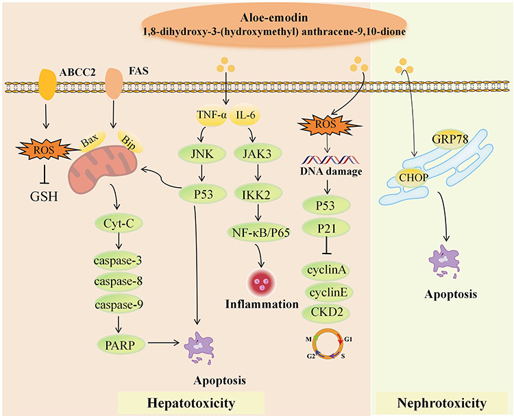 |
Figure 4 The toxic mechanism of aloe-emodin. |
Toxicity and Mechanism of Physcion
The toxicity of physcion mainly includes hepatotoxicity, nephrotoxicity, erythrotoxicity, and neurotoxicity. The mechanism involved in hepatotoxicity is the regulation of choline efflux transporters and drug-metabolizing enzymes. The nephrotoxicity of physcion has not been reported. Additionally, the erythrotoxicity and neurotoxicity of physcion also deserve further attention.
Hepatotoxicity of Physcion
Hepatotoxicity is the common toxic effect of physcion, but its nephrotoxicity has not been systematically reported. In addition, physcion may also exert erythrotoxicity and neurotoxicity.
MRP2 and Bsep are key transporters involved in bilirubin metabolism and choline efflux, and their inhibition can exacerbate cholestatic injury and induce hepatotoxicity.111,112 Kang L et al have demonstrated that physcion alters the distribution of total choline or individual choline molecules in the bile, liver, and plasma of mice by regulating choline efflux transporters, thereby inducing liver damage. Administration of physcion (100 mg/kg) to mice for 14 days resulted in mild inflammatory infiltration in the liver cell and induction of bile acid accumulation. The mechanism of toxicity involved down-regulation of Bsep and MRP2 expression.92 Physcion metabolites may be the main components of its hepatotoxicity. Studies have found that the metabolites of physcion had a strong inhibitory effect on Uridine diphosphate glucuronosyltransferase (UGT) 1A1 enzyme.113 Exogenous substances that inhibit the UGT1A1 enzyme will lead to an inability to convert bilirubin into conjugated bilirubin or a decrease in the amount of bilirubin converted. This results in bilirubin metabolism disorders, induces bilirubin accumulation in liver cells and blood, and ultimately causes liver toxicity.114 Zheng et al discovered that physcion exhibits high toxicity towards normal L02 cells. The mechanism underlying hepatotoxicity is associated with the inhibitory effect of physcion on CYP1A2, CYP2C9, CYP2D6, and CYP3A4, which competitively blocks the rate-limiting enzyme UGT1A1 in the bilirubin metabolism process.93
The potential toxicity of physcion to red blood cells and nerves still needs to be explored. Studies have demonstrated that physcion (50 and 100 µM) induces significant hemolysis in red blood cells, which is associated with programmed cell death.94 In terms of neurotoxicity, the survival rate of human SH-SY5Y neuroblastoma cells incubated with physcion (10 μM) for 24 hours dropped to approximately 55%, while under the same conditions, aloe-emodin-induced mortality was less than 15%, indicating that physcion was more neurotoxic than aloe-emodin. However, physcion still exhibits certain limitations in studies on erythrocyte toxicity and neurotoxicity. For instance, its toxicity mechanism has not been fully elucidated to date.115
In summary, the hepatotoxic mechanism of physcion involves the regulation of choline efflux transporters and drug-metabolizing enzymes. Although studies on erythrotoxicity and neurotoxicity are relatively limited, they also deserve attention.
Toxicity and Mechanism of Chrysophanol
Toxicity reports on chrysophanol are rare. At present, several studies suggest that it may possess hepatotoxicity, reproductive toxicity, and genotoxicity.
Chrysophanol seems to be the least toxic of the five AQs. Currently, there are very few studies on the toxicity of chrysophanol. Regarding hepatotoxicity, toxicity test results based on zebrafish indicate that chrysophanol is a potential hepatotoxicity marker of rhubarb.116 However, chrysophanol exhibits relatively low toxicity. The study revealed that chrysophanol (50 μM for 15 minutes) merely induced an elevation in Ca2⁺ concentration in rat hepatocytes, and it did not trigger apoptosis. However, following 30 minutes of exposure, cell viability was markedly diminished, indicating that the hepatotoxicity of chrysophanol is closely associated with the duration of exposure. Interestingly, exposure to physcion (50 μM) for 15–60 minutes exhibited no significant impact on hepatocyte viability, suggesting that chrysophanol demonstrates greater hepatotoxicity than physcion. Furthermore, following 10 minutes of treatment with rhein (50 μM), intracellular reduced GSH was entirely depleted. In contrast, chrysophanol (50 μM) reduced GSH levels to 60% of their initial value after 60 minutes of exposure. Therefore, the hepatotoxicity of chrysophanol is less severe than that of rhein.78 However, studies on the hepatotoxicity of chrysophanol are lacking or insufficient. With regard to reproductive toxicity, a study investigating the potential of chrysophanol to induce chromosome breakage in hamster ovary cells revealed that chrysophanol (30 μg/mL) exhibited almost no ability to cause chromosome breakage.117 A study using resonance light scattering spectroscopy to confirm the interaction between chrysophanol and DNA found that chrysophanol could insert into the base pairs of double-helix DNA in a manner similar to a DNA intercalator, thereby producing toxic effects. This finding indicated that chrysophanol exhibited certain genetic toxicity, and its toxicity was associated with environmental factors, including amino acids, sodium chloride, glucose, and pH. The genetic toxicity of chrysophanol is comparable to that of ethidium bromide, mitoxantrone, and doxorubicin.118
In summary, the above toxicity studies have obvious limitations: the hepatotoxic mechanism has not been thoroughly elucidated, the concentration settings for reproductive toxicity are restricted, and genotoxicity has not been verified in vivo or in vitro. These indicate that the toxicity of chrysophanol has not yet been comprehensively and systematically revealed, and many unknown safety risks require further exploration.
Relationship Between Pharmacokinetics and Toxicity of AQS
The quinone structure of AQs is fat-soluble, and the main absorption site is the intestine rather than the stomach.53 A study investigated the pharmacokinetics in rats after oral administration of rhubarb extract (300 mg/kg). The results showed that these five AQs were rapidly and widely distributed 10 minutes after oral administration, mainly in the stomach, liver, intestine, and kidneys. They were excreted unchanged in urine in small amounts and in feces in larger amounts. Thus, fecal excretion is the primary route of elimination for five AQs. However, the urinary excretion rate of rhein was much higher than that of other AQs. This may be related to the fact that other AQs are readily converted into rhein in the body.119,120 About 84 hours after administration, the cumulative urinary excretion of the five AQs reached more than 90% of the total excretion.119 In addition, the metabolic pathways of AQs in the body are complex, and the metabolism of the five AQs may affect each other’s Tmax, Cmax, T1/2, etc.121 Therefore, it is necessary to explore the relationship between the metabolic transformation and toxicity of AQs, although research in this area is currently insufficient. Drug metabolites produced in the body often increase polarity, enhance water solubility, and decrease or completely inactivate pharmacological activity.122 Therefore, metabolic reactions are generally considered to be biological detoxification processes. However, in some cases, therapeutic drugs can be converted into active metabolites under the action of various metabolic enzymes and produce toxicity.123 Drug biotransformation is a key factor in the early stages of new drug development and is divided into two types of reactions: Phase I (oxidation, reduction, and hydrolysis) and Phase II (conjugation). Phase I metabolic reactions are typically catalyzed by the CYP450 enzyme system in the liver. Phase II metabolic reactions are typically catalyzed by transferases in the liver, such as glucuronyltransferases, sulfotransferases, and methyltransferases.123
Metabolism and Toxicity of Emodin
Emodin rarely exists in its original form after entering the body. Following oral administration of polygonum cuspidatum to rats, free emodin was predominantly detected in the liver, whereas glucuronic acid/sulfate-conjugated emodin was found in the blood, kidneys, and lungs.124 A comprehensive study investigating the pharmacokinetic metabolism of emodin in rats has revealed that approximately 56% of emodin remained unabsorbed and was predominantly excreted in the feces in its original form. The original form and metabolites of emodin that entered the systemic circulation were mainly distributed in the kidneys. The hydroxylated metabolites were primarily excreted via urine and feces, whereas the glucuronidated metabolites were mainly excreted via urine and bile.125 The metabolism of emodin in the body is very complex. Bai et al employed UHPLC-Q-Exactive MS to detect the metabolites of emodin following oral administration to rats. The results showed that emodin underwent multiple metabolic pathways in addition to glucuronidation and hydroxylation. Moreover, these metabolites were extensively distributed in plasma, bile as well as heart, liver, spleen, lung, kidney, and brain tissues.126 Furthermore, upon intravenous injection of emodin (5.0 mg/kg) in rats, emodin was rapidly transformed into its hydroxylated derivatives or conjugates with glucuronic acid/sulfate. By contrast, following oral administration of emodin (20.0 mg/kg and 40.0 mg/kg), only emodin glucuronide was identifiable in the serum.127 In addition, gender is also an influential factor in the metabolism of emodin.128 The study demonstrated that regardless of whether rats were subjected to intraperitoneal injection (20 mg/kg or 40 mg/kg) or oral administration (20 mg/kg, 40 mg/kg and 80 mg/kg, 12 weeks) of emodin, the peak concentration of glucuronidated emodin was attained at 1 hour and was eliminated within 12 hours. It was noteworthy that female mice appeared to metabolize emodin more rapidly than male mice.129 These factors determine that the toxicity study of emodin metabolites is a difficult task.
The metabolic enzymes CYP1A1, CYP2C19, CYP3A4, UGT1A9, and UGT2B7 play a key role in the hepatotoxicity induced by emodin.64 5-hydroxy-emodin is a hydroxylated metabolite of emodin, which is primarily produced by CYP1A2, and is more toxic than emodin. The toxic mechanism is associated with the activation of the aromatic hydrocarbon receptor (AHR). The Ahr-Cyp1a1 pathway potentiates the ROS/ERS effect and CYP1A2 enzyme activity.63 In addition, promoting the oxidative metabolism of emodin by CYP2C19 and CYP3A4 enzymes and promoting the formation of hydroxyl compounds might be a mechanism for reducing the toxicity of emodin.130 Although this view requires further confirmation. Current research generally believes that the toxicity of emodin is stronger than its glucuronide derivative. Chen et al reported that UGT2B7 exhibited the most pronounced glucuronidation effect on emodin, presenting percentage distributions of 54.9% in human liver microsomes and 53.1% in human kidney microsomes. Long-term or high-dose use of emodin inhibited the activity of UGT2B7 by suppressing the expression of hepatocyte nuclear factor 4-α (HNF4α), which led to the accumulation and toxicity of emodin in hepatocytes.131 Therefore, inducing the simultaneous expression of HNF4A and UGT2B7 is showing promise as a novel detoxification means for emodin. Emodin-3-O-β-D-glucuronide is the main glucuronidation metabolite of emodin and has certain toxicity. In comparison, emodin had a larger area under the curve (AUC) and more pronounced hepatotoxicity, significantly increasing ALT and AST levels, indicating that emodin is more toxic. It was speculated that the higher hepatic accumulation of emodin might be the cause of its greater hepatotoxicity. In addition, GSH depletion has been associated with emodin-induced hepatotoxicity. Liu et al identified an emodin–cysteine adduct in cultures of normal human liver L-02 cells exposed to emodin, the level of which increased in an emodin-concentration-dependent manner. Decreased levels of GSH and oxidized glutathione (GSSG), together with elevated levels of glutamate, a GSH metabolite, were also observed in the cultures. Marked alterations in acylcarnitines suggested that GSH depletion-induced disorder of fatty acid oxidation contributes to hepatotoxicity.132 Acylcarnitines play an important role in mitochondrial function, and mitochondrial dysfunction has been reported to be associated with hepatotoxicity.133 Moreover, the differences in emodin metabolism between male and female rats account for the sex-related differences in the toxicokinetics of emodin.134 It is worth noting that the increased expression of MRP2 induced by emodin might accelerate the elimination of emodin-3-O-β-D-glucuronide, which was also one of the detoxification mechanisms.134
Therefore, the author speculates that to reduce the toxicity of emodin, in addition to adhering to conventional usage and dosage guidelines, it is of great importance to prevent emodin from accumulating in the body. For instance, a study has found that TSG significantly inhibits the phase II metabolism of emodin, thereby increasing the bioavailability of emodin and elevating the risk of cumulative poisoning.135 Moreover, it is also essential to avoid using emodin in combination with CYP3A enzyme inducers or UGT2B7 enzyme inhibitors.125 Additionally, AhR, HNF4A, and MRP2 are detoxification targets that warrant further investigation.
Metabolism and Toxicity of Rhein
Following oral administration to rats, mice, rabbits, and beagle dogs, rhein was predominantly distributed to the liver, followed by the stomach, intestines, lungs, spleen, kidneys, heart, and brain. The absorbed rhein was metabolized into glucuronides and sulfates, which were subsequently excreted in the bile and entered the enterohepatic circulation. In the intestine, glucuronides and sulfates might undergo conversion to other AQs.136 Rhein contains two hydroxyl groups and one carboxyl group, which endow it with strong polarity and electrochemical redox properties.103 Three metabolites were detected in the feces of rats administered rhubarb AQs using HPLC, and rhein was identified as the metabolite that accumulated to the greatest extent over time, which may be related to the in vivo conversion of aloe-emodin and sennoside A into rhein.133 Studies have demonstrated that the metabolites of rhein primarily consist of three monoglucuronides, corresponding to two hydroxy glucuronides and one acyl glucuronide (connected through the carboxyl group).137 Therapeutic drugs can be metabolized into active metabolites by various metabolic enzymes in the body and subsequently covalently bind to biological macromolecules (such as proteins or DNA), thereby leading to increased toxicity.123 A study also revealed that a monohydroxylated metabolite was detected in the urine and bile of rats administered with rhein. This metabolite was likewise detected in rat and human liver microsome cultures exposed to rhein. Meanwhile, a total of three GSH conjugates were identified in rat bile. These GSH conjugates might be formed through the combination of rhein or its metabolites.138 Furthermore, it is likely that the majority of rhein was converted into acyl glucuronides in the liver. Rhein acyl glucuronides covalently bound to GSH to form rhein-GSH adducts, which might eventually induce toxicity. UGT1A1, UGT1A9, and UGT2B7 are the enzymes that catalyze the conversion of rhein acyl glucuronides.137 CYP2C19, serving as the main metabolic enzyme of rhein, activated rhein and converted it into active metabolites. This enzyme formed electrophilic centers in rhein, which then combined with GSH to generate GSH-rhein adducts and monohydroxyrhein, ultimately leading to hepatotoxicity.123,139 Studies have demonstrated that rhein is primarily metabolized into chemically reactive acyl glucuronides in primate (human and monkey) liver microsomes. In contrast, the predominant metabolite of rhein in rats, mice, and dogs liver microsomes is a monoglucuronide formed on the hydroxyl group, which is unreactive. The interspecies differences in rhein glucuronidation are likely attributable to variations in the content of UGTs in the livers of different species.140 In addition, in vitro data from rat and human hepatic systems indicate that rhein metabolism involves a combination of weak Phase I and intensive Phase II processes. Almost all rhein elimination is mediated by hepatic metabolism, and total hepatic clearance comprises metabolic clearance via CYP450, UGT, and SULT enzymes.141 Following oral administration of rhein in beagle dogs, rhein glucuronides and sulfates were rapidly formed in plasma, pharmacokinetic parameters revealed that the AUC0-10h of rhein glucuronides/sulfates was significantly elevated, suggesting that these conjugates play a more prominent role in the systemic circulation of rhein in vivo.142 However, the potential toxic effects and mechanisms associated with rhein biotransformation via the above Phase I and Phase II metabolic pathways remain unclear and inadequately investigated.
Metabolism and Toxicity of Aloe-Emodin
After oral administration of rhubarb extract in rats, aloe-emodin was mainly distributed in the stomach, and was also found in tissues including the liver, kidney, lung, heart and brain. A large amount of aloe-emodin was converted into rhein. Aloe-emodin was mainly excreted in its prototype form, with a urinary excretion rate of 1.14% and a fecal excretion rate of 46.83%.119,120 After oral administration of aloe-emodin to rats, the main components in the blood were aloe-emodin glucuronide and rhein glucuronide, while the parent aloe-emodin was barely detectable in blood. However, when aloe-emodin was intravenously administered to rats, the main components in the blood were aloe-emodin glucuronide and rhein sulfate.143 Hence, the metabolites of aloe-emodin are likely to emerge as the key factors exerting a more pronounced impact on the bioactivity. The metabolites of aloe-emodin show considerable complexity. CYP1A2, 2B6, 2C19, 3A1 and 3A4 are the main enzymes involved in the metabolism of aloe-emodin.144 Studies have shown that the metabolites detected in rat and human liver microsomes exposed to aloe-emodin include two hydroxylated metabolites and four GSH conjugates. The GSH conjugates were derived from aloe-emodin or its hydroxylated metabolites, while the hydroxylation of aloe-emodin was mainly catalyzed by CYP 1A2, 3A4 and 3A5.145 In addition, CYP3A1 is the main metabolic enzyme in rat liver microsomes. Studies have shown that aloe-emodin is metabolized into hydroxy metabolites and rhein in rat liver microsomes.88 CYP3A4, as the main metabolic enzyme in human liver microsomes, is closely related to the hepatotoxicity of aloe-emodin. Inhibition of CYP3A4 would aggravate the damage of aloe-emodin to L02 and HepaRG cells.88 Quinones also induce toxicity through various other mechanisms, including protein binding, DNA binding, and interaction with GSH.146 Panigrahi et al found that the cytotoxicity of aloe-emodin might be attributed to its strong GSH depletion ability and DNA binding affinity.147 This was consistent with the observation that a large amount of GSH compounds were detected in liver microsomes incubated with aloe-emodin. Phase II metabolism of aloe-emodin contributes to its GSH depletion capacity. Sulfotransferases are responsible for mediating the Phase II metabolic activation of aloe-emodin. The benzyl hydroxyl group of aloe-emodin is likely to undergo sulfation reaction to form aloe-emodin sulfate, which was chemically reactive and could react with endogenous GSH to form aloe-emodin-GSH conjugates (SN1 mechanism) or be directly attacked by GSH (SN2 mechanism). Both pathways lead to intracellular GSH depletion.148 However, the toxicity research of aloe-emodin metabolites is relatively limited and needs further research. For example, the toxicity research of aloe-emodin metabolites is still insufficient, and the mechanism of hepatocellular damage caused by CYP3A4 inhibition still deserves further exploration. In addition, intestinal commensal microbes encode a variety of enzymes that perform structural modifications on drugs, mediating drug activation, inactivation, toxicity induction, altered stability, reduced bioavailability, and rapid excretion, thus playing an important role in drug metabolism.149 The gut microbiota is essential for the biotransformation of aloe-emodin. Aloe-emodin undergoes rapid oxidation and conjugation in the gastrointestinal tract before entering the systemic circulation, and the resulting metabolites may exhibit altered biological activity and bioavailability.150 However, studies on the gut microbiota–metabolism–toxicity axis of aloe-emodin remain scarce to date.
Metabolism and Toxicity of Physcion
There are few systematic studies on the pharmacokinetic properties of physcion. In studies of AQs co-cultured with human intestinal flora in vitro, physcion is likely to be metabolized by intestinal flora into aloe-emodin, emodin, chrysophanol, or other metabolites before entering the blood.151 Therefore, physcion was often difficult to detect after oral administration of different rhubarb preparations.152 Four monohydroxylated metabolites and one O-demethylated metabolite, as well as emodin, were detected in incubations of physcion with rat liver microsomes. Emodin was speculated to be the primary metabolite of physcion contributing to hepatotoxicity. CYP2C19, CYP1A2, CYP2B6, CYP3A4 predominantly participated in the hydroxylation reaction of physcion. Furthermore, three N-acetylcysteine (NAC) conjugates were observed in rat liver microsomes supplemented with NAC as a trapping agent.153 The metabolites of physcion displayed a strong inhibitory effect on the UGT1A1, while physcion showed minimal inhibitory activity towards the UGT1A1. It was hypothesized that the inhibition of UGT1A1-mediated bilirubin metabolism by metabolites might pose a potential risk of hepatotoxicity.113 In fact, physcion is converted into multiple metabolites in vivo. Nevertheless, the study of toxic mechanisms associated with metabolites is also insufficient. Future studies should focus on the effects of physcion metabolites on UGT1A1-mediated bilirubin metabolism and the hepatotoxicity caused by NAC depletion.
Metabolism and Toxicity of Chrysophanol
After oral administration of chrysophanol to rats, chrysophanol was widely distributed in the body, mainly in tissues with rich blood supply such as the heart, spleen and liver. The concentration of chrysophanol in the kidney was higher than that in the liver, which indicated that it was eliminated through excretion rather than metabolism.154 There are few reports on the toxicity of chrysophanol. Sun Y et al showed that hydroxylation of chrysophanol on alkyl carbon and aromatic carbon was the main metabolic pathway. Three hydroxylated metabolites, aloe-emodin, 7-hydroxy-emodin, and 2-hydroxy-emodin, were detected in rat and human liver microsomes exposed to chrysophanol. Chrysophanol and its oxidative metabolites reacted with GSH in both in vitro and in vivo conditions. CYP1A2 was the main enzyme for chrysophanol bioactivation, while CYP2B6 and CYP3A4 also contributed to the generation of its oxidative metabolites.155 Aloe-emodin is one of the main metabolites of chrysophanol.155 Studies have shown that aloe-emodin is more toxic to the liver and kidneys than chrysophanol.156 Although CYP450 is involved in the metabolism of chrysophanol, its toxicity mechanism has not yet been confirmed, and whether aloe-emodin is the main toxic metabolite of chrysophanol still needs further confirmation.
In summary, the metabolic pathways and toxicity mechanisms of AQs are highly complex. Both Phase I and Phase II metabolites of AQs are associated with toxicity. AQs have numerous metabolites, which are interconvertible and undergo multiple chemical reactions in vivo. This makes toxicity studies of AQs extremely challenging. Furthermore, the metabolism of AQs is dependent on the route of administration, gender, and species, making appropriate animal experimental design crucial. The following strategies are proposed for studies on AQs-induced toxicity: (1) Use advanced methods and technologies to analyze metabolites; (2) Use artificial intelligence (AI) technology to screen and predict metabolites; (3) Further explore inhibitors or activators of metabolic enzymes. The metabolic toxicity of AQs is shown in Figure 5.
 |
Figure 5 Metabolic toxicity of AQs. |
Application of Cutting-Edge Technologies in Toxicological Research on AQs
With the development of modern science and technology, multi-omics technologies, single-cell sequencing, AI and computer simulation technologies, as well as microfluidic technologies and mass spectrometry imaging (MPI), will serve as effective tools for the toxicological research of rhubarb AQs in the future. Progress in the application of cutting-edge technologies in toxicological research on AQs is presented in Figure 6.
 |
Figure 6 Application of cutting-edge technologies in toxicological research on five AQs. |
Multi-Omics Technologies and Single-Cell Sequencing
Omics is an interdisciplinary discipline integrating technologies from multiple fields including biology, statistics, and computer science, and such multi-omics integration technology plays an indispensable role in various scientific fields.157 Multi-omics is a comprehensive multi-level analytical method that encompasses genomics, transcriptomics, proteomics, and metabolomics.158 Transcriptomics enables the in-depth revelation of intrinsic correlations between the toxicological effects and detoxification of traditional Chinese medicines (TCMs) in terms of gene regulation and life activity processes, thereby acquiring more specific information regarding functional genes and related signaling pathways.159,160 Proteomics allows rapid and comprehensive analysis of protein expression differences in cells, tissues and organs under toxic conditions, which facilitates screening differentially expressed proteins, identifying functional biomarkers and clarifying their underlying molecular mechanisms.161 Metabolomics can dynamically monitor the progression of toxicity via continuous sampling, and more intuitively identify the target organs of TCM-induced toxicity as well as their associated mechanisms of action.162 Therefore, multi-omics enables targeted high-throughput screening for toxicological research of TCMs characterized by the “multi-component and multi-target” property. For instance, Xin Li et al integrated transcriptomics and metabolomics to define the biological pathways underlying liver toxicity induced by Cortex Dictamni, linking its toxic mechanisms to oxidative stress and mitochondrial dysfunction.163 Q Ran et al employed transcriptomics and metabolomics to identify key targets and metabolites associated with the liver toxicity of Rhododendron, laying a foundation for the safety evaluation of this herb in clinical application.164 Through combined hepatic transcriptomic, proteomic and metabolomic analyses, X Liu et al demonstrated that chronic hepatotoxicity caused by Emilia sonchifolia correlates with cholestasis, oxidative stress and ferroptosis, which are mediated by Cyp2c29, Cyp3a41a, Ugt2b1 and Hsd3b3.165 However, multi-omics-based toxicological research on rhubarb AQs remains scarce currently. We recommend that multi-omics be employed in future studies to rapidly, comprehensively and precisely characterize the toxic targets and mechanisms of rhubarb AQs. Unlike multi-omics, single-cell sequencing enables analysis of individual omics layers (transcriptome, epigenome, proteome) in single cells, facilitating in-depth insight into complex molecular alterations in compound-exposed single cells and offering novel perspectives on toxicant response mechanisms at cell-type resolution.166 For example, R Sun et al employed single-cell sequencing to identify key pathways underlying benzene-induced carcinogenesis and confirmed the hematopoietic cell lineage as a critical toxic target subpopulation.167 Yu et al investigated the toxicity of PFOA in nine cell populations of zebrafish, identifying cardiac cells as the most severely affected subpopulation, as well as the key targets and mechanisms underlying its cardiotoxicity.168 Furthermore, Chen et al demonstrated that DQ induces an oxidative stress microenvironment in endothelial and parenchymal cells, leading to multi-organ injury, based on a single-cell atlas covering over 270,000 cells from mouse lung, liver and kidney.169 Single-cell sequencing overcomes the averaging limitation of bulk sequencing, enabling accurate discrimination of heterogeneous cell subpopulations, identification of compound-targeted subpopulations, and precise dissection of cell-specific molecular responses and regulatory networks. Similarly, rhubarb AQs induce multi-organ injury with differential toxicity across target organs. Thus, we propose constructing a multi-organ single-cell atlas and applying single-cell sequencing to identify the core cell populations and injury mechanisms of drug toxicity, thereby further elucidating the pathophysiological and molecular mechanisms underlying rhubarb AQ-induced multi-organ injury.
Notably, the integration of scRNA-seq with other multi-omics has emerged as a research frontier. This integration not only accurately and comprehensively characterizes cellular states but also maps their intrinsic correlations, which are expected to become the standard toolkit for molecular and cellular biology research.170 Kiran Makhani et al employed single-cell multi-omics to analyze arsenic-induced transcriptomic and epigenetic alterations in distinct immune cells.171 Wu Zhi et al utilized scRNA-seq and multi-omics analysis to identify the mechanism of bladder tumorigenesis induced by tobacco carcinogens, facilitating integrated multi-omics insights into the risk assessment and toxic mechanisms of tobacco carcinogens.172 Thus, toxicological research based on single-cell multi-omics is likely to become one of the future research trends for rhubarb AQs.
AI and Computer Simulation Technology
With the continuous advancement of toxicology, omics technologies and AI prediction techniques, multiple specialized toxicology databases have been established. AI is exerting a profound impact on drug-induced toxicity assessment, with broad coverage ranging from the basic prediction of toxicity potential at different biological levels to the identification of toxic biomarkers and biological network analysis.173 A study summarized the datasets and tools applied in drug toxicity prediction, including 55 databases (toxicity, chemical, omics, and benchmark databases) and 12 tools, which can be used to evaluate drug-induced hepatotoxicity, nephrotoxicity, cardiotoxicity and neurotoxicity.174 These offer promising directions for machine learning-driven drug toxicity research, where the selection and development of high-quality models are critical. For example, the random forest model achieves 83% accuracy in cardiac datasets and 85% accuracy in hepatitis prediction.175 Furthermore, ensemble models may be more favorable. An ensemble model was generated via probability averaging of eight effective and widely used machine learning algorithms (Naïve Bayes, K-nearest neighbor, Kstar, AdaBoostM1, Bagging, J48, Random Forest, and Deeplearning4j). Compared with previous models, this model exhibits significant superiority and is applicable for DILI risk assessment in early drug development stages.176 In nephrotoxicity prediction, among the deep learning model based on 8 fingerprints and Rdkit descriptors and 27 machine learning models, the deep neural network model performed better in five-fold cross-validation, while the extra tree model exhibited superior performance on test data.177 In addition, the deephERG model constructed by the support vector machine algorithm and multitask deep neural network (DNN) algorithm is also recognized as a high-quality model for nephrotoxicity prediction.178 In cardiotoxicity prediction, the most commonly used models are support vector machine (SVM) and random forest (RF) classifiers.179 However, the deephERG model constructed by the multitask deep neural network (DNN) algorithm and the XGBoost model built via the ensemble method are proven to be suitable models for cardiotoxicity.180 Meanwhile, the extra tree regressor submodel and the predictive model (NB-03) based on eight molecular descriptors and ECFP_10 fingerprints exhibit favorable predictive performance for neurotoxicity.181,182 Specialized AI models are required for target organ specificity, compound physicochemical properties, and toxicity patterns.183,184 Therefore, with the support of big data, it is imperative to explore suitable AI models to integrating into the toxicity prediction of rhubarb AQs on various target organs.
Microfluidic Technology
Microfluidic technology features high throughput, low sample consumption and precise cell manipulation, enabling real-time in-situ monitoring of cellular process biomarkers.185 Microfluidic platforms for drug toxicity analysis include drug dilution generators, 3D cell co-cultures and organ chip.186 Organ chips developed based on microfluidic chip technology have attracted extensive attention; they can simulate the microenvironments of various living cells, tissues and organs in vitro, enable real-time dynamic monitoring of intracellular drug toxicity-related indicators, and reflect the key structural and functional characteristics of human tissues and organs.187 Currently, organ chips used for simulation include the lung, liver, heart, intestine, brain, kidney and others.188 For example, Xu et al established a analyzable high-throughput microparticle model integrating endocardial and myocardial functions via microfluidic technology based on 3D cell co-culture, which can accurately analyze the effects of metabolites on cardiotoxicity.189 YC Toh et al developed a microfluidic 3D hepatocyte chip (3D HepaTox Chip) designed to maintain the synthetic and metabolic functions of hepatocytes, which was applied for in vitro drug toxicity testing to predict in vivo drug-induced hepatotoxicity.190 Yang et al established an integrated microfluidic array system; using Danio rerio embryos as biological models, it enables accurate evaluation of time- and dose-dependent damage in chemically exposed embryos.191 In recent years, multi-organ chips have been regarded as capable of bridging the gap between the dynamic characteristics of human biology and achieved remarkable development. In a constructed liver-kidney chip model, hepatocytes can not only abundantly and stably express metabolism-related biomarkers but also enable the investigation of compound biotransformation and toxicity.192 Theobald et al established a tri-organ gut-vascular-neural axis chip, coupled with an integrated solid-phase extraction mass spectrometer, and found that metabolites of PFAS in intestinal epithelial cells could induce neurological dysfunction, promoting axial changes in oxidative stress responses and inflammatory signaling.193 Aleksander Skardal et al described a tri-tissue organ chip system consisting of liver, heart and lung for evaluating bleomycin-induced cardiotoxicity. Interestingly, this multi-organ chip revealed that bleomycin exhibits both cardiotoxicity and pulmonary toxicity, whereas the isolated cardiac system failed to recapitulate such cardiotoxicity, indicating that bleomycin may induce secondary mediators derived from other tissues rather than directly affecting cardiac organoids.194 Furthermore, Xin Wang developed a system-on-a-chip incorporating seven interacting microphysiological systems, encompassing the brain, pancreas, liver, lung, heart, intestine and endometrium, to identify changes in metabolites of compounds and biomarkers. This model reveals that oxidation, reduction and conjugation reactions are the most critical metabolic pathways for compounds.195 Given the characteristics of rhubarb AQs involving multi-organ toxicity and complex metabolism, multi-organ chips can not only track their in vivo metabolic pathways, identify toxic metabolites and toxicity mechanisms, but also investigate the toxicity mechanisms under systemic crosstalk, thus being well-suited for toxicity studies of rhubarb AQs.
Mass Spectrometry Imaging
Compared with conventional immunofluorescence microscopy, positron emission tomography and magnetic resonance imaging, mass spectrometry imaging (MSI) serves as a mature label-free technique for the simultaneous mapping of multiple molecular distributions in tissues.196 At the molecular level, MSI delivers key information regarding the spatial specificity of ADMET (absorption, distribution, metabolism, excretion, and toxicity) of xenobiotics.197 Cao M Z et al used DESI-MSI to reveal that the major metabolites of VX were distributed in metabolically active tissues (heart, liver, kidney, etc), with the heart showing the highest accumulation of VX metabolites during exposure. It was successfully applied to assessing the dynamic distribution of VX and its metabolites in zebrafish.198 Md Monirul Islam et al employed AP-MALDI-MSI to observe the pharmacokinetics of imipramine and chloroquine, as well as the spatially differential distribution of their metabolites, in the kidneys of male and female mice.199 Furthermore, the quantitative MALDI imaging method developed by Weiwei Tang et al not only meets the requirements of specificity, sensitivity and linearity but also reveals the tissue-specific spatiotemporal distribution patterns of tetraamines in rat lungs, livers, kidneys, spleens and hearts, which are of great significance for the study of drug-induced organ toxicity.200 Owing to the complex metabolism and abundant components of rhubarb AQs, MSI is proposed to track their real-time metabolic pathways and clarify their in vivo spatial distribution, improves the understanding of rhubarb AQ pharmacokinetics and plays a critical role in investigating metabolite toxicity.
In conclusion, cutting-edge technologies including multi-omics, single-cell sequencing, AI and computer simulation, microfluidics, and MSI possess unique technical advantages and application values in rhubarb AQ toxicity research, addressing intractable complex issues in traditional toxicology from multiple dimensions: molecular mechanisms, toxicity prediction, real-time monitoring, and in situ localization.
Discussion
Diacerein (for osteoarthritis) and mitoxantrone (for antitumor treatment), two clinically approved drugs, are both AQs. Clinical studies on rhubarb-based therapy have shown that rhubarb exhibits certain therapeutic efficacy in stage 3–4 chronic kidney disease, atherosclerosis, and acute pancreatitis.201,203 Furthermore, in a single-center, parallel, double-blind, randomized controlled trial, nano-emodin (n-Emo)-mediated photodynamic therapy (PDT) was shown to promote wound healing in patients undergoing free gingival graft (FGG) treatment. The clinical application of rhubarb AQs has gradually attracted increasing attention. However, related clinical toxicity has also been documented.204 A 72-year-old patient developed hepatocellular injury after taking an AQ-containing formula continuously for 14 days. Using the updated Roussel Uclaf Causality Assessment Method, this adverse event was presumed to be associated with AQs in the formula.205 In Japan, an analysis of herb-related drug-induced liver injury (DILI) was performed among 287 patients diagnosed with DILI, and rhubarb was identified as a common herb involved.206 Therefore, it is important to study and understand their toxicity and mechanism.
Among the five AQs, the toxic mechanisms of emodin, rhein, and aloe-emodin have been extensively investigated. Mitochondrial dysfunction, oxidative stress, and downstream apoptotic signaling constitute the most fundamental toxic mechanisms. These core pathways mediate organ damage, dominated by hepatotoxicity and nephrotoxicity, which are also the most commonly reported adverse effects of AQs. Emodin has been well studied with substantial toxicological evidence, including hepatotoxicity, nephrotoxicity, and reproductive toxicity. ERS and ferroptosis mediate its hepatotoxicity and nephrotoxicity, respectively, while IGF receptor-related pathways are involved in its reproductive toxicity. In contrast, rhein is the only AQ that has been definitively shown to induce cardiotoxicity, warranting caution in clinical applications. Its hepatotoxic mechanism is more complex, additionally involving autophagy and lipid imbalance. The hepatotoxicity and nephrotoxicity of aloe-modin have been reasonably confirmed, yet its genotoxicity remains inconclusive. Research on physcion has mainly focused on hepatotoxicity mediated by the inhibition of transporters and metabolic enzymes. Although evidence suggests potential erythrocyte toxicity and neurotoxicity, the underlying mechanisms remain to be elucidated. Chrysophanol is considered the least toxic among the five AQs. However, recent evidence indicates that it may exert potential genotoxicity via DNA intercalation, and relevant studies are still scarce.51,61,73,82 Aloe-emodin, rhein, and emodin are confirmed hepatotoxic components.207,208 while there are few studies on the toxicity of chrysophanol and physcion. A study used a combination of cytotoxicity tests and computer-simulated reverse dose methods to evaluate the potential human hepatotoxicity of 16 AQs and their derivatives. The results showed that rhein was identified as a potential hepatotoxic compound among the 16 AQs due to comprehensive factors such as cytotoxicity, plasma concentration and daily intake.208 In addition, Gati Krushna Panigrahi et al reported that the higher cytotoxicity observed for rhein, emodin, and aloe-emodin among AQs might be attributed to their strong binding affinity for DNA and GSH.147 Furthermore, different AQs exhibit varying degrees of hepatotoxicity. A study examined the effects of AQs on the proliferation of primary rat hepatocyte microtissues and found that their hepatotoxicity was dose- and time-dependent, with the order of hepatotoxicity being rhein > emodin > physcion.147 Similarly, an evaluation of the cytotoxicity of five AQs (rhein, emodin, aloe-emodin, physcion, and chrysophanol) in rat primary hepatocytes and HepG2 cells demonstrated that rhein exhibited the highest toxicity, followed by emodin, aloe-emodin, physcion, and chrysophanol in descending order of potency.209 A study compared the toxicity of rhein and chrysophanol in rat liver cells and revealed that rhein exhibited significantly higher efficacy in generating ROS and inducing apoptosis. In contrast, chrysophanol induced only a transient elevation in intracellular Ca2⁺ concentration without causing detectable oxidative damage, thereby indirectly supporting its lower toxicity relative to rhein.78
Numerous factors influence the toxicity of AQs, with dosage, treatment duration, and drug metabolism being the most extensively investigated. Chronic toxicity was observed in rats following continuous oral administration of rhubarb extract at doses ranging from 381 to 608 mg/kg over a 52-week period.210 In rats, emodin administered at doses of 80–144 mg/kg/day induced significant maternal toxicity, characterized by alterations in body weight. Conversely, no statistically significant maternal toxicity was observed at a dose of 57 mg/kg/day. In contrast, persistent maternal toxicity was evident in mice following chronic exposure to emodin at 1005 mg/kg/day.211 Furthermore, acute exposure to rhein at a single dose of 4000 mg/kg resulted in a 40% mortality rate in mice. In a subchronic toxicity study, oral administration of rhein at 375 mg/kg/day for 75 days induced a 55.5% mortality rate.74 Aloe-emodin administered at doses of 500, 1000, and 2000 mg/kg induced primary DNA damage in both hepatic and renal tissues.108 However, the dose levels of aloe-emodin found in food supplements or herbal remedies are unlikely to cause toxicity, ROS generation, or Nrf2 activation in the liver and kidneys.108 Therefore, the dosage and duration of administration must be carefully considered to ensure the safe and effective use of AQs in clinical or therapeutic applications. However, the safe dosage range and duration of administration required to achieve a satisfactory benefit/risk balance for AQs remain unknown and require further research.212 The use of CYP450 inhibitors to inhibit the toxicity of AQs may significantly elevate the plasma concentrations of other drugs. Furthermore, interindividual variability in CYP450 enzyme expression levels contributes to differential susceptibility to drug-drug interactions. Excessive CYP450 inhibition may cause serious side effects or even death.213,214 Therefore, when considering using CYP450 enzyme inhibitors to reduce the toxicity of AQs, attention should be paid to monitoring the plasma concentrations of other drugs. In addition, glucuronidation is the major metabolic pathway of AQs, and all glucuronides of aloe-emodin, emodin, chrysophanol, and physcion are formed by multiple human UGT isozymes, particularly UGT1A9. In contrast, the UGT2B subfamily exhibits high activity only towards β-OH.215 Therefore, it is recommended to conduct further research on the metabolic pathways related to UGT1A9 and UGT2B, with particular attention to toxicity mechanisms. It is important to note that factors including gender, species, disease status, and route of administration significantly influence the metabolism of AQs. The expression level of UGT2B1 in the liver of male mice is higher than that in female mice, resulting in the intestine of male mice being able to metabolize emodin more efficiently than that of female mice. In addition, notable differences exist in the glucuronidation metabolism of emodin across different species (mice, guinea pigs, dogs and humans).128 Interestingly, when rats were fed a Chinese herbal extract containing AQs such as emodin and chrysophanol as active ingredients, it was observed that long-term administration of AQs caused numerous adverse changes, and male rats appeared to be more vulnerable than females, with irreversible changes in the weight of the testicles, epididymis, and spleen relative to body weight.216 In addition, it has been reported that gender-related disparities in gastric emptying and gastric pH may contribute to variations in drug solubility and dissolution rate.217 Moreover, the impact of diseases on the pharmacokinetics of AQs should not be ignored. Plasma concentrations of rhein, emodin, aloe-emodin, and chrysophanol were significantly elevated in rats with acute liver injury when compared to those in normal rats.218 Normal and NAFLD rats were orally given different doses of AQs, and the concentrations of emodin, rhein, aloe-emodin, physcion and chrysophanol in plasma were determined individually. It was found that the AQs in the NAFLD rat group had a longer retention time, a slower absorption rate, but higher bioavailability and peak concentration.219 Furthermore, following intravenous administration of emodin, the identified metabolites encompassed emodin glucuronide, ω-hydroxyemodin (ω-OHE), and ω-OHE sulfate/glucuronide. In contrast, when emodin was administered orally, only emodin glucuronide was detected as a metabolite, whereas emodin, ω-OHE, and ω-OHE sulfate/glucuronide were not observed.127 Therefore, the toxicity study of AQs should consider multiple factors. It is advisable to select animal models similar to humans for research, design experimental groups with diverse genders, and choose appropriate administration methods.
In cases of toxicity induced by GSH depletion, it is advisable to supplement with specific antioxidants capable of preserving and elevating GSH levels in both serum and tissues, including NAC α-lipoic acid, silymarin flavonoids (derived from milk thistle) and L-glutamine. Consequently, concurrent administration of GSH-associated antioxidants alongside AQs is recommended. For example, concurrent administration of dihydromyricetin (a flavanone that increases GSH levels) at the same time as taking AQs showed significantly less liver damage than taking AQ monotherapy.220,221 Furthermore, Co‑treatment with the natural compound baicalin markedly attenuates rhein‑mediated oxidative damage and exerts cytoprotective effects.222 This provides a practical method for effectively preventing AQs-induced hepatotoxicity and nephrotoxicity.
Gut microbiota-mediated drug metabolism and toxicity deserve considerable attention. A large number of microorganisms colonize the human gastrointestinal tract. After oral administration, intestinal bacteria can enhance drug activity, attenuate toxicity, or induce toxicity via metabolism or biotransformation of the drug active ingredients.223 For example, gut microbiota dysbiosis affects the metabolism of indomethacin, thereby influencing intestinal injury induced by the drug.224 Irinotecan is converted to its active form in the liver; however, when it enters the intestine, the normally harmless gut microbiota can transform it into a toxic form.225 An imbalance of the gut microbiota caused by broad-spectrum antibiotics results in metabolic dysfunction of olanzapine in rats.226 Therefore, the gut microbiota play an equally important role as the liver in the metabolic transformation of drugs. A pharmacokinetic study on the excretion of RPM extract after oral administration in rats found that the excretion rate of emodin in bile and feces was higher than that in urine, indicating that emodin may be mainly metabolized in the liver. Rhein was excreted primarily through bile and was not detected in urine or feces, suggesting that rhein may undergo enterohepatic circulation and be further metabolized after intestinal reabsorption. In addition, the mean cumulative excretion-time curves of chrysophanol and aloe-emodin rose slowly after administration, which may be related to the elimination of individual components, gastrointestinal tissue hydrolysis of other components, or metabolic transformation by hepatic drug-metabolizing enzymes.227 Other studies have confirmed that metabolites of aloe-emodin produced under the action of gut microbiota in the gastrointestinal tract may exhibit certain biological activities,150 suggesting that the changes in toxicity of rhein and aloe-emodin mediated by gut microbiota warrant further investigation. However, studies on the toxicity of rhubarb AQs after metabolism by gut microbiota are still lacking at present.
In addition, MRP2 and HNF4α may be considered as important key regulatory factors for the detoxification of AQs. MRP2 plays an important role in the trans-plasma membrane excretion of various organic anions, GSH conjugates, conjugated sulfates and unconjugated metabolites, and phase II drug metabolites, and is believed to contribute to the excretion of environmental toxins.228 HNF4α predominantly regulates the expression of drug-metabolizing enzymes and transporters in human hepatocytes, coordinating the regulation of bile acid stasis and xenobiotic metabolism for drug metabolism and detoxification.229,230
AQs are characterized by complex metabolic profiles and intricate structure-activity relationships, and there is currently a lack of animal and human toxicology data. A rapid and accurate toxicological evaluation of AQs is needed.215,231 Cutting-edge technologies, including multi-omics, single-cell sequencing, AI and computer simulation, microfluidics, and MSI, can serve as promising future research directions for rhubarb AQs toxicity studies. Single-cell sequencing facilitates elucidation of cellular heterogeneity mechanisms, while multi-omics enables comprehensive dissection of molecular regulatory mechanisms. The combined application of multi-omics and single-cell sequencing represents the most cutting-edge strategy currently, realizing multi-dimensional correlation analysis of “cell subsets-genes-proteins-metabolites”; for instance, Hayley M. Bennett et al employed mass spectrometry-based single-cell proteomics to detect approximately 1,500/2,500 proteins.232 Building on single-cell sequencing and multi-omics technologies, we may attempt to establish a linked molecular regulatory network underlying rhubarb AQs toxicity, thereby comprehensively revealing its toxicity mechanisms spanning molecular, cellular and organ levels. Currently, AI-based network toxicology also serves as an important research direction in drug toxicity. This technology can successfully construct a complex network of “rhubarb AQs-targets-toxicity pathways”. By incorporating machine learning algorithms such as RF and SVM, it can quantify the interaction strength between components and targets, eliminate false-positive targets, and predict potential toxic targets of unknown AQ metabolites.233 Furthermore, deep learning-based synergistic toxicity prediction models can be used to establish concentration-toxicity response models and determine the concentration thresholds of AQs toxicity. Hu S et al constructed an integrated model consisting of four base models: RF, extreme gradient boosting (XGBoost), least absolute shrinkage and selection operator (Lasso), and multilayer perceptron (MLP). Their model predicted that liver toxicity would occur when the dose of emodin exceeded 45.74 mg/kg per day or the administration duration exceeded 30.41 days.234 This provides a reference for rational dosing regimens. In addition, molecular dynamics (MD) simulation, which mimics atomic-level experimental conditions, can well simulate the molecular-target docking mechanism, endowing studies with high efficiency, cost-effectiveness and predictability.235 Furthermore, MD simulation integrated with machine learning/deep learning can dynamically generate adaptive force fields.236 For example, Zhenping Bao et al integrated MD simulation, transcriptomics, metabolomics and machine learning to predict the toxic effects of Triton X.237 Furthermore, Hu S et al employed meta-analysis and machine learning prediction to reveal the balance between the hepatoprotective and hepatotoxic effects of emodin in rodent models. By identifying dose-and duration-dependent responses, they found that a dose exceeding 45.74 mg/kg/day or a treatment duration longer than 30.41 days may represent critical thresholds at which emodin shifts from exerting hepatoprotective effects to inducing hepatotoxicity. This finding may facilitate the development of more rational dosing and therapeutic regimens.234 Therefore, computer simulation technology incorporating network toxicology, machine learning and MD simulation can be used to simulate the interaction patterns between rhubarb AQs, their metabolites and toxic target proteins, thereby further revealing the molecular mechanisms of their toxic effects. At the same time, AI models can also predict the synergistic toxicity of rhubarb AQs with multiple components, resolving the long-standing challenge in traditional research of quantifying how multi-component interactions affect toxicity. It is worth noting that microfluidic technology coupled with mass spectrometry is increasingly prevalent in the field of biomarker discovery.238 By simulating the in vivo physiological microenvironment and constructing 3D organoid models, microfluidic chip technology can recapitulate the entire process of absorption, metabolism, and toxic effects of AQs. Through the real-time detection module of microfluidic chips, toxicological indicators such as cell viability, lactate dehydrogenase (LDH) release, and ROS production can be dynamically monitored,185,239 providing more accurate data for in vitro toxicity assessment. MSI technology enables the in situ localization and quantitative analysis of rhubarb AQs and their metabolites in vivo tissues,240 addressing the issue that traditional tissue homogenization methods fail to locate toxic target tissues and reveal tissue-specific metabolic differences. In the future, integrating microfluidics with MSI can achieve integrated analysis of “in vitro dynamic monitoring-in vivo in situ localization” for rhubarb AQs. For example, microfluidic chips can be used to acquire real-time response data of cells exposed to rhubarb AQs, combined with MSI to clarify their distribution and metabolic characteristics in vivo tissues, further correlating molecular mechanisms with tissue damage phenotypes and providing multi-dimensional technical support for comprehensive dissection of rhubarb AQs toxicity. However, several aspects of cutting-edge technological research still warrant improvement in the future. First, the interpretation of species differences remains insufficient. Most existing studies are limited to rodent models; it is necessary to clarify the species-specific metabolism and toxicity (such as differences in CYP450 enzyme activity) using humanized organoid models combined with human single-cell sequencing data.241 Second, research on indirect toxicity is inadequate. Single-cell multi-omics and MSI techniques should be employed to explore the regulatory effects of rhubarb AQs on the gut microbiota-toxicity site axis.242,243 Third, the technical system lacks standardization, and there are no unified protocols for multi-technology integrated research (eg., single-cell data processing, optimization standards for force-field parameters in MD simulations). Therefore, relevant standardized guidelines for toxicity evaluation need to be established to improve research quality.
Furthermore, exploring the application of innovative dosage forms emerges as a crucial avenue for alleviating the toxicity of AQs. Rhubarb total free AQs oral colon-specific drug delivery granules, which are formulated through a microbial-triggering system in conjunction with a pH-dependent system, exhibit a markedly lower level of nephrotoxicity.244,245 A novel detoxification method is injecting exosome-loaded miRNAs into toxic sites. Exosomes serve as natural kacarriers for drug delivery, possessing high affinity and inherent targeting capability, which enable the long-distance transport of miRNAs into cells and can thus be used as a new delivery vector for miRNA therapy.246 Ma Chunhua’s study found that B-exo-miRNA-499a-5p and C-B-exo-miRNA-499a-5p effectively ameliorated doxorubicin-induced cardiotoxicity by inhibiting the CD38/MAPK/NF-κB signaling pathway,247 providing new ideas and approaches for rhein-induced cardiotoxicity. In addition, it is recommended to use rhubarb that has been processed by traditional methods rather than raw rhubarb to prevent toxicity. Studies have found that the AQ content of rhubarb is significantly reduced after multiple cooking and drying processes.248 However, the clinical use of rhubarb lacks standardized guidelines for its processing and detoxification.249 Moreover, although the establishment of pharmacovigilance (PV) procedures for herbal medicines is gaining increasing attention,250 the development of standard guidelines and PV systems is challenging due to the inherent characteristics of natural products. Notably, the combined use of certain natural products considered to have “detoxification” effects with rhubarb can alleviate the toxic side effects of rhubarb while enhancing its therapeutic efficacy. For instance, glycyrrhizic acid (GL), glycyrrhetinic acid (GA), and liquiritigenin (LI), the main active components of licorice, interact with Pgp, BCRP, MRP2, and Claudin1, promoting the excretion of rhein and emodin and exerting a certain detoxification effect.251 Through synergistic interventions via multiple approaches including dosage form modification, standardized processing, and rational compatibility, it is expected to effectively reduce the toxicity of AQs while preserving the clinical efficacy of rhubarb, thereby providing a scientific basis for the safe and rational application of rhubarb and other similar AQs-containing herbs.
In conclusion, AQs exhibit significant medicinal research potential owing to their diverse pharmacological effects. However, AQs also possess inherent toxicity. The toxic target organs vary among different AQs, and the research on toxicity mechanism is still lacking. Current studies have clearly demonstrated that there is a significant association between the metabolites and the toxicity of AQs. Nevertheless, the in vivo metabolism of AQs presents a substantial challenge due to its complexity, which involves both phase I and phase II metabolisms. Furthermore, AQs can be converted into various chemical structures and produce a large number of metabolites in vivo. However, empirical studies and theoretical frameworks regarding the complex relationship between AQs metabolism and its toxicity are still lacking. Consequently, there is an urgent need for comprehensive research to clarify this relationship. These factors make the exploration of the toxicity of AQs a significant challenge. In the future, alongside the iteration and integration of diverse technologies, it is anticipated to construct an integrated technical framework for rhubarb AQs toxicity research, featuring multi-omics screening, single-cell precise localization, AI-based prediction and validation, microfluidic real-time monitoring, and MSI-mediated in situ visualization. This will achieve full-process coverage covering toxic target screening, mechanistic elucidation, risk prediction and detoxification strategy optimization.
Abbreviations
AQs, anthraquinones; UGT, Uridine diphosphate glucuronosyltransferase; NF-κB, nuclear factor kappa-light-chain-enhancer of activated B cells; GSH, glutathione; ERS, endoplasmic reticulum stress; ATP, adenosine triphosphate; mTOR, mammalian target of rapamycin; MAPK, Mitogen-activated protein kinase; Bcl-2, B-cell lymphoma/Leukemia-2; Nrf2, Nuclearfactor erythroidderived 2-like 2; ROS, reactive oxygen species; ERK, extracellular signal-regulated protein kinases; PI3K/AKT, phosphatidylinositol 3-kinase/protein kinase B; LPS, lipopolysaccharide; SOD, superoxide dismutase; CHOP, CCAAT/enhancer binding protein homologous protein; AI, artificial intelligence; MMP, mitochondrial membrane potential; GPX4, glutathione peroxidase 4; JNK, c-Jun N-terminal kinase; TLR4, Toll-like receptor 4.
Acknowledgments
Many thanks to Chengdu Medical College and Sichuan Provincial Maternal and Child Health Hospital for their support.
Author Contributions
All authors made a significant contribution to the work reported, whether that is in the conception, study design, execution, acquisition of data, analysis and interpretation, or in all these areas; took part in drafting, revising or critically reviewing the article; gave final approval of the version to be published; have agreed on the journal to which the article has been submitted; and agree to be accountable for all aspects of the work.
Funding
This work was supported by the Chengdu Medical College-Sichuan Province Women and Children’s Joint Research Fund in 2025 (25LHSFY3-60).
Disclosure
The authors report no conflicts of interest in this work.
References
1. Wen Y, Yan PJ, Fan PX, et al. The application of rhubarb concoctions in traditional Chinese medicine and its compounds, processing methods, pharmacology, toxicology and clinical research. Front Pharmacol. 2024;15:1442297. doi:10.3389/fphar.2024.1442297
2. Xiang H, Zuo J, Guo F, Dong D. What we already know about rhubarb: a comprehensive review. ChinMed. 2020;15:88. doi:10.1186/s13020-020-00370-6
3. Wei SY, Yao WX, Ji WY, Wei JQ, Peng SQ. Qualitative and quantitative analysis of AQs in rhubarbs by high performance liquid chromatography with diode array detector and mass spectrometry. Food Chem. 2013;141(3):1710–38. doi:10.1016/j.foodchem.2013.04.074
4. Mohamad Hesam S, Qi C, Wenli S. Wonderful natural drugs with surprising nutritional values, Rheum species, gifts of the nature. Letters Organic Chem. 2022;19(10):818–826. doi:10.2174/1570178619666220112115918
5. Zhang X, Wang L, Chen DC. Effect of rhubarb on gastrointestinal dysfunction in critically ill patients: a retrospective study based on propensity score matching. Chinese Med J. 2018;131(10):1142–1150. doi:10.4103/0366-6999.231523
6. Zhu YS, Huang Y, Cai LQ, et al. The Chinese medicinal herbal formula ZYD88 inhibits cell growth and promotes cell apoptosis in prostatic tumor cells. Oncol Reports. 2003;10(5):1633–1639.
7. Yang L, Wan Y, Li W, et al. Targeting intestinal flora and its metabolism to explore the laxative effects of rhubarb. Appl Microbiol Biotechnol. 2022;106(4):1615–1631. doi:10.1007/s00253-022-11813-5
8. Kuo IP, Lee PT, Nan FH. Rheum officinale extract promotes the innate immunity of Orange-spotted grouper (Epinephelus coioides) and exerts strong bactericidal activity against six aquatic pathogens. Fish Shellfish Immunol. 2020;102:117–124. doi:10.1016/j.fsi.2020.04.024
9. Qiu J, Xu F, Wei H, et al. Metabolic restoration: rhubarb polysaccharides as a shield against non-alcoholic fatty liver disease. Int J Biol Macromol. 2025;305(Pt 2):141151. doi:10.1016/j.ijbiomac.2025.141151
10. Kolodziejczyk-Czepas J, Czepas J. Rhaponticin as an anti-inflammatory component of rhubarb: a minireview of the current state of the art and prospects for future research. Phytochem Rev. 2019;18(5):1375–1386. doi:10.1007/s11101-019-09652-w
11. Yu L, Qin J, Zhang M, Gao Y, Zhao Y. Research progress on the anti-liver cancer mechanism and toxicity of rhubarb AQ. Drug Des Devel Ther. 2024;18:6089–6113. doi:10.2147/dddt.S489377
12. Miao H, Wang KE, Li P, Zhao YY. Rhubarb: traditional uses, phytochemistry, multiomics-based novel pharmacological and toxicological mechanisms. Drug Des Devel Ther. 2025;19:9457–9480. doi:10.2147/dddt.S557114
13. VanMen C, Jang YS, Zhu HM, et al. Chemical-based species classification of rhubarb using simultaneous determination of five bioactive substances by HPLC and LDA analysis. Phytochem Analysis. 2012;23(4):359–364. doi:10.1002/pca.1365
14. He D, Chen B, Tian Q, Yao S. Simultaneous determination of five AQs in medicinal plants and pharmaceutical preparations by HPLC with fluorescence detection. J Pharm Biomed Analysis. 2009;49(4):1123–1127. doi:10.1016/j.jpba.2009.02.014
15. Wang J, Li H, Jin C, Qu Y, Xiao X. Development and validation of a UPLC method for quality control of rhubarb-based medicine: fast simultaneous determination of five AQ derivatives. J Pharm Biomed Analysis. 2008;47(4–5):765–770. doi:10.1016/j.jpba.2008.03.011
16. Alves DS, Pérez-Fons L, Estepa A, Micol V. Membrane-related effects underlying the biological activity of the AQs emodin and barbaloin. Biochem Pharmacol. 2004;68(3):549–561. doi:10.1016/j.bcp.2004.04.012
17. Wen Q, Miao J, Lau N, et al. Rhein attenuates lipopolysaccharide-primed inflammation through NF-κB inhibition in RAW264.7 cells: targeting the PPAR-γ signal pathway. Can J Physiol Pharmacol. 2020;98(6):357–365. doi:10.1139/cjpp-2019-0389
18. Tan L, Geng DD, Hu FZ, Dong Q. rapid identification and quantification of natural antioxidants in the seeds of Rhubarb from different habitats in China using accelerated solvent extraction and HPLC-DAD-ESI-MSn-DPPH assay. J Chromatogr Sci. 2016;54(1):48–57. doi:10.1093/chromsci/bmv105
19. Wang Y, Yu H, Zhang J, et al. Anti-tumor effect of emodin on gynecological cancer cells. Cell Oncol. 2015;38(5):353–363. doi:10.1007/s13402-015-0234-8
20. S-m X, J-x Z, Kang W, et al. Emodin induces mitochondrial apoptosis in human hepatoma Hepg2 cells. Drug Eval Res. 2018;41(5):773–779. doi:10.7501/j.issn.1674-6376.2018.05.010
21. Lin W, Zhong M, Liang S, et al. Emodin inhibits migration and invasion of MHCC-97H human hepatocellular carcinoma cells. Exp Ther Med. 2016;12(5):3369–3374. doi:10.3892/etm.2016.3793
22. Yang L, Li J, Xu L, et al. Rhein shows potent efficacy against non-small-cell lung cancer through inhibiting the STAT3 pathway. Cancer Manage Res. 2019;11:1167–1176. doi:10.2147/cmar.S171517
23. Kuo PL, Hsu YL, Ng LT, Lin CC. Rhein inhibits the growth and induces the apoptosis of Hep G2 cells. Planta medica. 2004;70(1):12–16. doi:10.1055/s-2004-815448
24. Zhang H, Yi JK, Huang H, et al. Rhein suppresses colorectal cancer cell growth by inhibiting the mTOR pathway in vitro and in Vivo. Cancers. 2021;13(9). doi:10.3390/cancers13092176
25. Naqishbandi AM. Cytotoxic and apoptotic potential of gemini-chrysophanol nanoparticles against human colorectal cancer HCT-116 cell lines. BMC Pharmacol Toxicol. 2022;23(1):56. doi:10.1186/s40360-022-00597-z
26. Xi S, Ding W, Weng D, et al. Chrysophanol induces apoptosis and ferroptosis of gastric cancer cells by targeted regulation of mTOR. Chem Biol Drug Des. 2024;103(1):e14417. doi:10.1111/cbdd.14417
27. Shen F, Ge C, Yuan P. Aloe-emodin induces autophagy and apoptotic cell death in non-small cell lung cancer cells via Akt/mTOR and MAPK signaling. Eur J Pharmacol. 2020;886:173550. doi:10.1016/j.ejphar.2020.173550
28. Gao R, Wu X, Huang Z, et al. Anti-tumor effect of aloe-emodin on cervical cancer cells was associated with human papillomavirus E6/E7and glucose metabolism. Onco Targets Ther. 2019;12:3713–3721. doi:10.2147/ott.S182405
29. Trybus W, Król T, Trybus E, Stachurska A. Physcion induces potential anticancer effects in cervical cancer cells. Cells. 2021;10(8):2029. doi:10.3390/cells10082029
30. Pan XP, Wang C, Li Y, Huang LH. Physcion induces apoptosis through triggering endoplasmic reticulum stress in hepatocellular carcinoma. Biomed Pharmacother. 2018;99:894–903. doi:10.1016/j.biopha.2018.01.148
31. Zhang L, Dong R, Wang Y, et al. The anti-breast cancer property of physcion via oxidative stress-mediated mitochondrial apoptosis and immune response. Pharm Biol. 2021;59(1):303–310. doi:10.1080/13880209.2021.1889002
32. Gao J, Li Y, Chen J, et al. Emodin ameliorates acute radiation proctitis in mice by regulating AKT/MAPK/NF-κB/VEGF pathways. Int Immunopharmacol. 2024;132:111945. doi:10.1016/j.intimp.2024.111945
33. Zheng P, Tian X, Zhang W, et al. Rhein suppresses neuroinflammation via multiple signaling pathways in LPS-stimulated BV2 microglia cells. Evidence-Based Complementary Alternative Med. 2020;2020:7210627. doi:10.1155/2020/7210627
34. Yang D, Ge T, Zhou J, Li H, Zhang Y. Aloe-emodin alleviates inflammatory bowel disease in mice by modulating intestinal microbiome homeostasis via the IL-4/IL-13 axis. Heliyon. 2024;10(15):e34932. doi:10.1016/j.heliyon.2024.e34932
35. Dong X, Wang L, Song G, et al. Physcion protects rats Against cerebral ischemia-reperfusion injury via inhibition of TLR4/NF-kB signaling pathway. Drug Des Devel Ther. 2021;15:277–287. doi:10.2147/dddt.S267856
36. Li A, Liu Y, Zhai L, Wang L, Lin Z, Wang S. Activating peroxisome proliferator-activated receptors (PPARs): a new sight for chrysophanol to treat paraquat-induced lung injury. Inflammation. 2016;39(2):928–937. doi:10.1007/s10753-016-0326-2
37. Lu C, Wang H, Lv W, et al. Antibacterial properties of AQs extracted from rhubarb against Aeromonas hydrophila. Fish Sci. 2011;77(3):375–384. doi:10.1007/s12562-011-0341-z
38. Li T, Lu Y, Zhang H, et al. Antibacterial activity and membrane-targeting mechanism of aloe-emodin against Staphylococcus epidermidis. Front Microbiol. 2021;12:621866. doi:10.3389/fmicb.2021.621866
39. Ding Z, Da HH, Osama A, Xi J, Hou Y, Fang J. Emodin ameliorates antioxidant capacity and exerts neuroprotective effect via PKM2-mediated Nrf2 transactivation. Food Chem Toxicol. 2022;160:112790. doi:10.1016/j.fct.2021.112790
40. Yin Z, Geng X, Zhang Z, Wang Y, Gao X. Rhein relieves oxidative stress in an Aβ(1-42) Oligomer-burdened neuron model by activating the SIRT1/PGC-1α-regulated mitochondrial biogenesis. Front Pharmacol. 2021;12:746711. doi:10.3389/fphar.2021.746711
41. Saxton RA, Sabatini DM. mTOR signaling in growth, metabolism, and disease. Cell. 2017;168(6):960–976. doi:10.1016/j.cell.2017.02.004
42. Ancey PB, Contat C, Boivin G, et al. GLUT1 expression in tumor-associated neutrophils promotes lung cancer growth and resistance to radiotherapy. Cancer Res. 2021;81(9):2345–2357. doi:10.1158/0008-5472.Can-20-2870
43. Wang WB, Li JT, Hui Y, Shi J, Wang XY, Yan SG. Combination of pseudoephedrine and emodin ameliorates LPS-induced acute lung injury by regulating macrophage M1/M2 polarization through the VIP/cAMP/PKA pathway. ChinMed. 2022;17(1):19. doi:10.1186/s13020-021-00562-8
44. Simental-Mendía M, Lozano-Sepúlveda SA, Garza-Tapia M, et al. The effects of the combination of rhein and platelet-rich plasma on human articular chondrocytes. Life. 2023;13(8):1723. doi:10.3390/life13081723
45. Gu M, Zhou Y, Liao N, et al. Chrysophanol, a main AQ from Rheum palmatum L. (rhubarb), protects against renal fibrosis by suppressing NKD2/NF-κB pathway. Phytomedicine. 2022;105:154381. doi:10.1016/j.phymed.2022.154381
46. Yang Y, Sun J, You H, et al. Aloe-emodin relieves allergic contact dermatitis pruritus by inhibiting mast cell degranulation. Immunol Lett. 2024;270:106902. doi:10.1016/j.imlet.2024.106902
47. Wen Q, Mei L, Ye S, et al. Chrysophanol demonstrates anti-inflammatory properties in LPS-primed RAW 264.7 macrophages through activating PPAR-γ. Int Immunopharmacol. 2018;56:90–97. doi:10.1016/j.intimp.2018.01.023
48. Hu Y, Huang W, Luo Y, et al. Assessment of the anti-inflammatory effects of three rhubarb AQs in LPS-Stimulated RAW264.7 macrophages using a pharmacodynamic model and evaluation of the structure-activity relationships. J Ethnopharmacol. 2021;273:114027. doi:10.1016/j.jep.2021.114027
49. Li S, Tan HY, Wang N, et al. The role of oxidative stress and antioxidants in liver diseases. Int J Mol Sci. 2015;16(11):26087–26124. doi:10.3390/ijms161125942
50. Wu T, Zhang Y, Lv Y, et al. Beneficial Impact and molecular mechanism of bacillus coagulans on Piglets’ intestine. Int J Mol Sci. 2018;19(7). doi:10.3390/ijms19072084
51. Mihaylova R, Elincheva V, Simeonova R, Momekov G. Emodin and the AQ scaffold: therapeutic promise and strategies to overcome translational barriers. Molecules. 2026;31(5):833. doi:10.3390/molecules31050833
52. Qu J, Pei L, Wang X, et al. Acute and subchronic oral TOXICITY of AQ in sprague dawley rats. Int J Environ Res Public Health. 2022;19(16):10413. doi:10.3390/ijerph191610413
53. Wang D, Wang XH, Yu X, et al. Pharmacokinetics of AQs from medicinal plants. Front Pharmacol. 2021;12:638993. doi:10.3389/fphar.2021.638993
54. Lin L, Liu Y, Fu S, Qu C, Li H, Ni J. Inhibition of mitochondrial complex function-the hepatotoxicity mechanism of emodin based on quantitative proteomic analyses. Cells. 2019;8(3):263. doi:10.3390/cells8030263
55. Dong X, Ni B, Fu J, et al. Emodin induces apoptosis in human hepatocellular carcinoma HepaRG cells via the mitochondrial caspase‑dependent pathway. Oncol Rep. 2018;40(4):1985–1993. doi:10.3892/or.2018.6620
56. Yang X, Zhang Y, Liu Y, Chen C, Xu W, Xiao H. Emodin induces liver injury by inhibiting the key enzymes of FADH/NADPH transport in rat liver. Toxicol Res. 2018;7(5):888–896. doi:10.1039/c7tx00307b
57. Zhang Y, Yang X, Jia Z, et al. Proteomics unravels emodin causes liver oxidative damage elicited by mitochondrial dysfunction. Front Pharmacol. 2020;11:416. doi:10.3389/fphar.2020.00416
58. Cui YT, Liu B, Xie J, Xu P, Habte-Tsion HM, Zhang YY. The effect of emodin on cytotoxicity, apoptosis and antioxidant capacity in the hepatic cells of grass carp (Ctenopharyngodon idellus). Fish Shellfish Immunol. 2014;38(1):74–79. doi:10.1016/j.fsi.2014.02.018
59. Cui Y, Lu P, Song G, Liu Q, Zhu D, Liu X. Involvement of PI3K/Akt, ERK and p38 signaling pathways in emodin-mediated extrinsic and intrinsic human hepatoblastoma cell apoptosis. Food Chem Toxicol. 2016;92:26–37. doi:10.1016/j.fct.2016.03.013
60. Park SB, Cho GH, Park YE, Chun HS. Emodin, an emerging mycotoxin, induces endoplasmic reticulum stress-related hepatotoxicity through IRE1α-XBP1 axis in HepG2 cells. Toxins. 2023;15(7):455. doi:10.3390/toxins15070455
61. Qiu LZ, Yue LX, Ni YH, et al. Emodin-Induced Oxidative inhibition of mitochondrial function assists BiP/IRE1α/CHOP signaling-mediated ER-related apoptosis. Oxid Med Cell Longev. 2021;2021:8865813. doi:10.1155/2021/8865813
62. Jiang LL, Jiang Y, Zhao DS, et al. CYP3A Activation and Glutathione Depletion Aggravate Emodin-Induced Liver Injury. Chem Res Toxicol. 2018;31(10):1052–1060. doi:10.1021/acs.chemrestox.8b00117
63. Wang M, Zhang Z, Ruan P, et al. Emodin-induced hepatotoxicity is enhanced by 3-methylcholanthrene through activating aryl hydrocarbon receptor and inducing CYP1A1 in vitro and in vivo. Chem Biol Interact. 2022;365:110089. doi:10.1016/j.cbi.2022.110089
64. Wu L, Chen Y, Liu H, et al. Emodin-induced hepatotoxicity was exacerbated by probenecid through inhibiting UGTs and MRP2. Toxicol Appl Pharmacol. 2018;359:91–101. doi:10.1016/j.taap.2018.09.029
65. Xing M, Ma X, Wang X, et al. Emodin disrupts the Notch1/Nrf2/GPX4 antioxidant system and promotes renal cell ferroptosis. J Appl Toxicol. 2023;43(11):1702–1718. doi:10.1002/jat.4509
66. Wang C, Dai X, Liu H, et al. Involvement of PPARγ in emodin-induced HK-2 cell apoptosis. Toxicol vitro. 2015;29(1):228–233. doi:10.1016/j.tiv.2014.10.021
67. Wang C, Jiang Z, Yao J, et al. Participation of cathepsin B in emodin-induced apoptosis in HK-2 Cells. Toxicol Lett. 2008;181(3):196–204. doi:10.1016/j.toxlet.2008.05.013
68. Luo T, Li N, He YQ, et al. Emodin inhibits human sperm functions by reducing sperm [Ca(2+)]i and tyrosine phosphorylation. Reprod Toxicol. 2015;51:14–21. doi:10.1016/j.reprotox.2014.11.007
69. Oshida K, Hirakata M, Maeda A, Miyoshi T, Miyamoto Y. Toxicological effect of emodin in mouse testicular gene expression profile. J Appl Toxicol. 2011;31(8):790–800. doi:10.1002/jat.1637
70. Chang MH, Chang SC, Chan WH. Injurious effects of emodin on maturation of mouse oocytes, fertilization and fetal development via apoptosis. Int J Mol Sci. 2012;13(11):13911–13925. doi:10.3390/ijms131113911
71. Chang MH, Huang FJ, Chan WH. Emodin induces embryonic toxicity in mouse blastocysts through apoptosis. Toxicology. 2012;299(1):25–32. doi:10.1016/j.tox.2012.05.006
72. He Q, Liu K, Wang S, Hou H, Yuan Y, Wang X. Toxicity induced by emodin on zebrafish embryos. Drug Chem Toxicol. 2012;35(2):149–154. doi:10.3109/01480545.2011.589447
73. You L, Dong X, Yin X, et al. Rhein Induces Cell Death in HepaRG Cells through Cell Cycle Arrest and Apoptotic Pathway. Int J Mol Sci. 2018;19(4):1060. doi:10.3390/ijms19041060
74. Yang D, Huang WY, Li YQ, et al. Acute and subchronic toxicity studies of rhein in immature and d-galactose-induced aged mice and its potential hepatotoxicity mechanisms. Drug Chem Toxicol. 2022;45(3):1119–1130. doi:10.1080/01480545.2020.1809670
75. Bounda GA, Zhou W, Wang DD, Yu F. Rhein elicits in vitro cytotoxicity in primary human liver HL-7702 cells by inducing apoptosis through mitochondria-mediated pathway. Evidence-Based Complementary Alternative Med. 2015;2015:329831. doi:10.1155/2015/329831
76. Panigrahi GK, Yadav A, Srivastava A, Tripathi A, Raisuddin S, Das M. Mechanism of rhein-induced apoptosis in rat primary hepatocytes: beneficial effect of cyclosporine A. Chem Res Toxicol. 2015;28(6):1133–1143. doi:10.1021/acs.chemrestox.5b00063
77. KoraMagazi A, Wang D, Yousef B, Guerram M, Yu F. Rhein triggers apoptosis via induction of endoplasmic reticulum stress, caspase-4 and intracellular calcium in primary human hepatic HL-7702 cells. Biochem Biophys Res Commun. 2016;473(1):230–236. doi:10.1016/j.bbrc.2016.03.084
78. Kågedal K, Bironaite D, Ollinger K. AQ cytotoxicity and apoptosis in primary cultures of rat hepatocytes. Free Radical Res. 1999;31(5):419–428. doi:10.1080/10715769900300981
79. Li Y, Shen F, Bao Y, Chen D, Lu H. Apoptotic effects of rhein through the mitochondrial pathways, two death receptor pathways, and reducing autophagy in human liver L02 cells. Environ Toxicol. 2019;34(12):1292–1302. doi:10.1002/tox.22830
80. Mao Y, Zhang M, Yang J, et al. The UCP2-related mitochondrial pathway participates in rhein-induced apoptosis in HK-2 cells. Toxicol Res. 2017;6(3):297–304. doi:10.1039/c6tx00410e
81. Huang Q, Fan M, Ji F, et al. The safety evaluation of Shenze Shugan capsule and mechanism of apoptosis induced by five potentially nephrotoxic components. J Ethnopharmacol. 2024;324:117777. doi:10.1016/j.jep.2024.117777
82. Li GM, Chen JR, Zhang HQ, et al. Rhein activated Fas-induced apoptosis pathway causing cardiotoxicity in vitro and in vivo. Toxicol Lett. 2022;363:67–76. doi:10.1016/j.toxlet.2022.04.006
83. Hu Y, Zeng C, Liao F, et al. Embryotoxicity and Teratogenicity of Rhein. J Health Sci. 2014;2:180–184.
84. Huang CH, Chan WH. Rhein Induces Oxidative Stress and Apoptosis in Mouse Blastocysts and Has Immunotoxic Effects during Embryonic Development. Int J Mol Sci. 2017;18(9). doi:10.3390/ijms18092018
85. Liu DM, Yang D, Zhou CY, et al. Aloe-emodin induces hepatotoxicity by the inhibition of multidrug resistance protein 2. Phytomedicine. 2020;68:153148. doi:10.1016/j.phymed.2019.153148
86. Quan Y, Gong L, He J, et al. aloe-emodin induces hepatotoxicity by activating NF-κB inflammatory pathway and P53 apoptosis pathway in zebrafish. Toxicol Lett. 2019;306:66–79. doi:10.1016/j.toxlet.2019.02.007
87. Dong X, Fu J, Yin X, Yang C, Ni J. Aloe-emodin induces apoptosis in human liver HL-7702 cells through fas death pathway and the mitochondrial pathway by generating reactive oxygen species. Phytother Res. 2017;31(6):927–936. doi:10.1002/ptr.5820
88. Hu YH, Quan ZY, Li DK, Wang CY, Sun ZX. Inhibition of CYP3A4 enhances aloe-emodin induced hepatocyte injury. Toxicol vitro. 2022;79:105276. doi:10.1016/j.tiv.2021.105276
89. Zhu S, Jin J, Wang Y, et al. The endoplasmic reticulum stress response is involved in apoptosis induced by aloe-emodin in HK-2 cells. Food Chem Toxicol. 2012;50(3–4):1149–1158. doi:10.1016/j.fct.2011.12.018
90. Y-q LI, W-y HUANG, Y-s LIANG, et al. Mechanism of rhein on renal toxicity of mice. Chin J Exp Traditional Med Formulae. 2019;24:54–59.
91. Martus HJ, Suter W. Mutation research, genetic toxicology and environmental mutagenesis. Mutat Res. 2009;672(2):135. doi:10.1016/j.mrgentox.2008.11.001
92. Kang L, Li D, Jiang X, et al. Hepatotoxicity of the major AQs derived from polygoni multiflori radix based on bile acid homeostasis. Front Pharmacol. 2022;13:878817. doi:10.3389/fphar.2022.878817
93. Zheng X, Yu Z, Wang X, Zhou S. Hepatotoxicity of main components of polygonum multiflorum and its effects on drug metabolism enzyme. China Pharm. 2021;12:2619–2623.
94. Akiel M, Alsughayyir J, Basudan AM, et al. Physcion induces hemolysis and premature phosphatidylserine externalization in human erythrocytes. Biol Pharm Bull. 2021;44(3):372–378. doi:10.1248/bpb.b20-00744
95. Zhang G, Sun J, Liu M, et al. Polygoni multiflori radix exacerbates idiosyncratic inflammatory liver injury through the FXR-SHP pathway and altered pharmacokinetic behavior. Biomed Pharmacother. 2023;160:114233. doi:10.1016/j.biopha.2023.114233
96. Teka T, Wang L, Gao J, et al. Polygonum multiflorum: recent updates on newly isolated compounds, potential hepatotoxic compounds and their mechanisms. J Ethnopharmacol. 2021;271:113864. doi:10.1016/j.jep.2021.113864
97. Rao T, Liu YT, Zeng XC, Li CP, Ou-Yang DS. The hepatotoxicity of Polygonum multiflorum: the emerging role of the immune-mediated liver injury. Acta Pharmacol Sin. 2021;42(1):27–35. doi:10.1038/s41401-020-0360-3
98. Wang Y, Wang L, Saxena R, et al. Clinicopathological features of He Shou Wu-induced liver injury: this ancient anti-aging therapy is not liver-friendly. Liver International. 2019;39(2):389–400. doi:10.1111/liv.13939
99. Tran NHT. Dynamics and Structure of the ER Stress Sensor IRE1α in Cells: Windows Into the Role of Oligomerization in Signaling. San Francisco: University of California; 2020.
100. Cao C, Hui L, Li C, et al. In vitro study of the nephrotoxicity of total Dahuang (Radix Et Rhizoma Rhei Palmati) AQs and emodin in monolayer human proximal tubular epithelial cells cultured in a transwell chamber. J Tradit Chin Med. 2019;39(5):609–623.
101. Feng HY, Wang YQ, Yang J, Miao H, Zhao YY, Li X. AQs from rheum officinale ameliorate renal fibrosis in acute kidney injury and chronic kidney disease. Drug Des Devel Ther. 2025;19:5739–5760. doi:10.2147/dddt.S521265
102. Wang L, Hu C, Liu S, et al. Plasma lipidomics investigation of hemodialysis effects by using liquid chromatography-mass spectrometry. J Proteome Res. 2016;15(6):1986–1994. doi:10.1021/acs.jproteome.6b00170
103. Zhu Y, Yang S, Lv L, et al. Research progress on the positive and negative regulatory effects of rhein on the kidney: a review of its molecular targets. Molecules. 2022;27(19):6572. doi:10.3390/molecules27196572
104. Bironaite D, Ollinger K. The hepatotoxicity of rhein involves impairment of mitochondrial functions. Chem Biol Interact. 1997;103(1):35–50. doi:10.1016/s0009-2797(96)03747-7
105. Wang JQ, Yang Y, Cai CY, et al. Multidrug resistance proteins (MRPs): structure, function and the overcoming of cancer multidrug resistance. Drug Resist Updat. 2021;54:100743. doi:10.1016/j.drup.2021.100743
106. Anderson RD, Berger NA. Mutagenicity and carcinogenicity of topoisomerase-interactive agents. Mutat Res. 1994;309(1):109–142. doi:10.1016/0027-5107(94)90048-5
107. Müller SO, Eckert I, Lutz WK, Stopper H. Genotoxicity of the laxative drug components emodin, aloe-emodin and danthron in mammalian cells: topoisomerase II mediated? Mutat Res. 1996;371(3–4):165–173. doi:10.1016/s0165-1218(96)90105-6
108. Nesslany F, Simar-Meintières S, Ficheux H, Marzin D. Aloe-emodin-induced DNA fragmentation in the mouse in vivo comet assay. Mutat Res. 2009;678(1):13–19. doi:10.1016/j.mrgentox.2009.06.004
109. Galli CL, Cinelli S, Ciliutti P, Melzi G, Marinovich M. Aloe-emodin, a hydroxyanthracene derivative, is not genotoxic in an in vivo comet test. Regulatory Toxicol Pharmacol. 2021;124:104967. doi:10.1016/j.yrtph.2021.104967
110. Heidemann A, Völkner W, Mengs U. Genotoxicity of aloeemodin in vitro and in vivo. Mutat Res. 1996;367(3):123–133. doi:10.1016/0165-1218(95)00084-4
111. Abdel-Rasol MA, El-Sayed WM. Mechanistic insights into atorvastatin-induced hepatotoxicity: molecular pathways, clinical relevance, and strategies for safer personalized therapy. Clin Exp Med. 2026;26(1). doi:10.1007/s10238-026-02091-w
112. Kumar R, Kumar A, Alam A, Mundkur Y, Wingrove J. Risk assessment for drug-induced hyperbilirubinemia: a mechanistic approach. CPT Pharmacometrics Syst Pharmacol. 2026;15(3):e70037. doi:10.1002/psp4.70037
113. Wang Q, Wang YD, Yang JB, Liu Y, Wen HR, Ma SC. Study on hepatotoxicity of physcion based on liver metabolism in vitro. Zhongguo Zhong yao za zhi. 2019;44(11):2367–2372. doi:10.19540/j.cnki.cjcmm.20190129.002
114. Liu D, Yu Q, Li Z, et al. UGT1A1 dysfunction increases liver burden and aggravates hepatocyte damage caused by long-term bilirubin metabolism disorder. Biochem Pharmacol. 2021;190:114592. doi:10.1016/j.bcp.2021.114592
115. Ho SL, Poon CY, Lin C, et al. Inhibition of β-amyloid aggregation by albiflorin, aloeemodin and neohesperidin and their neuroprotective effect on primary hippocampal cells against β-amyloid induced toxicity. Current Alzheimer Res. 2015;12(5):424–433. doi:10.2174/1567205012666150504144919
116. Chen W, Yin Y, Zhang F, et al. Application of chemometric analysis with acute toxicity tests of zebrafish and network pharmacology for comparison of different species: rhubarb as an example. Chem Biodivers. 2025;22(10):e01000. doi:10.1002/cbdv.202501000
117. Mengs U, Schuler D, Marshall RR. No induction of chromosomal aberrations in Chinese hamster ovary cells by chrysophanol. Mutat Res. 2001;492(1–2):69–72. doi:10.1016/s1383-5718(01)00150-4
118. Yang XM, Li JS, Huang GX, Li QQ, Yan LJ. Study on potential toxic mechanism of chrysophanol binding DNA by saturation value binding DNA. Asian J Chem. 2012;24(2):551–557.
119. Zhao D, Feng SX, Zhang HJ, et al. Pharmacokinetics, tissue distribution and excretion of five rhubarb AQs in rats after oral administration of effective fraction of AQs from rheum officinale. Xenobiotica. 2021;51(8):916–925. doi:10.1080/00498254.2021.1940353
120. Wu W, Yan R, Yao M, Zhan Y, Wang Y. Pharmacokinetics of AQs in rat plasma after oral administration of a rhubarb extract. Biomed Chromatography. 2014;28(4):564–572. doi:10.1002/bmc.3070
121. Li RR, Liu XF, Feng SX, et al. Pharmacodynamics of Five AQs (Aloe-emodin, emodin, rhein, chysophanol, and physcion) and reciprocal pharmacokinetic interaction in rats with cerebral ischemia. Molecules. 2019;24(10). doi:10.3390/molecules24101898
122. Jian W, Liu HF, Zhao W, Jones E, Zhu M. Simultaneous screening of glutathione and cyanide adducts using precursor ion and neutral loss scans-dependent product ion spectral acquisition and data mining tools. J Am Soc Mass Spectrometry. 2012;23(5):964–976. doi:10.1007/s13361-012-0354-6
123. Liu X, Lv H, Guo Y, et al. Structure-Based Reactivity Profiles of Reactive Metabolites with Glutathione. Chem Res Toxicol. 2020;33(7):1579–1593. doi:10.1021/acs.chemrestox.0c00081
124. Lin SP, Chu PM, Tsai SY, Wu MH, Hou YC. Pharmacokinetics and tissue distribution of resveratrol, emodin and their metabolites after intake of Polygonum cuspidatum in rats. J Ethnopharmacol. 2012;144(3):671–676. doi:10.1016/j.jep.2012.10.009
125. Zhou L, Hu X, Han C, et al. Comprehensive investigation on the metabolism of emodin both in vivo and in vitro. J Pharm Biomed Analysis. 2023;223:115122. doi:10.1016/j.jpba.2022.115122
126. Bai J, Su H, Liao B, et al. In-vivo metabolic profiling of the Natural products Emodin and Emodin-8-O-β-D-glucoside in rats using liquid chromatography quadrupole Orbitrap mass spectrometry. Arabian J Chem. 2024;17(9):105905. doi:10.1016/j.arabjc.2024.105905
127. Shia CS, Hou YC, Tsai SY, Huieh PH, Leu YL, Chao PD. Differences in pharmacokinetics and ex vivo antioxidant activity following intravenous and oral administrations of emodin to rats. J Pharmaceut Sci. 2010;99(4):2185–2195. doi:10.1002/jps.21978
128. Liu W, Tang L, Ye L, et al. Species and gender differences affect the metabolism of emodin via glucuronidation. AAPS J. 2010;12(3):424–436. doi:10.1208/s12248-010-9200-6
129. Sougiannis AT, Enos RT, VanderVeen BN, et al. Safety of natural AQ emodin: an assessment in mice. BMC Pharmacol Toxicol. 2021;22(1):9. doi:10.1186/s40360-021-00474-1
130. Wang X, Dong L, Zhao G, Li W, Peng Y, Zheng J. 2,3,5,4’-Tetrahydroxy stilbene-2-O-β-D-glucoside, a mechanism-based inactivator of CYP2C19 and CYP3A4, potentiates hepatic protein adduction and hepatotoxicity induced by emodin in vivo. Chem Biol Interact. 2022;368:110234. doi:10.1016/j.cbi.2022.110234
131. Chen Y, Zhang T, Wu L, et al. Metabolism and toxicity of emodin: genome-wide association studies reveal hepatocyte nuclear factor 4α regulates UGT2B7 and emodin glucuronidation. Chem Res Toxicol. 2020;33(7):1798–1808. doi:10.1021/acs.chemrestox.0c00047
132. Liu X, Liu Y, Qu Y, Cheng M, Xiao H. Metabolomic profiling of emodin-induced cytotoxicity in human liver cells and mechanistic study. Toxicol Res. 2015;4:948–955. doi:10.1039/C4TX00246F
133. Modanloo M, Shokrzadeh M. Analyzing mitochondrial dysfunction, oxidative stress, and apoptosis: potential role of L-carnitine. Iran J Kidney Dis. 2019;13(2):74–86.
134. Wu L, Han W, Chen Y, et al. Gender differences in the hepatotoxicity and toxicokinetics of emodin: the potential mechanisms mediated by UGT2B7 and MRP2. Mol Pharmaceut. 2018;15(9):3931–3945. doi:10.1021/acs.molpharmaceut.8b00387
135. Yu Q, Jiang LL, Luo N, et al. Enhanced absorption and inhibited metabolism of emodin by 2, 3, 5, 4’-tetrahydroxystilbene-2-O-β-D-glucopyranoside: possible mechanisms for polygoni multiflori radix-induced liver injury. Chinese J Nat Med. 2017;15(6):451–457. doi:10.1016/s1875-5364(17)30067-5
136. Fu Y, Yang L, Liu L, et al. Rhein: an updated review concerning its biological activity, pharmacokinetics, structure optimization, and future pharmaceutical applications. Pharmaceuticals. 2024;17(12). doi:10.3390/ph17121665
137. Yuan Y, Zheng J, Wang M, Li Y, Ruan J, Zhang H. Metabolic activation of Rhein: insights into the potential toxicity induced by Rhein-containing herbs. J Agric Food Chem. 2016;64(28):5742–5750. doi:10.1021/acs.jafc.6b01872
138. Xu Y, Mao X, Qin B, Peng Y, Zheng J. In vitro and in vivo metabolic activation of rhein and characterization of glutathione conjugates derived from rhein. Chem Biol Interact. 2018;283:1–9. doi:10.1016/j.cbi.2018.01.001
139. He LN, Yang AH, Cui TY, et al. Reactive metabolite activation by CYP2C19-mediated rhein hepatotoxicity. Xenobiotica. 2015;45(4):361–372. doi:10.3109/00498254.2014.984794
140. Xiao SL, Guan LJ, Jiang RF, Wang XG, Li X, Cai W. The metabolism and pharmacokinetics of Rhein and Aurantio-obtusin. Current Drug Metabolism. 2020;21(12):960–968. doi:10.2174/1389200221666200719002128
141. Hao K, Qi Q, Wan P, et al. Prediction of human pharmacokinetics from preclinical information of rhein, an antidiabetic nephropathy drug, using a physiologically based pharmacokinetic model. Basic Clin Pharmacol Toxicol. 2014;114(2):160–167. doi:10.1111/bcpt.12148
142. Wang Z, Hu H, Chen F, Lan K, Wang A. Reduced system exposures of total rhein and baicalin after combinatory oral administration of rhein, baicalin and berberine to beagle dogs and rats. J Ethnopharmacol. 2013;145(2):442–449. doi:10.1016/j.jep.2012.11.008
143. Yu CP, Shia CS, Lin HJ, Hsieh YW, Lin SP, Hou YC. Analysis of the pharmacokinetics and metabolism of aloe-emodin following intravenous and oral administrations in rats. Biomed Chromatography. 2016;30(10):1641–1647. doi:10.1002/bmc.3735
144. Wang Z, Yang H, Yuan M, Li H. Characterization of metabolic kinetics and CYP phenotyping of aloe-emodin in liver microsomes. J Int Pharm Res. 2017;6:442–447.
145. Wang X, Xin X, Sun Y, et al. Chemical Reactivity of Aloe-emodin and its hydroxylation metabolites to thiols. Chem Res Toxicol. 2019;32(2):234–244. doi:10.1021/acs.chemrestox.8b00248
146. Bolton JL, Dunlap T. Formation and biological targets of quinones: cytotoxic versus cytoprotective effects. Chem Res Toxicol. 2017;30(1):13–37. doi:10.1021/acs.chemrestox.6b00256
147. Panigrahi GK, Verma N, Singh N, et al. Interaction of AQs of Cassia occidentalis seeds with DNA and glutathione. Toxicol Rep. 2018;5:164–172. doi:10.1016/j.toxrep.2017.12.024
148. Li R, Li W, You Y, Guo X, Peng Y, Zheng J. Metabolic activation and cytotoxicity of aloe-emodin mediated by sulfotransferases. Chem Res Toxicol. 2019;32(6):1281–1288. doi:10.1021/acs.chemrestox.9b00081
149. Pant A, Maiti TK, Mahajan D, Das B. Human gut microbiota and drug metabolism. Microb Ecol. 2023;86(1):97–111. doi:10.1007/s00248-022-02081-x
150. Walia R, Chaudhuri SR, Dey P. Reciprocal interaction between gut microbiota and aloe-emodin results in altered microbiome composition and metabolism of aloe-emodin. Food Bioscience. 2025;70:107061. doi:10.1016/j.fbio.2025.107061
151. Huang Z, Xu Y, Wang Q, Gao X. Metabolism and mutual biotransformations of AQs and anthrones in rhubarb by human intestinal flora using UPLC-Q-TOF/MS. J Chromatogr B. 2019;1104:59–66. doi:10.1016/j.jchromb.2018.10.008
152. Fang F, Wang JB, Zhao YL, et al. A comparative study on the tissue distributions of rhubarb AQs in normal and CCl4-injured rats orally administered rhubarb extract. J Ethnopharmacol. 2011;137(3):1492–1497. doi:10.1016/j.jep.2011.08.028
153. Qin X, Peng Y, Zheng J. In vitro and in vivo studies of the electrophilicity of physcion and its oxidative metabolites. Chem Res Toxicol. 2018;31(5):340–349. doi:10.1021/acs.chemrestox.8b00026
154. Chen Q, He H, Luo S, Xiong L, Li P. A novel GC-MS method for determination of chrysophanol in rat plasma and tissues: application to the pharmacokinetics, tissue distribution and plasma protein binding studies. J Chromatogr B Anal Technol Biomed Life Sci. 2014;973c:76–83. doi:10.1016/j.jchromb.2014.10.011
155. Sun Y, Xin X, Zhang K, Cui T, Peng Y, Zheng J. Cytochrome P450 mediated metabolic activation of chrysophanol. Chem Biol Interact. 2018;289:57–67. doi:10.1016/j.cbi.2018.04.015
156. Wang JB, Ma YG, Zhang P, et al. [Effect of processing on the chemical contents and hepatic and renal toxicity of rhubarb studied by canonical correlation analysis]. Acta Pharmaceutica Sinica. 2009;44(8):885–890. Chinese.
157. Poinsignon T, Poulain P, Gallopin M, Lelandais G. Working with omics data: an interdisciplinary challenge at the crossroads of biology and computer science. In: Colliot O, editor. Machine Learning for Brain Disorders. Humana Copyright 2023, The Author(s); 2023:313–330.
158. Hayes CN, Nakahara H, Ono A, Tsuge M, Oka S. From omics to multi-omics: a review of advantages and tradeoffs. Genes. 2024;15(12):1551. doi:10.3390/genes15121551
159. Yadi Y, Ziyi S, Bin H, et al. Application of proteomics in the study of toxicology and toxic markers of traditional Chinese medicines. Biomed Chromatogr. 2025;39(10):e70216. doi:10.1002/bmc.70216
160. Xie C, Che Y, Zhou Y, Wu J, Shen J. Multi-omics research strategies in traditional Chinese medicine: a review. Medicine. 2025;104(44):e45479. doi:10.1097/md.0000000000045479
161. Kennedy S. The role of proteomics in toxicology: identification of biomarkers of toxicity by protein expression analysis. Biomarkers. 2002;7(4):269–290. doi:10.1080/13547500210127318
162. Shi J, Cao B, Wang XW, et al. Metabolomics and its application to the evaluation of the efficacy and toxicity of traditional Chinese herb medicines. J Chromatogr B. 2016;1026:204–216. doi:10.1016/j.jchromb.2015.10.014
163. Li X, Dong L, Cui X, et al. Elucidation of Cortex Dictamni hepatotoxicity mechanisms through toxic component identification and integrated multi-omics analysis. J Ethnopharmacol. 2026;358:120947. doi:10.1016/j.jep.2025.120947
164. Ran Q, Huang M, Wang L, et al. Integrated bioinformatics and multi-omics to investigate the mechanism of Rhododendron molle Flos-induced hepatotoxicity. J Ethnopharmacol. 2025;341:119308. doi:10.1016/j.jep.2024.119308
165. Liu X, Chen G, Yang Y, et al. Comprehensive multi-omics analysis reveals the mechanism of hepatotoxicity induced by Emilia sonchifolia (L.) DC. J Ethnopharmacol. 2025;342:119371. doi:10.1016/j.jep.2025.119371
166. Diamante G, Ha SM, Wijaya D, Yang X. Single cell multiomics systems biology for molecular toxicity. Curr Opin Toxicol. 2024;39:100477. doi:10.1016/j.cotox.2024.100477
167. Sun R, Xu K, Ji S, et al. Toxicity in hematopoietic stem cells from bone marrow and peripheral blood in mice after benzene exposure: single-cell transcriptome sequencing analysis. Ecotoxicol Environ Saf. 2021;207:111490. doi:10.1016/j.ecoenv.2020.111490
168. Yu J, Cheng W, Jia M, et al. Toxicity of perfluorooctanoic acid on zebrafish early embryonic development determined by single-cell RNA sequencing. J Hazard Mater. 2022;427:127888. doi:10.1016/j.jhazmat.2021.127888
169. Chen Z, Lin G, Ye K, et al. Single-cell analysis of diquat-induced oxidative stress and its impact on organ-specific toxicity. Ecotoxicol Environ Saf. 2025;297:118246. doi:10.1016/j.ecoenv.2025.118246
170. Wu X, Yang X, Dai Y, et al. Single-cell sequencing to multi-omics: technologies and applications. Biomarker Res. 2024;12(1):110. doi:10.1186/s40364-024-00643-4
171. Makhani K, Yang X, Dierick F, et al. Unveiling the impact of arsenic toxicity on immune cells in atherosclerotic plaques: insights from single-cell multi-omics profiling. bioRxivorg. 2023:
172. Wu J, Gao F, Meng R, et al. Single-cell and multi-omics analyses highlight cancer-associated fibroblasts-induced immune evasion and epithelial mesenchymal transition for smoking bladder cancer. Toxicology. 2024;504:153782. doi:10.1016/j.tox.2024.153782
173. Tran TTV, Surya Wibowo A, Tayara H, Chong KT. Artificial intelligence in drug toxicity prediction: recent advances, challenges, and future perspectives. J Chem Inf Model. 2023;63(9):2628–2643. doi:10.1021/acs.jcim.3c00200
174. Bai C, Wu L, Li R, Cao Y, He S, Bo X. Machine learning-enabled drug-induced toxicity prediction. Adv Sci. 2025;12(16):e2413405. doi:10.1002/advs.202413405
175. C AUH, Khan MS, Shah M. Comparison of machine learning algorithms in data classification. In:
176. He S, Ye T, Wang R, et al. An in silico model for predicting drug-induced hepatotoxicity. Int J Mol Sci. 2019;20(8):1897. doi:10.3390/ijms20081897
177. Lempicki C, Milosavljevic J, Laggner C, et al. Discovery of a small molecule with an inhibitory role for RAB11. Int J Mol Sci. 2024;25(23):13224. doi:10.3390/ijms252313224
178. Gong Y, Teng D, Wang Y, et al. In silico prediction of potential drug-induced nephrotoxicity with machine learning methods. J Appl Toxicol. 2022;42(10):1639–1650. doi:10.1002/jat.4331
179. Han JY, Kim MJ, Kim H, Choi K, Ju S, Kim MG. Machine learning for predicting human drug-induced cardiotoxicity: a scoping review. Toxics. 2025;13(12). doi:10.3390/toxics13121087
180. Cai C, Guo P, Zhou Y, et al. Deep Learning-Based Prediction of Drug-Induced Cardiotoxicity. J Chem Inf Model. 2019;59(3):1073–1084. doi:10.1021/acs.jcim.8b00769
181. Jiang C, Zhao P, Li W, Tang Y, Liu G. In silico prediction of chemical neurotoxicity using machine learning. Toxicol Res. 2020;9(3):164–172. doi:10.1093/toxres/tfaa016
182. Zhang H, Mao J, Qi HZ, et al. Developing novel computational prediction models for assessing chemical-induced neurotoxicity using naïve Bayes classifier technique. Food Chem Toxicol. 2020;143:111513. doi:10.1016/j.fct.2020.111513
183. Zhang R, Wen H, Lin Z, Li B, Zhou X. Artificial intelligence-driven drug toxicity prediction: advances, challenges, and future directions. Toxics. 2025;13(7). doi:10.3390/toxics13070525
184. Seal S, Mahale M, García-Ortegón M, et al. Machine learning for toxicity prediction using chemical structures: pillars for success in the real world. Chem Res Toxicol. 2025;38(5):759–807. doi:10.1021/acs.chemrestox.5c00033
185. Lou C, Yang H, Hou Y, et al. Microfluidic platforms for real-time in situ monitoring of biomarkers for cellular processes. Adv Mater. 2024;36(6):e2307051. doi:10.1002/adma.202307051
186. Zhao P, Wang J, Chen C, et al. Microfluidic applications in drug development: fabrication of drug carriers and drug toxicity screening. Micromachines. 2022;13(2). doi:10.3390/mi13020200
187. Gonçalves IM, Carvalho V, Rodrigues RO, et al. Organ-on-a-chip platforms for drug screening and delivery in tumor cells: a systematic review. Cancers. 2022;14(4):935. doi:10.3390/cancers14040935
188. An L, Liu Y, Liu Y. Organ-on-a-chip applications in microfluidic platforms. Micromachines. 2025;16(2):201. doi:10.3390/mi16020201
189. Xu T, Zhang Y, Chen S, et al. Biomimetic microparticles with myocardial and endocardial integration for drug toxicity studies. Anal Chem. 2025;97(19):10369–10377. doi:10.1021/acs.analchem.5c00672
190. Toh YC, Lim TC, Tai D, Xiao G, van Noort D, Yu H. A microfluidic 3D hepatocyte chip for drug toxicity testing. Lab Chip. 2009;9(14):2026–2035. doi:10.1039/b900912d
191. Yang F, Chen Z, Pan J, Li X, Feng J, Yang H. An integrated microfluidic array system for evaluating toxicity and teratogenicity of drugs on embryonic zebrafish developmental dynamics. Biomicrofluidics. 2011;5(2):24115. doi:10.1063/1.3605509
192. Theobald J, Ghanem A, Wallisch P, et al. Liver-kidney-on-chip to study toxicity of drug metabolites. ACS Biomater Sci Eng. 2018;4(1):78–89. doi:10.1021/acsbiomaterials.7b00417
193. Ding X, Xu N, Zhang W, Wang P. Integrated microfluidic three-organ chip for real-time toxicity analysis of fluorotelomer alcohols in the gut-vascular-nerve axis. Lab Chip. 2025;25(23):6170–6176. doi:10.1039/d5lc00631g
194. Skardal A, Murphy SV, Devarasetty M, et al. Multi-tissue interactions in an integrated three-tissue organ-on-a-chip platform. Sci Rep. 2017;7(1):8837. doi:10.1038/s41598-017-08879-x
195. Wang X, Cirit M, Wishnok JS, Griffith LG, Tannenbaum SR. Analysis of an integrated human multiorgan microphysiological system for combined tolcapone metabolism and brain metabolomics. Anal Chem. 2019;91(13):8667–8675. doi:10.1021/acs.analchem.9b02224
196. Zhang H, Lu KH, Ebbini M, Huang P, Lu H, Li L. Mass spectrometry imaging for spatially resolved multi-omics molecular mapping. Npj Imaging. 2024;2(1):20. doi:10.1038/s44303-024-00025-3
197. Qiu TA. Mass spectrometry imaging for spatial toxicology research. J Mass Spectrometry. 2024;59(12):e5104. doi:10.1002/jms.5104
198. Cao M, Zhang Y, Dong M, et al. Toxicity assessment of VX in zebrafish: multi-organ toxicity evaluation and tissue distribution visualization using DESI-MSI. Arch Toxicol. 2025;99(11):4555–4573. doi:10.1007/s00204-025-04148-3
199. Islam MM, Rahman MF, Islam A, et al. Elucidating gender-specific distribution of imipramine, chloroquine, and their metabolites in mice kidney tissues through AP-MALDI-MSI. Int J Mol Sci. 2024;25(9):4840. doi:10.3390/ijms25094840
200. Tang W, Chen J, Zhou J, et al. Quantitative MALDI imaging of spatial distributions and dynamic changes of tetrandrine in multiple organs of rats. Theranostics. 2019;9(4):932–944. doi:10.7150/thno.30408
201. Khan IA, Nasiruddin M, Haque SF, Khan RA. Evaluation of rhubarb supplementation in stages 3 and 4 of chronic kidney disease: a randomized clinical trial. Int J Chronic Dis. 2014;2014:789340. doi:10.1155/2014/789340
202. Liu YF, Yu HM, Zhang C, et al. Treatment with rhubarb improves brachial artery endothelial function in patients with atherosclerosis: a randomized, double-blind, placebo-controlled clinical trial. Am J Chin Med. 2007;35(4):583–595. doi:10.1142/s0192415x07005089
203. Wan B, Fu H, Yin J, Xu F. Efficacy of rhubarb combined with early enteral nutrition for the treatment of severe acute pancreatitis: a randomized controlled trial. Scand J Gastroenterol. 2014;49(11):1375–1384. doi:10.3109/00365521.2014.958523
204. Yaghobee S, Pourhajibagher M, Bahrami R, Isaabadi M. Nano-emodin mediated photodynamic therapy for wound healing of donor site after free gingival graft: a parallel clinical trial. Photodiagnosis Photodyn Ther. 2024;45:103958. doi:10.1016/j.pdpdt.2023.103958
205. Liang X, Yang X, Luo T, Dun W. A case report and a literature review of drug-induced liver injury caused by Huzhang according to the updated RUCAM. Medicine. 2026;105(5):e47385. doi:10.1097/md.0000000000047385
206. Nakamura R, Arakawa N, Tanaka Y, et al. Significant association between HLA-B*35:01 and onset of drug-induced liver injury caused by Kampo medicines in Japanese patients. Hepatol Res. 2023;53(5):440–449. doi:10.1111/hepr.13874
207. Quan -Y-Y, Zhou Y-M, Liu M-C, Li Y-X. Hepatotoxic material basis of polygoni multiflori radix with zebrafish model. Chin J Exp Traditional Med Formulae. 2019;06(25):52–57.
208. Lin L, Lin H, Zhang M, et al. A novel method to analyze hepatotoxic components in Polygonum multiflorum using ultra-performance liquid chromatography-quadrupole time-of-flight mass spectrometry. J Hazard Mater. 2015;299:249–259. doi:10.1016/j.jhazmat.2015.06.014
209. Panigrahi GK, Ch R, Mudiam MK, Vashishtha VM, Raisuddin S, Das M. Activity-guided chemo toxic profiling of Cassia occidentalis (CO) seeds: detection of toxic compounds in body fluids of CO-exposed patients and experimental rats. Chem Res Toxicol. 2015;28(6):1120–1132. doi:10.1021/acs.chemrestox.5b00056
210. Yang W, Liu J, Zheng Y, et al. Study on chronic toxicity of rhubarb extract in Sprague-Dawley rats. Heliyon. 2022;8(10):e10907. doi:10.1016/j.heliyon.2022.e10907
211. Jahnke GD, Price CJ, Marr MC, Myers CB, George JD. Developmental toxicity evaluation of emodin in rats and mice. Birth Defects Res Part B. 2004;71(2):89–101. doi:10.1002/bdrb.20002
212. Zamora R, Hidalgo FJ. Chapter 70-AQs in tea and implications for toxicology. In: Preedy VR, Patel VB, editors. Tea in Health and Disease Prevention (Second Edition). Academic Press; 2025:821–828.
213. Zhao M, Ma J, Li M, et al. Cytochrome P450 Enzymes and Drug Metabolism in Humans. Int J Mol Sci. 2021;22(23):12808. doi:10.3390/ijms222312808
214. Furge LL, Guengerich FP. Cytochrome P450 enzymes in drug metabolism and chemical toxicology: an introduction. Biochem Mol Biol Educ. 2006;34(2):66–74. doi:10.1002/bmb.2006.49403402066
215. Wu W, Hu N, Zhang Q, et al. In vitro glucuronidation of five rhubarb AQs by intestinal and liver microsomes from humans and rats. Chem Biol Interact. 2014;219:18–27. doi:10.1016/j.cbi.2014.05.006
216. Islam R, Mamat Y, Ismayil I, et al. Toxicity of AQs: differential effects of rumex seed extracts on rat organ weights and biochemical and haematological parameters. Phytother Res. 2015;29(5):777–784. doi:10.1002/ptr.5317
217. Donovan MD. Sex and racial differences in pharmacological response: effect of route of administration and drug delivery system on pharmacokinetics. J Women’s Health. 2005;14(1):30–37. doi:10.1089/jwh.2005.14.30
218. Li P, Lu Q, Jiang W, et al. Pharmacokinetics and pharmacodynamics of rhubarb AQs extract in normal and disease rats. Biomed Pharmacother. 2017;91:425–435. doi:10.1016/j.biopha.2017.04.109
219. Zhang F, Wu R, Liu Y, et al. Comparative pharmacokinetic study of Rhubarb AQs in normal and nonalcoholic fatty liver disease rats. Eur Drug Metabolism Pharmacokinetics. 2024;49(1):111–121. doi:10.1007/s13318-023-00875-z
220. Gao J, Shi N, Guo H, et al. UPLC-Q-TOF/MS-based metabolomics approach to reveal the hepatotoxicity of emodin and detoxification of dihydromyricetin. ACS omega. 2021;6(8):5348–5358. doi:10.1021/acsomega.0c05488
221. Zhao Y, Liu X, Ding C, Gu Y, Liu W. Dihydromyricetin reverses thioacetamide-induced liver fibrosis through inhibiting NF-κB-mediated Inflammation and TGF-β1-regulated of PI3K/Akt signaling pathway. Front Pharmacol. 2021;12:783886. doi:10.3389/fphar.2021.783886
222. Liu J, Vaziri ND, Miao H, Zhao Y. The renoprotective effect, nephrotoxicity, and molecular mechanisms of emodin. Integrat Med Nephrol Androl. 2026;13(1):e25–00036. doi:10.1097/imna-d-25-00036
223. Currò D. The role of gut microbiota in the modulation of drug action: a focus on some clinically significant issues. Expert Rev Clin Pharmacol. 2018;11(2):171–183. doi:10.1080/17512433.2018.1414598
224. Liang X, Bittinger K, Li X, Abernethy DR, Bushman FD, FitzGerald GA. Bidirectional interactions between indomethacin and the murine intestinal microbiota. eLife. 2015;4:e08973. doi:10.7554/eLife.08973
225. Wallace BD, Wang H, Lane KT, et al. Alleviating cancer drug toxicity by inhibiting a bacterial enzyme. Science. 2010;330(6005):831–835. doi:10.1126/science.1191175
226. Davey KJ, Cotter PD, O’Sullivan O, et al. Antipsychotics and the gut microbiome: olanzapine-induced metabolic dysfunction is attenuated by antibiotic administration in the rat. Transl Psychiatry. 2013;3(10):e309. doi:10.1038/tp.2013.83
227. Cheng W, Wu S, Yuan Z, et al. Pharmacokinetics, tissue distribution, and excretion characteristics of a radix polygoni multiflori extract in rats. Front Pharmacol. 2022;13:827668. doi:10.3389/fphar.2022.827668
228. Jedlitschky G, Hoffmann U, Kroemer HK. Structure and function of the MRP2 (ABCC2) protein and its role in drug disposition. Expert Opin Drug Metab Toxicol. 2006;2(3):351–366. doi:10.1517/17425255.2.3.351
229. Hwang-Verslues WW, Sladek FM. HNF4α--role in drug metabolism and potential drug target? Curr Opin Pharmacol. 2010;10(6):698–705. doi:10.1016/j.coph.2010.08.010
230. Chiang JY. Hepatocyte nuclear factor 4alpha regulation of bile acid and drug metabolism. Expert Opin Drug Metab Toxicol. 2009;5(2):137–147. doi:10.1517/17425250802707342
231. Wang Q. Hepatotoxicity of eight components of radix polygonum multiflorum thunb. in rat liver microsome system in vitro. Chin Pharm J. 2018;24:589–593.
232. Bennett HM, Stephenson W, Rose CM, Darmanis S. Single-cell proteomics enabled by next-generation sequencing or mass spectrometry. Nature Methods. 2023;20(3):363–374. doi:10.1038/s41592-023-01791-5
233. Zhang J, Li H, Zhang Y, et al. Computational toxicology in drug discovery: applications of artificial intelligence in ADMET and toxicity prediction. Brief Bioinform. 2025;26(5). doi:10.1093/bib/bbaf533
234. Hu S, Guo X, Tu L, et al. The efficacy and toxicity equilibrium of emodin for liver injury: a bidirectional meta-analysis and machine learning. Phytomedicine. 2025;142:156650. doi:10.1016/j.phymed.2025.156650
235. Fu K, Xie Q, Lü F, et al. Molecular dynamics simulation and experimental studies on the thermomechanical properties of epoxy resin with different anhydride curing agents. Polymers. 2019;11(6):975. doi:10.3390/polym11060975
236. Sinha S, Tam B, Wang SM. Applications of molecular dynamics simulation in protein study. Membranes. 2022;12(9):844. doi:10.3390/membranes12090844
237. Bao Z, Liu R, Wu Y, et al. Screening structure and predicting toxicity of pesticide adjuvants using molecular dynamics simulation and machine learning for minimizing environmental impacts. Sci total environ. 2024;942:173697. doi:10.1016/j.scitotenv.2024.173697
238. Slusher GA, Kottke PA, Culberson AL, Chilmonczyk MA, Fedorov AG. Microfluidics enabled multi-omics triple-shot mass spectrometry for cell-based therapies. Biomicrofluidics. 2024;18(1):011302. doi:10.1063/5.0175178
239. Dervisevic E, Tuck KL, Voelcker NH, Cadarso VJ. Recent progress in lab-on-a-chip systems for the monitoring of metabolites for mammalian and microbial cell research. Sensors. 2019;19(22):5027. doi:10.3390/s19225027
240. Zhang J, Mao Z, Zhang D, Guo L, Zhao H, Miao M. Mass spectrometry imaging as a promising analytical technique for herbal medicines: an updated review. Front Pharmacol. 2024;15:1442870. doi:10.3389/fphar.2024.1442870
241. Zushin PH, Mukherjee S, Wu JC. FDA Modernization Act 2.0: transitioning beyond animal models with human cells, organoids, and AI/ML-based approaches. J Clin Invest. 2023;133(21). doi:10.1172/jci175824
242. Zhu B, Bai Y, Yeo YY, et al. A multi-omics spatial framework for host-microbiome dissection within the intestinal tissue microenvironment. Nat Commun. 2025;16(1):1230. doi:10.1038/s41467-025-56237-7
243. Suo Y, Thimme R, Bengsch B. Spatial single-cell omics: new insights into liver diseases. Gut. 2025;gutjnl–2024–332105. doi:10.1136/gutjnl-2024-332105
244. Liu P, Wei H, Chang J, et al. Oral colon-specific drug delivery system reduces the nephrotoxicity of rhubarb AQs when they produce purgative efficacy. Exp Ther Med. 2017;14(4):3589–3601. doi:10.3892/etm.2017.4959
245. Zhang L, Chang JH, Zhang BQ, et al. The pharmacokinetic study on the mechanism of toxicity attenuation of rhubarb total free AQ oral colon-specific drug delivery system. Fitoterapia. 2015;104:86–96. doi:10.1016/j.fitote.2015.05.018
246. Huang L, Wu E, Liao J, Wei Z, Wang J, Chen Z. Research advances of engineered exosomes as drug delivery carrier. ACS omega. 2023;8(46):43374–43387. doi:10.1021/acsomega.3c04479
247. Ma C, Yang Z, Wang J, et al. Exosomes miRNA-499a-5p targeted CD38 to alleviate AQ induced cardiotoxicity: experimental research. Int J Surg. 2024;110(4):1992–2006. doi:10.1097/js9.0000000000001118
248. Liu T, Yu M, Dai Y, Xiao Y, Li L. Traditional method of rhubarb processing optimized by combining flavor analysis with AQ content determination. Front Nutrition. 2024;11:1406430. doi:10.3389/fnut.2024.1406430
249. Cui H, Jiang W. Aloe-emodin upregulates DUSP1 to alleviate aristolochic acid-induced renal tubular epithelial cell damage. Toxicol Mech Methods. 2025;35(8):1074–1088. doi:10.1080/15376516.2025.2515071
250. Sar S, Banerjee T, Baidya A, et al. Pharmacovigilance of herbal medicines for lifestyle diseases. In: Dhara AK, Mandal SC, editors. Role of Herbal Medicines: Management of Lifestyle Diseases. Springer Nature Singapore; 2023:525–543.
251. Li X, Li X, Michael AF, et al. Investigation of the attenuation effect of licorice on the toxicity of rhubarb using a P-gp lipid raft bioaffinity chromatography. J Ethnopharmacol. 2025;349:119929. doi:10.1016/j.jep.2025.119929
 © 2026 The Author(s). This work is published and licensed by Dove Medical Press Limited. The
full terms of this license are available at https://www.dovepress.com/terms
and incorporate the Creative Commons Attribution
- Non Commercial (unported, 4.0) License.
By accessing the work you hereby accept the Terms. Non-commercial uses of the work are permitted
without any further permission from Dove Medical Press Limited, provided the work is properly
attributed. For permission for commercial use of this work, please see paragraphs 4.2 and 5 of our Terms.
© 2026 The Author(s). This work is published and licensed by Dove Medical Press Limited. The
full terms of this license are available at https://www.dovepress.com/terms
and incorporate the Creative Commons Attribution
- Non Commercial (unported, 4.0) License.
By accessing the work you hereby accept the Terms. Non-commercial uses of the work are permitted
without any further permission from Dove Medical Press Limited, provided the work is properly
attributed. For permission for commercial use of this work, please see paragraphs 4.2 and 5 of our Terms.