Back to Journals » Clinical, Cosmetic and Investigational Dermatology » Volume 16
Complete Remission of Vulvar Squamous Cell Carcinoma After Volumetric Modulated Arc Therapy in Copper Smelting and Purification Workers: A Case Report
Received 18 November 2022
Accepted for publication 13 January 2023
Published 20 January 2023 Volume 2023:16 Pages 185—192
DOI https://doi.org/10.2147/CCID.S398275
Checked for plagiarism Yes
Review by Single anonymous peer review
Peer reviewer comments 2
Editor who approved publication: Dr Jeffrey Weinberg
Xiaoting Yang,1 Mei Cai,1 Nan Li2
1Department of Dermatology, The Second Affiliated Hospital of Kunming Medical University, Kunming, People’s Republic of China; 2Department of Oncology, The Second Affiliated Hospital of Kunming Medical University, Kunming, People’s Republic of China
Correspondence: Mei Cai, Department of Dermatology, The Second Affiliated Hospital of Kunming Medical University, No. 374, Yunnan-Myanmar Avenue, Kunming, Yunnan, 650101, People’s Republic of China, Tel +86 13888532488, Email [email protected]
Abstract: Vulvar squamous cell carcinoma (VSCC) is the most frequent vulvar neoplasia, with invasiveness and metastasis. Typically, surgery is the preferred treatment. Radiotherapy is commonly used for unresectable locally advanced tumors and for early-stage patients who are at risk of serious complications from surgery or have a severe concomitant disease that prevents them from undergoing surgery. Compared to external irradiation, three-dimensional conformal radiotherapy (3D-CRT), and intensity-modulated radiotherapy (IMRT), various studies using volumetric modulated arc therapy (VMAT) alone in early-stage VSCC have been reported rarely. In this case, the patient had a large skin lesion and no lymph node metastasis. Surgical excision would seriously affect the urinary function and vulvar shape, so radical radiotherapy was given. To ensure the radiation dose for the radical treatment effect and to avoid high-dose radiation to normal organs, the volumetric intensity-modulated radiotherapy technique was chosen. After treatment, the patient’s vulvar appearance returned to normal, and the tumor achieved complete remission without further surgery or chemotherapy, with no local recurrence or associated toxic side effects. This suggests that the efficacy of VMAT alone in early-stage VSCC is accurate and worthy of clinical promotion. The patient had been engaged in copper smelting and purification for many years, and it is unusual for her to have skin lesions with such a large surface area. In conjunction with her previous history of nasal basal cell carcinoma, the mechanism of oxidative stress during metal exposure should be further clinically examined, as it may be crucial in the formation and progression of malignancies.
Keywords: radiotherapy, skin malignancies, squamous cell carcinoma, oxidative stress, metal
Introduction
Vulvar cancer is one of the uncommon gynecologic malignancies, accounting for 3–5% of gynecologic cancers, and the incidence is on the rise.1 Especially in older women aged 75 years and older, it may be associated with non-neoplastic epithelial lesions such as sclerosing mossy lesions of the vulva and atypical proliferation of epithelial cells due to advanced age. The incidence of vulvar intraepithelial neoplasia (VIN) is also on the rise in women aged 50 years and above. In vulvar cancer associated with human papillomavirus (HPV) infection (mainly HPV 16 and HPV 18 types), VIN is the precancerous lesion.2 Pruritus, ulceration, and swelling of the vulva are the most common presenting features. The most common histopathological subtype is squamous cell carcinoma, which accounts for 85–90% of all cases.1 Surgery is the mainstay of treatment, however, radiotherapy plays an important role in cases of inoperable locally advanced tumors or where there are risk factors for local recurrence.3 Due to the high exposure of normal organs, external irradiation and 3D-CRT are not recommended at present, but mainly IMRT is used.2 The IMRT radiation field is consistent with the shape of the target area while making the dose uniform within the target area, the tumor area produces a higher dose, the tumor periphery may not need the same dose as the tumor area, and the amount of irradiation to normal tissue is smaller, especially suitable for the target area’s irregular shape and surrounded by important tissues and organs. VMAT offers these same advantages and additionally enables simultaneous dose increase of the tumor area. The patient in this case had a large vulvar lesion area that was not easy to cut, and the recurrence rate of surgery was very high. After VMAT, the lesion basically subsided, and the biopsy reached the standard of complete remission without further surgery. There were no obvious side effects of radiotherapy.
The patient had previous nasal basal cell carcinoma, and the size of the vulvar lesion was extremely rare. Considering that she had worked for many years in copper refining and smelting, exposure to heavy metals throughout her work could lead to an increase in free radicals, resulting in a decrease in antioxidant levels and oxidative stress, which could contribute to tissue damage.4 Whereas oxidative stress has been shown to play an important role in the pathogenesis of SCC.5 Previous studies in animals and humans suggest that exposure to arsenic and copper may increase the risk of various skin cancers.6 However, very few studies have examined the relationship between VSCC and heavy metal exposure, and the pathophysiological mechanisms remain unclear. Therefore, further clinical studies are needed to consider and investigate whether the metal smelting environment drives the development and progression of VSCC through mechanisms such as oxidative stress.
Case Report
An 80-year-old woman came to our hospital with vulvar discomfort for 2 years and bilateral labia majora plaques and ulcers for more than 3 months. Two years ago, she felt localized vulvar thickening with discomfort and scrubbed herself excessively 2–3 times a day. Three months ago, she found flesh-colored ulcerative plaques on the right labia majora with some marginal lesions of dark brown color, accompanied by itching and burning-like pain. The two histopathological examinations performed in our clinic on the dark brown lesion were suggestive of seborrheic keratosis. After the operation, the wound was red and swollen with pain, the ulcerative plaque expanded rapidly and involved the left labia majora. “Mupirocin ointment and povidone-iodine” was given as topical rubbing but had no significant efficacy, then our outpatient clinic admitted the patient with “skin infection”. She had a long history of hypertension and diabetes mellitus, and had long days of metal smelting in a copper mine. Two years after radical surgery for basal cell carcinoma of the nose, no recurrence was observed. Physical examination: irregularly shaped ulcerative plaques from the lateral labia majora to the medial labia minora were seen on both sides, about 6cmX5cm on the right side and 5cmX3cm on the left side, with a flushed ulcer base and a little yellow thick discharge covering the surface, slightly elevated edges, and obvious tenderness. The surrounding skin was red and swollen, and a dark-brown papule could be seen on the right side at the lower middle of the outer edge of the lesion. The labia minora appeared to be covered with multiple pus moss. Several lymph nodes could be palpated in the groin bilaterally and were not enlarged (Figure 1). Histopathological examination of ulcerated lesions: irregular epidermal hyperplasia and infiltration into the dermis, formation of squamous cell masses, visible nuclear heterogeneity, lymphocytic infiltration in the superficial dermis (Figure 2A and B). Immunohistochemical staining:Ki-67 (Lower one-third layer), P16(+) (Figure 3A and B); PD-L1 CPS>1.High-risk HPV ribosomal acid typing suggests HPV l6 positive (+).Diagnosis:PD-L1-positive HPV-associated basaloid squamous cell carcinoma of the vulva.
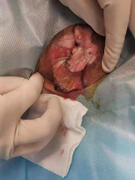 |
Figure 1 Clinical photographs prior to treatment. |
 |
Figure 2 (A and B) Basophilic cell-forming tumor masses can be seen, with dense cellular arrangements and a lack of the classic fenestrated arrangement around them. (Hematoxylin and eosin;×20). |
 |
Figure 3 (A) Ki-67(+), (B) P16(+). (Immunohistochemical staining;×20). |
We gave her a full course of VMAT:45Gy/1.8Gy/25f, and 1 month later added local skin electron beam radiotherapy:24Gy/2.0Gy/12f, radiotherapy machine: ELEKTA Infinity. After radical radiotherapy, the patient felt significantly better in terms of itching and painful symptoms.At the follow-up after one month, the vulva was widely erythematous and swollen, the patches and ulcers at the bilateral labia majora were significantly reduced and thinned. After three months, the plaques and ulcers were basically healed and no new lesions appeared.The biopsy was taken again from the central location of the original ulcer and showed: no tumor cells were seen (Figure 4). Regular follow-up checkups every three months.The skin lesions of the patients before and after radical radiotherapy are shown in (Figure 5).
 |
Figure 4 Pathology sampling biopsy three months after the end of treatment.(Hematoxylin and eosin;×20). |
 |
Figure 5 (A)Just after the VMAT, (B) three months after completion of treatment, (C) six months after completion of treatment. |
Discussion
VSCC is highly malignant, not absolutely excised, and easily metastasized.7 In addition, the misdiagnosis rate of SCC is high.Especially crater-like seborrheic keratoses with redundancy, hyperkeratosis, echinoderm and pseudocyst keratoses can be immensely similar to SCC.8 In this case, the pathology of two successive biopsies was seborrheic keratosis, the result probably originated from the random occurrence of collision tumors. The third biopsy diagnosed VSCC.In terms of treatment options, the patient was diagnosed with early-stage, very high-risk vulvar SCC according to the American Joint Committee on Cancer 8th edition TNM staging,9 and the National Comprehensive Cancer Network Guidelines have previously recommended complete surgical resection as the first choice.9 However, the patient’s lesion was large and wrapped around the urethral orifice, so complete surgery required partial removal of the urethra, resulting in long-term postoperative retention of a urinary catheter, which greatly reduced her quality of life and was against her will. Thus surgical treatment was not used. Six months after radical VMAT treatment, the patient had no significant recurrence and achieved a radical tumor cure without surgery.
This result may result from the high conformality, precision, and effectiveness of VMAT compared to conventional radiotherapy. Previous studies suggest that the effect of radiotherapy alone for vulvar cancer is poor and the local recurrence rate is high. This result is considered to be related to factors such as a moist vulva, poor tolerance of radiation by a skin mucosa, large vulvar tumors, a lack of satisfactory dose distribution of radiotherapy, and difficulty in receiving irradiation doses for vulvar cancer to achieve the radical therapeutic effect. The advantage of VMAT is that it can address these issues by continuously varying the dose release rate, rack speed, and multileaf collimator position so that the dose is delivered in a better conformation to reduce toxicity, significantly increase the release rate, and enhance biological effects. It also shortens treatment time to avoid patient movement affecting peripheral tumor coverage and leading to marginal recurrence.1,10 The literature shows similar planning target volume (PTV) coverage and homogeneity for IMRT and VMAT, while VMAT has significantly improved bowel dose and treatment delivery time. However, dosimetric studies comparing fixed-field IMRT and VMAT in gynecologic malignancies are limited. VMAT has been proven to be better than IMRT for gynaecological malignancies. In comparison to IMRT, which has been shown to be superior to conventional 3D-CRT, VMAT displayed a reduction in dose to the bladder, bowel and rectum and achieved better PTV coverage with improved conformity and homogeneity.11 Deng et al12 showed no significant differences between organs at risk doses but better dose conformity, slightly less monitor units and shorter delivery times with VMAT. In addition, the patient’s HPV infection and P16-positive status enhanced the sensitivity of the tumor to radiation and made it less likely to re-proliferate after radiotherapy. The mechanisms underlying the enhanced radiosensitivity to HPV positivity have been identified in head and neck cancers, namely impaired DNA repair and enhanced apoptotic response.13 These mechanisms may also be applicable in VSCC.Among those with vulvar SCC, P16-positive patients respond better to radical radiotherapy than P16-negative ones, and P16 overexpression is a prognostic factor independent of HPV status.14 In this patient with high PD-L1 expression, pointers exist for the addition of immune-targeted therapy to prevent a recurrence, but our successful treatment without recurrence by intensity-modulated radiotherapy alone suggests that appropriate choices in the use of radiotherapy methods and doses can remove the unnecessary burden. The woman was of advanced age, in poor physical condition, and badly tolerated radiation, but there were no associated post-radiotherapy complications after treatment, demonstrating the safety of VMAT. A recent international study has published recommendations on how to better standardize the indications for radiotherapy and radiotherapy techniques.9 Considering the patient’s history of diabetes and poor wound healing ability, topical electron wire treatment provides better dose distribution, and weak penetration, but works only on superficial tissues, protecting deeper tissues in a superior way while promoting skin healing.15 Nevertheless, it remains to be seen whether the same excellent results can be achieved with VMAT alone in HPV- and P16-negative and lymph node-positive VSCC. An analysis of data from the National Cancer Data Base showed that radiation therapy combined with concurrent chemotherapy was superior to radiation therapy alone in lymph node-positive vulvar cancer.2 In some cases, proximal margins are more common, such as when the disease is close to structures such as the clitoris, urethra, or vulva, however, the NCCN guidelines recommend re-excision of positive margins or those that are classified as proximal (<8 mm). If surgery is not possible, the guidelines recommend adjuvant vulvar radiation therapy. This approach is now being questioned as more studies aim to determine if this improves overall outcomes in patients with closer margins, especially given the impact of surgery and vulvar radiotherapy on quality of life thereafter.16 The safety of cutting margins with smaller margins is also the subject of research.17
Hence, in the future, clinicians should be more flexible in the use of radiotherapy and assess whether patients with VSCC of different etiological pathways need enhanced doses of VMAT or combined chemotherapy. Whether patients treated with surgery need routine postoperative adjuvant VMAT to avoid the side effects of radiotherapy while shortening the surgical margins to protect vulvar structures and functions and reduce the marginal recurrence rate.
The patient had a large skin lesion area and a previous history of nasal basal cell carcinoma, which may be related to the working environment of metal refining and smelting.She mentioned that the pungent odor could be smelled at work, so she could not exclude occupational exposure to heavy metals caused by inhalation. Studies over the past two decades have shown that the redox activity of metals can lead to oxidative stress, a state of imbalance between oxidation and antioxidation in the body, causing DNA damage, lipid peroxidation (LPO), and protein changes triggering other effects that can eventually lead to cancer.4,18 Among them, copper can directly accelerate the formation of reactive oxygen species (ROS) through reactions such as the Fenton reaction and the Haber-Weiss reaction, and exposure to high levels of copper can significantly reduce glutathione levels.18,19 Redox reactions of the inactive metals arsenic, lead and cadmium show their toxic effects by binding to sulfhydryl groups of proteins and reducing glutathione.4,20 A decrease in glutathione and an increase in ROS can reduce the antioxidant defense system of cells and alter the composition and integrity of cell membranes to increase the sensitivity of cells to oxidative attack. Glutathione peroxidase (GPX) and catalase (CAT) are recognized as major scavengers of hydrogen peroxide and the total thiol molecule (TTM) has the antioxidant function of thiol groups to lower the free-radical volume and is used to evaluate the degree of protein oxidation. Alishahi M et al4 detected reduced serum GPX, CAT, and TTM levels in copper smelters, confirming the decrease in antioxidant levels despite the presence of increased metal-generated free radicals. There is increasing evidence in recent years that oxidative stress plays a key role in the pathogenesis of squamous cell carcinoma,21–23 but the blood oxidative profile has not yet been determined and further studies are needed. In basal cell carcinoma, there is no clear evidence linking metal exposure to the disease, but it is rare for individuals to have two different pathological types of primary skin tumors at the same time, and oxidative stress mechanisms are also applicable in basal cell carcinoma.24 Consequently, more meta-analyses are needed to explore the correlation between metal exposure and skin tumors.
Conclusion
VMAT is a promising treatment method for VSCC, which may play a better effect than surgery, especially when facing patients with HPV infection and P16 positivity.It is worth further research and promotion to provide new ideas for the therapy of VSCC. The occupational health department should pay attention to the physical health condition of workers engaged in metal purification work in the copper mining environment, and clarify the relationship between heavy metal occupational exposure and skin tumors at a deeper level to provide more protection for workers.
Ethics Statement
The publications of images were included in the patient’s consent for publication of the case. Institutional approval has been obtained to publish the case details.
Consent Statement
The authors certify that they have obtained all appropriate patient consent forms. The patient signed a consent form for the publication of the case details and images.
Disclosure
The authors report no conflicts of interest in this work.
References
1. Mazumder K, Elangovan A, Rai B, et al. Conventional radiotherapy and intensity-modulated radiotherapy in carcinoma vulva: an experience from a tertiary medical center of India. South Asian J Cancer. 2019;8(1):41–43. doi:10.4103/sajc.sajc_66_17
2. Gynecological Tumor Committee of China Anti-Cancer Association. Guidelines for the diagnosis and treatment of vulvar malignancies (2021 edition). China Oncol. 2021;31(6):533–545. doi:10.19401/j.cnki.1007-3639.2021.06.11
3. Chargari C, Petit A, Escande A, et al. Role of radiotherapy in the management of vulvar cancer: recommendations of the French society for radiation oncology. Cancer Radiother. 2022;26(1–2):286–291. doi:10.1016/j.canrad.2021.08.014
4. Alishahi M, Malekirad A, Mandegary A, et al. A study on oxidative stress, hematological and biochemical parameters in copper smelter workers. Toxin Rev. 2020;8:1–6. doi:10.1080/15569543.2017.1278708
5. Liu W, Yang HS, Zheng SY, et al. Oxidative stress genes in patients with esophageal squamous cell carcinoma: construction of a novel prognostic signature and characterization of tumor microenvironment infiltration. BMC Bioinform. 2022;23(1):406. doi:10.1186/s12859-022-04956-9
6. Schipani G, Del DE, Todaro G, et al. Arsenic and chromium levels in hair correlate with actinic keratosis/non-melanoma skin cancer: results of an observational controlled study. Ital J Dermatol Vene. 2021;156(6):703–708. doi:10.23736/S2784-8671.20.06600-6
7. Hinten F, Molijn A, Eckhardt L, et al. Vulvar cancer: two pathways with different localization and prognosis. Gynecol Oncol. 2018;149(2):310–317. doi:10.1016/j.ygyno.2018.03.003
8. Ogita A, Ansai SI. What is a solitary keratoacanthoma? A benign follicular neoplasm, frequently associated with squamous cell carcinoma. Diagnostics. 2021;11(10):1848. doi:10.3390/diagnostics11101848
9. Abu-rustum NR, Yashar CM, Bradley K, et al. NCCN guidelines version 1.2022 vulvar cancer (squamous cell carcinoma). National Comprehensive Cancer Network; 2021. Available from: https://www.nccn.org/guidelines/guidelines-detail?category=1&id=1476.
10. Moncharmont C, Vallard A, Guy JB, et al. Real-life efficacy of volumetric modulated arc therapy in head and neck squamous cell carcinoma. Eur Ann Otorhinolary. 2017;134(3):165–169. doi:10.1016/j.anorl.2016.12.005
11. Knapp P, Eva B, Reseigh G, et al. The role of volumetric modulated arc therapy (VMAT) in gynaecological radiation therapy: a dosimetric comparison of intensity modulated radiation therapy versus VMAT. J Med Radiat Sci. 2019;66(1):44–53. doi:10.1002/jmrs.311
12. Deng X, Han C, Chen S, et al. Dosimetric benefits of intensity-modulated radiotherapy and volumetric-modulated arc therapy in the treatment of postoperative cervical cancer patients. J Appl Clin Med Phys. 2017;18(1):25–31. doi:10.1002/acm2.12003
13. Proctor L, Hoang L, Moore J, et al. Association of human papilloma virus status and response to radiotherapy in vulvar squamous cell carcinoma. Int J Gynecol Cancer. 2020;30(1):100–106. doi:10.1136/ijgc-2019-000793
14. Yap ML, Allo G, Cuartero J, et al. Prognostic significance of human papilloma virus and p16 expression in patients with vulvar squamous cell carcinoma who received radiotherapy. Clin Oncol-Uk. 2018;30(4):254–261. doi:10.1016/j.clon.2018.01.011
15. Yan TT, Hou JJ, Zhang SY, et al. Analysis of the efficacy of surgical excision combined with electron beam irradiation in the treatment of keloid scars. J Tissue Eng Reconstr Surg. 2021;17(4):317–319.
16. Milliken S, May J, Sanderson PA, et al. Reducing the radicality of surgery for vulvar cancer: are smaller margins safer? Minerva Obstet Gynec. 2021;73(2):160–165. doi:10.23736/S2724-606X.20.04743-7
17. Giannini A, D’Oria O, Chiofalo B, et al. The giant steps in surgical downsizing toward a personalized treatment of vulvar cancer. J Obstet Gynaecol Re. 2022;48(3):533–540. doi:10.1111/jog.15103
18. Malekirad AA, Mirabdollahi M, Pilehvarian AA, et al. Status of neurocognitive and oxidative stress conditions in iron-steel workers. Toxicol Ind Health. 2015;31(7):670–676. doi:10.1177/0748233713483196
19. Babu MY, Palanikumar L, Nagarani N, et al. Cadmium and copper toxicity in three marine macroalgae: evaluation of the biochemical responses and DNA damage. Environ Sci Pollut R. 2014;21(16):9604–9616. doi:10.1007/s11356-014-2999-0
20. Ajsuvakova OP, Tinkov AA, Aschner M, et al. Sulfhydryl groups as targets of mercury toxicity. Coordin Chem Rev. 2020;(8):417. doi:10.1016/j.ccr.2020.213343
21. Shi N, Chen F, Zhang X, et al. Suppression of oxidative stress and NFκB/MAPK signaling by lyophilized black raspberries for esophageal cancer prevention in rats. Nutrients. 2017;9(4):413. doi:10.3390/nu9040413
22. O’Farrell NJ, Phelan JJ, Feighery R, et al. Differential expression profiles of oxidative stress levels, 8-oxo-dG and 4-HNE, in barrett’s esophagus compared to esophageal adenocarcinoma. Int J Mol Sci. 2019;20(18). doi:10.3390/ijms20184449
23. Carrara IM, Melo GP, Bernardes SS, et al. Looking beyond the skin: cutaneous and systemic oxidative stress in UVB-induced squamous cell carcinoma in hairless mice. J Photoch Photobio B. 2019;195:17–26. doi:10.1016/j.jphotobiol.2019.04.007
24. Sander CS, Hamm F, Elsner P, et al. Oxidative stress in malignant melanoma and non-melanoma skin cancer. Brit J Dermatol. 2003;148(5):913–922. doi:10.1046/j.1365-2133.2003.05303.x
 © 2023 The Author(s). This work is published and licensed by Dove Medical Press Limited. The
full terms of this license are available at https://www.dovepress.com/terms
and incorporate the Creative Commons Attribution
- Non Commercial (unported, 3.0) License.
By accessing the work you hereby accept the Terms. Non-commercial uses of the work are permitted
without any further permission from Dove Medical Press Limited, provided the work is properly
attributed. For permission for commercial use of this work, please see paragraphs 4.2 and 5 of our Terms.
© 2023 The Author(s). This work is published and licensed by Dove Medical Press Limited. The
full terms of this license are available at https://www.dovepress.com/terms
and incorporate the Creative Commons Attribution
- Non Commercial (unported, 3.0) License.
By accessing the work you hereby accept the Terms. Non-commercial uses of the work are permitted
without any further permission from Dove Medical Press Limited, provided the work is properly
attributed. For permission for commercial use of this work, please see paragraphs 4.2 and 5 of our Terms.