
Back to Journals » Clinical Ophthalmology » Volume 19
Comparison of Post-Operative Outcomes between IOP-20 and IOP-50 in Phacoemulsification with Active Fluidics System: Randomized Single Blinded Trial
Authors Wavikar CM, Tanna MN, Wavikar GC, Kale CB, Setia MS 
Received 22 May 2025
Accepted for publication 25 September 2025
Published 30 September 2025 Volume 2025:19 Pages 3573—3582
DOI https://doi.org/10.2147/OPTH.S542284
Checked for plagiarism Yes
Review by Single anonymous peer review
Peer reviewer comments 2
Editor who approved publication: Dr Scott Fraser
Chandrashekhar M Wavikar,1 Mamta N Tanna,1 Gargi C Wavikar,2 Chandrashekhar B Kale,1 Maninder Singh Setia3
1Department of Ophthalmology, Wavikars Eye Hospital and Wavikar Eye Institute, Thane, Maharashtra, India; 2Department of Ophthalmology, Sri Sankaradeva Nethralaya, Guwahati, Assam, India; 3Department of Epidemiology, Wavikars Eye Hospital and Wavikar Eye Institute, Thane, Maharashtra, India
Correspondence: Maninder Singh Setia, Wavikars Eye Hospital and Wavikar Eye Institute, 4 th and 5 th Floor, Amber Arcade, Near Lodha Paradise, Bhivandi Bypass Road, Thane, MH, India, 400601, Email [email protected]
Purpose: To compare the central corneal thickness, mitigation events, post-operative vision, and complications in patients who underwent cataract surgery using the CENTURION ® Vision System with the ACTIVE SENTRY ® (Alcon Industries, Inc.) system and were assigned to one of two groups: intraocular pressure (IOP) group 20 or 50.
Setting: Wavikar Eye Institute, Thane, India.
Design: A prospective comparative two arm, randomized, single blinded, interventional study.
Methods: A total of 107 eyes were randomized to either the IOP-20 (n=55) or IOP-50 (n=52) group. All patients underwent phacoemulsification with an active fluidics system performed by a single surgeon; intraocular pressure (IOP) was set at either 20 or 50 mmHg.
Results: The mean [SD] baseline CCT in the IOP-20 group was 511.9 [35.7] μm and it was 521.1 [34.8] μm in the IOP 50 group (p=0.18). It had changed to 530.6 [39.8] μm in the IOP-20 group and 539.5 [39.2] μm in the IOP 50 (p=0.24). The mean [SD] ECD at the end of day 90 was 2423.0 [204.6] cells/mm2 in the IOP-20 group and 2476.1 [236.0] cells/mm2 in the IOP-50 group (p=0.22). Approximately 22% of the eyes in the IOP-20 group and 77% of the eyes in the IOP-50 group required active surge mitigation; this difference was statistically significant (p< 0.001).
Conclusion: We found that the phacoemulsification with an active fluidics system handpiece at 20 mmHg provided a safe and stable environment for cataract surgery.
Keywords: active fluidics system, IOP-20, IOP-50, safety, effectiveness
Introduction
Phacoemulsification was first introduced over fifty years ago. Although it initially faced some resistance, it has become an important procedure of choice for cataract surgeries.1,2 Over the years, there have been many changes to improve the efficacy of surgery and improvisation of the phaco delivery systems.3–5 While on one hand there were changes to improve the cutting efficiency, whereas, on the other hand there have been many improvisations in fluidics systems to improve the surgical efficiency and to reduce the surgical trauma.5,6 One of the major improvements in fluidics was the conversion of the gravity fluidics system (GFS) to the active fluidics system (AFS).7,8 The major objective of this system is to reduce fluctuations in the anterior chamber with changing intraocular pressure along with changes in the vacuum. The GFS requires a high bottle height of 70–110 cm. A higher bottle height ensures greater flow into the anterior chamber; however, this may also lead to an increase in intraocular pressure.9 This high intraocular pressure may lead to injuries, such as subtle retinal ischaemia, endothelial cell loss, inflammation, or posterior capsule rupture, and phacoemulsification with a lower bottle height has been reported to be favourable for the corneal endothelium.10–12
Therefore, search was on to identify a fluidics system which would allow the surgeon to operate at low intraocular pressures. This was needed because one of the prime concerns of surgeons was to decrease the anterior chamber surge after occlusion breaks so as to reduce the risk of potential complications such as posterior capsular rent and endothelial damage. As research progressed, better AFS was introduced which allowed the surgeon to work at a lower bottle height. It also allowed surgeons to have a preset IOP and maintain it with active replenishment of fluids.13 One of the important mechanisms to reduce surge during phacoemulsification maneuvers is the introduction of ACTIVE SENTRY ® (Alcon Industries, Inc.) with Quick-valve Technology.14 Previous studies have compared various outcomes between GFS and AFS. Some authors have found that the total aspiration time and corrected distance visual acuity (CDVA) were better in the GFS group than in the AFS group.8 Other authors found no significant differences in CDVA, cumulative dissipated energy (CDE), fluidics, or viscoelastic device use between AFS and GFS groups.15 A systematic review by Su and colleagues concluded that although the AFS is superior to GFS in terms of CDE and estimated fluid usage, there was no significant difference in post-operative endothelial cell density or central corneal thickness.16 The active fluidics system allows different intraocular pressure (IOP) settings, such as higher IOP levels (in the range of 50–80 mm of Hg) or a more physiologic one in the range of 20–30 mm of Hg.15,17 A surgeon may choose the former because of its perioperative stabilisation of IOP or the latter because of its intra-operative and post-operative outcomes, and patient satisfaction.18,19 A major concern in the use of lower IOPs may be stabilisation of the anterior chamber during surgery. However, there is limited information in the surgeons’ understanding of whether the new AFS is safe enough from the point of view of surgical efficiency, tissue injury, and surgical complications at different IOP levels.13,16
Thus, we designed this trial to compare the central corneal thickness in patients who underwent cataract surgery using phacoemulsification with an active fluidics system and assigned them to one of two groups: intraocular pressure group 20 or 50. We also compared the number of mitigation events, post-operative vision, and complications between the two groups.
Methods
This was a prospective, comparative, two-arm, randomised, single-blinded, interventional study of 107 eyes (55 in the IOP-20 group and 52 in the IOP-50 group) in a single centre in Thane, India.
Study Site, Population, and Procedures
This study was conducted at the Wavikar Eye Institute in Thane, India. It is a private eye hospital with all ophthalmological specialties. We included patients who were assigned to undergo cataract surgery at our centre. The inclusion criteria were as follows: a) age 50–85 years; b) immature cataract (Grade NS 2 according to the Lens Opacities Classification System), c) dilated pupil size – 6.00 mm or above; and d) anterior chamber depth of 2.6 to 4.5 mm. The exclusion criteria were as follows: 1) any other preexisting ocular pathologies such as Fuchs’ endothelial dystrophy, pseudo-exfoliation, and glaucoma; 2) any intraocular complications such as capsular rent or detachment of Descemet’s membrane; and 3) post-operative complications such as infection.
The two arms in this study were the IOP-20 and IOP-50 group. All patients who consented to participate in this study were randomly assigned to one of the two groups using a computer-generated randomization schedule. The operating surgeon was aware of the group; however, the participants were not. Therefore, this was a single-blind study. The primary endpoint of the study was the change in central corneal thickness (CCT) post-operatively on day one. The other endpoints were as follows: endothelial cell density (ECD) changes, active surge mitigation (ASM) events corrected distance visual acuity (CDVA), cumulative disseminated energy, ultrasound time, pain/comfort during surgery, and anterior chamber flare and cells. We assessed the parameters on post-operative days 1, 30 (± 7 days), and 90 (± 10 days). The CCT and ECD were measured using EM-4000 Specular Microscope (Tomey GmBH, Germany). The assessor of these parameters at follow-up was not blinded.
Based on a mean difference of 20 μm in CCT between the two groups post-operatively, an alpha of 0.05, a power of 80%, and a sample size of 51 in each group was estimated. To account for any losses to follow-up, we proposed to recruit 55 participants in each group. In the 10P-50 group, three participants could not complete their 30 days follow-up and were excluded from the analysis. Thus, the final sample was 55 in the IOP-20 group and 52 in the IOP-20 group, respectively.
Surgical Methods
All the patients underwent phacoemulsification surgery with an active fluidics system handpiece by a single surgeon The IOP was set at either 20 mm Hg or 50 mm Hg. These surgeries were performed under topical anaesthesia with 0.5% proparacaine. A clear corneal incision was made using a 2.4 mm keratome blade. The “soft shell” technique with two viscoelastic devices were used to support the anterior chamber as well as protect the corneal endothelium. Continuous curvilinear capsulorhexis (5∼5.5 mm in diameter) was performed. The standardised parameters for each stem of phacoemulsification and irrigation/aspiration are presented in Table 1. The vacuum in the IOP-20 group was 450 mm Hg and it was 500 mm Hg in the IOP-50 group. The aspirational flow rate was 20 cc/min in the IOP-20 group and it was 28 cc/min in the IOP-50 group. These parameters were chosen by the surgeon after careful titration for the most stable anterior chamber with these two IOPs.
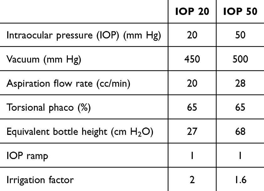 |
Table 1 Standardized Parameters for Each Step of Phacoemulsification and Irrigation/Aspiration |
Phacoemulsification was performed using the direct chop technique. A hydrophobic intraocular lens was implanted into the capsular bag, and viscoelastics were aspirated from the anterior chamber and behind the lens using the bimanual irrigation –aspiration technique. The clear corneal incision and sideport incisions were closed by hydrating the anterior stroma. A Likert scale (numerical pain rating scale) was used immediately at the end of the surgery to measure intra-operative pain. Intra-operative parameters including cumulative dissipated energy (CDE), ultrasound time, active surge mitigation (ASM) instances, and patient pain levels were recorded. We used topical medication 0.5% moxifloxacin for 7 days, and 1.0% prednisolone acetate and sodium hyaluronate (0.18%) for one month post-operatively.
Statistical Methods
We assessed the normality of the data using the Shapiro–Wilk test. We estimated the mean and standard deviation (SD) for normal data and median and interquartile range (IQR) for non-normal data. The means between the two groups were compared using the unpaired t-test, and the medians were compared using the Mann–Whitney Wilcoxon test. The proportions were compared using the chi-square test or Fisher’s exact test for low expected cell counts. We used logistic regression models for multivariate analysis of binary outcomes and estimated the odds ratios (OR) and 95% confidence intervals (CI). Linear regression models were also used for multivariate analysis of linear outcomes. We performed a complete case analysis, and a p value of < 0.05 was considered statistically significant. Data were analysed using Stata Version 17 (StataCorp, College Station, Texas, USA).
This study was approved by the Ethics Committee of Ethicare Independent Ethics Committee. EEC/WEI/2023/001 (dated 06 September 2023). This trial was registered in the Clinical Trials Registry of India (CTRI: 2023/10/058235). All participants provided written informed consent before participating in the study. The study was conducted in accordance with the Declaration of Helsinki and the International Good Clinical Practice Guidelines.
Results
The mean [SD] age of the participants was 65.0 [7.2] years, and there was no significant difference between the IOP-20 and IOP-50 groups (65.3 [6.8] vs 64.8 [7.7]; p=0.72). Approximately 48% of the eyes included in this study were right eyes, and 52% were left eyes; there was no significant difference between the two groups. The mean [SD] anterior chamber depth (ACD) was 3.14 [0.39] mm; there was no significant difference in the mean [SD] ACD between these two groups (3.18 [0.44] vs 3.09 [0.34]; p=0.22). The details are presented in Table 2.
 |
Table 2 Baseline and Certain Surgical Characteristics in 107 Eyes from the Two Groups (IOP 20 and IOP 50), Thane, India |
The mean [SD] baseline CCT in the IOP-20 group was 511.9 [35.7] μm and it was 521.1 [34.8] μm in the IOP-50 group (p=0.18). It had changed to 530.6 [39.8] μm in the IOP-20 group and 539.5 [39.2] μm in the IOP-50 (p=0.24). There was no significant difference in the median (IQR) change between the two groups [17 (6, 32) vs 18 (5.5, 25.5); p=0.72)] (Table 3). In the multivariate analysis, after adjusting for age, ACD, ultrasound time, and preoperative CDVA, we did not find any significant difference in CCT in the IOP-20 group compared to the IOP-50 group (estimate: −13.2, 95% CI: −28.3, 1.8; p=0.09).
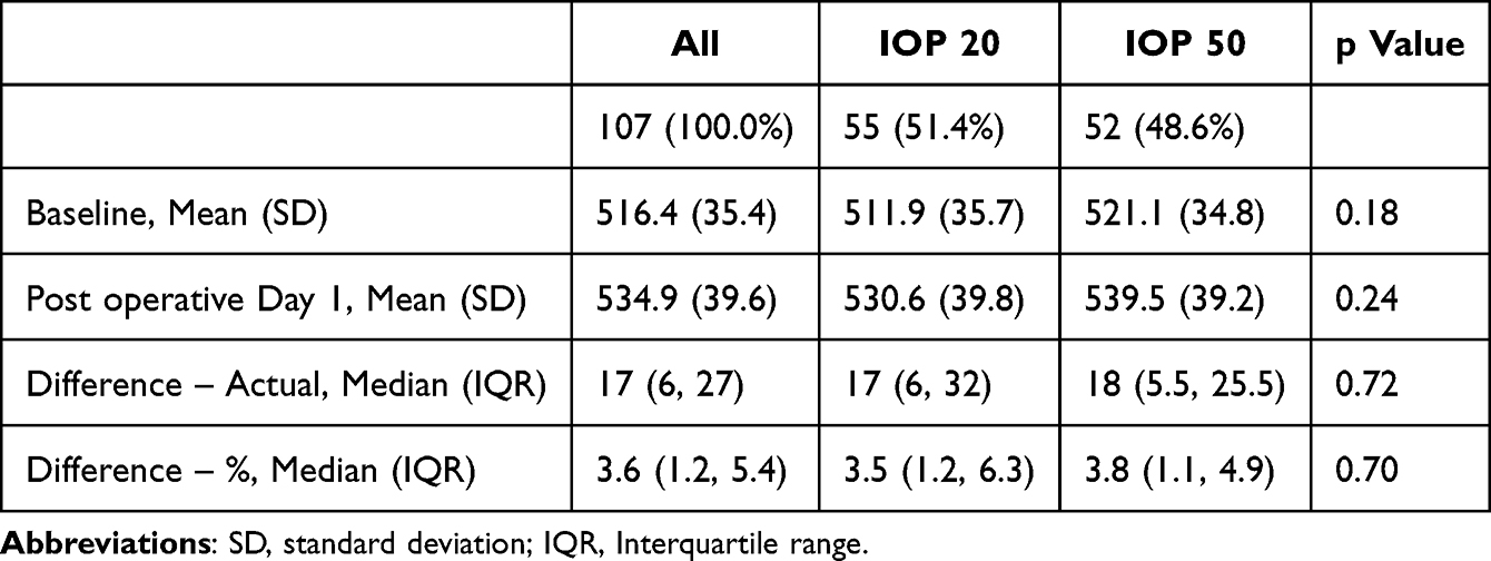 |
Table 3 Values of Central Corneal Thickness (CCT) μm in Both Groups (IOP 20 and IOP 50) |
The mean [SD] endothelial cell density (ECD) in the IOP-20 group was 2534.0 [200.7] cells/mm2 and it was 2585.8 [234.7] cells/mm2 in the IOP-50 group (p=0.22). The ECD reduced post-operatively in both groups (Figure 1). However, we found that the change was not significantly different between the groups on post-operative days 1, 30, and 90 (Table 4). The mean [SD] ECD at the end of day 90 post-operatively was 2423.0 [204.6] cells/mm2 in the IOP-20 group and it was 2476.1 [236.0] cells/mm2 in the IOP-50 group (p=0.22). The details of each visit are presented in Table 4. In the multivariate analysis, after adjusting for age, ACD, ultrasound time, and preoperative CDVA, we did not find any significant difference in ECD in the IOP-20 group compared with the IOP-50 group on day 1 (estimate: −70.4, 95% CI: −155.2, 14.3; p=0.10), day 30 (estimate: −54.0, 95% CI: −138.6, 30.5; p=0.21), or day 90 (estimate: −55.9, 95% CI: −138.3, 26.5; p=0.18).
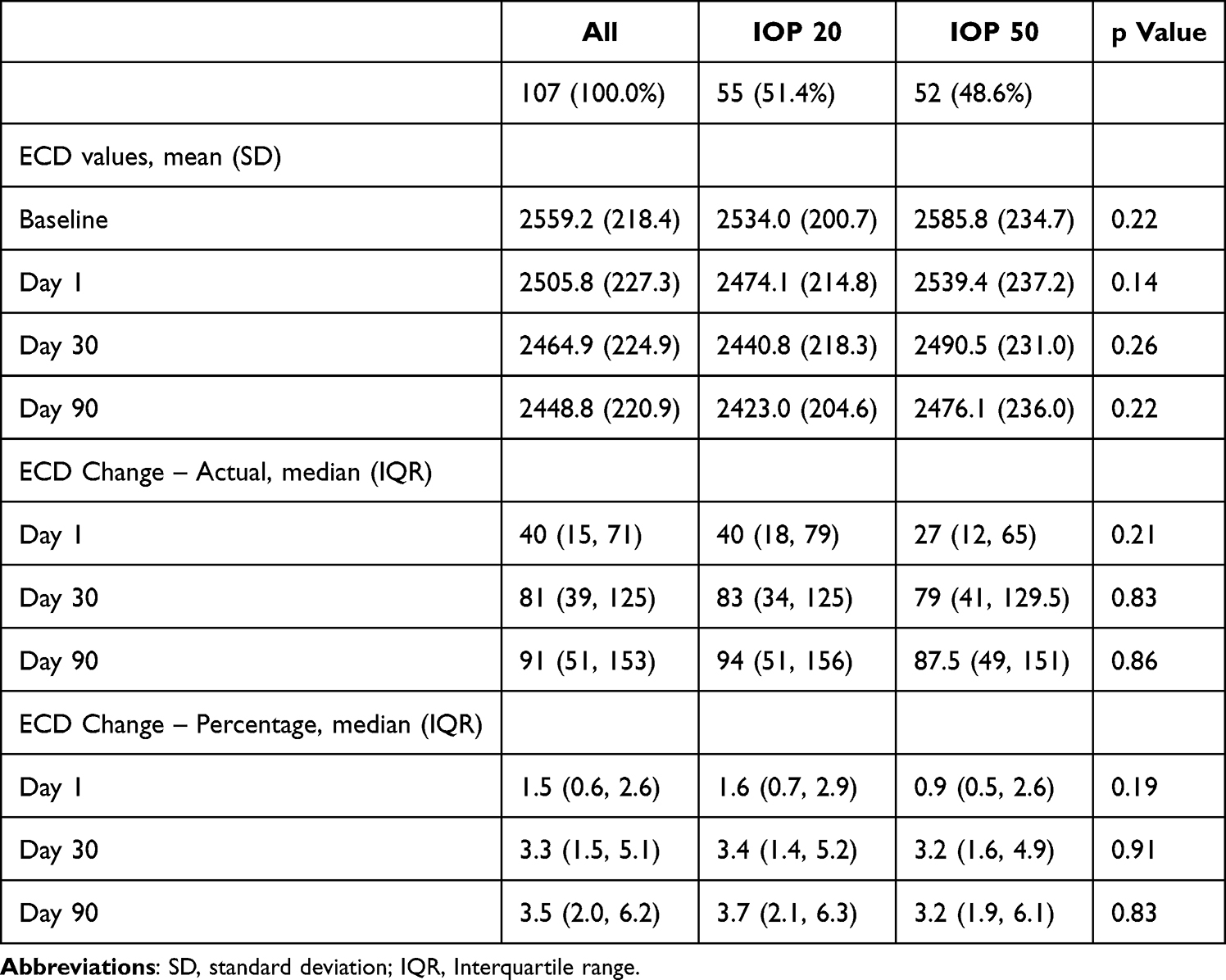 |
Table 4 Pre and Post-Operative (Day 1, Day 30, and Day 90) Endothelial Cell Density (ECD, Cells/mm2) in Both Groups (IOP 20 and IOP 50), Thane, India |
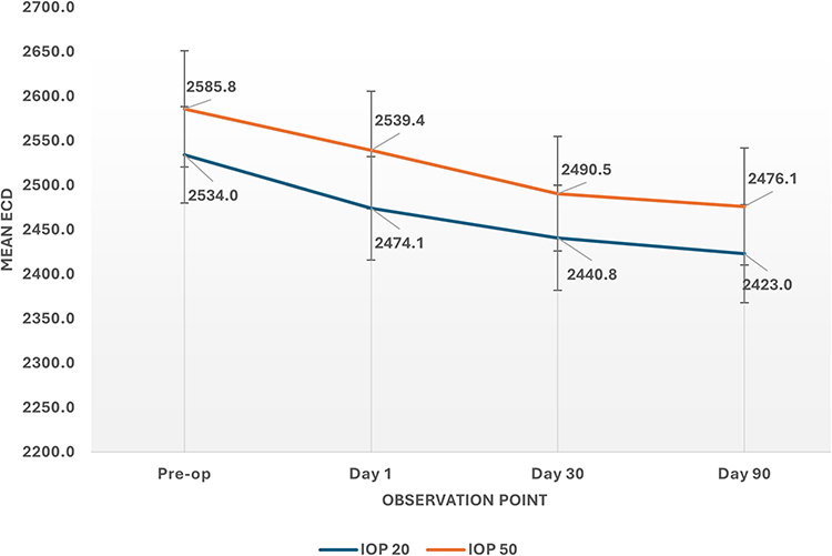 |
Figure 1 Figure showing the changes in mean endothelial cell density (ECD) (cells/mm2) in the IOP 20 and IOP 50 group, Thane, India. |
Approximately 22% of the eyes required active surge mitigation in the IOP-20 group and 77% of the eyes required active surge mitigation in the IOP-50 group; the difference was statistically significant (p<0.001). Approximately 40.4% of eyes in the IOP-50 group required six or more mitigation events compared with 1.8% in the IOP-20 group; the difference was statistically significant (p<0.001). In the logistic regression models, even after adjusting for age, ultrasound time, ACD, and preoperative CDVA, we found that the odds for mitigation events were significantly lower in the IOP-20 group compared with the IOP-50 group (OR, 0.08; 95% CI: 0.03, 0.20; p<0.001). Even though the OR was higher for age (OR: 1.06, 95% CI: 1.00, 1.14; p=0.059), the association was not statistically significant. There was no significant difference in the mean ultrasound time or CDE between the two groups. The proportion of eyes with corrected distance visual acuity (CDVA) of 6/6 and 6/9 on the Snellen’s chart was not significantly different between the two groups (96.4% vs 100.0%; p=0.50). There was a reduction in the median values of the refraction parameters (sphere, cylinder, and spherical equivalent) in both groups one-month post-operatively compared with baseline (Figure 2A–C). However, there were no significant differences in these parameters (sphere, cylinder, and spherical equivalent) between the two groups, either at baseline or post-operatively. The mean [SD] quality of life post-surgery was 9.25 [1.25] in the IOP-20 group and 9.63 [0.86] in the IOP-50 group (p=0.07). There was no significant difference in post-surgery heaviness or pain post-surgery in both these groups (Table 5).
 |
Table 5 Intraoperative Parameters, Active Mitigation Events, Vision, and Overall Quality of Surgery in Both These Groups |
 |
Figure 2 (A–C) Figure showing the refractive parameters (Sphere [2A], Cylinder [2B], and Spherical equivalent [2C]) at baseline and post-operatively at 1 month in the IOP 20 and 50 groups, Thane, India. |
Discussion
Thus, in this trial, we found that the mean CCT was not significantly different post-operatively between the IOP-20 and IOP-50 groups, and the increase in CCT was also not significantly different between these groups. Similarly, we found that the change in ECD did not differ between groups. Visual acuity and post-surgery quality of life were also not significantly different between groups. However, the number of active surge mitigation instances was significantly higher in the IOP-50 group compared with the IOP-20 group.
The main advantages of the active fluidics system are its role in chamber stability and improved responsiveness during the phacoemulsification procedure.18,20,21 It detects pressure changes in real time, responds to fluctuations, and helps minimize pressure surges, thus adequately stabilizing IOP during procedures.22,23 A study by Kim and colleagues compared the Active Sentry handpiece with the Centurion Ozil handpiece and reported active surge mitigation actuations as one of the outcomes.14 They found that ASM was activated for a mean (SD) of 16.99 (9.64) times per surgery in those operated with Active Sentry. Furthermore, they also found that the mean number of ASM actuations increased with the grade of cataract and was associated with age, preoperative CDVA, and ultrasound time. Jirásková and Stěpanov also reported that IOP was significantly lower in the Active Sentry group compared with the Centurion Ozil group during phacoemulsification, and they found that a post-occlusion surge was not observed in the Active Sentry group.24 Wang and coworkers also reported lower ASM in eyes operated at 20 mm Hg than in eyes operated at 60 mm Hg.19 ASM actuations may be due to: 1) surgeon/surgical technique (stop and chop vs direct chop), 2) factors related to the eye (such as age, grade of cataract, or condition of the pupil), or 3) phacoemulsification parameters set during the procedure.14,25,26 We had controlled for the surgeon factors and the factors related to the eye, as all cases were operated upon by a single surgeon using the same chopping technique and we only included Grade NS 2 cataracts with well-dilating pupils (careful case selection as per the inclusion criteria). Therefore, the only difference between the two groups responsible for the differences in ASM instances was the third factor, that is, phacoemulsification parameters. Between the two groups, the major and significantly different parameter was the IOP of 50 and 20 mm Hg. Fewer ASM events indicated that there were fewer instances of micro flutters (and therefore flutters), which are a prerequisite for the activation of ASM. A smaller number of micro flutters or surges in the anterior chamber indicates a safer milieu for surgeons during phacoemulsification. As seen in our data, the IOP-20 group had a significantly lower likelihood of ASM actuations, even after adjusting for age, preoperative CDVA, ultrasound time, and ACD. Thus, the IOP-20 group, which had a significantly lower number of ASM incidents, had a lower number of micro flutters, thereby providing a safer environment for the surgeon during cataract surgery. This point is important as one of the main concerns while operating at a low IOP is the stability of the anterior chamber.
Other than the number of ASM actuations, we did not find any significant differences in the post-operative central corneal thickness, endothelial cell density, ultrasound time, cumulative dissipated energy, and post-operative vision between the two groups. Vasavada and colleagues compared the intra-operative and post-operative outcomes between the traditional and Active Sentry groups. They found that Active Sentry had lower CDE and lower post-operative increase in CCT.27 Another systematic review reported that active fluidics system is better than gravity fluidics systems in terms of CDE, fluid usage, and total aspiration time; however, there were no significant differences in post-operative CCT or ECD.16 Loss of endothelial cell density may be associated with age, cataract grade, and surgical factors (such as surgical time, ultrasound, irrigation volume, or use of viscoelastic devices).28–31 Liu and colleagues found that a significantly higher proportion of individuals experienced “no pain” in the low IOP-AFS group than in the normal IOP-GFS group.15 While operating at a low IOP of 20 mmHg, another major concern is whether low IOP will increase the endothelial cell loss during cataract surgery. A previous study by Rauen and coworkers found a smaller reduction in ECD in low-IOP settings compared with high-IOP settings.32 Another study by Scarfone and colleagues found that endothelial cell loss was not significantly different between low and high infusion groups.25 Our study showed that there was no statistically significant difference in the endothelial cell loss between both groups, even after adjusting for age, ACD, ultrasound power, and preoperative CDVA. Thus, change in IOP or lowering the IOP, potentially, did not adversely affect endothelial cell loss.
We conducted this study as a single-blinded study, and we did not blind the operating surgeon (it was necessary for the surgeon to be aware of the phacoemulsification settings from the view of patient’s safety). Some other studies have also assessed retinal parameters, such as retinal vessel density and retinal thickness.8,33,34 However, these studies compared the active fluidics system with gravity fluidics system. We compared the refractory, vision, corneal parameters, and surge mitigation for two different IOP values of the active fluidics system. Although we assessed the pain scores, it is likely that the patient may have considered the entire procedure and post-procedure for reporting the scores, rather than the actual phacoemulsification portion of the procedure.
Despite these limitations, our study makes a useful contribution to literature. To establish a new technique, it is important to assess the effectiveness as well as safety of this technique compared with the current procedures. In our study, the 20-mm Hg intraocular pressure group was the new technique or the interventional group, and the 50-mm Hg intraocular pressure group was the control group. The effectiveness between these two groups, as assessed by ultrasound time and CDE, did not show any significant differences. Safety was compared by measuring the differences in CCT, endothelial cell loss, and CDVA, which also did not show any significant differences. Patient comfort was assessed by comparing the pain scores, and no significant differences were found. The assessment of safe milieu or intraocular environment for cataract surgeons was done by comparing episodes of active surge mitigation in these two groups; these episodes were significantly lower in the IOP-20 group compared with the IOP-50 group. Thus, we found that the phacoemulsification with an active fluidics system handpiece at 20 mm Hg provided a safe and stable environment for cataract surgery in our patients.
What is Known?
- One of the major improvements in the fluidics of the phacoemulsification system was the conversion of the gravity fluidics system (GFS) to the active fluidics system (AFS).
- Intraocular pressure (IOP) in the AFS may be associated with post-operative outcomes, including vision.
What Does This Study Add?
- The mean central corneal thickness (CCT) was not significantly different post-operatively between the IOP-20 and IOP-50 groups. The increase in CCT was also not significantly different between these groups.
- Changes in endothelial cell density, visual acuity, and quality of life after surgery were not significantly different between the groups.
- The number of active surge mitigation instances was significantly higher in the IOP-50 group than that in the IOP-20 group.
- Thus, we found that the phacoemulsification with an active fluidics system handpiece at 20 mm Hg provided a safe and stable environment for cataract surgery in our patients.
Data Sharing Statement
Deidentified data may be made available after reasonable request. The requests can be made at [email protected]. The data will be made available after ethical and administrative approval and in accordance with local data protection rules.
Funding
This study was an investigator-initiated trial funded by a research grant from Alcon # 88131349. The funder did not have any role in design, analysis, and interpretation of results.
Disclosure
The authors have no financial or proprietary interest in the product, method, or material described herein.
References
1. Pandey SK, Milverton EJ, Maloof AJ. A tribute to Charles David Kelman MD: ophthalmologist, inventor and pioneer of phacoemulsification surgery. Clin Exp Ophthalmol. 2004;32(5):529–533.
2. Vasumathi R. Remembering Dr. Charles D. Kelman and development of phacoemulsification. TNOA J Ophthalmic Sci Res. 2018;56(1):45–50. doi:10.4103/tjosr.tjosr_36_18
3. Gurnani B, Kaur K. Phacoemulsification. StatPearls [Internet]. 2023.
4. Davis G. The evolution of cataract surgery. Mo Med. 2016;113(1):58–62.
5. Howard Fine I. the evolution of phacoemulsification. September 2007. Available from: https://crstoday.com/articles/2007-sep/crst0907_05-php.
6. Gimbel HV, da Reitz Pereira C. Advances in phacoemulsification equipment. Curr Opin Ophthalmol. 2002;13(1):30–32. doi:10.1097/00055735-200202000-00007
7. Kasturi N, Jacob N, Jossy A, Chakrabarti A. Emerging innovations in cataract surgery. Delhi J Ophthalmol. 2022;32(6):61–71.
8. Luo Y, Li H, Chen W, et al. A prospective randomized clinical trial of active-fluidics versus gravity-fluidics system in phacoemulsification for age-related cataract (AGSPC). Ann Med. 2022;54(1):1977–1987. doi:10.1080/07853890.2022.2098375
9. Stodola E. A look at phaco fluidics Cataract: back to basics. September 2022. Available from: https://www.eyeworld.org/2022/a-look-at-phaco-fluidics/.
10. Meyer JJ, Kuo AF, Olson RJ. The risk of capsular breakage from phacoemulsification needle contact with the lens capsule: a laboratory study. Am J Ophthalmol. 2010;149(6):882–6e1.
11. Tsinopoulos IT, Lamprogiannis LP, Tsaousis KT, et al. Surgical outcomes in phacoemulsification after application of a risk stratification system. Clin Ophthalmol. 2013;7:895–899. doi:10.2147/OPTH.S42726
12. Suzuki H, Oki K, Shiwa T, Oharazawa H, Takahashi H. Effect of bottle height on the corneal endothelium during phacoemulsification. J Cataract Refract Surg. 2009;35(11):2014–2017.
13. Luo Y, Xu G, Li H, Ma T, Ye Z, Li Z. Application of the active-fluidics system in phacoemulsification: a review. J Clin Med. 2023;12(2):611. doi:10.3390/jcm12020611
14. Kim JY, Kim H, Jun I, Kim TI, Seo KY. Effect and safety of pressure sensor-equipped handpiece in phacoemulsification system. Korean J Ophthalmol. 2023;37(5):387–394. doi:10.3341/kjo.2022.0157
15. Liu Y, Hong J, Chen X. Comparisons of the clinical outcomes of Centurion((R)) active fluidics system with a low IOP setting and gravity fluidics system with a normal IOP setting for cataract patients with low corneal endothelial cell density. Front Med Lausanne. 2023;10:1294808. doi:10.3389/fmed.2023.1294808
16. Su YC, Lee YY, Su YC. Active-fluidics versus gravity-fluidics in lens extraction: a systematic review and meta-analysis of randomized controlled trials. Eur J Ophthalmol. 2023;33(1):247–256. doi:10.1177/11206721221107512
17. Brezin AP, Monnet D, Lignereux F, Rozot P, Jilet L, Abdoul H. Impact of a handpiece with a built-in fluidics pressure sensor on phacoemulsification: a multicentre prospective comparative study. BMJ Open Ophthalmol. 2023;8(1):e001431. doi:10.1136/bmjophth-2023-001431
18. Lubeck D. Enhancing the safety profile of cataract procedures with the active sentry handpiece and intrepid hybrid tip. 2019. Available from: https://crstoday.com/articles/key-insights-centurion-with-active-sentry-handpiece-and-intrepid-hybrid-tip/enhancing-the-safety-profile-of-cataract-procedures-with-the-active-sentry-handpiece-and-intrepid-hybrid-tip-3.
19. Wang S, Tao J, Yu X, Diao W, Bai H, Yao L. Safety and prognosis of phacoemulsification using active sentry and active fluidics with different IOP settings - a randomized, controlled study. BMC Ophthalmol. 2024;24(1):350. doi:10.1186/s12886-024-03626-z
20. Berdahl JP. Cataract Surgery Gets Two Valuable Incremental Improvements. 2019. Available from: https://crstoday.com/articles/key-insights-centurion-with-active-sentry-handpiece-and-intrepid-hybrid-tip/cataract-surgery-gets-two-valuable-incremental-improvements-2.
21. Cionni RJ. Evaluating two key safety advances in the CENTURION vision system. 2019. Available from: https://crstoday.com/articles/key-insights-centurion-with-active-sentry-handpiece-and-intrepid-hybrid-tip/evaluating-two-key-safety-advances-in-the-centurion-vision-system-3.
22. Brézin AP. Surgical performance of CENTURION® vision system with ACTIVE SENTRY®. 2024. Available from: https://crstoday.com/articles/surgical-performance-of-centurion-vision-system-with-active-sentry/surgical-performance-of-centurion-vision-system-with-active-sentry?toc=true.
23. Early AD, Ifantides C, Melendez RF, Zhu D. More physiologic IOP in cataract surgery matters: here’s why. 2024. Available from: https://crstoday.com/articles/more-physiologic-iop-in-cataract-surgery-matters-heres-why/more-physiologic-iop-in-cataract-surgery-matters-heres-why?toc=true.
24. Jiraskova N, Stepanov A. Our experience with active sentry and centurion ozil handpieces. Cesk Slov Oftalmol. 2021;77(1):18–21. doi:10.31348/2021/1
25. Scarfone HA, Rodriguez EC, Rufiner MG, et al. Vitreous-lens interface changes after cataract surgery using active fluidics and active sentry with high and low infusion pressure settings. J Cataract Refract Surg. 2024;50(4):333–338. doi:10.1097/j.jcrs.0000000000001359
26. Sabur H, Unsal U. The efficacy and safety profile of cataract procedures performed with a sensor-embedded handpiece and a new hybrid phaco tip. Eur J Ophthalmol. 2022;32(6):3438–3443. doi:10.1177/11206721221084764
27. Vasavada V, Agrawal D, Vasavada SA, Vasavada AR, Yagnik J. Intraoperative performance and early postoperative outcomes following phacoemulsification with three fluidic systems: a randomized trial. J Refract Surg. 2024;40(5):e304–e12. doi:10.3928/1081597X-20240314-02
28. O’Brien PD, Fitzpatrick P, Kilmartin DJ, Beatty S. Risk factors for endothelial cell loss after phacoemulsification surgery by a junior resident. J Cataract Refract Surg. 2004;30(4):839–843. doi:10.1016/S0886-3350(03)00648-5
29. Hayashi K, Hayashi H, Nakao F, Hayashi F. Risk factors for corneal endothelial injury during phacoemulsification. J Cataract Refract Surg. 1996;22(8):1079–1084. doi:10.1016/S0886-3350(96)80121-0
30. Morya AK, Gurnani B, Mishra D, et al. Comparison of corneal endothelial cell loss during manual small-incision cataract surgery using viscoelastic-assisted nucleus removal versus continuous balanced salt solution plus technique - randomized controlled trial. Indian J Ophthalmol. 2022;70(11):3960–3966. doi:10.4103/ijo.IJO_1871_22
31. Chen HC, Huang CW, Yeh LK, et al. Accelerated corneal endothelial cell loss after phacoemulsification in patients with mildly low endothelial cell density. J Clin Med. 2021;10(11).
32. Rauen MP, Joiner H, Kohler RA, O’Connor S. Phacoemulsification using an active fluidics system at physiologic vs high intraocular pressure: impact on anterior and posterior segment physiology. J Cataract Refract Surg. 2024;50(8):822–827. doi:10.1097/j.jcrs.0000000000001457
33. Zhao Y, Wang D, Nie L, et al. Early changes in retinal microcirculation after uncomplicated cataract surgery using an active-fluidics system. Int Ophthalmol. 2021;41(5):1605–1612. doi:10.1007/s10792-021-01694-4
34. Liu X, Fang Y, Zhou Y, Wang M, Luo Y. Dynamic changes in retinal vessel density observed by optical coherence tomography angiography after phacoemulsification: active vs gravity fluidics system. Arq Bras Oftalmol. 2022;85(2):205–207. doi:10.5935/0004-2749.20220093
 © 2025 The Author(s). This work is published and licensed by Dove Medical Press Limited. The
full terms of this license are available at https://www.dovepress.com/terms
and incorporate the Creative Commons Attribution
- Non Commercial (unported, 4.0) License.
By accessing the work you hereby accept the Terms. Non-commercial uses of the work are permitted
without any further permission from Dove Medical Press Limited, provided the work is properly
attributed. For permission for commercial use of this work, please see paragraphs 4.2 and 5 of our Terms.
© 2025 The Author(s). This work is published and licensed by Dove Medical Press Limited. The
full terms of this license are available at https://www.dovepress.com/terms
and incorporate the Creative Commons Attribution
- Non Commercial (unported, 4.0) License.
By accessing the work you hereby accept the Terms. Non-commercial uses of the work are permitted
without any further permission from Dove Medical Press Limited, provided the work is properly
attributed. For permission for commercial use of this work, please see paragraphs 4.2 and 5 of our Terms.