
Back to Journals » International Journal of Women's Health » Volume 18

Comparison of Ovarian Stimulation Protocols in Hypogonadotropic Hypogonadism: Case Series and Review of Literature
Authors Zulfa F  , Tjahyadi D, Riyadi AS, Sasotya RMS, Rachmawati A
Received 10 May 2026
Accepted for publication 11 July 2026
Published 23 July 2026 Volume 2026:18 612954
DOI https://doi.org/10.2147/IJWH.S612954
Checked for plagiarism Yes
Review by Single anonymous peer review
Peer reviewer comments 2
Editor who approved publication: Dr Marta Barba
Video abstract of “Ovarian stimulation protocols in hypogonadotropic hypogonadism” [612954].
Views: 2
Fadhilah Zulfa, Dian Tjahyadi, Anthony Sudono Riyadi, Raden Mas Sonny Sasotya, Anita Rachmawati
Department of Obstetrics and Gynecology, Faculty of Medicine, Universitas Padjadjaran, Dr. Hasan Sadikin General Hospital, Bandung, Indonesia
Correspondence: Fadhilah Zulfa, Department of Obstetrics and Gynecology, Faculty of Medicine, Universitas Padjadjaran, Dr. Hasan Sadikin General Hospital, Jl. Pasteur No. 38, Bandung, West Java, 40161, Indonesia, Tel +6222 203 2530 ext 3240, Email [email protected]
Introduction: Hypogonadotropic hypogonadism (HH) is one of the least common etiologies for female infertility and needs different approach when dealing with this condition, especially in the presence of low ovarian reserve. Standard measures are unlikely to be of use and therefore personalized and customized ovarian stimulation must be employed for this patient because the threshold of response to stimulation may differ substantially from that of normal patient. Moreover age-dependent decline in ovarian response for this subset of patients has not been established. Facing with this situation, the question arises at what regimens and at what appropriate dose to start stimulation as previous studies reported a higher dose of gonadotrophins and longer day of injections are needed than other IVF patients because of "dormant” ovaries that need to be primed before follicular response is achieved.
Case Illustration: We present two cases of infertile women who had hypogonadotropic hypogonadism and underwent In Vitro Fertilization (IVF) procedures with different stimulation protocols. Both patient exhibit clinical and biochemical features of hypogonadotropic hypogonadism and underwent individualized ovarian stimulation protocol. The first patient had diminished ovarian reserve and concomitant male factor infertility, received combination of follitropin delta, menotropin, and adjuvant of growth hormone during stimulation and subsequent intrauterine pregnancy was achieved. While in the second patient, despite a relatively more favorable ovarian reserve and normal semen parameters, underwent stimulation with recombinant FSH and LH but failed to achieve clinical pregnancy.
Conclusion: Hypogonadotropic hypogonadism remains a challenging cause of infertility requiring individualized ovarian stimulation strategies. This case series demonstrated heterogenous reproductive outcomes following different stimulation protocols, suggesting that tailored gonadotropin-based therapy may be effective in selected patients with HH. The potential benefit of adjunctive growth hormone requires further evaluation in larger prospective studies.
Keywords: reproductive techniques, endocrinology, ovarian stimulation protocols, hypogonadotropic hypogonadism
Introduction
Hypogonadotropic hypogonadism (HH) is an endocrine disorder resulting from insufficient secretion of gonadotropin-releasing hormone (GnRH) by the hypothalamus or inadequate production of gonadotropins by the pituitary gland. It is classified as a WHO Group I ovulatory disorder and accounts for approximately 10% of all anovulatory disorders, making it a relatively uncommon but clinically significant cause of female infertility.1 This hormonal insufficiency leads to impaired gonadotropin secretion, resulting in dysregulation of the hypothalamic–pituitary–gonadal (HPG) axis and subsequent ovarian dysfunction. Women with hypothalamic hypogonadism (HH) frequently develop anovulation and infertility, necessitating hormonal treatment.2
In routine clinical practice, ovulation induction using daily administration of low-dose exogenous gonadotropins constitutes the standard therapeutic approach for women with HH and is applicable across all etiological subtypes. A defining feature of HH in women is the absence or profound deficiency of endogenous luteinizing hormone (LH), rendering gonadotropin preparations containing an LH component essential for effective treatment. The introduction of urinary-derived gonadotropins, particularly human menopausal gonadotropin (hMG), which provides both follicle-stimulating hormone (FSH) and LH activity, represented a significant advancement by enabling physiologic replacement of deficient endogenous hormones and facilitating follicular recruitment and development.3,4
Substantial progress in assisted reproductive technology (ART), including in vitro fertilization (IVF), has expanded therapeutic options for patients who fail to conceive following ovulation induction alone. However, due to the low prevalence of HH, no single protocol was accepted worldwide for the success of IVF procedures in patients with HH.4,5
Patient with HH needs different approach when dealing with this condition especially in the presence of low ovarian reserve. Standard measures are unlikely to be of use and therefore personalized and customized ovarian stimulation must be employed for this patient because the threshold of response to stimulation may differ substantially from that of normal patient.6 We present two cases of infertile women who had hypogonadotropic hypogonadism and underwent IVF procedures with different stimulation protocols. One of the women was successfully conceived but the other was not. We would like to analyze the efficacy of different procedures to determine which one has the most potential for achieving high in vitro fertilization (IVF) success rates in individuals with hypogonadotropic hypogonadism (HH).
Case Illustration
Case 1
A 41-year-old woman, married for 13 years and a normal sexual life, was admitted to our reproductive endocrinology and infertility clinic in February 2024, seeking treatment for infertility. She had a history of failed intrauterine insemination and laparoscopic cystectomy one year before. Baseline hormonal evaluation revealed diminished ovarian reserve with anti-Mullerian Hormone (AMH) level of 0.48 ng/mL. Serum follicle-stimulating hormone (FSH) level and estradiol (E2) level were 0.81 mIU/mL and 5.00 pg/mL, respectively. Semen analysis of her husband resulted in teratozoospermia. From transvaginal ultrasound, her uterus was hypoplasia and with low antral follicle count (1 follicle in the left ovary and 3 follicles in the right ovary). She was then diagnosed with hypogonadotropic hypogonadism and planned for IVF using short-protocol.
Controlled ovarian stimulation was initiated using recombinant FSH (rFSH; Rekovelle®) 20 µg/day, human menopausal gonadotropin (hMG; Menopur®) 150 IU/day, and recombinant growth hormone (GH; Saizen®) 9 IU/day for six consecutive days (Table 1a). Ultrasound evaluation on stimulation day 7 demonstrated two developing follicles in the left ovary, while no dominant follicle was observed in the right ovary. A GnRH antagonist (0.25 mg/day subcutaneously) was subsequently added for five days to prevent premature luteinizing hormone surge. On stimulation day 12, ovulation was triggered with recombinant human chorionic gonadotropin (r-hCG) 250 µg subcutaneously, and GH administration was discontinued. At trigger day, serum E2 and progesterone levels were 127.09 pg/mL and 0.56 ng/mL, respectively. Oocyte retrieval was performed 36 hours later, yielding 2 oocytes from the left ovary. An embryo transfer was performed 3 days afterward, during which two embryos (7-cell and 8-cell grade embryos) were transferred. Clinical pregnancy was successfully achieved.
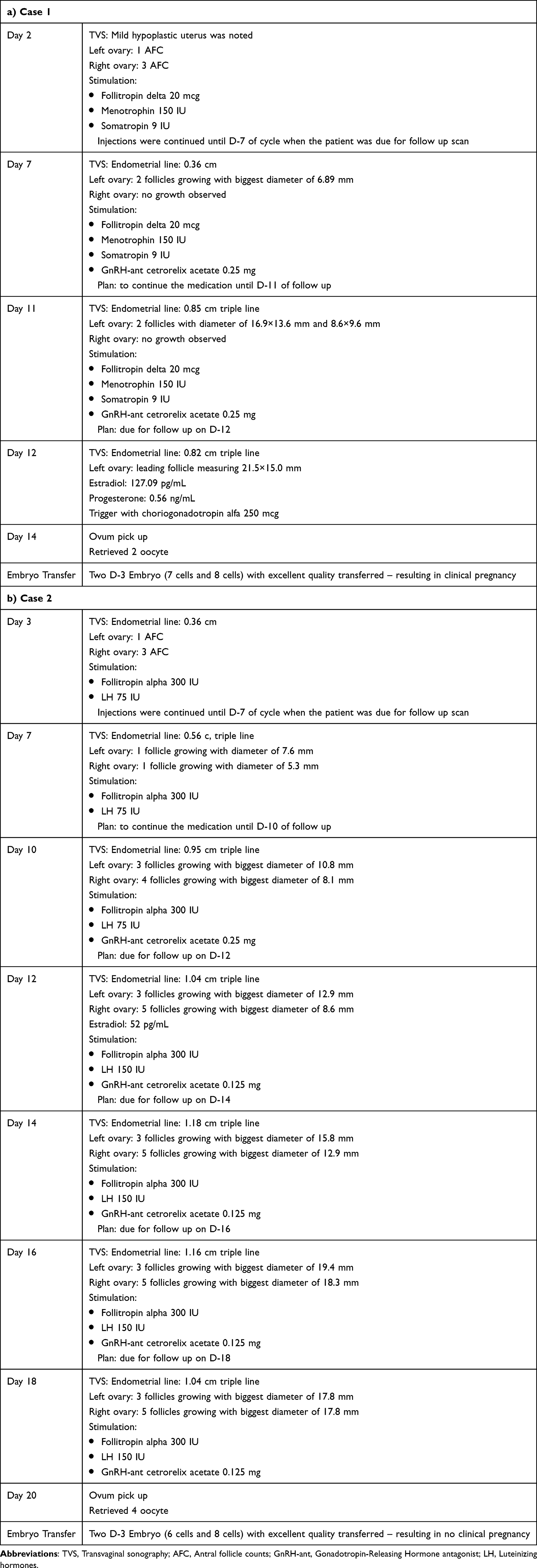 |
Table 1 Summary of in vitro Fertilization Stimulation |
Case 2
A 36-year-old woman with a 9-year history of infertility presented to our fertility clinic in July 2022 for assisted reproductive treatment. She had a known history of secondary amenorrhea secondary to hypogonadotropic hypogonadism and had previously received cyclic estrogen-progesterone replacement therapy. Baseline hormonal assessment performed one year prior to IVF demonstrated profoundly suppressed gonadotropin levels, with FSH of 0.25 mIU/mL, luteinizing hormone (LH) of 0.10 mIU/mL, and estradiol level of 15.68 pg/mL. Thyroid-stimulating hormone (TSH) and prolactin levels were within normal ranges at 2.51 mIU/mL and 5.58 ng/mL, respectively. AMH level was 1.08 ng/mL, suggesting relatively preserved ovarian reserve. Her partner’s semen analysis was normal. Initial transvaginal ultrasound revealed a thin endometrium measuring 3.6 mm, with three antral follicles in the right ovary and one follicle in the left ovary. The patient was subsequently scheduled for IVF with combined FSH and LH stimulation.
Ovarian stimulation was initiated using recombinant FSH (rFSH) 300 IU/day combined with recombinant LH (rLH) 75 IU/day (Table 1b). After four days of stimulation, transvaginal ultrasonography demonstrated minimal follicular response, with persistence of only one follicle in each ovary. Stimulation was therefore continued, and repeat ultrasound evaluation on stimulation day 8 demonstrated improved ovarian response, including four follicles in the right ovary and three follicles in the left ovary, accompanied by trilaminar endometrial development with endometrial thickness of 9.5 mm. A GnRH antagonist (0.25 mg/day subcutaneously) was subsequently initiated. On stimulation day 10, serum estradiol level remained low at 52 pg/mL despite the presence of eight developing follicles (five in the right ovary and three in the left ovary). The rLH dose was therefore increased to 150 IU/day, while the GnRH antagonist dose was reduced to 0.125 mg/day. Final oocyte maturation was triggered on stimulation day 16 using recombinant hCG 250 µg subcutaneously, followed by oocyte retrieval 36 hours later. A total of four oocytes were successfully retrieved, consisting of two oocytes from each ovary. Due to clinical considerations, all embryos were cryopreserved, and frozen embryo transfer was performed two months after oocyte retrieval. However, serum β-hCG measured two weeks after embryo transfer was <2.3 mIU/mL, indicating unsuccessful implantation and absence of clinical pregnancy. The clinical profile of both cases was presented in Table 2.
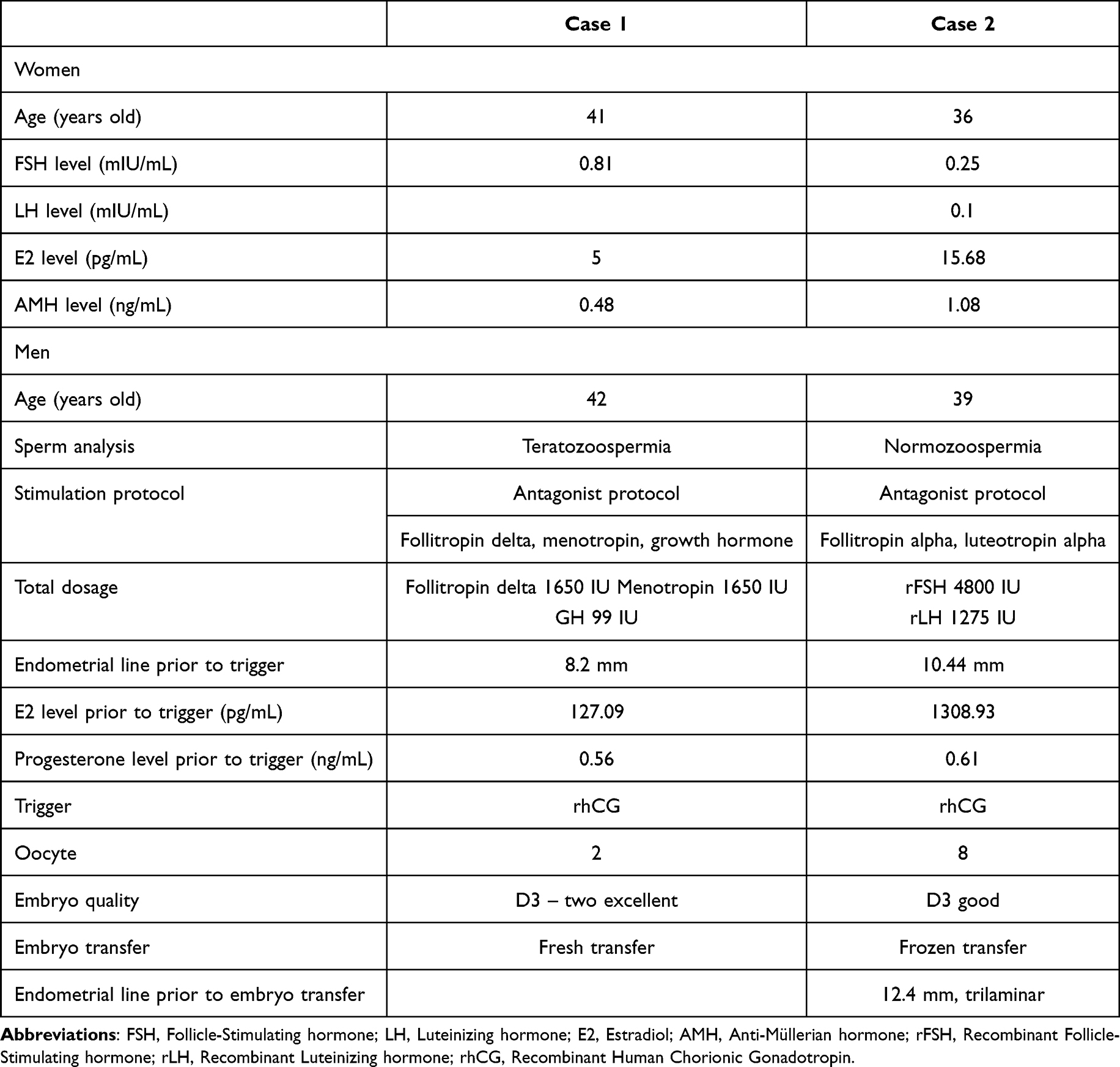 |
Table 2 Clinical Profile of Case 1 and Case 2 |
Discussion
Hypogonadotropic hypogonadism (HH) is a condition where the gonads fail to function properly due to abnormal gonadotropin levels. It is a rare factor contributing to female infertility. Clinicians face different situation when dealing with fertility issue of HH as this group of patient have different response to ovarian stimulation compared to other ART patients especially when this condition is accompanied by diminished ovarian reserve.7 Baseline gonadotropin levels, commonly employed to predict ovarian response in other infertility etiologies, are unreliable predictors in this population, and ovarian responsiveness can only be adequately assessed following exposure to stimulation.8
In the present case series, both patients demonstrated markedly suppressed gonadotropin levels consistent with HH, yet showed different ovarian responses and reproductive outcomes following individualized IVF stimulation protocols. These findings emphasize the heterogeneity of ovarian responsiveness in HH patients and highlight the importance of tailored stimulation strategies based on age, ovarian reserve, and follicular dynamics.
In Case 1, clinical pregnancy was successfully achieved despite advanced maternal age and diminished ovarian reserve, as reflected by the low AMH level and poor baseline follicular count. The favorable outcome observed may be associated with the combined use of recombinant FSH (rFSH), human menopausal gonadotropin (hMG), and adjuvant growth hormone (GH).
The choice of gonadotropin given is important in HH patient. The use of hMG paved the way in replacing the absent of endogenous hormone. Because hMG provides both FSH and LH activity, it is particularly advantageous in LH-deficient women. Chronic LH deficiency in HH has been implicated in impaired follicular and oocyte development. Stimulation with FSH alone in this population has been associated with increased gonadotropin requirements, suboptimal follicular development, reduced estradiol production, and lower ovulation rates. The pregnancy rate with hMG preparations was reported as 25% to 30% after an average of 3 treatment cycles in HH woman.9
Beyond its supportive role in folliculogenesis, LH is also essential for the proliferation and differentiation of theca cells and subsequent androgen synthesis, which enhances estrogen production via aromatization in granulosa cells. During the late follicular phase, LH also contributes to progesterone production, facilitating positive estrogen feedback necessary for follicular maturation.10 Numerous studies have emphasized the importance of LH supplementation during controlled ovarian stimulation in improving follicular development and clinical outcomes. Therefore, hMG is often favored over rFSH monotherapy in women with HH who require combined FSH and LH activity.11
In case 2, follicular recruitment enhanced after increasing the rLH dose, indicating that inadequate LH activity may restrict folliculogenesis. LH increases the availability of androgen substrates, helps granulosa cells respond to FSH, and encourages the production of estradiol. Clinical studies have shown that women with severe LH deficiency who take LH supplements have better estradiol production, more oocytes, and better embryo development.12,13 Meta-analytic evidence also has demonstrated significantly higher clinical pregnancy rates (OR: 2.03, p = 0.003), implantation rate (OR: 2.62, p = 0.004) and oocytes (weight mean difference: 1.98, p = 0.03) yield in hypo-responsive patients receiving recombinant LH supplementation compared with those undergoing FSH monotherapy. Hence supplementation with LH is justified in this situation, especially when estradiol response is insufficient despite follicular development.14 Nevertheless, despite relatively preserved AMH levels and higher gonadotropin stimulation doses, pregnancy was not achieved in the second case, which may reflect the complex endocrine characteristics of HH, in which ovarian reserve markers alone do not necessarily predict follicular responsiveness because adequate follicular maturation depends heavily on sufficient LH activity. Figure 1 depicts the importance of LH in promoting the growth of the follicle. Above the ceiling and below the threshold, abnormal follicular development are observed.15
 |
Figure 1 The LH window concept in follicular growth.16 |
Another adjuvant therapy which could improve oocyte yield and pregnancy rates in poor responders is GH supplementation. Case 1 included growth hormone during stimulation, which could affect follicular responsiveness by increasing granulosa cell FSH sensitivity through insulin-like growth factor-1 (IGF-1). Growth hormone has been suggested as an adjunct for poor responders although evidence is still inconsistent.17 GH and GH/IGF have been proposed to enhance ovarian function through modulation of intracellular signaling pathways, including MAPK/ERK, Jak/STAT, and PI3K/Akt, thereby improving cellular proliferation, steroidogenesis, and oocyte competence.18–20
Moreover, evidence stated that the administration of recombinant human growth hormone (rhGH) has been associated with significant increases in the total number of oocytes retrieved, the proportion of metaphase II (MII) oocytes, intracellular adenosine triphosphate (ATP) content, mitochondrial membrane potential, and the prevalence of uniform mitochondrial distribution within oocytes.18 Recent meta-analysis by Chinese investigator in 2023 revealed that the addition of GH was associated with an increase in clinical pregnancy rate (RR: 1.63, 95% CI [1.31–2.03]), and a greater number of oocytes retrieved (MD: 0.91, 95% CI [0.47–1.35]). Moreover, the addition of GH resulted in an increase in the fertilization rate (RR: 1.33, 95% CI [1.18–1.50]), embryo implantation rate (RR: 1.56, 95% CI [1.21–2.01]) and correlate with shorter days and lower dose of gonadotrophins injection during ovarian stimulation.21 Owing to improved clinical outcomes, reduced duration of ovarian stimulation, and lower cumulative gonadotropin requirements, this approach has emerged as a promising therapeutic strategy.
Diminished ovarian reserve (DOR) also constitutes a major limiting factor for treatment success and is characterized by reduced oocyte quantity and quality, decreased AMH levels, diminished antral follicle count, and elevated basal FSH concentrations.22 The American Society for Reproductive Medicine (ASRM) has emphasized that DOR lacks definitive diagnostic criteria and is primarily manifested as a reduction in reproductive potential.23 An additional consideration in HH is the interpretation of ovarian reserve markers. Pandurangi et al highlighted that AMH concentrations may underestimate the true ovarian reserve in women with HH because chronic gonadotropin deficiency suppresses follicular recruitment and growth. Consequently, low AMH values in HH should be interpreted with caution, as ovarian responsiveness may improve substantially following prolonged gonadotropin exposure.6 To overcome these challenges, strategies such as high-dose FSH stimulation (300–450 IU/day) and the use of hMG to provide LH activity like we used in case 1, have been employed to enhance follicular development and oocyte maturation.24
Another important point to note is that GnRH antagonist protocols offer an effective means of preventing premature LH surges without prolonging stimulation duration, which is particularly advantageous in women with HH. Meta-analyses have demonstrated comparable pregnancy rates between antagonist protocols and short agonist protocols in poor responders, owing to the initial flare effect exerted to help in recruitment of the follicles.12
Both patients needed prolonged stimulation, especially in the second case, which reached the trigger point after a long period of exposure to gonadotropins. This delayed response is typical of HH cycles because follicular recruitment relies solely on external stimulation. Prior research has indicated stimulation durations longer in HH patients, yielding satisfactory oocyte retrieval results.25 These findings are consistent with the observations reported by Pandurangi et al, who evaluated ART outcomes in seven women with HH and demonstrated that only one patient responded within the conventional 12-day stimulation period, whereas the remaining patients required more than 12 days of gonadotropin exposure, with stimulation extending up to 54 days in some cases.6 Despite the prolonged treatment duration, oocyte maturity, embryo quality, fertilization rates, and cumulative pregnancy outcomes remained favorable. So, a bad response early on should not lead to an early cycle cancellation in HH, and doctors should expect the stimulation to last longer.
Even though the endocrine pathology was the same, the reproductive outcomes were different in each case. Case 1 successfully underwent embryo transfer and clinical pregnancy, while case 2 did not attain clinical pregnancy subsequent to frozen embryo transfer. This difference shows that the response of the ovaries alone does not predict whether implantation will be successful. Differences in outcomes were probably caused by the age of patient, the embryo’s ability to survive, factors in the uterus, and the endometrium’s ability to accept the embryo. These results correspond with earlier studies suggesting that HH patients may attain sufficient oocyte yield while still encountering inconsistent pregnancy outcomes.26
These cases collectively underscore that ovarian stimulation in HH necessitates personalized gonadotropin dosing, sufficient LH support, extended stimulation, and comprehensive monitoring. Estradiol alone should not dictate cycle management; ultrasound-based evaluation should take precedence. Additionally, successful oocyte retrieval does not ensure pregnancy, highlighting the multifactorial nature of reproductive outcomes in HH.
There are a few limitations in this case series. The limited sample size restricts generalizability, and the heterogeneity among cases complicates interpretation. Hormonal monitoring was also restricted. Furthermore, adjuvant growth hormone therapy in case 1 presents potential confounding variables. Nonetheless, these cases offer clinically pertinent insights into ovarian stimulation in HH.
Conclusion
Hypogonadotropic hypogonadism remains a rare and clinically challenging cause of infertility that requires individualized ovarian stimulation strategies during assisted reproductive treatment. In this case series, differing ovarian stimulation protocols resulted in heterogeneous reproductive outcomes, reflecting the multifactorial nature of treatment response in women with HH. Although ovarian stimulation incorporating hMG and adjunctive growth hormone was associated with successful pregnancy in one patient, the heterogeneity of patient characteristics and treatment regimens precludes any definitive conclusion regarding its efficacy. These findings suggest that tailored gonadotropin-based stimulation may be feasible in HH, while the potential role of adjunctive growth hormone warrants further investigation in larger controlled studies.
Informed Consent Patient Statement
Formal ethical approval was not required for the publication of this case series. Written informed consent was obtained from both patients for publication of their clinical information and accompanying data. Patient confidentiality and anonymity were maintained throughout the manuscript.
Acknowledgments
All authors were involved in the conceptualization and design of the study. FZ, DT, ASR, and AR contributed to data curation, material preparation, and formal analysis. FZ, DT, ASR, and RMSS contributed to manuscript drafting. All authors contributed to the conceptualization and study design, critically revised the manuscript for important intellectual content, approved the final version for publication.
This publication charge is funded by Unpad through the Indonesian Endowment Fund for Education (LPDP) on behalf of the Indonesian Ministry of Higher Education, Science and Technology and managed under the EQUITY Program (Contract No. 4303/ B3/DT.03.08/2025 and 3927/UN6. RKT/HK.07.00/2025).
Disclosure
The authors report no conflicts of interest in this work.
References
1. de Jong SM, de Kat AC, Schoonmade LJ, Hillebrand JJ, van Wely M, van Loendersloot LL. Ovarian reserve markers in World Health Organisation type 1 anovulatory disorders: a systematic review and meta-analysis. Reprod Biomed Online. 2026. doi:10.1016/j.rbmo.2026.105768
2. Munro MG, Balen AH, Cho S, et al. The FIGO ovulatory disorders classification system. Hum Reprod. 2022;37(10):2446–10. doi:10.1002/ijgo.14331
3. Fatemi HM, Lawrenz B. Individualized In-Vitro Fertilization: Delivering Precision Fertility Treatment. Cambridge University Press; 2021.
4. Ghumman S. Principles and Practice of Controlled Ovarian Stimulation in ART. Springer New Delhi; 2015.
5. Zhang C-M, Zhang H, Yang R, et al. The reproductive outcome of women with hypogonadotropic hypogonadism in IVF. Front Endocrinol. 2022;13:850126. doi:10.3389/fendo.2022.850126
6. Pandurangi M, Tamizharasi M, Reddy NS. Pregnancy outcome of assisted reproductive technology cycle in patients with hypogonadotropic hypogonadism. J Hum Reprod Sci. 2015;8(3):146–150. doi:10.4103/0974-1208.165141
7. Loutradis D, Drakakis P, Vomvolaki E, Antsaklis A. Different ovarian stimulation protocols for women with diminished ovarian reserve. J Assist Reprod Genet. 2007;24(12):597–611. doi:10.1007/s10815-007-9181-2
8. Lewit N, Kol S. The low responder, hypogonadotropic hypogonadism female patient in IVF: do not give up! Fertil Steril. 2000;74(2):401–402. doi:10.1016/S0015-0282(00)00599-9
9. Mignini Renzini M, Brigante C, Coticchio G, et al. Retrospective analysis of treatments with recombinant FSH and recombinant LH versus human menopausal gonadotropin in women with reduced ovarian reserve. J Assist Reprod Genet. 2017;34(12):1645–1651. doi:10.1007/s10815-017-1034-z
10. Oduwole OO, Huhtaniemi IT, Misrahi M. The roles of luteinizing hormone, follicle-stimulating hormone and testosterone in spermatogenesis and folliculogenesis revisited. Int J Mol Sci. 2021;22(23):12735. doi:10.3390/ijms222312735
11. Jirge PR, Patil MM, Gutgutia R, et al. Ovarian stimulation in assisted reproductive technology cycles for varied patient profiles: an indian perspective. J Hum Reprod Sci. 2022;15(2):112–125. doi:10.4103/jhrs.jhrs_59_22
12. Lunenfeld B, Bilger W, Longobardi S, Alam V, D’Hooghe T, Sunkara SK. The development of gonadotropins for clinical use in the treatment of infertility. Front Endocrinol. 2019;10:429. doi:10.3389/fendo.2019.00429
13. Setti AS, Braga DPDAF, Júnior AI, Júnior EB. Improving implantation rate in 2nd ICSI cycle through ovarian stimulation with FSH and LH in GnRH antagonist regimen. Rev Bras Ginecol Obstet. 2021;43(10):749–758. doi:10.1055/s-0041-1736306
14. Conforti A, Esteves SC, Di Rella F, et al. The role of recombinant LH in women with hypo-response to controlled ovarian stimulation: a systematic review and meta-analysis. Reprod Biol Endocrinol. 2019;17(1):18. doi:10.1186/s12958-019-0460-4
15. Di Segni N, Busnelli A, Secchi M, Cirillo F, Levi-Setti PE. Luteinizing hormone supplementation in women with hypogonadotropic hypogonadism seeking fertility care: insights from a narrative review. Front Endocrinol. 2022;13:907249. doi:10.3389/fendo.2022.907249
16. Palermo R. Differential actions of FSH and LH during folliculogenesis. Reprod Biomed Online. 2007;15(3):326–337. doi:10.1016/s1472-6483(10)60347-1
17. Kolibianakis E, Venetis C, Diedrich K, Tarlatzis B, Griesinger G. Addition of growth hormone to gonadotrophins in ovarian stimulation of poor responders treated by in-vitro fertilization: a systematic review and meta-analysis. Hum Reprod Update. 2009;15(6):613–622. doi:10.1093/humupd/dmp026
18. Pan P, Huang X. The clinical application of growth hormone and its biological and molecular mechanisms in assisted reproduction. Int J Mol Sci. 2022;23(18):10768. doi:10.3390/ijms231810768
19. Homburg R, Ostergaard H. Clinical applications of growth hormone for ovarian stimulation. Hum Reprod Update. 1995;1(3):264–275. doi:10.1093/humupd/1.3.264
20. Carlsson B, Bergh C, Bentham J, et al. Expression of functional growth hormone receptors in human granulosa cells. Hum Reprod. 1992;7(9):1205–1209. doi:10.1093/oxfordjournals.humrep.a137827
21. Cozzolino M, Cecchino GN, Troiano G, Romanelli C. Growth hormone cotreatment for poor responders undergoing in vitro fertilization cycles: a systematic review and meta-analysis. Fertil Steril. 2020;114(1):97–109. doi:10.1016/j.fertnstert.2020.03.007
22. Jirge PR. Poor ovarian reserve. J Hum Reprod Sci. 2016;9(2):63–69. doi:10.4103/0974-1208.183514
23. Yin J, Chang H-M, Li R, Leung PC. Recent progress in the treatment of women with diminished ovarian reserve. Gynecol Obstet Clin Med. 2021;1(4):186–189. doi:10.1016/j.gocm.2021.10.004
24. Leijdekkers JA, Torrance HL, Schouten NE, et al. Individualized ovarian stimulation in IVF/ICSI treatment: it is time to stop using high FSH doses in predicted low responders. Hum Reprod. 2020;35(9):1954–1963. doi:10.1093/humrep/dez184
25. Jiang S, Kuang Y. The effects of low‐dose human chorionic gonadotropin combined with human menopausal gonadotropin protocol on women with hypogonadotropic hypogonadism undergoing ovarian stimulation for in vitro fertilization. Clin Endocrinol. 2018;88(1):77–87. doi:10.1111/cen.13481
26. Abdelaal AE, Behery MA, Abdelkawi AF. Reproductive outcomes in women with hypogonadotrophic hypogonadism, a case series study. Middle East Fertil Soc J. 2021;26(1):11. doi:10.1186/s43043-021-00055-6
 © 2026 The Author(s). This work is published and licensed by Dove Medical Press Limited. The
full terms of this license are available at https://www.dovepress.com/terms
and incorporate the Creative Commons Attribution
- Non Commercial (unported, 4.0) License.
By accessing the work you hereby accept the Terms. Non-commercial uses of the work are permitted
without any further permission from Dove Medical Press Limited, provided the work is properly
attributed. For permission for commercial use of this work, please see paragraphs 4.2 and 5 of our Terms.
© 2026 The Author(s). This work is published and licensed by Dove Medical Press Limited. The
full terms of this license are available at https://www.dovepress.com/terms
and incorporate the Creative Commons Attribution
- Non Commercial (unported, 4.0) License.
By accessing the work you hereby accept the Terms. Non-commercial uses of the work are permitted
without any further permission from Dove Medical Press Limited, provided the work is properly
attributed. For permission for commercial use of this work, please see paragraphs 4.2 and 5 of our Terms.