
Back to Journals » Clinical Ophthalmology » Volume 20
Comparison of Anterior Chamber Depth Changes of Two Hydrophobic Acrylic Intraocular Lenses in the Capsular Bag: A Prospective, Randomized, Contralateral Eye, Multicenter Study
Authors Nakano S  , Arimoto A, Sasaki H
Received 5 November 2025
Accepted for publication 25 January 2026
Published 9 March 2026 Volume 2026:20 579432
DOI https://doi.org/10.2147/OPTH.S579432
Checked for plagiarism Yes
Review by Single anonymous peer review
Peer reviewer comments 2
Editor who approved publication: Dr Scott Fraser
Shinichiro Nakano,1 Atsushi Arimoto,2 Hiroshi Sasaki3
1Department of Ophthalmology, Ryugasaki Saiseikai Hospital, Ryugasaki, Ibaraki, Japan; 2Department of Ophthalmology, Anamizu General Hospital, Anamizu, Ishikawa, Japan; 3Department of Ophthalmology, Kanazawa Medical University, Uchinada, Ishikawa, Japan
Correspondence: Shinichiro Nakano, Department of Ophthalmology, Ryugasaki Saiseikai Hospital, Ryugasaki, Ibaraki, Japan, Tel +81-297-63-7111, Email [email protected]
Purpose: To evaluate postoperative changes in anterior chamber depth (ACD), intraocular lens (IOL) tilt, and decentration of the Clareon IOL in a capsular bag compared with those of the AcrySof IQ IOL.
Patients and Methods: In this prospective multicenter study, we randomly assigned patients scheduled for bilateral cataract surgery to receive the Clareon IOL in one eye and the AcrySof IQ IOL (both manufactured by Alcon Vision LLC, Fort Worth, TX, USA) in the fellow eye. ACD, IOL tilt, decentration, visual acuity, and subjective refraction were measured 1 day, 1 week, and 1, 3, and 6 months after surgery. The primary objective was to demonstrate the equivalence between Clareon and AcrySof IQ IOL regarding ACD changes from 1 day to 6 months.
Results: Overall, 64 eyes from 32 patients were included. The Clareon IOL met the equivalence criteria; the 95% confidence interval for the difference in ACD change (0.026 mm; 95% confidence interval, − 0.02 to 0.072) was within − 0.15 and 0.15 mm. At 6 months, the means ± standard deviations of the ACD, IOL tilt, and decentration were 4.26 ± 0.30 mm, 4.3 ± 1.9°, and 0.17 ± 0.11 mm for Clareon IOL, and 4.23 ± 0.30 mm, 4.0 ± 1.9°, and 0.16 ± 0.11 mm for AcrySof IQ IOL, respectively (P = 0.052, 0.421, and 0.916, respectively). The subjective spherical equivalence at 6 months was − 0.01 ± 1.03 and − 0.08 ± 0.90 D for Clareon IOL and AcrySof IQ IOL, respectively.
Conclusion: The Clareon IOL was equivalent to the AcrySof IQ IOL regarding ACD changes from 1 day to 6 months. No significant differences were observed in ACD, IOL tilt, or refractive outcomes at 6 months between the two IOLs in the same patients. Both IOLs showed good stability in the bag through 6 months.
Keywords: monofocal intraocular lens, anterior chamber depth, tilt, decentration, refraction
Introduction
Cataract surgery is among the most common surgical procedures performed worldwide. After removing the crystalline lens, an intraocular lens (IOL) is implanted. Although the use of multifocal IOLs has recently increased, monofocal IOLs remain predominant and account for more than 90% of all IOL implantations.1,2 Achieving postoperative refraction that aligns with the target refraction based on the lifestyle of the patient is essential to enhance postoperative patient satisfaction. Moreover, postoperative IOL stability within the capsular bag is crucial for ensuring refractive stability after cataract surgery.
Post-operative capsular-bag shrinkage is well-recognized and can compromise IOL stability within the bag.3–6 Additionally, the capsulorhexis size can have an influence on the postoperative IOL position.7 IOL instability in the capsular bag leads to shifts in postoperative anterior chamber depth (ACD), which are associated with changes in subjective refraction.8–11 Furthermore, IOL tilt and decentration can influence astigmatism, coma, and higher-order aberrations, thereby reducing visual image quality.12–15
Evidence indicates that aspheric IOL decentration exceeding 0.4 mm or tilt greater than 7° may adversely affect visual function.16 Additionally, postoperative axial ACD shifts significantly influence refractive outcomes. Olsen et al17 reported that a 0.1-mm change in ACD can result in a refractive error of at least 0.27 diopters (D), whereas Goto et al18 reported a change of 0.13 D for the same variation. Therefore, achieving stable postoperative visual acuity and high patient satisfaction requires implanting IOLs with high in-the-bag stability, minimizing tilt, decentration, and ACD changes.
The Clareon IOL (SY60WF, Alcon Vision LLC, Fort Worth, TX, USA) is based on the AcrySof IQ platform (SN60WF, Alcon Vision LLC); however, it incorporates enhancements aimed at reducing the incidence of glistening and subsurface nanoglistening, as well as modifications to the edge design. Although several studies have reported the long-term clinical outcomes and stability of the Clareon IOL, comparative data on postoperative in-the-bag stability (specifically, ACD, IOL tilt, and decentration) between the Clareon and AcrySof IQ IOLs remain limited.19–22 In particular, no previous reports have examined the equivalence of ACD, tilt, and decentration in the same patients.
Therefore, this study evaluated the equivalence of postoperative IOL stability between Clareon and AcrySof IQ IOLs in the same patients using anterior segment optical coherence tomography (OCT). Although AcrySof IQ IOL remains available in some countries, it may be discontinued in the future; thus, these findings may help ensure that surgeons can transition to Clareon IOLs without concerns regarding postoperative outcomes.
Materials and Methods
Study Design
This was a prospective, randomized, contralateral eye, multicenter study approved by the Kanazawa Medical Hospital Clinical Research Review Board (Approval Number: T012). The study was performed according to the tenets of the Declaration of Helsinki and the Clinical Trials Act and was registered in the Japan Registry of Clinical Trials (jRCTs042200009). All the patients provided written informed consent before participating in the study.
Inclusion criteria were patients aged ≥20 years, age-related bilateral cataract, and patients planning to undergo bilateral cataract surgery with implantation of Clareon or AcrySof IQ IOLs. Patients were excluded from the study if they (1) had additional ocular surgery scheduled between cataract extraction and the 6-month postoperative visit; (2) received toric intra-ocular lenses (IOLs); (3) possessed ocular pathologies or a history of disease that could affect postoperative visual acuity other than cataract; (4) underwent continuous curvilinear capsulorhexis that did not completely cover the optical edge of the IOL; or (5) exhibited capsular-bag or ciliary-zonule abnormalities.
Sixty-four eyes from 32 patients scheduled for bilateral cataract surgery were enrolled. With a one-sided significance level (α) of 0.025 and an equivalence margin of 0.15 mm for the difference in ACD change from 1 day to 6 months between IOLs, assuming an expected paired difference in the ACD change from 1 day to 6 months postoperatively of 0 and a standard deviation (SD) of 0.28 mm, 32 eyes per group were required to achieve a statistical power of 80%.3,23
IOL
The AcrySof IQ IOL (SN60WF) is a single-piece hydrophobic monofocal IOL with a posterior aspheric surface. It is composed of phenylethyl methacrylate and a phenylethyl acrylate copolymer with a water content and refractive index of 0.4% and 1.55, respectively, with an overall diameter of 13.0 mm and a biconvex optic with a 6.0 mm diameter. Additionally, the material incorporates both ultraviolet light–blocking and blue-light filtering chromophores.24
The Clareon IOL (SY60WF) is a single-piece hydrophobic monofocal IOL with an anterior aspheric surface. It is based on the AcrySof IQ IOL platform; nonetheless, it is made of 2-hydroxyethyl methacrylate instead of the phenylethyl methacrylate of the AcrySof IQ IOL. The material difference leads to a water content 1.5%, in situ refractive index of 1.55 at 35° and 550 nm, and glass transition temperature of 9.1°C.25,26 Furthermore, the edge design of Clareon IOL is more precise and sharper than that of the AcrySof IQ IOL.27
Surgical Procedure and Outcomes
Each surgeon performed the routine phacoemulsification procedures. Clareon and AcrySof IQ IOLs were implanted contralaterally in the same patient. Randomization was conducted using the envelope method to determine the IOL model to be implanted in the right or left eye. The first eye to be operated on for each patient was selected based on a comprehensive clinical evaluation. In all patients, complete coverage of the IOL optic edge by the continuous curvilinear capsulorrhexis was confirmed. The IOL power was calculated to target emmetropia or slight myopia using either the SRK/T or the Barrett Universal II formula based on measurements from an IOL-Master 500 or 700 biometer, respectively. The surgery was performed within 1 week of the pre-operative assessment.
Follow-up examinations were performed 1 day, 1 week, and 1, 3, and 6 months postoperatively. At each visit, monocular best-corrected visual acuity (BCVA) was measured using a Landolt ring chart at a 5-m distance, subjective refraction, and slit-lamp examinations. IOL tilt and ACD were measured using anterior segment OCT with either SS-1000 CASIA or CASIA 2 (Tomey, Aichi, Japan). ACD was defined as the distance from the posterior surface of the cornea to the anterior surface of the IOL. IOL decentration was measured using CASIA 2 at each follow-up. Anterior segment OCT allows three-dimensional imaging and automatically calculates IOL tilt and decentration. All measurements were conducted by masked examiners who were unaware of the IOL model implanted in each eye.
The primary endpoint was the change in ACD for both IOLs from 1 day to 6 months postoperatively. The other endpoints included monocular BCVA, subjective refraction, IOL tilt, decentration, and ACD at each follow-up visit.
Statistical Analyses
Statistical analyses were performed using the JMP Pro software (version 18.0; SAS Institute, Inc., Cary, NC, USA). Descriptive statistics, including the mean and SD, summarized patient demographics and endpoints. Visual acuity was converted into logarithmic minimum angle of resolution (logMAR) values. The 95% confidence interval (CI) for the change in ACD from 1 day to 6 months was calculated for each IOL group.
The change in ACD from 1 day to 6 months was compared between the IOL groups using a two-sided 95% CI. Equivalence was concluded if the 95% CI for the difference in ACD change between Clareon and AcrySof IQ IOL lay within the equivalence margin of −0.15 and 0.15 mm.
Normality of the data was confirmed by the Shapiro–Wilk test. A repeated measure analysis of variance was used to analyze changes over time within each IOL. A P-value from a paired t-test was reported at each visit, with significance defined as P < 0.05.
Results
Patients
Although 35 patients provided written informed consent, 34 were enrolled and completed the 6-month postoperative follow-up. One patient was excluded because the surgeon identified a violation of the exclusion criteria before the surgery. Patient demographic data are presented in Table 1. The mean ± SD of age was 75.9 ± 7.3 years. No significant difference was found in preoperative BCVA between the IOL groups (P = 0.498). No intra-or postoperative complications were observed.
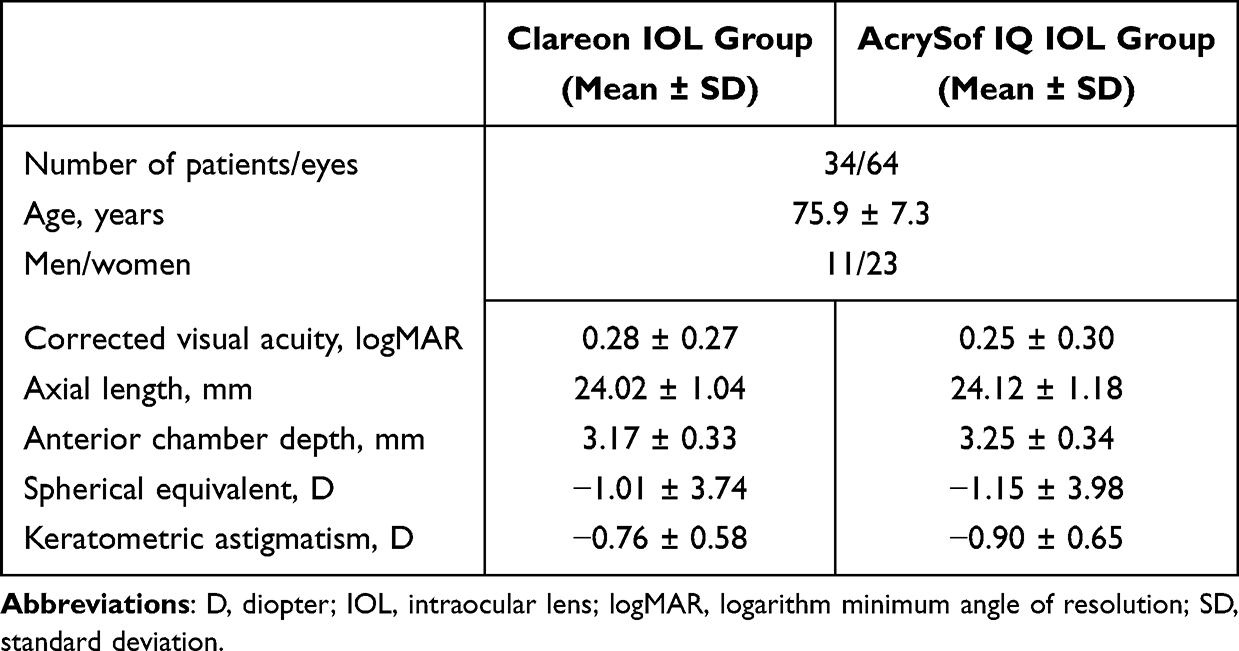 |
Table 1 Preoperative Patient Demographic Data |
ACD
For the primary endpoint, the mean ± SD of ACD change from 1 day to 6 months postoperatively was −0.100 ± 0.129 mm (95% CI, −0.054 to −0.146) for Clareon IOL and −0.126 ± 0.145 mm (95% CI, −0.075 to −0.177) for AcrySof IQ IOL. Equivalence was confirmed, as the 95% CI for the difference in ACD change between Clareon IOL and AcrySof IQ IOL (0.026 mm; 95% CI, −0.020 to 0.072) was within the equivalence margin of −0.15 to 0.15 mm. The ACD values at each follow-up and changes in ACD from 1 day are shown in Figure 1A and B. At 6 months, the mean ± SD for ACD was 4.263 ± 0.300 mm for Clareon IOL and 4.232 ± 0.304 mm for AcrySof IQ IOL (P = 0.0516). No significant difference in ACD or change in ACD from 1 day was observed between the IOL groups at any follow-up visit. In addition, there were no within-group differences at each follow-up examination (P > 0.05).
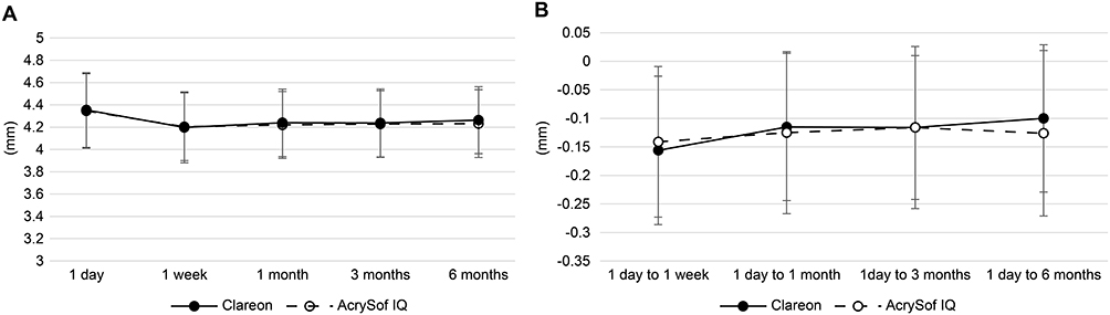 |
Figure 1 Anterior chamber depth changes from day 1 to month 6. (A) Anterior chamber depth after Clareon and AcrySof IQ intraocular lens implantation. (B) Change in anterior chamber depth from 1 day after Clareon and AcrySof IQ intraocular lens implantation. |
IOL Tilt and Decentration
The mean ± SD of IOL tilt for Clareon IOL at 1 day, and 1, 3, and 6 months postoperatively were 4.1 ± 1.7°, 4.0 ± 1.7°, 4.2 ± 1.9°, and 4.2 ± 1.9°, respectively (Figure 2A). There was no significant difference over time (P = 0.963). For AcrySof IQ IOL, the corresponding values were 3.9 ± 1.6°, 4.1 ± 1.8°, 4.1 ± 1.8°, and 4.0 ± 1.9°, respectively (Figure 2A). There was no significant difference in AcrySof IQ IOL over time (P = 0.981). No significant differences were noted in the IOL tilt between the two IOLs at any follow-up visit.
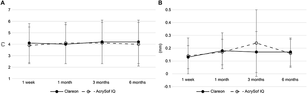 |
Figure 2 Tilt and decentration change from day 1 to month 6. (A) Intraocular lens tilt after Clareon and AcrySof IQ intraocular lens implantation. (B) Intraocular lens decentration after Clareon and AcrySof IQ intraocular lens implantation. |
IOL decentration was evaluated in 10 patients using CASIA 2. The mean ± SD of IOL decentration for Clareon and AcrySof IQ was 0.13 ± 0.09 and 0.14 ± 0.14 mm at 1 day (P = 0.8924), 0.18 ± 0.14 and 0.17 ± 0.10 mm at 1 week (P = 0.7132), 0.17 ± 0.16 and 0.24 ± 0.26 mm at 3 months (P = 0.3554), and 0.17 ± 0.11 and 0.16 ± 0.11 mm at 6 months postoperatively (P = 0.9155), respectively (Figure 2B).
Refraction and Visual Acuity
The mean ± SD of the subjective spherical equivalent for the eyes inserted with Clareon IOL at 1 day, 1 week, and 1, 3, and 6 months postoperatively were 0.17 ± 1.06, −0.09 ± 1.18, 0.00 ± 1.06, −0.02 ± 1.02, and −0.01 ± 1.03 D, respectively (Figure 3). For the eyes inserted with AcrySof IQ IOL, the corresponding values were 0.08 ± 0.96, −0.09 ± 0.90, −0.07 ± 1.01, −0.06 ± 0.89, and −0.08 ± 0.90 D, respectively (Figure 3). Both IOLs showed no significant changes over time (P > 0.05).
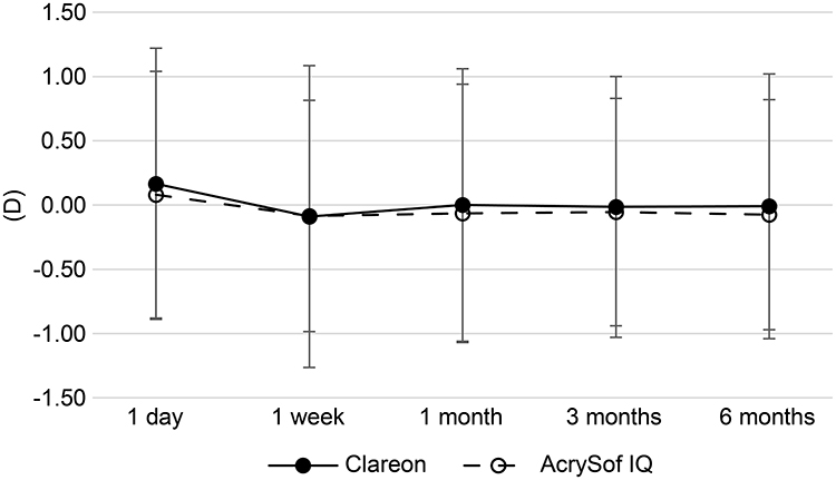 |
Figure 3 Subjective spherical equivalent after Clareon and AcrySof IQ intraocular lens implantation. |
The BCVA remained stable for 6 months in both the Clareon and AcrySof IQ IOL groups. The mean ± SD of BCVA at 6 months postoperatively was −0.06 ± 0.08 logMAR and −0.05 ± 0.10 logMAR for the Clareon IOL and AcrySof IQ IOL groups, respectively (Figure 4).
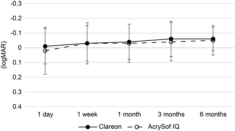 |
Figure 4 Corrected visual acuity after Clareon and AcrySof IQ intraocular lens implantation. |
Discussion
Postoperative in-the-bag IOL stability is a key determinant of patient satisfaction after cataract surgery. However, direct comparisons between Clareon and AcrySof IQ IOLs remain limited, particularly when implanted in the same patients and ACD, tilt, and decentration using anterior segment OCT are evaluated. In this prospective, randomized, contralateral eye multicenter study, we evaluated the in-the-bag stability of both IOLs, revealing that the changes in ACD from 1 day to 6 months postoperatively were comparable. Both IOLs demonstrated minimal postoperative tilt and decentration, with refractive stability and visual acuity well-maintained throughout the follow-up period.
In this study, the ACD change from 1 day to 6 months postoperatively was −0.100 ± 0.129 and −0.126 ± 0.145 mm for Clareon IOL and AcrySof IQ IOL, respectively, indicating comparable axial stability between the two IOLs. These findings are consistent with those of previous studies.22,26,28 Lane et al26 experimentally evaluated the IOL stability of five different IOLs, reporting that both Clareon and AcrySof IQ IOLs demonstrated the lowest level of axial displacement, IOL tilt, and decentration. Moreover, Werner et al28 conducted a contralateral implantation of Clareon and AcrySof IQ IOLs in rabbit eyes and found no significant difference in ACD between the two IOLs. Ullrich et al22 evaluated the axial change in ACD from 1 hour to 6 months postoperatively in 80 eyes of 40 patients who received contralateral implantation of Clareon and AcrySof IQ IOLs, reporting a change of −0.28 ± 0.21 and −0.21 ± 0.13 mm for the Clareon IOL and AcrySof IQ IOL, respectively.
Regarding axial shift in ACD following surgery, Ullrich et al22 reported that both Clareon and AcrySof IQ IOLs exhibited a forward IOL movement in ACD from 1 hour to 1 week, followed by a slight IOL shift backward from 1 week to 6 months. Similarly, several studies on ACD changes have reported a tendency for a forward shift early after surgery, followed by slight backward movement3,29,30 and our study demonstrated a similar change in ACD. Goto et al8 reported that IOLs with hinged haptics achieved earlier in-the-bag stability than those without hinges As both Clareon and AcrySof IQ IOLs incorporate hinged haptic designs, the early postoperative stability in anterior chamber depth observed here likely reflects this shared structural feature.
Further, we evaluated IOL tilt and decentration, demonstrating that both Clareon and AcrySof IQ IOLs exhibited minimal changes, with no significant difference between the IOLs, and remained stable in the bag at day 1 through 6 months postoperatively. These findings are consistent with those of previous research on the Clareon IOL tilt and decentration.20,31 Waster et al31 reported that the mean of IOL tilt for the Clareon IOL at 8 weeks postoperatively was 5.06 ± 1.14°, revealing only a change of 0.29 ± 1.32° from the tilt of the preoperative crystalline lens. Similarly, the mean of IOL decentration at 8 weeks was 0.26 ± 0.13 mm, with a change of 0.09 ± 0.146 mm from the preoperative lens position. Furthermore, Nuijts et al20 conducted a prospective, multicenter study across 19 sites in seven countries, evaluating IOL tilt and decentration in 424 eyes implanted with Clareon IOLs. Over a 3-year postoperative period, only one eye exhibited an IOL tilt greater than 10°, and another eye with a 4.5° tilt at 6 months postoperatively showed resolution thereafter. Regarding IOL decentration, one eye demonstrated 0.5 mm of IOL decentration from 1 to 3 years postoperatively, and another eye showed 1 mm of decentration at 2 and 3 years due to capsular phimosis. As aspheric IOLs have been reported to impair visual function when decentered by more than 0.4 mm or tilted beyond 7°, the degrees of tilt and decentration observed for Clareon and AcrySof IQ IOLs here are unlikely to exert clinically relevant effects on visual performance and were comparable between the two models.6
Previous studies have reported a correlation between ACD changes and subjective refraction postoperatively.8–11 Here, both eyes with Clareon or AcrySof IQ IOLs showed a slight myopic shift from 1 day to 1 week postoperatively, followed by a subtle hyperopic shift from 1 week to 1 month. These refractive shifts corresponded to the ACD changes observed in both IOLs, with a slight forward movement occurring between 1 day and 1 week, followed by minimal backward movement from 1 week to 1 month.
BCVA remained stable from 1 day to 6 months postoperatively in both the Clareon and AcrySof IQ IOL groups, without significant differences observed between the two groups. These findings are consistent with the results reported by Ullrich et al,22 who conducted a contralateral randomized study and found comparable BCVA at 6 months postoperatively: −002 ± 0.07 for the Clareon IOL and −0.03 ± 0.07 for the AcrySof IQ IOL, without significant differences between the two lenses.
Although some studies have reported a correlation between postoperative refractive changes and shifts in the ACD,8–11 others have found no such association, suggesting that refractive changes rely mainly on alterations in the corneal curvature.32 Additionally, postoperative capsular bag shrinkage and capsulorhexis size may affect in-the-bag IOL stability.3–7 In this study, we did not measure the corneal curvature, capsular-bag size, or capsulorhexis size, all of which could have influenced postoperative ACD between eyes. The secondary outcomes, particularly IOL decentration, were assessed in a relatively small sample, which may limit statistical power; therefore, these findings should be interpreted as exploratory. Further studies incorporating postoperative measurements of corneal curvature, capsular bag size, capsulorhexis size, and IOL decentration with a sufficient sample size may be warranted to strengthen refractive and IOL stability in the Clareon IOL bag. Nevertheless, a major strength of this study was that it was conducted as a contralateral, randomized, prospective, multicenter study with an a priori sample size calculation, which enhanced the reliability and generalizability of the findings. However, the contralateral-eye design introduces inter-eye dependency, which remains a methodological limitation.
Conclusion
The Clareon IOL demonstrated postoperative stability equivalent to that of the AcrySof IQ IOL regarding ACD changes between day 1 and month 6. The anterior segment OCT findings from the same patients demonstrated no significant differences in ACD, IOL tilt, or refractive outcomes at 6 months between the two IOLs. Both IOLs had good stability in the bag throughout the 6-month follow-up period.
Abbreviations
ACD, anterior chamber depth; BCVA, best-corrected visual acuity; CI, confidence interval; D, diopters; IOL, intraocular lens; logMAR, logarithm of the minimum angle of resolution; OCT, optical coherence tomography; SD, standard deviation.
Data Sharing Statement
Data supporting the findings of this study are available from the corresponding author (S.N.) upon reasonable request.
Acknowledgments
This study was presented as an electronic poster at the 2023 European Society of Cataract and Refractive Surgery (ESCRS) Annual Meeting. All the authors thank Apex LLC for editorial assistance during manuscript preparation.
Author Contributions
All authors made a significant contribution to the work reported, whether that is in the conception, study design, execution, acquisition of data, analysis and interpretation, or in all these areas; took part in drafting, revising or critically reviewing the article; gave final approval of the version to be published; have agreed on the journal to which the article has been submitted; and agree to be accountable for all aspects of the work.
Funding
This study was supported by an investigator-initiated study grant (#51718191) from Alcon Vision LLC (Fort Worth, TX, USA). The funder had no involvement in the study design.
Disclosure
Dr. Shinichiro Nakano received research grants from Alcon Vision LLC, Johnson & Johnson Vision, and HOYA. Dr. Hiroshi Sasaki received research grants from Alcon Vision LLC, TOMIKI MEDICAL INSTRUMENTS Co., Ltd, Santen Pharmaceutical Co., Ltd, Sanwa Medical Co., Ltd., Senju Pharmaceutical Co., Ltd, Otsula Pharmaceutical Co., Ltd, IQVIA Inc., Bayer AG, and HOYA Corporation. The authors report no other conflicts of interest in this work.
References
1. Kohnen T, Findl O, Nuijts R, Ribeiro F, Cochener-Lamard B. ESCRS clinical trends survey 2016-2021: 6-year assessment of practice patterns among society delegates. J Cataract Refract Surg. 2023;49(2):133–9. doi:10.1097/j.jcrs.0000000000001053
2. Tabuchi H, Sato M, Kamiya K, et al. JSCRS clinical Survery. IOL&RS. 2024;38(3):382–401.
3. Wirtitsch MG, Findl O, Menapace R, et al. Effect of haptic design on change in axial lens position after cataract surgery. J Cataract Refract Surg. 2004;30(1):45–51. doi:10.1016/S0886-3350(03)00459-0
4. Modesti M, Pasqualitto G, Appolloni R, Pecorella I, Sourdille P. Preoperative and postoperative size and movements of the lens capsular bag: ultrasound biomicroscopy analysis. J Cataract Refract Surg. 2011;37(10):1775–1784. doi:10.1016/j.jcrs.2011.04.035
5. Dick HB, Conrad-Hengerer I, Schultz T. Intraindividual capsular bag shrinkage comparing standard and laser-assisted cataract surgery. J Refract Surg. 2014;30(4):228–233. doi:10.3928/1081597X-20140320-01
6. Tehrani M, Dick HB, Krummenauer F, Pfirrmann G, Boyle T, Stoffelns BM. Capsule measuring ring to predict capsular bag diameter and follow its course after foldable intraocular lens implantation. J Cataract Refract Surg. 2003;29(11):2127–2134. doi:10.1016/S0886-3350(03)00352-3
7. Çekiç O, Batman C. The relationship between capsulorhexis size and anterior chamber depth relation. Ophthalmic Surg Lasers Imaging. 1999;30(3):185–190. doi:10.3928/1542-8877-19990301-06
8. Goto S, Maeda N, Ohnuma K, Noda T. Comparison of two one-piece acrylic foldable intraocular lenses: short-term change in axial movement after cataract surgery and its effect on refraction. PLoS One. 2022;17(8):e0273431. doi:10.1371/journal.pone.0273431
9. Koeppl C, Findl O, Kriechbaum K, et al. Postoperative change in effective lens position of a 3-piece acrylic intraocular lens. J Cataract Refract Surg. 2003;29(10):1974–1979. doi:10.1016/S0886-3350(02)02049-7
10. Nejima R, Miyai T, Kataoka Y, et al. Prospective intrapatient comparison of 6.0-millimeter optic single-piece and 3-piece hydrophobic acrylic foldable intraocular lenses. Ophthalmology. 2006;113(4):585–590. doi:10.1016/j.ophtha.2005.10.064
11. Teshigawara T, Meguro A, Mizuki N. Relationship between postoperative intraocular lens shift and postoperative refraction change in cataract surgery using three different types of intraocular lenses. Ophthalmol Ther. 2021;10(4):989–1002. doi:10.1007/s40123-021-00390-x
12. Lawu T, Mukai K, Matsushima H, Senoo T. Effects of decentration and tilt on the optical performance of 6 aspheric intraocular lens designs in a model eye. J Cataract Refract Surg. 2019;45(5):662–668. doi:10.1016/j.jcrs.2018.10.049
13. McKelvie J, McArdle B, McGhee C. The influence of tilt, decentration, and pupil size on the higher-order aberration profile of aspheric intraocular lenses. Ophthalmology. 2011;118(9):1724–1731. doi:10.1016/j.ophtha.2011.02.025
14. Yan W, Auffarth GU, Khoramnia R, Łabuz G. Blue-light filtering monofocal intraocular lenses: a study on optical function and tolerance to misalignment. J Refract Surg. 2024;40(2):e79–e88. doi:10.3928/1081597X-20240112-02
15. Taketani F, Yukawa E, Ueda T, Sugie Y, Kojima M, Hara Y. Effect of tilt of 2 acrylic intraocular lenses on high-order aberrations. J Cataract Refract Surg. 2005;31(6):1182–1186. doi:10.1016/j.jcrs.2004.11.048
16. Holladay JT, Piers PA, Koranyi G, van der Mooren M, Norrby NES. A new intraocular lens design to reduce spherical aberration of pseudophakic eyes. J Refract Surg. 2002;18(6):683–691. doi:10.3928/1081-597X-20021101-04
17. Olsen T. Calculation of intraocular lens power: a review. Acta Ophthalmol Scand. 2007;85(5):472–485. doi:10.1111/j.1755-3768.2007.00879.x
18. Goto S, Maeda N, Koh S, et al. Prediction of postoperative intraocular lens position with angle-to-angle depth using anterior segment optical coherence tomography. Ophthalmology. 2016;123(12):2474–2480. doi:10.1016/j.ophtha.2016.09.005
19. Kinoshita K, Miyata K, Nejima R, Honbo M, Mori Y, Minami K. Surface light scattering from 1-piece hydrophobic acrylic intraocular lenses with hydroxyethyl methacrylate: contralateral observation for 7 years. J Cataract Refract Surg. 2021;47(6):702–705. doi:10.1097/j.jcrs.0000000000000621
20. Nuijts RMMARMMA, Bhatt U, Nanavaty MA, Roberts TV, Peterson R, Teus MA. Three-year multinational clinical study on an aspheric hydrophobic acrylic intraocular lens. J Cataract Refract Surg. 2023;49(7):672–678. doi:10.1097/j.jcrs.0000000000001173
21. Oshika T, Fujita Y, Inamura M, Miyata K. Mid-term and long-term clinical assessments of a new 1-piece hydrophobic acrylic IOL with hydroxyethyl methacrylate. J Cataract Refract Surg. 2020;46(5):682–687. doi:10.1097/j.jcrs.0000000000000142
22. Ullrich M, Ruiss M, Hienert J, et al. Anterior chamber depth variability between 2 hydrophobic acrylic 1-piece intraocular lenses: randomized trial. J Cataract Refract Surg. 2021;47(11):1460–1465. doi:10.1097/j.jcrs.0000000000000668
23. Eom Y, Kang S-YS-Y, Song J-SJ-S, Kim HM. Comparison of the actual amount of axial movement of 3 aspheric intraocular lenses using anterior segment optical coherence tomography. J Cataract Refract Surg. 2013;39(10):1528–1533. doi:10.1016/j.jcrs.2013.04.040
24. Werner L. Glistenings and surface light scattering in intraocular lenses. J Cataract Refract Surg. 2010;36(8):1398–1420. doi:10.1016/j.jcrs.2010.06.003
25. Werner L, Thatthamla I, Ong M, et al. Evaluation of clarity characteristics in a new hydrophobic acrylic IOL in comparison to commercially available IOLs. J Cataract Refract Surg. 2019;45(10):1490–1497. doi:10.1016/j.jcrs.2019.05.017
26. Lane S, Collins S, Das KK, et al. Evaluation of intraocular lens mechanical stability. J Cataract Refract Surg. 2019;45(4):501–506. doi:10.1016/j.jcrs.2018.10.043
27. Yamashita K, Hayashi K, Hata S. Clinical performance and shape analysis of trifocal intraocular lenses via scanning electron microscopy. BMC Ophthalmol. 2024;24(1):86. doi:10.1186/s12886-024-03355-3
28. Werner L, Ellis N, Heczko JB, et al. In vivo evaluation of a new hydrophobic acrylic intraocular lens in the rabbit model. J Cataract Refract Surg. 2018;44(12):1497–1502. doi:10.1016/j.jcrs.2018.07.040
29. Negishi K, Masui S, Torii H, Nishi Y, Tsubota K. Refractive stability of a new single-piece hydrophobic acrylic intraocular lens and corneal wound repair after implantation using a new automated intraocular lens delivery system. PLoS One. 2020;15(9):e0238366. doi:10.1371/journal.pone.0238366
30. Sato T, Shibata S, Yoshida M, Hayashi K. Short-term dynamics after single- and three-piece acrylic intraocular lens implantation: a swept-source anterior segment optical coherence tomography study. Sci Rep. 2018;8(1):10230. doi:10.1038/s41598-018-28609-1
31. Waser K, Straßmair K, Pomberger L, et al. Capsular bag performance of a novel hydrophobic single-piece intraocular lens. Ophthalmol Ther. 2025;14(2):295–308. doi:10.1007/s40123-024-01075-x
32. Klijn S, Sicam VA, Reus NJ. Long-term changes in intraocular lens position and corneal curvature after cataract surgery and their effect on refraction. J Cataract Refract Surg. 2016;42(1):35–43. doi:10.1016/j.jcrs.2015.08.015
 © 2026 The Author(s). This work is published and licensed by Dove Medical Press Limited. The
full terms of this license are available at https://www.dovepress.com/terms
and incorporate the Creative Commons Attribution
- Non Commercial (unported, 4.0) License.
By accessing the work you hereby accept the Terms. Non-commercial uses of the work are permitted
without any further permission from Dove Medical Press Limited, provided the work is properly
attributed. For permission for commercial use of this work, please see paragraphs 4.2 and 5 of our Terms.
© 2026 The Author(s). This work is published and licensed by Dove Medical Press Limited. The
full terms of this license are available at https://www.dovepress.com/terms
and incorporate the Creative Commons Attribution
- Non Commercial (unported, 4.0) License.
By accessing the work you hereby accept the Terms. Non-commercial uses of the work are permitted
without any further permission from Dove Medical Press Limited, provided the work is properly
attributed. For permission for commercial use of this work, please see paragraphs 4.2 and 5 of our Terms.