Back to Journals » Journal of Experimental Pharmacology » Volume 18
“Comparative Evaluation of Saussurea Lappa Root Extract Ointment and Standard Therapy on Second-Degree Burn Wound Healing in Rabbits”
Authors Al-Mehdar AA, Maad AH, Al-Shami AS ![]() , Al-Kadsi J, Al-Saban AM, Mohammed Saad H, Al-Worafi YM
, Al-Kadsi J, Al-Saban AM, Mohammed Saad H, Al-Worafi YM ![]()
Received 5 November 2025
Accepted for publication 19 January 2026
Published 23 January 2026 Volume 2026:18 575002
DOI https://doi.org/10.2147/JEP.S575002
Checked for plagiarism Yes
Review by Single anonymous peer review
Peer reviewer comments 3
Editor who approved publication: Prof. Dr. Abdelwahab Omri
Ali A Al-Mehdar,1 Abdullah H Maad,2 Ali Salman Al-Shami,3 Jalal Al-Kadsi,4 Abdulhakim M Al-Saban,3 Hammood Mohammed Saad,5 Yaser M Al-Worafi6
1Department of Pharmacology, Faculty of Medicine, Thamar University, Thamar City, Republic of Yemen; 2Department of Pharmaceutical, College of pharmacy, University of Al-Ameed, Karbala, Iraq; 3Derament of Pharmacy, Jiblah University for Medical and Health Science, Ibb City, Republic of Yemen; 4Department of Medicinal-Chemistry, Sanaa University, Sanaa City, Republic of Yemen; 5Department of Pediatrics, Althawra Hospital, Al Hudaydah City, Republic of Yemen; 6College of Medical sciences, Azal University for Human Development, Sanaa City, Republic of Yemen
Correspondence: Jalal Al-Kadsi, Department of Medicinal-Chemistry, Sanaa university, Sanaa City, Republic of Yemen, Email [email protected]
Background: Saussurea lappa is a traditional medicinal herb valued for its anti-inflammatory, antimicrobial, and antioxidant properties. Burn wounds are highly susceptible to infection and oxidative stress, which delay healing; therefore, developing effective plant-based alternatives for burn management is of clinical importance.
Objective: To investigate the phytochemical composition and healing effects of Saussurea lappa extract and its formulated ointment on second-degree burns in rabbit models.
Methods: Roots of S. lappa were dried, powdered, and extracted with methanol using a Soxhlet apparatus. The extract was incorporated into ointment formulations containing 1%, 5%, and 10% concentrations. Phytochemical screening confirmed the presence of alkaloids, flavonoids, tannins, phenols, and saponins. Twenty rabbits were divided into four groups: untreated control, base ointment, standard Mebo® ointment, and 10% S. lappa ointment. Second-degree burns were induced under anesthesia, and treatments were applied daily for 21 days. Wound contraction was measured using ImageJ software, and histopathological assessments were performed on days 7, 14, and 21.
Results: The 10% S. lappa ointment demonstrated a wound closure rate of 95.1% by day 20, comparable to Mebo (96.1%) and superior to control groups. Histological analysis revealed complete re-epithelialization, dense collagen formation, and well-developed granulation tissue. The formulation remained physically stable under various storage conditions.
Conclusion: The methanolic root extract of Saussurea lappa significantly enhanced burn wound healing in rabbits, likely due to its antioxidant, antimicrobial, and anti-inflammatory effects. The 10% S. lappa ointment exhibited efficacy comparable to the standard treatment, suggesting its potential as a safe, natural, and cost-effective topical therapy for burn wound management.
Keywords: phytochemical screening, Saussurea lappa, burn wound healing, second-degree burns, topical herbal formulation, antimicrobial and anti-inflammatory activity
Introduction
Historically, ethnomedicine has utilised the entire range of plant parts, including bark, fruit, leaves, stems, and roots, all of which possess therapeutic properties. Individuals acknowledge the use of herbal medicine and botanical extracts as alternatives to manufactured or pharmaceutical drugs, frequently due to their diminished adverse effects. The findings suggest that the use of herbal medical approaches corresponds with a revival of natural medicines that generally exhibit minimal or no negative effects1,2 This plant has been employed for millennia as both a preventive and therapeutic remedy for numerous maladies, as well as a highly nutritious vegetable.3 It is thoroughly detailed in Vedic literature for the management of diverse ailments.4 Burn injuries remain a major global health issue, particularly in developing regions where access to advanced wound care is limited. According to the World Health Organization (2023), an estimated 11 million people suffer severe burns annually, resulting in substantial morbidity and economic burden5. Burn wounds heal through overlapping phases—haemostasis, inflammation, proliferation, and remodelling—each influenced by cytokine signalling, oxidative stress, and microbial infection.6,7 Conventional treatments such as silver sulfadiazine and topical antibiotics are widely used but may cause cytotoxicity, allergic reactions, and delayed epithelialization, underscoring the need for safer, plant-derived alternatives (Exploring Trends in Natural Product-Based Treatments for Skin Burns, 2025). Medicinal plants have long served as sources of wound-healing agents due to their abundance of secondary metabolites—flavonoids, terpenoids, phenolics, tannins, and alkaloids—that exhibit antioxidant, anti-inflammatory, and antimicrobial activities.These compounds enhance fibroblast proliferation, collagen deposition, and angiogenesis while modulating inflammatory mediators such as TNF-α and NF-κB (Anti-inflammatory and Wound-Healing Activities of Medicinal Plants).8,9 Among these herbs, Saussurea lappa C.B. Clarke—commonly known as costus—is particularly promising. Native to the Himalayas, it has been used in Ayurvedic and Unani medicine to treat respiratory, digestive, and skin disorders10,11. Phytochemical analyses reveal that S. lappa roots contain sesquiterpene lactones (costunolide and dehydrocostus lactone), flavonoids, phenolic acids, and essential oils responsible for its antimicrobial, antioxidant, and anti-inflammatory properties12,13. Experimental studies have demonstrated that methanolic extracts of S. lappa accelerate wound contraction and inhibit Staphylococcus aureus and Pseudomonas aeruginosa growth.11 While emulgel formulations promote faster re-epithelialization and collagen formation14. Moreover, nanofiber and nanoparticle systems incorporating S. lappa extract enhance antimicrobial efficacy and upregulate angiogenic factors such as VEGF and TGF-β1, further supporting tissue regeneration.15,16.
Plant-based remedies have been utilized in traditional medicine for a considerable duration; nonetheless, thorough scientific studies regarding their efficacy, especially in the realm of burn wound healing, are limited. Saussurea lappa (Costus root), known for its anti-inflammatory, antioxidant, and antimicrobial properties, has shown promise in several therapeutic areas; however, its effectiveness for treating second-degree burns has yet to be thoroughly investigated. The existing research gap includes a lack of comprehensive studies on its effectiveness in burn wounds, limited preclinical validation in regulated animal models, and insufficient evidence regarding its therapeutic potential for burns as silver sulfadiazines become less effective because of antimicrobial resistance and cytotoxicity, it is clear that we need safer, more effective treatments. This study aims to address these deficiencies by investigating the effects of a 10% S. lappa ointment on second-degree burn wounds in rabbits, assessing its efficacy and safety relative to Mebo ointment. We want to find out if S. lappa works to heal burns, and has antimicrobial properties as well as good, cheap alternative to standard treatments. The results will yield substantial data for the management of burn wounds and support the future utilization of S. lappa as a viable alternative, particularly in resource-limited settings.
Methodology
Plant Material and Extraction
Roots of Saussurea lappa C.B. Clarke were obtained from a certified herbal supplier in Sana’a, Yemen, and authenticated by the Department of Pharmacognosy, Faculty of Medicine, Sana’a University. The roots were washed, shade-dried, and pulverized into coarse powder using a mechanical grinder. The powdered material (2000 g) was extracted with methanol using a Soxhlet apparatus for 72 hours, maintaining the temperature at 60°C11. The extract was filtered, concentrated under reduced pressure using a rotary evaporator, and dried at 40°C to yield a viscous dark-brown residue (yield 6.9% w/w). The crude extract was stored at 4°C in an airtight container until use.
Plant Authentication and Identification
The plant material utilized in this study was Saussurea lappa C.B. Clarke (Family: Asteraceae). A botanist in the Department of Pharmacognosy at Sana’a University was able to identify and confirm the species.
A voucher specimen has been prepared and stored in the Public Herbarium of the Department of Pharmacognosy, Faculty of Medicine, Sana’a University, with the accession number SL-2024-05-YEM.
The Royal Botanic Gardens, Kew (POWO) citation is the most internationally accepted for taxonomy. (Royal Botanic Gardens Kew, 2024).17
Phytochemical Screening
Qualitative phytochemical analysis was carried out following standard procedures to detect the presence of major secondary metabolites including alkaloids, flavonoids, glycosides, phenols, tannins, saponins, steroids, terpenoids, and resins8. Tests such as Dragendorff’s reagent (for alkaloids), Ferric chloride (for phenols and tannins), Liebermann-Burchard (for steroids), and foam tests (for saponins) were applied. The results were recorded as presence (+) or absence (–) of each constituent.
While qualitative analysis provided useful insights into the chemical composition of Saussurea lappa, we acknowledge the importance of quantitative characterization to more accurately determine the concentrations of bioactive compounds. In future studies, we plan to employ quantitative methods such as total phenolic content assays, flavonoid assays, and HPLC quantification of key constituents like costunolide and dehydrocostus lactone to better understand the relationship between the phytochemical profile and the wound-healing activity of S. lappa.
Formulation of Ointment
Table 1 shows that the methanolic extract of S. lappa was added to an ointment base made of white soft paraffin, liquid paraffin, cetostearyl alcohol, and propylene glycol using the fusion method14. Initially, the extract was dissolved in propylene glycol, and the resulting mixture was slowly added to the molten base while stirring constantly until it was completely mixed. Formulations F1, F2, and F3 were made with three different concentrations: 1%, 5%, and 10% w/w. The formulations were put into sterilized aluminum tubes and kept at room temperature until they could be tested.
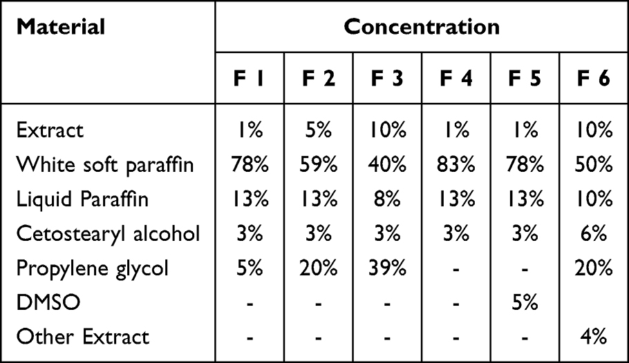 |
Table 1 Formulation Composition of Saussurea lappa Extract Ointments with Varying Concentrations and Excipient Ratios |
There were a number of reasons for choosing to focus on the 10% concentration. Previous research on S. lappa and analogous herbal formulations indicates that elevated concentrations frequently demonstrate enhanced therapeutic effects, especially in wound healing models.11,13 The 10% concentration has been determined to offer optimal effectiveness in facilitating wound healing while adhering to safe parameters for topical application. The 10% ointment concentration was selected based on other published studies indicating that concentrations between 5% and 10% yielded optimal outcomes in promoting wound healing.12,14
Evaluation of Ointment Stability
The physical stability of the prepared formulations was assessed over a 3-month period under different temperature conditions (5°C, 25°C, 30°C, and 40°C) as described by10. Parameters evaluated included color, odor, homogeneity, and consistency. No phase separation or microbial contamination was observed during the study period, confirming formulation stability.
Ethical Approval
All animal procedures in this study were approved by the Institutional Animal Ethical Committee of Sanaa University (Reg. No. SN/IAEC/02/05-24/01/15-05-24). The study adhered to ethical guidelines for the care and use of laboratory animals, in accordance with the American Veterinary Medical Association (AVMA) recommendations for anesthesia and euthanasia.
Study Design and Animals
Twenty healthy adult male and female rabbits (weighing 1500–2000 g) were used in this study. The animals were housed under standard laboratory conditions (temperature 22 ± 2°C, relative humidity 50–60%, 12-h light/dark cycle) and had free access to a standard diet and water.
Randomization and Blinding
The animals were randomly assigned to one of four treatment groups using a computer-generated randomization list. This ensured that each rabbit had an equal chance of being placed in any of the experimental groups (negative control, base control, standard treatment, or test group). The randomization process minimized selection bias and helped ensure that the groups were comparable at baseline.
Blinding was applied during both the treatment and evaluation phases of the study. The researchers responsible for applying the treatments were blinded to the group assignments to prevent bias in treatment administration. Furthermore, the evaluation of wound healing and histopathological outcomes was performed by an independent assessor who was also blinded to the treatment groups. This blinding ensured that the assessment of the treatment outcomes was unbiased and objective.
Experimental Design
Animals were randomly divided into four groups (n = 5 per group):
- Group I (Negative control): No treatment applied.
- Group II (Base control): Treated with ointment base without extract.
- Group III (Standard control): Treated with commercial Mebo ointment (β-sitosterol-based).
- Group IV (Test group): Treated with 10% S. lappa extract ointment. Treatments were applied topically once daily for 21 days.
Sample Size Calculation and Justification
Five rabbits were utilized per group in this study. This sample size was selected based on prior animal studies of burn wound healing, which demonstrated that comparable group sizes yield reliable and statistically significant outcomes11,13.A formal statistical power calculation was not conducted due to limited resources; however, the chosen sample size was considered adequate to identify significant differences between the treatment groups while also taking into account practical factors such as animal availability and the study’s duration.We recognize the significance of statistical power and propose that subsequent studies with larger sample sizes would strengthen the results and augment the statistical power of the findings.
Anaesthesia and Euthanasia Method Details
The second-degree burns in our research were inflicted under mild ketamine anesthesia (35 mg/kg, intramuscular). This procedure was chosen due to its well-established application in animal research and was performed by a qualified anaesthesiologist. The ketamine dosage and administration procedure were meticulously followed in accordance with the standards established by the American Veterinary Medical Association (AVMA) for the humane treatment of animals in research. Animals were carefully observed throughout the procedure to confirm that sufficient anesthesia was maintained and that they were not exposed to unnecessary stress or discomfort.
Induction of Burn Wound
Second-degree burns were induced using light ketamine anesthesia (35 mg/kg, intramuscular) as described by11. with slight changes. Ensure that the animals were completely anesthetized prior to the burn induction technique. A skilled anaesthesiologist will administer it to guarantee appropriate anesthesia during the burn induction phase in accordance with the American Veterinary Medical Association’s (AVMA) recommendations for anesthesia. The ketamine dose was determined using published guidelines for similar procedures in rabbits. Throughout the surgery, the anaesthesiologist evaluated the depth of anaesthesia to ensure that the animals were completely sedated and painless.
Wound Area Measurement
The wound area was measured on days 0, 4, 8, 12, 16, and 20 using a transparent millimeter graph and analyzed with ImageJ software. The percentage of wound contraction was calculated according to the following formula.14.
The healing percentage of each wound was calculated as below:

Histopathological Examination
On days 7, 14, and 21, cutaneous tissue samples were obtained from euthanized animals and preserved in 10% neutral buffered formalin. The tissues were prepared, embedded in paraffin, sectioned at a thickness of 5 μm, and stained with hematoxylin and eosin (H&E) for microscopic examination. Sections were analyzed for epithelial regeneration, granulation tissue development, collagen accumulation, and the process of angiogenesis.13
Statistical Analysis
Data are expressed as mean ± standard deviation (SD). One-way ANOVA was conducted to analyze the differences in wound healing percentages across the groups: Negative Control, Blank Excipients, 10% S. lappa ointment, and Mebo ointment. Tukey’s post-hoc test was utilized to determine significant differences among the groups. Levene’s test was performed to evaluate the homogeneity of variance among groups, (p > 0.05) revealing no significant differences.
Result
Figure 1 shows that the creams exhibited color variations ranging from off-white to brownish, corresponding to the concentration of the extract. All formulations were smooth, homogeneous, and possessed acceptable aesthetic characteristics.
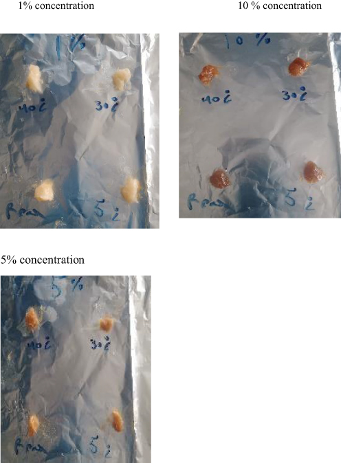 |
Figure 1 Physical Appearance of Saussurea lappa Ointments Containing 1%, 5%, and 10% Extract Concentrations. |
Table 2 shows that the stability of the ointment formulations was assessed for color, odor, homogeneity, and consistency at different temperature conditions (5°C, 25°C, 30°C, and 40°C) over a 3-month period. All formulations, including the 10% Saussurea lappa ointment, remained stable with no phase separation, no microbial contamination, and consistent texture and appearance. This indicated excellent physical stability. Confirming the suitability of the formulations for topical application.
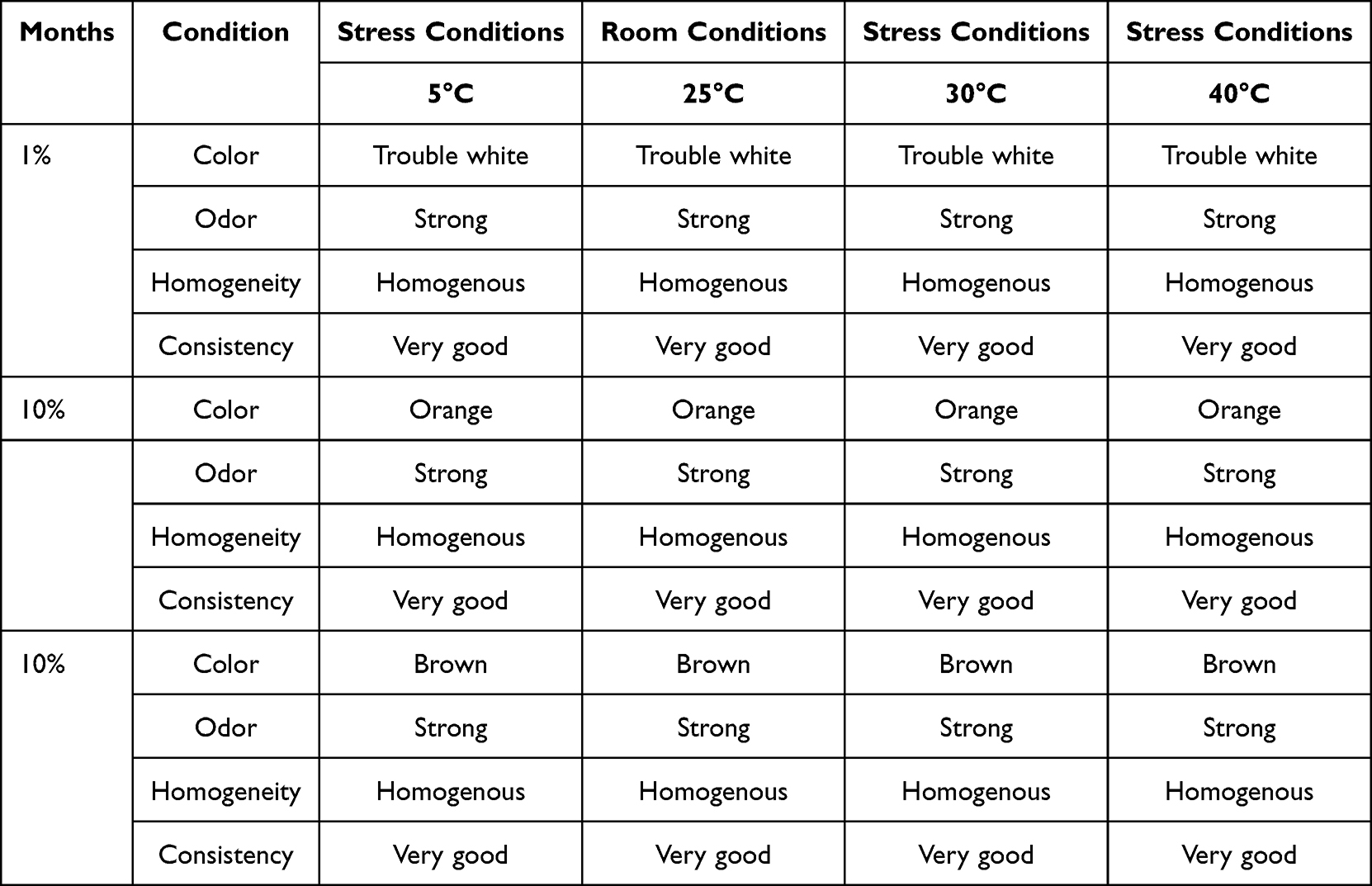 |
Table 2 Physicochemical Stability of Saussurea lappa Ointment Formulations Stored at Different Temperatures (°C) Over a 3-Month Period |
Table 3 shows that the 10% S. lappa ointment closed 95.1% of the wounds by day 20, which was statistically identical to the Mebo ointment (96.1%). This means that both treatments worked equally well to help the wounds heal. The negative control group (not treated) had a wound closure rate of 92.6%, and the blank excipients group (base ointment) had a rate of 90.1%. Both groups healed far more slowly than the treatment groups. The statistical analysis indicated no significant difference between the 10% S. lappa ointment and Mebo ointment (p > 0.05), implying that S. lappa is as effective as the usual treatment. A statistically significant difference (p < 0.05) in wound closure was seen between the treatment groups (10% S. lappa and Mebo) and the negative control and blank excipients groups, thereby validating the extract’s wound-healing efficacy.
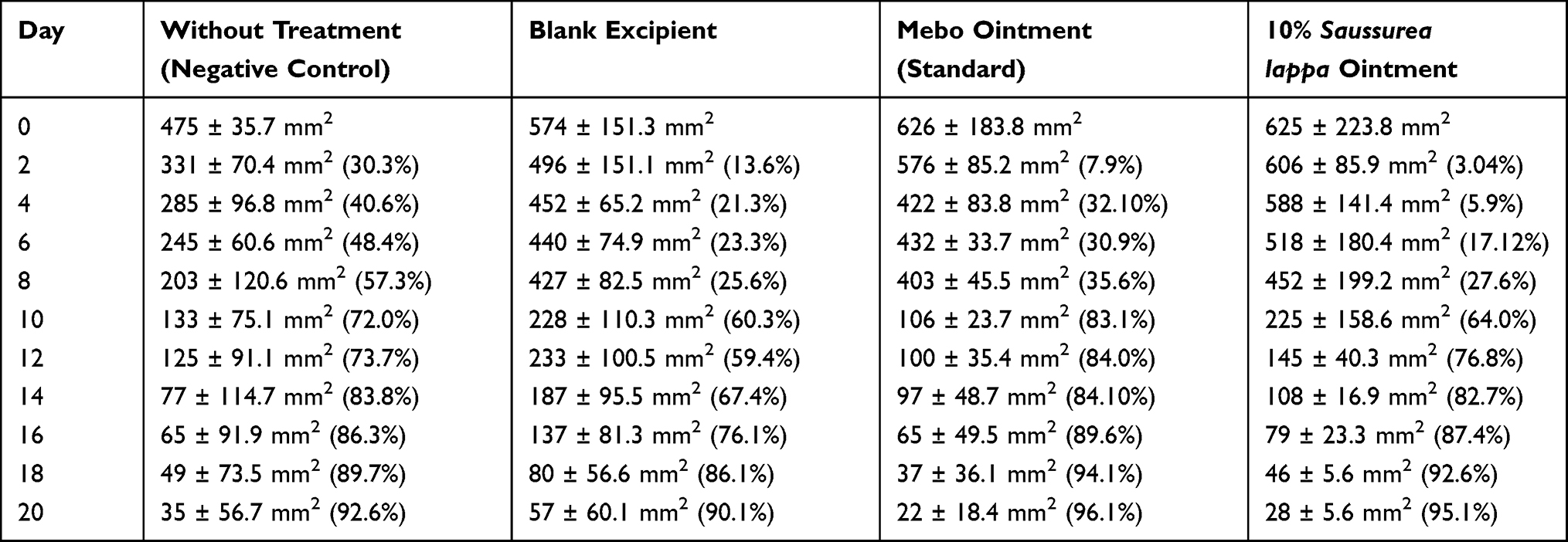 |
Table 3 Effect of Saussurea lappa Extract Ointment on Wound Area Reduction (mm2) and Healing Percentage (%) in Rabbits with Second-Degree Burns |
Table 4 shows Tukey’s post-hoc test revealed both Mebo and 10% Saussurea lappa ointment greatly promoted wound closure compared to the Negative Control and Blank Base groups. No significant difference was observed between Mebo and 10% S. lappa, suggesting that the herbal formulation achieved wound-healing efficacy comparable to the standard treatment.
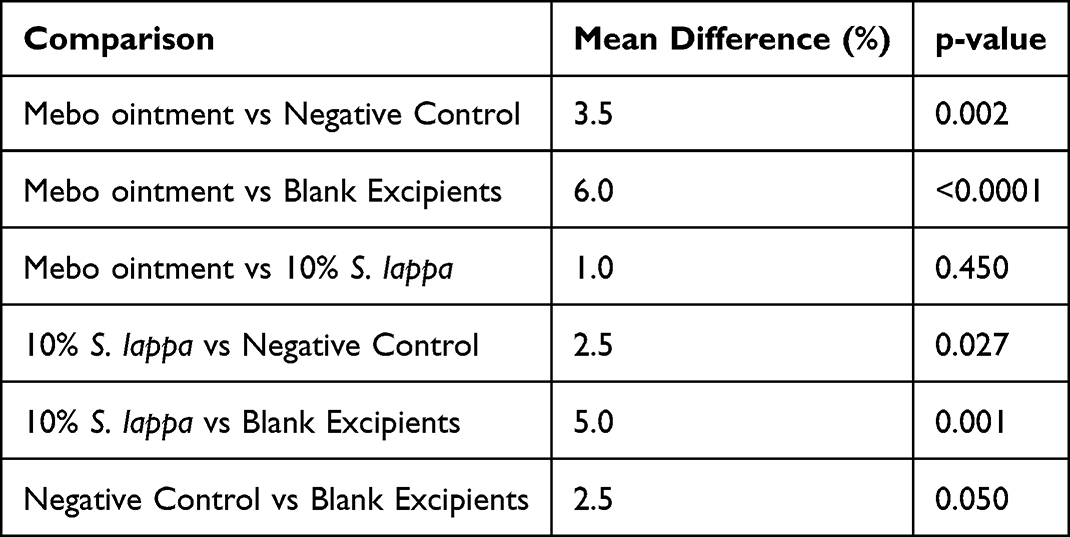 |
Table 4 Tukey’s Post-Hoc Analysis of Wound Healing Percentages (%) on Day 20: Comparison of Saussurea lappa Extract Ointment, Mebo Ointment, and Control Groups |
Figure 2 demonstrates that the wound contraction curves for Mebo and the 10% S. lappa formulation were nearly identical, with both treatments significantly surpassing the wound contraction observed in the control groups. A notable acceleration in healing was evident after day 10. These findings support that the S. lappa ointment facilitates rapid and sustained wound contraction, showing efficacy comparable to the standard treatment.
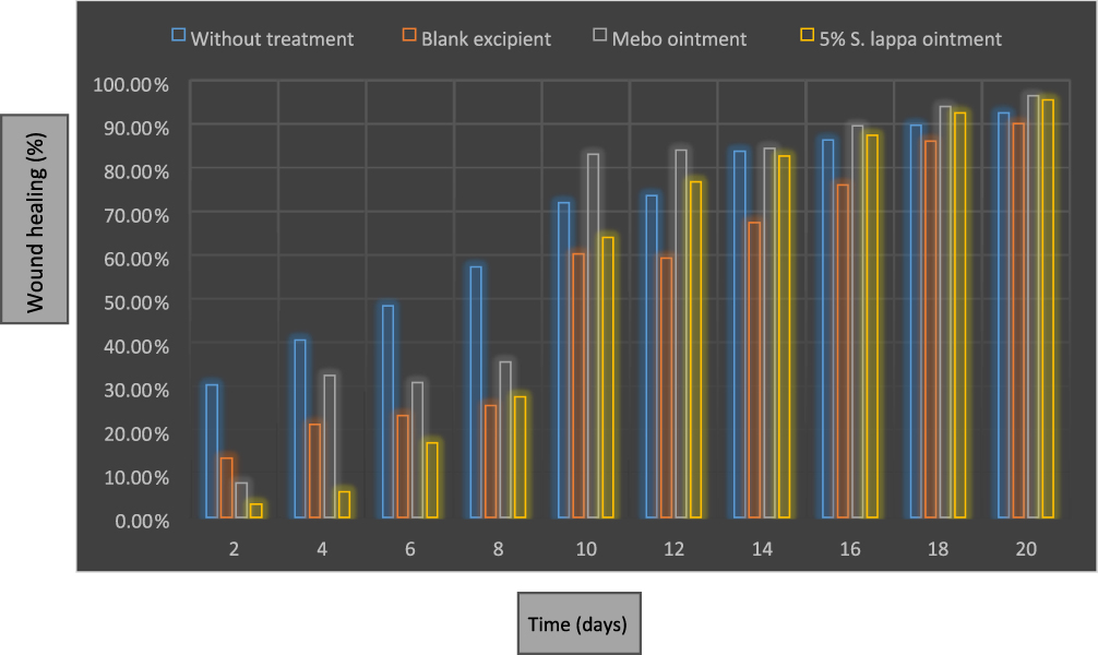 |
Figure 2 Percentage of Wound Healing (%) Over 20 Days in Control and Treatment Groups: Saussurea lappa Extract Ointment vs Mebo Ointment and Controls. |
Figures 3–5 present histopathological images of skin sections from rabbits treated with various ointments (10% Saussurea lappa ointment, Mebo ointment, Blank Excipients, and no treatment) on days 6, 14, and 22. Macroscopic observations revealed partial scab formation across all groups by day 6. By day 14, both the 10% S. lappa ointment and Mebo ointment groups showed extensive epithelialization, suggesting accelerated wound healing. By day 20, wounds treated with 10% S. lappa ointment and Mebo ointment were nearly completely healed, with no visible lesions, whereas the Negative Control and Blank Excipients groups still exhibited residual wound areas.
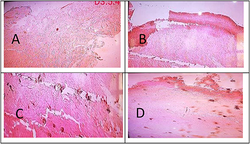 |
Figure 3 Histopathological Section of Burn Wound Tissue on Day 6 (Early Inflammatory Phase, 5 µm Sections). (A) Negative Control (Untreated group). (B) Blank Excipient (Ointment base only). (C) Standard Treatment (Mebo® ointment). (D) 10% Saussurea lappa Ointment. |
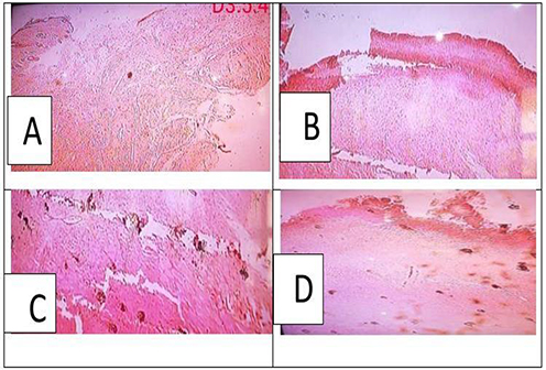 |
Figure 4 Histopathological Section of Burn Wound Tissue on Day 14 (Proliferative Phase, 5 µm Sections). (A) Negative Control (Untreated group). (B) Blank Excipient (Ointment base only). (C) Standard Treatment (Mebo® ointment). (D) 10% Saussurea lappa Ointment. |
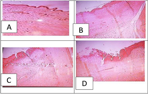 |
Figure 5 Histopathological Section of Burn Wound Tissue on Day 22 (Remodeling Phase, 5 µm Sections). (A) Negative Control (Untreated group). (B) Blank Excipient (Ointment base only). (C) Standard Treatment (Mebo® ointment). (D) 10% Saussurea lappa Ointment. |
Day 6: Figure 3 depicts Macroscopic observations revealed that all groups had formed partial scabs by day 6. (A) The negative control (untreated) had substantial inflammatory cell infiltration and tissue damage, while (B) the blank excipient group had moderate inflammation with minimal tissue organization. (C) The Mebo ointment-treated group exhibited less inflammatory infiltration and early granulation tissue development. (D) The 10% Saussurea lappa ointment-treated group demonstrated a significant reduction in inflammation and early evidence of tissue regeneration.
Day 14: Figure 4 depicts histological slices of second-degree burn wound tissue on day 14 (proliferative phase). (A) The untreated negative control shows inadequate re-epithelialization and immature granulation tissue, whereas (B) the blank excipient group demonstrates limited epithelium regrowth and considerable collagen deposition. (C) The Mebo ointment-treated group exhibited substantial re-epithelialization, well-developed granulation tissue, and enhanced collagen production. Finally (D), the 10% Saussurea lappa ointment-treated group had nearly full re-epithelialization, thick collagen deposition, and active angiogenesis, indicating faster wound healing.
Figure 5 shows histopathological sections of second-degree burn wound tissue at day 22 (remodeling phase). (A) The negative control (untreated) shows inadequate tissue remodeling and disordered collagen fibers. (B) The blank excipient group demonstrates incomplete tissue healing with uneven collagen organization. (C) The Mebo ointment-treated group exhibited complete re-epithelialization, mature collagen fibers, and restored skin structure. (D) The 10% Saussurea lappa ointment-treated group had complete epithelial regeneration, thick and well-organized collagen bundles, and significant angiogenesis, which closely resembled normal skin structure.
Notes: by day 20, the 10% S. lappa ointment and Mebo ointment groups demonstrated near-complete wound healing, with no visible lesions, whereas the Negative Control and Blank Excipients groups still displayed residual wound areas. By day 22, the S. lappa-treated group exhibited complete epithelial regeneration, dense collagen fiber deposition, and active angiogenesis. The restored tissue architecture closely resembled normal skin, indicating full structural recovery.
Discussion
The current study examined the wound-healing efficiency of a methanolic root extract of Saussurea lappa prepared as a topical ointment in a rabbit second-degree burn model. The results showed that the 10% S. lappa ointment greatly increased wound contraction, epithelial regeneration, granulation tissue formation, and collagen deposition when compared to the untreated and base-treated groups. These results are consistent with previous studies on the plant’s anti-inflammatory and antibacterial phytoconstituents.10,11,13
While the untreated control group had an unexpectedly high rate of spontaneous wound closure by day 20, Tukey’s post-hoc test revealed that the differences between the 10% S. lappa ointment and the negative control remained statistically significant. Furthermore, the wound-healing efficacy of 10% S. lappa was statistically comparable to that of standard Mebo ointment (p > 0.05), demonstrating that the herbal formulation performed similarly to a commercially available product.
The enhanced rate of wound closure and tissue repair in the S. lappa-treated group may be attributed to the plant’s elevated concentration of bioactive compounds, including sesquiterpene lactones, flavonoids, phenolic acids, tannins, and saponins.10,12 These phytoconstituents have been shown to speed up wound healing through a variety of mechanisms, including fibroblast proliferation, collagen synthesis stimulation, inflammatory mediator modulation, and angiogenesis enhancement.6,14
The comparatively high wound healing rate of 92.6% in the Negative Control group implies that burn wounds can heal spontaneously, especially in animal models like rabbits. Despite this natural recovery, the 10% S. lappa ointment showed statistically significant superior wound healing compared to the untreated control, indicating the efficacy of Saussurea lappa in burn wound treatment. This finding highlights the plant’s potential as a viable alternative for burn wound management, as it produced comparable healing results to Mebo ointment, a popular treatment. This finding is consistent with recent studies on Saussurea lappa’s antibacterial and anti-inflammatory capabilities11.which may contribute to its capacity to speed up wound healing. The effectiveness of S. lappa ointment in this study suggests that plant-based formulations may serve as a safe and economical alternative to conventional treatments.
The sesquiterpene lactones costunolide and dehydrocostus lactone, found in S. lappa, have been demonstrated to reduce NF-κB activation and block pro-inflammatory cytokines like TNF-α and IL-1β, preventing excessive inflammation and tissue necrosis (Kolahalam et al, 2021). This anti-inflammatory effect is believed to have contributed to the current study’s early transition from the inflammatory to the proliferative phase of recovery. Furthermore, the observed increase in granulation tissue and collagen fiber density in the S. lappa group is consistent with previous reports that flavonoids and phenolic compounds increase fibroblast activity and collagen deposition, resulting in improved tensile strength and re-epithelialization.8,13
Histopathological findings indicated full epithelium regeneration with mature collagen bundles and angiogenesis by day 22, corroborating the macroscopic observations.11 Reported similar histological changes, stating that S. lappa extract promoted fast re-epithelialization and structured granulation tissue in excision wound models.
When viewed within the larger framework of herbal burn-healing research, the findings support previous publications on the therapeutic advantages of plant extracts in burn wound treatment11 found that S. lappa increased re-epithelialization and collagen deposition in excision wound models, which is consistent with our findings; however, their study did not include thermal burns, which involve deeper tissue injury and slower healing kinetics. Other medicinal plants with similar pharmacological profiles, such as Aloe vera, Curcuma longa, Calendula officinalis, and Nigella sativa, have been demonstrated to improve burn healing by reducing inflammation, increasing fibroblast activity, and stimulating angiogenesis. For example, Aloe vera gel dramatically boosted collagen synthesis and reduced inflammation in a rat burn model, whereas Curcuma longa curcumin formulations improved antioxidant capacity and epithelium regeneration in thermal burns. S. lappa outperforms other extracts in terms of healing potential, possibly due to its synergistic effect on NF-κB signaling, TNF-α reduction, and growth factor regulation.
S. lappa’s antibacterial capabilities may also have contributed to faster wound closure. The plant’s methanolic extracts and essential oils have been shown to have excellent antibacterial action against Staphylococcus aureus, Pseudomonas aeruginosa, and other wound infections. By reducing microbial contamination, S. lappa may minimize infection-induced burn healing delays, fostering a suitable milieu for tissue regeneration. Furthermore, the extract’s antioxidant capacity, primarily due to phenolic and flavonoid constituents, may have reduced reactive oxygen species (ROS) accumulation at the wound site, protecting newly formed tissues from oxidative damage.6,10
Nanomaterials have garnered significant attention due to their distinctive physical, chemical, and biological characteristics that facilitate wound healing at various stages18. Shin et al created a highly effective antioxidant hydrogel by including antioxidants into its matrix. This integration increased antioxidant activity and the efficiency of reactive oxygen species (ROS) clearance, which is crucial in limiting oxidative damage to newly created tissues during wound healing19. Furthermore, Liu et al enhanced traditional gel-based wound dressings with freeze-dried oxygen-encapsulated nanoparticles. These nanoparticles delivered dissolved oxygen directly to the wound surface, and experimental findings indicated that they facilitated wound healing by enhancing oxygen availability in hypoxic wound environments20. The potential of nanomaterials, such as those described, is consistent with the findings of this study, in which bioactive compounds in S. lappa extract contributed to improved wound healing via angiogenesis and collagen synthesis, similar to how these nanomaterial-based systems promote tissue regeneration.
Recent research has strengthened the therapeutic efficacy of S. lappa in wound treatment. Kolahalam et al found that zinc oxide nanoparticles made from S. lappa rhizome extract have strong antibacterial and cytocompatible properties15. Khalil et al found that S. costus-loaded polycaprolactone-gelatine nanofibers improved epithelial regeneration by up-regulating VEGF and TGF-β1.16 These molecular insights are congruent with the current findings, which indicate enhanced granulation and collagen production in S. lappa-treated wounds, implying that its bioactive chemicals activate growth factor-mediated pathways involved in angiogenesis and tissue remodeling. Furthermore, the physical stability of the manufactured S. lappa ointment at varied temperature settings indicates acceptable pharmaceutical quality and topical applicability, supporting the findings of.10
The 10% concentration offered the best healing efficacy, which is consistent with dose-dependent findings in prior investigations with S. lappa formulations.14
The 10% S. lappa ointment greatly improved burn wound healing, most likely due to its active ingredients, which include sesquiterpene lactones, flavonoids, and phenolics. These chemicals reduce inflammation, ameliorate oxidative stress, and increase fibroblast activity and collagen formation. Furthermore, its antibacterial qualities may aid in preventing infection-related delays in recovery. The healing results with S. lappa were substantially equal to those with Mebo ointment, indicating that S. lappa is a potential natural alternative for burn therapy. This study contributes novel insights by investigating S. lappa within a thermal burn model, an area that has not been extensively explored in prior research on excision wounds. However, one significant drawback of this study is the small sample size (n = 5 per group), which may affect the precision and generalizability of the findings. While post-hoc power analysis suggested that the trial was well-powered to detect significant treatment effects, bigger confirmatory studies with rigorous a priori power calculations are required to validate and expand on these findings.
Conclusion
This study found that the methanolic root extract of Saussurea lappa, applied as a 10% topical ointment, dramatically hastened the healing of second-degree burn wounds in rabbits. The extract increased wound contraction, accelerated re-epithelialization, encouraged granulation tissue formation, and enabled dense collagen deposition, yielding statistically comparable results to the standard Mebo ointment. These therapeutic activities are directly linked to the extract’s diverse phytochemical profile, which includes sesquiterpene lactones, flavonoids, and phenolic compounds that collectively exert anti-inflammatory, antioxidant, antibacterial, and pro-regenerative properties. Given its proven efficacy, S. lappa is a good natural choice for burn wound treatment. Overall, these data give solid scientific evidence that Saussurea lappa is an effective, accessible, and safe botanical alternative for burn wound treatment.
Recommendation
Future investigations should incorporate quantitative phytochemical analyses, including HPLC or GC-MS profiling of major bioactive constituents such as costunolide and dehydrocostus lactone, to strengthen correlations between chemical composition and biological activity. Additional dose–response studies are needed to identify the optimal extract concentration for therapeutic use. Mechanistic assays involving cytokine regulation, oxidative stress biomarkers, and growth factor pathways such as VEGF and TGF-β1. Additionally, expanded preclinical trials with larger sample size. Comprehensive toxicological evaluations should be conducted to establish long-term safety, particularly for repeated dermal application. Comparative studies examining S. lappa alongside other standard or emerging wound-healing agents would further contextualize its therapeutic potential. Ultimately, clinical trials in human subjects are essential to validate its efficacy, safety, and applicability in real-world burn management. Development of advanced delivery systems, such as hydrogels or nanofiber formulations, may further enhance the wound-healing performance of S. lappa.
Data Sharing Statement
The data that support the findings of this study are available on request from the corresponding author.
Acknowledgments
Our gratitude goes out to every one of the study participants.
Author Contributions
All authors made a significant contribution to the work reported, whether that is in the conception, study design, execution, acquisition of data, analysis and interpretation, or in all these areas; took part in drafting, revising or critically reviewing the article; gave final approval of the version to be published; have agreed on the journal to which the article has been submitted; and agree to be accountable for all aspects of the work.
Funding
The authors of this study declare that no funding for this study.
Disclosure
The authors report no conflicts of interest in this work.
References
1. Al-Shami AS, Alzomor M. “Antimicrobial activity of the latex of Jatropha curcas against cutaneous wound and burns infection”. Infect Drug Resist. 2025;18:3591–13. doi:10.2147/IDR.S531926
2. Al-Saban A, Maad AH, Al-Shami AS, Saad HM. Antimicrobial and antioxidant properties of Boswellia sacra resin: extraction, evaluation, and formulation into a topical cream for dermatological applications. Front Med Lausanne. 2025;12:1664265. doi:10.3389/fmed.2025.1664265
3. Khadka J, Cyriac KS, Al-Shami AS, Elyasi Z, Babukutty BE. Evaluation of herbal wound gel in experimental animal models. J Pharm Res Int. 2022;34(51B):1–11.Articleno.JPRI.91773. doi:10.9734/jpri/2022/v34i51B7204
4. Al-Shami AS, Alkadsi J. Synthesis and biological evaluation of acridone derivatives for antimicrobial and antifungal applications. J Exp Pharmacol. 2025;17:497–506. doi:10.2147/JEP.S530376
5. World Health Organization (WHO). Global Burn Statistics and Prevention. Geneva: WHO; 2023.
6. Cedillo-Cortezano M, Martinez-Cuevas LR, López JAM, Barrera López IL, Escutia-Perez S, Petricevich VL. Use of medicinal plants in the process of wound healing: a literature review. Pharmaceuticals. 2024;17(3):303. doi:10.3390/ph17030303
7. Morsy SM. Role of medicinal herbs and phytochemicals in post-burn management. Inflamm Res. 2023;72(9):1123–1138. doi:10.1007/s10787-023-01246-5
8. Yadav R, Agarwala M. Phytochemical analysis of some medicinal plants. J Phytol. 2011;3(12):20–30.
9. Zahara K, Tabassum S, Sabir S, et al. A review of the therapeutic potential of Saussurea lappa. Asian Pac J Trop Med. 2014;7(Suppl 1):S60–9. doi:10.1016/S1995-7645(14)60204-9
10. Bagheri S, Ebadi N, Taghipour Z, et al. Preparation of Saussurea costus traditional oil and standardization. Res J Pharmacognosy. 2018;5(2):51–56.
11. Ahsan A, Miana GA, Naureen H, Rehman MU, Anum K, Malik I. Extraction, phytochemical screening and wound-healing activity of Saussurea lappa. Proc Pakistan Acad Sci B Life Environ Sci. 2019;56(3):83–96.
12. Vadaparthi PR, Ciddi V, Reddy CS, et al. Estimation of costunolide and dehydrocostus lactone in Saussurea lappa by HPLC-DAD. Pharmacogn Mag. 2015;11(41):180–185. doi:10.4103/0973-1296.149739
13. Chang KM, Choi SI, Chung SJ, Kim GH. Antimicrobial activity of Saussurea lappa roots. Prev Nutr Food Sci. 2011;16(4):376–380. doi:10.3746/jfn.2011.16.4.376
14. Ahsan A, Miana GA, Naureen H, Rehman MU, Anum K, Malik I. Formulation, characterization and wound-healing potential of emulgel and in-situ gel containing root extract of Saussurea lappa Clarke (Asteraceae). Trop J Pharm Res. 2020;19(1):1–9. doi:10.4314/tjpr.v19i1.1
15. Kolahalam LA, Kasi Viswanath I, Diwakar BS, et al. Saussurea lappa rhizome extract-based zinc oxide nanoparticles: synthesis and biological evaluation. Heliyon. 2021;7(6):e07234. doi:10.1016/j.heliyon.2021.e07234
16. Khalil E, El-Sherbiny M, Hassan A, et al. Saussurea costus extract-loaded nanofibers enhance burn wound healing via VEGF and TGF-β1 modulation. RSC Adv. 2025;15:11245–11258.
17. Royal Botanic Gardens Kew. Saussurea lappa (Decne.) Sch.Bip. ex C.B.Clarke. Plants of the World Online; 2024. Available from: https://powo.science.kew.org/taxon/urn:lsid:ipni.org:names:240629-1.
18. Fu W, Sun S, Cheng Y, et al. Opportunities and challenges of nanomaterials in wound healing: advances, mechanisms, and perspectives. Chem Eng J. 2024;495:153640. doi:10.1016/j.cej.2024.153640
19. Shi L, Song D, Meng C, Cheng Y, Wang B, Yang Z. Opportunities and challenges of engineered exosomes for diabetic wound healing. Giant. 2024;18:100251. doi:10.1016/j.giant.2024.100251
20. Liu C-T, Wang -J-J, Yin G-L, et al. Nano-biomimetic fibronectin/lysostaphin-co-loaded silk fibroin dressing accelerates full-thickness wound healing via ECM-mimicking microarchitecture and dual-function modulation. Int J Nanomed. 2025;20:7469–7487. doi:10.2147/IJN.S521956
 © 2026 The Author(s). This work is published and licensed by Dove Medical Press Limited. The
full terms of this license are available at https://www.dovepress.com/terms
and incorporate the Creative Commons Attribution
- Non Commercial (unported, 4.0) License.
By accessing the work you hereby accept the Terms. Non-commercial uses of the work are permitted
without any further permission from Dove Medical Press Limited, provided the work is properly
attributed. For permission for commercial use of this work, please see paragraphs 4.2 and 5 of our Terms.
© 2026 The Author(s). This work is published and licensed by Dove Medical Press Limited. The
full terms of this license are available at https://www.dovepress.com/terms
and incorporate the Creative Commons Attribution
- Non Commercial (unported, 4.0) License.
By accessing the work you hereby accept the Terms. Non-commercial uses of the work are permitted
without any further permission from Dove Medical Press Limited, provided the work is properly
attributed. For permission for commercial use of this work, please see paragraphs 4.2 and 5 of our Terms.