
Back to Journals » International Journal of Women's Health » Volume 17
Comparative Evaluation of Focused Ultrasound Ablation Combined with Curettage and Transvaginal Repair in the Management of Cesarean Scar Pregnancy: A Retrospective Comparative Study
Authors Feng M, Shang H, Chen X, Chen J, Cui Y, Wang H, Ou Y, Huang X
Received 21 May 2025
Accepted for publication 8 August 2025
Published 27 August 2025 Volume 2025:17 Pages 2685—2697
DOI https://doi.org/10.2147/IJWH.S536488
Checked for plagiarism Yes
Review by Single anonymous peer review
Peer reviewer comments 2
Editor who approved publication: Dr Matteo Frigerio
Minqing Feng,1,* Huiling Shang,1,* Xiangdong Chen,1,* Jiebo Chen,1 Yunneng Cui,2 Hao Wang,1 Yuyi Ou,1 Xiaobin Huang1
1Department of Gynecology, Foshan Women and Children Hospital Affiliated to Guangdong Medical University, Foshan, Guangdong Province, 528000, People’s Republic of China; 2Department of Radiology, Foshan Women and Children Hospital Affiliated to Guangdong Medical University, Foshan, Guangdong Province, 528000, People’s Republic of China
*These authors contributed equally to this work
Correspondence: Xiaobin Huang, Department of Gynecology, Foshan Women and Children Hospital Affiliated to Guangdong Medical University, Foshan, Guangdong Province, 528000, People’s Republic of China, Tel +86-18038830078, Email [email protected] Yuyi Ou, Department of Gynecology, Foshan Women and Children Hospital Affiliated to Guangdong Medical University, Foshan, Guangdong Province, 528000, People’s Republic of China, Tel +86-13760693623, Email [email protected]
Background: Cesarean scar pregnancy (CSP) is an uncommon and potentially life-threatening form of ectopic pregnancy characterized by embryo implantation within the scar tissue of a prior cesarean delivery.
Objective: The aim of this study is to compare clinical outcomes between focused ultrasound ablation surgery (FUAS) combined with suction curettage under hysteroscopic guidance and transvaginal debridement and repair surgery (TDRS) in the treatment of CSP.
Methods: A retrospective analysis was conducted on 78 patients with CSP and treated between 2017 and 2023. Among them, 25 received FUAS followed by hysteroscopic suction curettage, and 53 underwent TDRS. Key clinical indicators included intraoperative parameters, postoperative recovery, treatment costs, complications, and subsequent pregnancy outcomes.
Results: No significant differences were observed between the two groups in terms of intraoperative blood loss, decline rates of β-human chorionic gonadotropin and hemoglobin, or menstrual recovery. FUAS was associated with a significantly shorter operative time (34.96 ± 28.90 vs 60.13 ± 22.87 minutes, p < 0.001), but also with a longer hospital stay (7.92 ± 2.98 vs 5.38 ± 1.61 days, p < 0.001) and higher treatment costs (Ren Min Bi (RMB) 15,278 ± 3980 vs RMB 9443 ± 1570, p < 0.001). The treatment success rate was 76.00% for FUAS and 96.23% for TDRS (p=0.078). Among patients seeking fertility, post-treatment pregnancy rates were 71.43% in the FUAS group and 76.47% in the TDRS group (p > 0.05). No procedure-related complications were reported in either group.
Conclusion: Both FUAS combined with curettage and TDRS demonstrated safety and effectiveness in the treatment of CSP, with favorable post-treatment fertility outcomes. TDRS was associated with shorter hospitalization and lower medical costs and may be preferable for certain CSP subtypes, such as type III. Treatment selection should be individualized based on clinical characteristics.
Keywords: cesarean scar pregnancy, focused ultrasound ablation surgery, suction curettage under hysteroscopic guidance, transvaginal debridement and repair surgery
Introduction
Cesarean scar pregnancy (CSP) is a rare form of ectopic pregnancy in which the gestational sac (GS) implants within the myometrial tissue at the site of a previous cesarean section scar. This condition was initially described by Larsen and Solomon in 1978.1 According to the 2016 Chinese Expert Consensus, CSP is classified into three types (type I/II/III) based on the implantation pattern of the GS and the thickness of the myometrium between the GS and the urinary bladder. The reported incidence of CSP ranges from approximately 1 in 1800 to 1 in 2656 pregnancies.2 Although uncommon, CSP poses significant clinical risks, including life-threatening hemorrhage, placenta accreta spectrum, uterine rupture, and maternal mortality, primarily due to the compromised structural integrity of the cesarean scar.
Given these potential complications, early and accurate diagnosis, followed by timely and appropriate intervention, is essential. However, a universally accepted guideline for the diagnosis and management of CSP has not yet been established.
Management strategies for CSP are generally guided by the principles of complete removal of the gestational sac, minimization of intraoperative blood loss, preservation of future fertility, and overall maternal safety. Available treatment methods encompass both medical and surgical options. Medical approaches include local or systemic administration of methotrexate (MTX), while procedural interventions may involve suction curettage, hysteroscopic curettage, or uterine artery embolization (UAE) along with curettage. Surgical methods—such as laparotomy, laparoscopy, and transvaginal debridement and repair—aim to excise the gestational sac and reconstruct the uterine scar.
Focused ultrasound ablation surgery (FUAS), a non-invasive therapeutic modality, has been used over the past two decades for the treatment of uterine fibroids, adenomyosis, and non-normally invasive placentation.2–4 Recent evidence has further demonstrated the safety and efficacy of FUAS in the management of CSP.5,6 FUAS can kill CSP tissues and destroy small blood vessels around the CSP, So that FUAS may help reduce intraoperative bleeding and increase the safety of the surgery. A meta-analysis showed that FUAS followed by curettage had less blood loss, a higher treatment success rate, fewer adverse events and better fertility protection.7 Transvaginal surgery for CSP is a novel surgical approach. It has several advantages, including a thorough one-time treatment lesion clearance, short operation time, minimized trauma, minimal intraoperative blood loss, quick reduction of blood b-HCG, and rapid menstruation recovery. It is a simple and feasible surgical approach of great clinical value and few treatment-related complications.
Although the use of FUAS in managing CSP has increased, direct comparative analyses between FUAS and transvaginal debridement and repair surgery (TDRS) remain limited. The aim of this study concluding 78 patients was to conduct a retrospective evaluation of clinical outcomes associated with FUAS along with hysteroscopic curettage when compared to TDRS. The findings contribute to the evidence base for guiding optimal treatment strategies for patients diagnosed with CSP.
Methodologies and Materials
Patients
Between January 2017 and June 2023, a retrospective analysis was conducted involving 25 patients who underwent FUAS along with hysteroscopic-guided suction curettage and 53 patients who received TDRS at the Department of Gynecology, Foshan Women and Children Hospital Affiliated to Guangdong Medical University.
Inclusion criteria were: (1) a history of cesarean section, (2) a positive pregnancy test, (3) confirmation of CSP through transvaginal ultrasound or magnetic resonance imaging (MRI), characterized by the exclusion of intrauterine or cervical pregnancy and identification of gestational tissue located on the anterior uterine wall with a myometrial defect at the site of a previous cesarean section scar (Figure 1), and (4) a gestational age of less than 10 weeks.
 |
Figure 1 Identification of gestational tissue on the anterior uterine wall with a myometrial defect (As indicated by the red arrow) at the site of a prior cesarean section scar. |
Exclusion criteria were: (1) significant vaginal bleeding, (2) acute pelvic inflammatory disease, (3) severe comorbid conditions, and (4) unstable vital signs.
Patient demographic and clinical characteristics—including age, interval since the most recent cesarean delivery, number of previous cesarean sections, gestational age, thickness of the uterine scar, distance between the lower margin of the gestational sac and the internal cervical os, maximum diameter of the gestational sac, presence of fetal cardiac activity, and pre-treatment serum β-human chorionic gonadotropin (β-hCG) levels—were retrieved from the hospital’s electronic medical records for subsequent analysis.
FUAS
FUAS was conducted using the Model JC200 Focused Ultrasound Tumor Therapeutic System (Chongqing Haifu Medical Technology Co., Ltd., Chongqing, China). Preoperative preparation included bowel cleansing, which involved a semi-liquid diet followed by a liquid diet for two days, 12 hours of fasting, and oral administration of magnesium sulfate. The abdominal area between the umbilicus and the upper border of the pubic symphysis was shaved, cleansed, and cleared of air bubbles. A urinary catheter was inserted to facilitate continuous bladder volume control during treatment.
Each individual underwent a single session of FUAS. During the procedure, patients were positioned prone on the treatment platform under conscious sedation, which was achieved using midazolam hydrochloride (20 ug/kg) and fentanyl (1 ug/kg). A sagittal ultrasound scanning mode was used to visualize the GS (Figure 2A). The treatment plan involved segmenting the GS into slices approximately 3 mm in thickness, with the ultrasound focal point targeted near the cesarean scar region and the location of fetal cardiac activity. Sonication was delivered at a power range of 300 to 400 W.
 |
Figure 2 Intraoperative imagine and assessment of FUAS combined with hysteroscopic-guided suction curettage for CSP. (A) A sagittal ultrasound scan mode was selected to monitor the gestational sac (GS, As indicated by the green area marked). (B) Real-time contrast-enhanced ultrasound showed a lack of blood supply (As indicated by the green area marked) at the implantation site of the gestational sac. (C) Hysteroscopy assessed the uterine cavity, including the GS’s position, shape, extent, and surface vascularity (As indicated by the red arrow). |
When a significant reduction in the vascular signal within the GS was observed, or when changes in grayscale were detected in the lesion, real-time contrast-enhanced ultrasound was conducted using a microbubble contrast agent (Sonovue, Bracco, Milan, Italy) to assess the extent of ablation and determine the optimal endpoint for the procedure (Figure 2B).
One to two days following FUAS, hysteroscopic-guided suction curettage was performed. Hysteroscopic evaluation of the uterine cavity was carried out to assess the location, morphology, extent, and vascularization of the GS (Figure 2C). The gestational sac was extracted using a No. 7 or No. 8 suction cannula under transabdominal ultrasound guidance. The procedure was deemed complete once both hysteroscopic and ultrasound examinations confirmed the absence of residual abnormal tissue. In cases of significant vaginal bleeding, hemostatic interventions—including administration of oxytocin or hypophysin and insertion of a Foley catheter—were used to achieve bleeding control.
TDRS
TDRS was conducted under using spinal and epidural anesthesia, with patients positioned in the dorsal lithotomy position. Bladder evacuation was achieved using a metal catheter. For hydrodissection and hemostasis, 0.2 mg of epinephrine diluted in 200 mL of saline was administered submucosally at the cervicovaginal junction.
Following a transverse incision of the vaginal mucosa, dissection of the vesicocervical space was carried out. A purplish-blue bulging mass was visualized in the anterior wall of the lower uterine segment (Figure 3A). To promote myometrial contraction, 6 units of hypophysin diluted in 20 mL of saline were injected circumferentially around the bulging mass. A transverse incision was made along the inferior border of the bulging tissue, and the underlying gestational tissue embedded in the cesarean scar was excised (Figure 3B).
 |
Figure 3 Intraoperative findings and tranvaginal surgical procedures for cesarean scar pregnancy. (A) A purplish-blue bulging mass (As indicated by the red arrow) in the anterior wall of the lower uterine segment was identified. (B) The gestational tissue (As indicated by the red arrow) beneath the scar was excised. (C) The uterine scar tissue was trimmed and sutured with 2–0 absorbable sutures (As indicated by the red arrow). |
Subsequently, the defective scar tissue of the uterus was debrided and repaired using 2–0 absorbable sutures (Figure 3C). Closure of the vaginal mucosa was conducted with the same type of suture material. A urinary catheter was retained for 24 hours postoperatively. Additionally, two gauze pads were inserted into the vaginal canal to reduce the risk of postoperative bleeding and were removed after 24 hours.
Results Evaluation and Follow-up
Data were collected on adverse events, FUAS-related therapeutic parameters, operative duration, intraoperative blood loss, the decline rate of serum β-hCG one day postoperatively, length of hospital stay, and hospitalization expenses. Upon discharge, patients were instructed to monitor serum β-hCG levels weekly until normalization was achieved.
Follow-up evaluations included the time to β-hCG normalization, return of menstruation, reproductive outcomes, and overall treatment success rates. Treatment success was defined as complete clinical recovery including HCG decreased to normal after 3–4 weeks and no residue found in ultrasound examination, without the need for secondary interventions such as methotrexate administration, mifepristone therapy, or additional surgical procedures.
All follow-up assessments were conducted via telephone interviews by an experienced gynecological nurse who was blinded to the treatment allocation, thereby minimizing potential bias in outcome reporting.
Statistical Analysis
Statistical analysis was conducted using SPSS version 23.0 (IBM Corp., Armonk, NY, USA). Continuous variables with normal distributions were expressed as mean ± standard deviation ( ), whereas non-normally distributed variables were reported as median values with interquartile ranges. Categorical variables were summarized as frequencies and percentages. Comparisons of categorical data were conducted using the chi-square test. For continuous variables, independent samples t-tests were applied when the data met normality assumptions; otherwise, non-parametric tests were employed. Using G*Power 3.1, with α = 0.05 and β = 0.2, the calculated total sample size required for detecting this difference is 68, which is close to our actual sample size (n = 78). This indicates that our sample size was sufficient to detect clinically meaningful differences between groups. All p-values were two-tailed, with statistical significance defined as p < 0.05.
Results
Baseline Demographics of Patients with CSP
The baseline demographic characteristics of patients diagnosed with CSP are presented in Table 1. No statistically significant differences were observed between the FUAS and TDRS groups with respect to age, number of previous cesarean deliveries, total gravidity, history of abortion, or the interval since the most recent cesarean section.
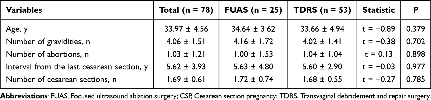 |
Table 1 Baseline Demographics of Patients with CSP |
Preoperative Clinical Characteristics
Preoperative clinical characteristics of the two groups are summarized in Table 2. No statistically significant differences were identified between the FUAS and TDRS groups with respect to gestational age, distance between the GS and the internal cervical os, or presence of fetal cardiac activity. However, the FUAS group demonstrated a smaller maximum diameter of the GS or mass and lower pre-treatment serum β-hCG levels compared to the TDRS group. In contrast, the thickness of the intervening myometrium was significantly greater in the FUAS group.
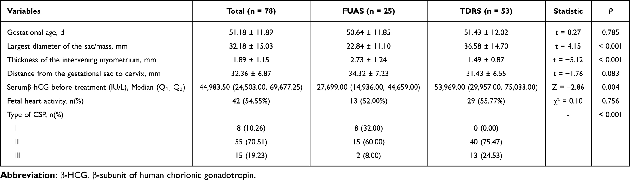 |
Table 2 Preoperative Clinical Characteristics of Patients with CSP |
A significant difference was observed in the distribution of CSP classification types between the two groups. Among patients in the FUAS group, 8 (32%) were classified as type I, 15 (60%) as type II, and 2 (8%) as type III. In the TDRS group, no type I cases were reported; 40 patients (75.47%) were classified as type II, and 13 (24.53%) as type III.
Comparison of Clinical Efficacy Between the Two Cohorts
Clinical efficacy outcomes are presented in Table 3 and Table 4.
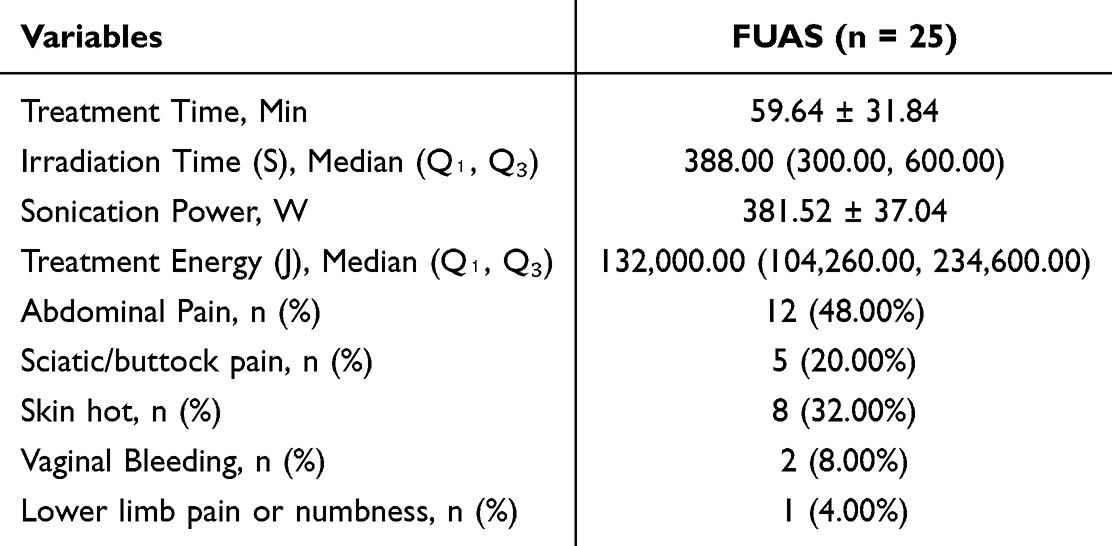 |
Table 3 Clinical Outcomes of FUAS |
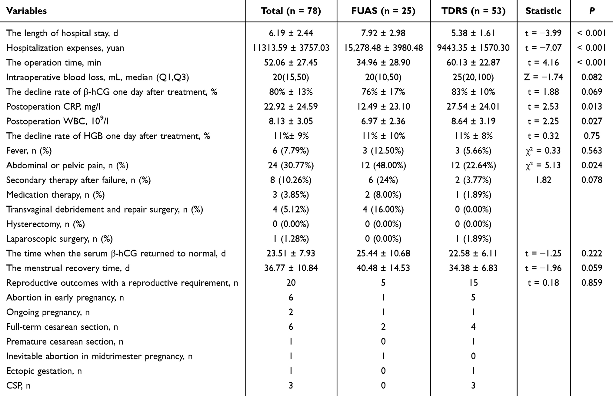 |
Table 4 Clinical Outcomes of Two Groups |
As detailed in Table 3, the mean treatment duration for the FUAS group was 59.64 ± 31.84 minutes, with a mean sonication power of 381.52 ± 37.04 W. The median irradiation time was 388.00 seconds (interquartile range: 300.00–600.00 seconds), and the median treatment energy was 132,000 J (interquartile range: 104,260–234,600 J). All ablation procedures were completed without the occurrence of severe complications. Post-treatment discomfort was reported in some cases, with 12 patients (48%) experiencing lower abdominal pain, 5 (20%) reporting sciatic or buttock discomfort, and 8 (32%) describing a “skin hot” sensation, though no instances of skin burns were observed. Minor vaginal bleeding occurred in 2 patients (8%), and lower limb pain or numbness was reported in 1 case (4%) during FUAS treatment. All symptoms resolved within two hours without the need for additional medical intervention. All patients in the FUAS group subsequently underwent suction curettage under hysteroscopic guidance.
As presented in Table 4, no statistically significant differences were found between the FUAS and TDRS groups with respect to intraoperative blood loss (median: 20 mL vs 25 mL, p = 0.082), the one-day post-treatment β-hCG decline rate (76% ± 17% vs 83% ± 10%, p = 0.069), or the hemoglobin decline rate (11% ± 8% vs 11% ± 10%, p = 0.75). However, hospital stay was significantly longer in the FUAS group compared to the TDRS group (7.92 ± 2.98 days vs 5.38 ± 1.61 days, p < 0.001), while operation time was notably shorter (34.96 ± 28.90 minutes vs 60.13 ± 22.87 minutes, p < 0.001). Additionally, hospitalization expenses were significantly higher for the FUAS group (15,278.48 ± 3980.48 CNY vs 9443.35 ± 1570.30 CNY, p < 0.001). Postoperative laboratory findings indicated lower levels of C-reactive protein and white blood cell counts in the FUAS group than in the TDRS group (p = 0.013 and p = 0.027, respectively).
No severe postoperative complications were reported in either group. However, the incidence of abdominal or pelvic pain was higher in the FUAS group, affecting 12 patients (48%), compared to 12 patients (22.64%) in the TDRS group. Postoperative fever rates did not differ significantly between the groups (p > 0.05).
The success rate in the FUAS group was 76%, which was lower than the 96.23% observed in the TDRS group. In the FUAS group, six patients required additional therapeutic intervention due to severe bleeding during curettage or retained pregnancy tissue. Specifically, four patients (16%) underwent transvaginal debridement and repair surgery for significant hemorrhage, one individual (4%) received oral mifepristone 22 days post-treatment for retained gestational tissue, and one individual (4%) underwent intramuscular MTX administration followed by curettage 28 days post-treatment due to substantial bleeding.
In the TDRS group, two patients required further intervention. One case was converted to laparoscopic surgery due to difficulty in cervical manipulation during transvaginal access, and another individual underwent systemic chemotherapy for suspected choriocarcinoma following persistent intrauterine residue and elevated β-hCG levels three months after surgery. To further explore factors associated with FUAS failure, a subgroup analysis comparing successful versus failed FUAS cases was performed (Table 5).
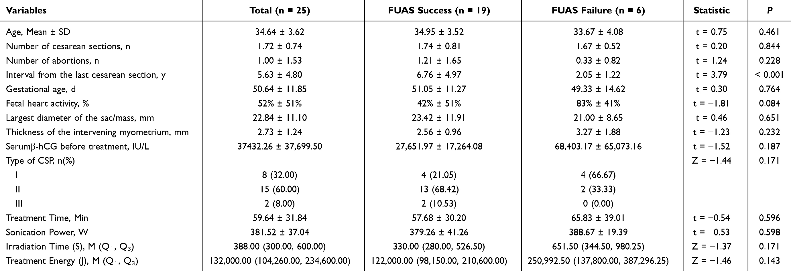 |
Table 5 Comparison of FUAS Success with FUAS Failure |
Follow up
During the follow-up period ranging from 6 to 72 months, no statistically significant differences were observed between the FUAS and TDRS groups regarding the time to menstrual recovery or the duration required for serum β-hCG levels to normalize.
Among 24 patients not using contraception during follow-up (7 in the FUAS group and 17 in the TDRS group), a total of 20 pregnancies were recorded in 18 patients (5 in the FUAS group and 13 in the TDRS group), with two patients experiencing two pregnancies each. The overall pregnancy rate was 75.00% (18/24). The pregnancy rate in the FUAS group was slightly lower at 71.43% (5/7) compared to 76.47% (13/17) in the TDRS group.
In the FUAS group, pregnancy outcomes included two full-term cesarean deliveries, one early pregnancy loss, one inevitable abortion during the second trimester, and one ongoing pregnancy. In the TDRS group, outcomes consisted of four full-term cesarean deliveries, one cesarean delivery at 33 weeks due to pernicious placenta accreta, five early pregnancy losses, one ongoing pregnancy, one tubal ectopic pregnancy, and three cases of recurrent cesarean scar pregnancy (RCSP).
The intrauterine pregnancy rate was 100% (5/5) in the FUAS group and 73.33% (11/15) in the TDRS group. The recurrence rate of CSP was 0% in the FUAS group and 20.0% (3/15) in the TDRS group. However, no statistically significant differences were identified between the two groups in terms of overall pregnancy outcomes (p > 0.05).
Discussion
According to the 2016 Chinese Expert Consensus, CSP is classified into three types based on the implantation pattern of the GS and the thickness of the myometrium between the GS and the urinary bladder: (1) Type I involves partial implantation within the cesarean scar, with a myometrial thickness exceeding 3 mm; (2) Type II involves partial implantation, but the myometrial thickness is less than 3 mm; and (3) Type III is defined by complete implantation into the scar with the GS bulging toward the bladder or forming an irregular hypervascular mass at the cesarean scar, with a myometrial thickness less than 3 mm or not measurable.8
Despite the availability of more than 30 clinical treatment modalities, encompassing both medical and surgical options, a universally accepted management guideline for CSP has not yet been established.9 Therapeutic decisions should be individualized, taking into account the CSP classification, myometrial thickness, clinical presentation, serum β-hCG levels, and the reproductive preferences of the patient.
FUAS combined with suction curettage under hysteroscopic guidance and TDRS are minimally invasive techniques that have demonstrated improved efficacy when compared to traditional treatment approaches. To the best of current knowledge, this study represents the first comparative analysis published in English assessing the clinical outcomes of these two treatment strategies.
FUAS is an emerging non-invasive treatment modality that has gained significant validation for its safety and efficacy in the management of CSP in recent years.10,11 The proposed therapeutic mechanisms of FUAS in CSP management include: (1) induction of coagulative necrosis within the implanted chorionic villi, (2) thermal damage to small-caliber blood vessels (diameter < 2 mm) surrounding the implantation site within the cesarean scar, and (3) disruption of adhesion between the GS and the myometrium at the scar site via acoustic cavitation, thereby facilitating subsequent suction curettage.
Zhu et al were the first to report the clinical effectiveness and safety of FUAS in combination with hysteroscopic suction curettage for CSP cases with gestational age less than 8 weeks.5 Their findings indicated a median intraoperative blood loss of 20 mL (range: 10–400 mL), a mean time to menstrual recovery of 35.1 ± 8.1 days, a mean duration to β-hCG normalization of 27.5 ± 6.4 days, and a mean hospital stay of 7.8 ± 1.5 days.
There were several comparable outcomes observed in this study. In the FUAS group, the median irradiation time was 388.00 seconds (interquartile range: 300.00–600.00 seconds), and the median intraoperative blood loss was 20 mL (interquartile range: 10–50 mL). The mean menstrual recovery time was 34.38 ± 6.83 days, the mean time to β-hCG normalization was 25.44 ± 10.68 days, and the mean duration of hospitalization was 7.92 ± 2.98 days. All FUAS procedures were completed without the occurrence of severe complications.
The classification of CSP is considered a significant risk factor for intraoperative hemorrhage. Yuan et al reported that FUAS along with ultrasound-guided suction curettage achieved a 100% success rate in treating CSP types I and II; however, 25% of type III cases experienced delayed severe bleeding necessitating UAE or additional surgical intervention.12 Similarly, Mu et al identified GS size and the myometrial thickness between the GS and urinary bladder as high-risk factors for intraoperative blood loss during FUAS combined with curettage.13 In a separate study, Zeng et al demonstrated that pituitrin-assisted curettage under ultrasound and hysteroscopic guidance is effective and relatively safe for CSP types I and II.9 However, surgical approaches such as laparoscopic, transvaginal, or transabdominal cesarean scar resection have been considered more appropriate for the management of type III CSP due to its higher hemorrhagic risk.
The findings of this study align with these observations. Within the FUAS group, 92% of cases were classified as CSP types I and II, whereas the TDRS group consisted entirely of types II and III. In addition, the FUAS group demonstrated smaller GS diameters and greater intervening myometrial thickness compared to the TDRS group. Given the increased risk profile associated with type III CSP, TDRS has been more frequently adopted at the study center to enhance patient safety and improve clinical outcomes.
As an emerging surgical approach for the management of CSP, TDRS offers several distinct advantages.14 By using the natural vaginal orifice, TDRS minimizes surgical invasiveness. The transvaginal route provides the shortest anatomical access to the lesion, thereby allowing the procedure to be completed more efficiently. Furthermore, the technique is conducted under direct visualization, which facilitates complete excision of the gestational lesion and simultaneous repair of the cesarean scar.
Initially introduced by Kang et al in 2009, subsequent studies have demonstrated that TDRS is a safe, effective, and minimally invasive option for CSP treatment.14–16 In a comparative study by Chen et al, TDRS was associated with a higher success rate compared to UAE (100% vs 94.7%).17
The findings from this study are consistent with prior literature in terms of both clinical efficacy and safety. In the TDRS group, the mean operative time was 60.13 ± 22.87 minutes, and the median intraoperative blood loss was 25 mL (range: 20–100 mL). Normalization of serum β-hCG levels occurred within an average of 22.58 ± 6.11 days, while menstrual recovery was achieved within 34.38 ± 6.83 days. No major adverse events were reported, including surgical or anesthetic complications, bladder injury, postoperative infection, or impaired wound healing.
Additionally, the TDRS technique does not require extensive preoperative preparation, contributing to reduced hospitalization time and lower overall treatment costs. Although no statistically significant differences were observed between the FUAS and TDRS cohorts with regard to the time required for β-hCG normalization or menstrual recovery, the TDRS group demonstrated significantly shorter hospital stays (5.38 ± 1.61 days vs 7.92 ± 2.98 days) and lower hospitalization expenses (RMB 9443.35 ± 1570.30 vs RMB 15,278.48 ± 3980.48).
The clinical success rate remains a critical parameter in the evaluation of treatment efficacy for CSP. Xi Wang et al reported success rates of 96.10% (148/154) for the high-intensity focused ultrasound ablation group and 92.40% (109/118) for the UAE group.18 Similarly, Fang et al observed cure rates of 97.89% for surgical treatment (n = 95), 43.74% for UAE (n = 32), and 70.37% for FUAS (n = 27), with statistically significant differences among the groups (p < 0.05).6 The lower success rate of HIFU was attributed to factors such as prolonged gestational age (> 55 days) and increased GS diameter (> 30 mm), both of which were associated with a heightened risk of treatment failure.
Petersen et al recommended transvaginal hysterotomy as a first-line therapy for CSP, reporting a success rate of 99.2% and a hysterectomy rate of only 0.8% (1/118).19 In this study, the clinical success rate for the FUAS group was 76%, which was notably lower than the 96.23% observed in the TDRS group.
Within the FUAS group, six patients required additional therapeutic interventions. Specifically, four patients (16%) underwent TDRS due to severe bleeding during suction curettage; one patient (4%) received oral mifepristone 22 days after treatment due to retained pregnancy tissue; and another patient (4%) was treated with intramuscular MTX followed by curettage 28 days post-treatment for persistent hemorrhage. All six cases ultimately resulted in successful clinical recovery.
The preoperative serum level of β - human chorionic gonadotropin (β-hCG) of these 6 patients was 68403.17 ± 65073.16IU/L, and the ratio of fetal heart activity was 83% ± 41%, which was higher than that of successful cases within the group (β - hCG:27651.97 ± 17264.08, the ratio of fetal heart activity 42% ± 51%). In terms of blood flow signals, 4 cases had abnormally rich blood flow signals at the scar site.These characteristics suggest that caution should be exercised when selecting FUAS for cases with high levels of β-hCG (>30000 IU/L), and with fetal heart activity and accompanied by high vascularization. Relevant supplementary analysis will provide more specific references for clinical patient screening.
However, due to the limited sample size, further research is warranted to examine the factors associated with treatment failure in FUAS for CSP. A deeper understanding of these factors may facilitate refinement of patient selection criteria and contribute to improved clinical outcomes.
Subsequent pregnancy outcomes were evaluated as a key aspect of this study. Several previous studies have indicated that FUAS offers advantages over UAE in preserving fertility, due to its non-destructive effects on uterine and ovarian function.7,20 Zhang et al reported a post-treatment pregnancy rate of 82.14% (23/28) following FUAS for CSP.21 In addition, a systematic review and meta-analysis involving 15 studies and 403 individuals with fertility intentions indicated an overall pregnancy rate of 76.2%, with 83.4% achieving intrauterine pregnancies and 15.3% experiencing RCSP.22 Stratified analyses indicated intrauterine pregnancy rates of 93.1% for conservative management, 80.1% for surgical treatments without cesarean scar resection, and 86.0% for surgical treatments with resection. Corresponding RCSP rates were 6.9%, 15.6%, and 14.0%, respectively. These findings indicate variability in reproductive outcomes depending on treatment modality, making definitive conclusions regarding the optimal approach for fertility preservation challenging.
Consistent with existing literature, post-treatment pregnancy rates were 71.43% (5/7) in the FUAS cohort and 76.47% (13/17) in the TDRS group among patients with fertility requirements. Intrauterine pregnancy rates were 100% in the FUAS group and 73.33% in the TDRS group, while the RCSP rates were 0% and 20.0%, respectively. These results indicate that favorable reproductive outcomes can be achieved following either FUAS or TDRS in patients with a history of CSP. Intracystic injection of potassium chloride (KCl) combined with methotrexate (MTX) exerts its effect by directly destroying embryonic tissue and inhibiting trophoblast cell proliferation, with the advantages of simple operation and low cost. Large sample studies have confirmed its effectiveness and safety in some low-risk cesarean scar pregnancies (such as those with small gestational sacs and low blood β - hCG levels).23 Compared with FUAS and TDRS in this study, intracystic injection is more suitable for early and low-risk cases who wish to avoid surgical intervention. However, for pregnancies with large gestational sacs, abundant blood supply, or type III cesarean scar, the treatment failure rate and secondary intervention rate may be higher. TDRS has more advantages in completely clearing lesions and controlling bleeding, while FUAS provides a supplement to non-invasive options. By comparing the applicable scenarios of different treatment methods, a more comprehensive reference can be provided for individualized clinical decision-making.
Both FUAS and TDRS represent viable treatment options for patients with CSP who have future fertility intentions. However, the elevated rate of RCSP observed in the TDRS cohort raises concerns regarding the potential benefits of cesarean scar resection and repair on long-term reproductive outcomes. To reduce the risk of RCSP, patients who conceive following CSP treatment should undergo early transvaginal ultrasound evaluation to determine the location of the gestational sac, enabling timely detection and intervention if recurrence occurs.
This study is subject to several limitations. The retrospective study design, limited sample size, potential recall bias, and single-center data collection may restrict the generalizability of the findings. Moreover, there were differences in the composition of CSP types between the two groups of patients in this study. The imbalance in the distribution of types made it difficult to completely rule out the interference of confounding factors on the study conclusions. For example, it was impossible to accurately distinguish the effects of the treatment method itself and the inherent characteristics of CSP types on clinical outcomes (such as surgery time and hospitalisation costs). This study did not statistically adjust for differences in preoperative clinical characteristics between the two groups, such as maximum gestational sac diameter, preoperative serum β - hCG levels, uterine scar muscle layer thickness, and CSP type distribution. Due to the potential impact of these baseline features on treatment outcomes, unadjusted analysis may to some extent affect the rigor of interpreting the results. Therefore, future studies involving well-designed, multicenter randomized controlled trials are warranted to comprehensively evaluate the clinical efficacy of FUAS and TDRS in CSP management and to clarify their impact on subsequent pregnancy outcomes.
Conclusion
Evidence from existing literature along with the findings of the present study indicate that both FUAS combined with suction curettage under hysteroscopic guidance and TDRS are effective and safe treatment modalities for CSP, with no severe complications reported in either group. Furthermore, patients with a history of CSP demonstrated favorable post-treatment pregnancy rates following either intervention, supporting the use of both approaches in patients with fertility preservation needs.
Notably, the TDRS cohort had a higher clinical success rate, offerd superior efficacy in complete lesion excision and hemostasis, especially for type III CSP. Based on these findings, individualized treatment plans should be developed after a comprehensive evaluation, with TDRS potentially being more suitable for type III CSP.
Given the retrospective nature and subtype imbalance, prospective, multicenter randomized controlled trials are urgently needed to validate these findings. Such studies should stratify by CSP type to clarify the comparative efficacy of FUAS and TDRS across subtypes, ultimately guiding personalized treatment algorithms.
Abbreviations
FUAS, focused ultrasound ablation surgery; TDRS, transvaginal debridement and repair surgery; CSP, cesarean scar pregnancy; GS, gestational sac; MTX, methotrexate; UAE, uterine artery embolization; MRI, magnetic resonance imaging; β-HCG, β-subunit of human chorionic gonadotropin.
Data Sharing Statement
All data generated or analysed during this study are included in this article. Further enquiries can be directed to the corresponding author (Xiaobin Huang and Yuyi Ou).
Ethics Approval and Consent to Participate
The investigation was sanctioned by the Ethics Committee of Foshan Women and Children Hospital (Approval No: FSFY-MEC-2025-006). This study was conducted in accordance with the declaration of Helsinki. Written informed consent was obtained from all patients.
Acknowledgments
We would like to acknowledge the hard and dedicated work of all the staff that implemented the intervention and evaluation components of the study.
Funding
Foshan Self-Funded Science and Technology Program (2220001004616); Foundation of State Key Laboratory of Ultrasound in Medicine and Engineering (Grant No.2022KFKT008).
Disclosure
The authors declare that they have no competing interests in this work.
References
1. Larsen JV, Solomon MH. Pregnancy in a uterine scar sacculus--an unusual cause of postabortal haemorrhage. A case report. South Afr Med J Suid-Afr Tydskr Vir Geneeskd. 1978;53:142–143.
2. Liu Y, Wang L, Zhu X. Efficacy and safety of high-intensity focused ultrasound compared with uterine artery embolization in cesarean section pregnancy: a meta-analysis. J Minim Invasive Gynecol. 2023;30(6):446–454. doi:10.1016/j.jmig.2023.02.021
3. Abd Elazeem HAS, Saad MM, Ahmed IA, et al. High-intensity focused ultrasound in management of placenta accreta spectrum: a systematic review. Int J Gynaecol Obstet off Organ Int Fed Gynaecol Obstet. 2020;151(3):325–332. doi:10.1002/ijgo.13391
4. Lee J-S, Hong G-Y, Lee K-H, Song J-H, Kim T-E. Safety and efficacy of ultrasound-guided high-intensity focused ultrasound treatment for uterine fibroids and adenomyosis. Ultrasound Med Biol. 2019;45(12):3214–3221. doi:10.1016/j.ultrasmedbio.2019.08.022
5. Zhu X, Deng X, Wan Y, et al. High-intensity focused ultrasound combined with suction curettage for the treatment of cesarean scar pregnancy. Medicine. 2015;94(18):e854. doi:10.1097/MD.0000000000000854
6. Fang S, Zhang P, Zhu Y, Wang F, He L. A retrospective analysis of the treatment of cesarean scar pregnancy by high-intensity focused ultrasound, uterine artery embolization and surgery. Front Surg. 2020;7:23. doi:10.3389/fsurg.2020.00023
7. Li Y, Hua C. Is high-intensity focused ultrasound superior to uterine artery embolization in cesarean scar pregnancy and subsequent pregnancy outcomes? A meta-analysis of the Chinese population. J Minim Invasive Gynecol. 2023;30(3):180–191. doi:10.1016/j.jmig.2022.11.015
8. Family Planning Subgroup, Chinese Society of Obstetrics and Gynocology, Chinese Medical Association. [Expert opinion of diagnosis and treatment of cesarean scar pregnancy (2016)]. Zhonghua Fu Chan Ke Za Zhi. 2016;51(8):568–572. Tonga. doi:10.3760/cma.j.issn.0529-567X.2016.08.003
9. Zeng S, Wang Y, Ye P, et al. Comparing the clinical efficacy of three surgical methods for cesarean scar pregnancy. BMC Womens Health. 2023;23(1):271. doi:10.1186/s12905-023-02415-y
10. Wang W, Chen Y, Yang Y, Qu D, Jiang J. High-intensity focused ultrasound compared with uterine artery chemoembolization with methotrexate for the management of cesarean scar pregnancy. Int J Gynaecol Obstet off Organ Int Fed Gynaecol Obstet. 2022;158(3):572–578. doi:10.1002/ijgo.14036
11. Yin Y, Pan F, He M, Zhang C, Liu Y. High intensity focused ultrasound combined with ultrasound-guided suction curettage treatment for cesarean scar pregnancy: a comparison of different HIFU sonication strategies. Int J Hyperth off J Eur Soc Hyperthermic Oncol North Am Hyperth Group. 2022;39(1):390–396. doi:10.1080/02656736.2022.2044078
12. Yuan Y, Pu D, Zhan P, et al. Focused ultrasound ablation surgery combined with ultrasound-guided suction curettage in the treatment and management of cesarean scar pregnancy. Eur J Obstet Gynecol Reprod Biol. 2021;258:168–173. doi:10.1016/j.ejogrb.2020.12.031
13. Mu L, Weng H, Wang X. Evaluation of the treatment of high intensity focused ultrasound combined with suction curettage for exogenous cesarean scar pregnancy. Arch Gynecol Obstet. 2022;306(3):769–777. doi:10.1007/s00404-022-06487-3
14. OuYang Z, Xu Y, Li H, Zhong B, Zhang Q. Transvaginal hysterotomy: a novel approach for the treatment of cesarean scar pregnancy. Taiwan J Obstet Gynecol. 2019;58(4):460–464. doi:10.1016/j.tjog.2019.05.005
15. Kang S-Y, Park B-J, Kim Y-W, Ro D-Y. Surgical management of cesarean scar ectopic pregnancy: hysterotomy by transvaginal approach. Fertil Steril. 2011;96(1):e25–28. doi:10.1016/j.fertnstert.2011.04.025
16. Chen Y-Q, Liu H-S, Li W-X, et al. Efficacy of transvaginal debridement and repair surgery for cesarean scar pregnancy: a cohort study compared with uterine artery embolism. Int J Clin Exp Med. 2015;8(11):21187–21193.
17. Chen H, Zhou J, Wang H, et al. The treatment of cesarean scar pregnancy with uterine artery embolization and curettage as compared to transvaginal hysterotomy. Eur J Obstet Gynecol Reprod Biol. 2017;214:44–49. doi:10.1016/j.ejogrb.2017.04.032
18. Wang X, Yang B, Chen W, Chen J. Clinical efficacy and re-pregnancy outcomes of patients with previous cesarean scar pregnancy treated with either high-intensity focused ultrasound or uterine artery embolization before ultrasound-guided dilatation and curettage: a retrospective cohort study. BMC Pregnancy Childbirth. 2023;23(1):85. doi:10.1186/s12884-023-05376-0
19. Birch Petersen K, Hoffmann E, Rifbjerg Larsen C, Svarre Nielsen H. Cesarean scar pregnancy: a systematic review of treatment studies. Fertil Steril. 2016;105(4):958–967. doi:10.1016/j.fertnstert.2015.12.130
20. Xiao X, Feng Z, Li T, et al. Comparing the efficacy and safety of high-intensity focused ultrasound and uterine artery embolization in caesarean scar pregnancy: a meta-analysis. Adv Ther. 2019;36(6):1314–1325. doi:10.1007/s12325-019-00959-w
21. Zhang C, Zhang Y, He J, Zhang L. Outcomes of subsequent pregnancies in patients following treatment of cesarean scar pregnancy with high intensity focused ultrasound followed by ultrasound-guided dilation and curettage. Int J Hyperth off J Eur Soc Hyperthermic Oncol North Am Hyperth Group. 2019;36:926–931.
22. Wu J, Ye J, OuYang Z, et al. Outcomes of reproduction following cesarean scar pregnancy treatment: a systematic review and meta-analysis. Eur J Obstet Gynecol Reprod Biol. 2021;262:80–92. doi:10.1016/j.ejogrb.2021.05.010
23. Swiercz G, Kabza J, Zacharska K, Mlodawska M, Młodawski J. Caesarean scar pregnancy. Analysis of medical intragestational therapy in a cohort of 35 patients. Medical Studies/Studia Medyczne. 2023;39(4):359–363. doi:10.5114/ms.2023.134087
 © 2025 The Author(s). This work is published and licensed by Dove Medical Press Limited. The
full terms of this license are available at https://www.dovepress.com/terms
and incorporate the Creative Commons Attribution
- Non Commercial (unported, 4.0) License.
By accessing the work you hereby accept the Terms. Non-commercial uses of the work are permitted
without any further permission from Dove Medical Press Limited, provided the work is properly
attributed. For permission for commercial use of this work, please see paragraphs 4.2 and 5 of our Terms.
© 2025 The Author(s). This work is published and licensed by Dove Medical Press Limited. The
full terms of this license are available at https://www.dovepress.com/terms
and incorporate the Creative Commons Attribution
- Non Commercial (unported, 4.0) License.
By accessing the work you hereby accept the Terms. Non-commercial uses of the work are permitted
without any further permission from Dove Medical Press Limited, provided the work is properly
attributed. For permission for commercial use of this work, please see paragraphs 4.2 and 5 of our Terms.