Back to Journals » Journal of Experimental Pharmacology » Volume 13
Combinatorial Therapies to Overcome BRAF/MEK Inhibitors Resistance in Melanoma Cells: An in vitro Study
Authors Pópulo H, Domingues B, Sampaio C, Lopes JM , Soares P
Received 29 December 2020
Accepted for publication 20 March 2021
Published 24 May 2021 Volume 2021:13 Pages 521—535
DOI https://doi.org/10.2147/JEP.S297831
Checked for plagiarism Yes
Review by Single anonymous peer review
Peer reviewer comments 3
Editor who approved publication: Professor Bal Lokeshwar
Helena Pópulo,1– 3 Beatriz Domingues,1,2 Cristina Sampaio,1,2 José Manuel Lopes,1– 4 Paula Soares1– 3
1Institute of Molecular Pathology and Immunology, IPATIMUP, University of Porto, Porto, Portugal; 2Instituto de Investigação e Inovação em Saúde, Universidade do Porto, Porto, Portugal; 3Department of Pathology, Medical Faculty, University of Porto, Porto, Portugal; 4Department of Pathology, Hospital São João, Porto, Portugal
Correspondence: Helena Pópulo
Institute of Molecular Pathology and Immunology, IPATIMUP, University of Porto, Rua Júlio Amaral de Carvalho, 45, Porto, 4200-135, Portugal
Tel +351 225570700
Fax +351 225570799
Email [email protected]
Background: Melanoma accounts for only 1% of all skin malignant tumors; however, it is the deadliest form of skin cancer. Since 2011, FDA (Food and Drug Administration) approved several novel therapeutic strategies, such as MAPK pathway targeted therapies, to treat cutaneous melanoma patients. However, their improvements in overall survival were limited, due to the development of resistance.
Methods: In this work, several combinations of therapies, including the metabolic modulator DCA, were tested in melanoma cell lines, considering that MAPK and PI3K/AKT/mTOR pathways are deregulated and interconnected in melanoma and that the presence of the Warburg effect in melanoma cells may influence the response to therapy. The effect of the treatments was assessed in the proliferation and survival of melanoma cell lines with different genetic profiles. Also, the possibility to overcome resistance to the treatment with vemurafenib was tested.
Results: In general, higher decrease in cell viability and cell proliferation and increase in apoptosis were obtained after the combination treatments, comparing with single treatments, in all the studied cell lines. The combination of cobimetinib and everolimus appear to be the best treatment option. The BRAFV600E -vemurafenib resistant melanoma cell line showed to retain sensitivity to both everolimus and DCA.
Discussion and Conclusion: Our results suggest that the combination of MAPK pathway inhibitors with mTOR pathway inhibitors and DCA should be considered as therapeutic options to treat melanoma patients, as the combinations potentiated the effects of each drug alone. In a cell line resistant to vemurafenib, we verified that combined MAPK inhibitors with inhibition of mTOR pathway and/or DCA metabolism modulation might constitute possible strategies in order to overcome resistance to MAPK inhibition.
Keywords: melanoma, vemurafenib, cobimetinib, everolimus, DCA, metabolism
Introduction
MAPK and PI3K/AKT/mTOR pathways are frequently deregulated and related to melanoma development, by the modulation of cell growth, proliferation and apoptosis.1 The mitogen-activated protein kinase pathway (MAPK) is frequently activated in melanoma,2 through BRAFV600 and NRASQ61 mutations, which constitute the most frequent identified mutations in cutaneous melanoma, 50% and 25%, respectively. PI3K/AKT/mTOR pathway overactivation is also frequent in melanoma, through PTEN loss or AKT overexpression,3,4 and can lead to melanoma progression.5 Higher mTOR pathway activation in cutaneous melanoma has been reported, connected with MAPK pathway and/or BRAF activation.6 The simultaneous occurrence of BRAF mutations and activation of PI3K/AKT/mTOR pathway was connected with cutaneous melanoma aggressiveness, worse prognosis and short patient’s progression-free and overall survival.6
Besides activation of these signaling pathways, metabolic alterations are also crucial for melanoma biology. Melanoma cells present the Warburg effect relying in glycolysis, independently of the oxygen level,7 which characterizes the metabolic activity of cancer cells.8 Aerobic glycolysis confers tumor cells' growth advantage and is required for evolution of invasive tumors, supplying metabolites and fast ATP for cell proliferation and conferring resistance to apoptosis through limited mitochondrial oxidative phosphorylation activity.9,10 Skin melanocytes are located in a naturally mild-hypoxic environment (10% or less of oxygen), which could pre-adapt melanoma to hypoxia.11 At low oxygen levels, hypoxia-inducible factor 1 α (HIF1-α) induces the glycolytic metabolism.12,13 In cutaneous melanoma, overexpression of HIF1–α associates with the activation of mTOR.14–17 The BRAF or NRAS mutations, frequent in melanoma, may also affect cell metabolism via activation of HIF1-α.11,15 The Warburg effect present in the cancer cells may be reversed by the metabolic modulator dichloroacetate (DCA), that shift cancer cell metabolism towards aerobic respiration.18 DCA inhibits the pyruvate dehydrogenase kinase (PDK) function,19 favoring pyruvate to enter the mitochondria, to participate in the TCA cycle. In breast cancer, non-small-cell lung cancer and glioblastoma models, DCA treatment, that favors the metabolic shift from glycolysis to oxidative phosphorylation, lead to an increase in apoptosis and a decrease in cell growth, glucose oxidation, mitochondrial membrane depolarization, and angiogenesis, probably through indirect HIF1–α inhibition.20,21 We and others have previously demonstrated in vitro that DCA downregulated tumor cell proliferation, induced mTOR pathway downregulation, and promoted an increase of apoptosis of melanoma cells.13,18,22,23 It was also reported that BRAFV600 melanoma cells resistant to vemurafenib are still sensitivity to DCA.24
The majority of the cutaneous melanoma cases are detected in an early stage, reaching 98% on 5–year survival rate.25 However, patients diagnosed with distant metastatic melanomas have a median survival of 8–9 months and 10–year overall survival less than 10%.26–28 In the last 10 years, FDA (Food and Drug Administration) approved several novel therapeutic approaches, targeted therapies and immune therapies, for stage IV (distant metastases) cutaneous melanoma patients.
Several combinatorial therapies of BRAFV600 inhibitors and MEK inhibitors (as vemurafenib + cobimetinib, dabrafenib + trametinib and encorafenib + binimetinib, respectively) were approved by FDA, for patients with unresectable or metastatic malignant melanoma harboring BRAF mutations.29,30 Ipilimumab, an anti-CTLA4 antibody was approved by FDA in 2011.31 Then, nivolumab and pembrolizumab, monoclonal antibodies against the programmed death receptor-1 (PD–1), were approved by FDA for the treatment of patients with metastatic melanoma.32–34 FDA approved in 2015 an oncolytic virus, talimogene laherparepvec (T–VEC), a genetically modified herpes simplex virus type 1.35–37 More recently, in 2017 and 2019, nivolumab and pembrolizumab, respectively, were approved to use as adjuvant treatment of melanoma patients with involvement of lymph nodes after complete resection.38,39
As the approved therapies still present limitations, and considering that MAPK and PI3K/AKT/mTOR pathways are frequently deregulated and interconnected in melanoma pathogenesis and that the Warburg effect found frequently in melanoma cells may affect the response to therapy, in the present study, several combinations of therapies were tested in vitro. The effect of the treatments was assessed in the proliferation and survival of melanoma cell lines with different genetic profiles. Moreover, the possibility to overcome resistance to the treatment with vemurafenib was also tested in a resistant cell line.
Materials and Methods
Cell Lines and Culture Conditions
Four skin melanoma cell lines were used: Mewo (BRAF-wildtype), A375 (BRAFV600E), ED013 (BRAFV600E) and ED013R2 (BRAFV600E; vemurafenib resistant). Mewo cell line was provided by Dr. Marc Mareel, from the Department of Radiotherapy and Nuclear Medicine, Ghent University Hospital, Belgium. A375 cell line was provided by Dr. Madalena Pinto, from CEQUIMED, Faculty of Pharmacy, University of Porto, Portugal. Vemurafenib-sensitive and -resistant cell lines (ED013 and ED013R2) were provided by Prof. Per Guldberg, from the Danish Cancer Society Research Center, Copenhagen, Denmark. The parental BRAFV600E cell line (ED013) was exposed to increasing concentrations of vemurafenib in order to create the vemurafenib-resistant cell line. The new cell line (ED013R2) was considered resistant when the cells could be propagated at a concentration of vemurafenib above the IC50 of the parental cell line.24 As the cell lines have not been purchased from an accredited commercial source, their identity was verified by i3S Cell Line Bank and the use was approved by the i3S Unit for Responsible Conduct in Research. A375 and ED013 were maintained in RPMI medium (Gibco/BRL – Invitrogen). ED013R2 was maintained in the same medium supplemented with additional 1 μM of vemurafenib (Absource Diagnostics GmbH). Mewo was maintained in DMEM medium (Gibco/BRL – Invitrogen). The mediums were complemented with 10% of fetal bovine serum, 100 U/mL Penicillin and 100 µg/mL Streptomycin. All the cell lines were tested for the presence of mycoplasma and were cultured as a monolayer and maintained at 37°C, in a humidified atmosphere (5% CO2).
Treatment of Melanoma Cell Lines Using Vemurafenib, Cobimetinib, Everolimus and DCA
Vemurafenib (Absource Diagnostics GmbH, München, Deutschland), molecular weight 489.92 g/mol, cobimetinib (Genentech, Roche Group, San Francisco, California, USA), molecular weight 531.32 g/mol, and everolimus (Novartis Pharma AG, Basel, Switzerland), molecular weight 958.22 g/mol were dissolved in dimethyl sulfoxide (DMSO). DCA (Sodium dichloroacetate, Sigma-Aldrich, St. Louis, Missouri, USA), molecular weight 128.94 g/mol, was dissolved in distilled water (dH2O) and filtered, with a 0.22 µm filter. To treat melanoma cell lines, drugs were added to the respective culture medium and incubated during 48 and 72 h. For control, cell lines were incubated with the respective culture medium and culture medium with DMSO and/or dH2O, according to the drug solvent.
Cell Viability Assay
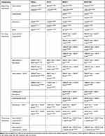
PrestoBlue (PB) assay was used to evaluate the effects of the treatments in melanoma cell lines. Cells were seeded in 96-well plates, according to cell line growth rate, at a density of 9x10³ (Mewo), 7x10³ (A375), 5x10³ (ED013) and 7.5x10³ (ED013R2) in 200 μL of medium. After 24 h, the medium was replaced by media containing different treatment concentrations. Concerning IC50 determination for vemurafenib, Mewo and A375 cells were treated with 10, 50, 100, 1000, 2500, 5000 nM, ED013 cells were treated with 500, 1000, 2000, 3000, 4000 and 5000 nM and ED013R2 cells were treated with 1000, 2500, 4000, 5000, 6000, 7500 and 10,000 nM. For cobimetinib, ED013 cells were treated with 10, 20, 30, 40, 50, 100 and 200 nM and ED013R2 cells were treated with 1000, 2500, 4000, 5000, 6000, 7500 and 10,000 nM. For DCA, the IC50 was previously established for Mewo and A375 cell lines,13 and for ED013 and ED013R2, both cell lines were treated with 5, 10, 20 and 40 mM. To test the combinations, half of the IC50 and the IC50, determined for each cell line was used for vemurafenib and cobimetinib, except for ED013R2, were the IC50 determined for ED013 cells was used. For everolimus, half of the recommended concentration and the recommended concentration by the manufacture was used (10 and 20 nM). For DCA the IC50 value of each cell line was used. The already approved combination for melanoma treatment (vemurafenib with cobimetinib) was tested in the vemurafenib resistant cell line (ED013R2), compared with the parental cell line (ED013), and tested against all the other combinations.
Cells were incubated for 48 or 72 h, washed with PBS (pH 7.4) and cell growth was evaluated using PB, according to the manufacturer’s instructions. A microplate reader (Synergy HT Multi-Mode Microplate Reader, BioTek Instruments Inc., Winooski, VT, USA) was used to measure fluorescence, at 560 nm excitation and 590 nm emission wavelengths. As control, the absorbance of the wells with culture medium and tumor cells was used and each experimental condition was evaluated with triplicates and repeated three times. Dose-response profiles and IC50 determination (the concentration that inhibits survival in 50%) were performed using GraphPadPrism5.0 (GraphPad Software, Inc., La Jolla, CA). Each experimental condition was repeated three times.
Cell Cycle and Apoptosis Analysis
Melanoma cells were seeded in 6-well plates, according to cell line growth rate, at a final density of 2×10⁵ (Mewo), 1.5×10⁵ (A375), 1.8x10⁵ (ED013) and 2x10⁵ (ED013R2) cells/well and incubated at 37°C for 24 h. Cells were then incubated with the treatments described above for 72 h of treatment. For cell cycle analysis, cells were collected and fixed overnight in ice-cold 70% ethanol. Before analysis, cells were resuspended in PBS with 0.1 mg/mL RNase A and 5 μg/mL propidium iodide. For apoptosis quantification, cells were collected and analyzed by flow cytometry using the Annexin-V FITC Apoptosis Kit (Clontech Laboratories, Inc., Saint-Germain-en-Laye, France) according to the manufacturer’s instructions. The flow cytometer BD Accuri C6 (BD Biosciences, Franklin Lakes, New Jersey, USA) was used to analyze the cellular DNA content and phosphatidylserine externalization, plotting at least 20,000 events per sample. The data was evaluated using the FlowJo 7.6.5 software (Tree Star, Inc., Ashland, USA). Each experimental condition was repeated three times.
Statistical Analysis
STAT VIEW-J 5.0 (SAS Institute, Inc., Cary, NC) was used to performe the statistical analysis. The data from the cell line experiments, comparing control vs treatment and treatment vs treatment, was analyzed by the two-tailed unpaired Student’s t-test. A p value <0.05 was considered statistically significant.
Results
Determination of the IC50 of Vemurafenib, Cobimetinib and DCA
Mewo, A375, ED013 and ED013R2 melanoma cell lines were incubated with increasing concentrations of vemurafenib, cobimetinib and DCA to determine the IC50, using the PrestoBlue cell viability assay.
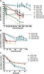
After 48 h of treatment, vemurafenib reduced the viability of Mewo, A375 and ED013 cell lines in a dose-dependent manner, with IC50 values ranging from 173 to 5000 nM (Table 1). A significant effect of vemurafenib in cell viability was observed in Mewo after treatment with 100 nM (p = 0.04), in A375 cell line with 10 nM (p = 0.03), and in ED013 cell line after treatment with 500 nM (p < 0.01) (Figure 1A). The IC50 was not reached for ED013R2 vemurafenib resistant cell line, until 10,000 nM of vemurafenib (Figure 1A).
 |
Table 1 IC50 Values for the Melanoma Cell Lines Analyzed |
Given the resistance of ED013R2 to vemurafenib, we tested the effects of cobimetinib, a MEK inhibitor, in cell viability in these cells and in the parental cell line. ED013 cell viability was reduced after cobimetinib treatment (Figure 1B), in a dose- and time-dependent manner. After 48 h of treatment, a significant effect of cobimetinib on ED013 cell viability was observed after treatment with 10 nM (p < 0.01) (Figure 2B). The IC50 was calculated as 40 ± 2.63 nM, after 48 h of treatment (Table 1). ED013R2 cells treated with 1000 to 10,000 nM of cobimetinib did not reach the IC50 (Figure 1B).
The IC50 of DCA was previously calculated as ~35 mM for Mewo and A375 cell lines (Table 1).13 DCA reduced the viability of ED013 and ED013R2 in a dose-dependent manner, and the effect of the treatment on cell viability was observed with 5 mM (p < 0.01) (Figure 1C). The IC50 values were calculated as 20 and 14 mM for ED013 and ED013R2 cells, respectively (Table 1).
The Effect of Vemurafenib, Cobimetinib, Everolimus, DCA and Combinatory Treatments in Melanoma Cell Line Viability
To evaluate melanoma cell viability, the previously established IC50 and half of the IC50 concentrations were used for each targeted drug: 2500 and 5000 nM for Mewo, 88 and 175 nM for A375, and 900 and 1800 nM for ED013 of vemurafenib, and 20 and 40 nM of cobimetinib for ED013, for 48 h and 72 h. For ED013R2 cells, resistant to both vemurafenib and cobimetinib, the IC50 calculated for the ED013 cells were used. Similarly, everolimus was used at the recommended concentration by the manufacture and half of it, 10 and 20 nM, in all cell lines. DCA was used at the IC50, 35 mM for Mewo and A375, 20 mM for ED013 and 14 mM for ED013R2 cells. The drugs were tested alone and in combination of two and three drugs (Table 2).
 |
Table 2 Treatment Combinations Doses Applied to Each Melanoma Cell Line |
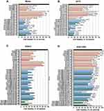
In general, the cell lines responded in a dose and time-dependent manner to treatments (Figure 2). Comparing to the control, treatments decreased the viability of all cell lines, with significant differences, at both time points (p < 0.01), with higher decrease with drug combinations than single-drug treatments (p < 0.01) (Figure 2).
Ten and 20 nM of everolimus achieved a significant decrease on cell viability of all cell lines compared to the control (p ≤ 0.01 for all cell lines, except for 72 h of treatment in A375) (Figure 2).
In Mewo cell line, combined treatment of vemurafenib with everolimus decrease significantly the percentage of viable cells compared to the control (p < 0.01) and also compared with vemurafenib combined with DCA treatments (p = 0.01, except for vemurafenib 5000 nM with DCA 35 mM, 72h of treatment), and with everolimus 20 nM combined with DCA 35 mM treatments (p < 0.01) (Figure 2A).
A375 cells treated with combined therapy of vemurafenib with DCA decreased viability compared with the control (p < 0.01), and also compared with the treatment with vemurafenib combined with everolimus (p < 0.01 at 48 h and 72 h), and with the treatment with everolimus with DCA (p < 0.01 at 72 h) (Figure 2B).
In ED013 cell line, significant differences on cell viability were observed comparing the effects of cobimetinib with everolimus with all the other treatments, after 48 h and 72 h of treatment (p < 0.01), even when compared with the combinations of vemurafenib with cobimetinib (Figure 2C). The combination of vemurafenib with DCA led to a higher decrease of cell viability compared with everolimus combined with DCA treatments (p ≤ 0.01), vemurafenib 900 nM with everolimus 20 nM (p < 0.01 to p = 0.02) and also all the combinations of vemurafenib with cobimetinib at 48h treatment (p < 0.01 to p = 0.04), except for vemurafenib 1800 nM with cobimetinib 20 nM (not significant). From all the treatments tested, the three-drug combination of vemurafenib, cobimetinib and everolimus triggered the most evident damage on ED013 cell viability, at both time points (p < 0.01) (Figure 2C).
Despite the resistance observed, ED013R2 cells responded in a time-dependent manner to vemurafenib treatment (p < 0.01), and to cobimetinib treatment (p < 0.01) (Figure 2D). In ED013R2 cells, the only two-drug treatment capable to induce a reduction on cell viability consistently higher than 50%, at both time points, was cobimetinib with everolimus (p < 0.01) (Figure 2D). The combination of everolimus with DCA led to a higher decrease of cell viability compared with vemurafenib with DCA (p < 0.01) and all the combinations of vemurafenib with cobimetinib (p < 0.01 to p = 0.02). For the three-drug approaches, in ED013R2 cells, combining vemurafenib, cobimetinib and everolimus was more effective than combining the two melanoma-approved drugs with DCA. Compared to the approved combination for melanoma treatment (vemurafenib combined with cobimetinib), the treatments that achieved the highest significant reduction on ED013R2 cell viability, at both time points, were vemurafenib 1800 nM with everolimus, cobimetinib 40 nM with everolimus, everolimus 20 nM with DCA, and both three-drug combinations (p < 0.01).
Comparing ED013 and ED103R2 melanoma cell lines, at 48 h, the treatments more significantly efficient in the resistant cells were everolimus 10 nM (p < 0.01) and everolimus 20 nM (p = 0.01). At 72 h of treatment, everolimus 10 nM induced a significantly higher effect in ED013R2 cells compared with ED013 cells (p < 0.01).
The Effect of Vemurafenib, Cobimetinib, Everolimus, DCA and Combinatory Treatments in Melanoma Cell Cycle
In general, and comparing to control, single-drug treatments and combinations increased the percentage of cells in G0/G1 phase and decreased the percentage of cells in S and G2/M phase in melanoma cell lines, but not all effects were statistically significant. Combined treatments led to higher effects in the cell cycle in comparison with single-drug treatments, although again not all reaching statistical significance (Table 2 and Figure 3).
In Mewo cell line, the highest percentage of cells in G0/G1 phase was observed in response to vemurafenib plus everolimus (p < 0.01), although it was similar to the effect of vemurafenib with DCA (p = 0.04 and p = 0.02), compared to the control (Figure 3A). The significant lowest percentages of Mewo cells in the S and G2/M phases were observed in response to vemurafenib with everolimus (p < 0.01) and with everolimus combined with DCA (p = 0.01), respectively, with significant differences comparing to the control (Figure 3A).
In A375 cell line, comparing with the control, treatment with DCA in combination with everolimus and vemurafenib showed a higher percentage of cells in G0/G1 phase (p = 0.03, p = 0.05, p = 0.02 and p = 0.02, respectively). The lowest percentages of A375 cells in the S and G2/M phases were observed in response to vemurafenib with DCA (not significant and p = 0.04) and to everolimus with DCA (not significant and p = 0.03), respectively (Figure 3B).
In ED013, among two-drug treatments, the highest percentages of ED013 cells in the G0/G1 phase were observed in response to cobimetinib with everolimus treatment (p = 0.01), followed by vemurafenib 1800 nM with everolimus treatment (p = 0.02) and vemurafenib 1800 nM with cobimetinib 40 nM treatment (p = 0.03), with significant differences comparing to the control. Regarding the three-drug treatments, vemurafenib, cobimetinib and everolimus (p = 0.01, comparing to control) was the more efficient in increasing the number of cells in G0/G1 phase (Figure 3C). The lowest percentages of ED013 cells in the S and G2/M phases were observed in response to cobimetinib with everolimus treatment (p = 0.01) and to the three-drug treatment of vemurafenib, cobimetinib and everolimus (p ≤ 0.01), with significant differences comparing to the control. In the ED013 cells, comparing to the approved combinations of vemurafenib and cobimetinib, the treatment that was capable to induce more significant effects, in all the phases, was cobimetinib with everolimus (p < 0.01 to 0.03), which was associated with a significant increase in the percentage of ED013 cells in the G0/G1 phase (p < 0.01 to p = 0.01), a significant decrease in the S phase (p < 0.01 to p = 0.02), and a decrease in the G2/M phase, but without statistical differences (Figure 3C).
In ED013R2 cells, cobimetinib with everolimus treatment (p < 0.01), followed by vemurafenib 1800 nM with everolimus treatment (p = 0.01) and the three-drug treatment vemurafenib, cobimetinib and everolimus (p = 0.01) were associated with a higher percentage of cells in G0/G1 phase, with significant differences from the percentage of cells in the control (Figure 3D). In the S phase, cobimetinib with everolimus treatment (p < 0.01) and to the three-drug treatment of vemurafenib, cobimetinib and everolimus (p < 0.01), were associated with a lower percentage of cells, with significant differences comparing to the control (Figure 3D). The observed decrease of the percentage of cells in G2/M phase after all the treatments was not statistically significantly different from the control. In the ED013R2 cells, the treatment that consistently surpass the effects induced by the approved combination for melanoma, in all the phases, was cobimetinib with everolimus, that was associated with a significant increase in the percentage of cells in the G1/G0 phase (p = 0.03 to 0.05), a significant decrease in the S phase (p < 0.01 to p = 0.02) and a decrease in the G2/M phase, but without statistical differences (Figure 3D).
Several treatments affected the cell cycle differently, comparing the two melanoma cell lines, and in general, the effects of the treatments were more evident on the ED013 cells than on the ED013R2 cells.
The Effect of Vemurafenib, Cobimetinib, Everolimus, DCA and Combinatory Treatments in Melanoma Cell Lines Apoptosis
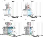
Comparing to control, single-drug treatments and combinations increased the percentage of apoptotic cells in general, although not all the differences were statistically significant. Combined treatments led to higher effects in comparison with single drug treatment, but again not all reaching statistical significance (Table 2 and Figure 4).
In Mewo cell line, treatment with the combination of vemurafenib with everolimus led to the higher increase in the percentage of apoptotic cells compared to the control (p < 0.01 and p = 0.01), similarly to the percentage found with the treatment of vemurafenib combined with DCA (p = 0.01 and p = 0.02, respectively) (Figure 4A).
In A375 cell line, the percentage of apoptotic cells was significantly increased in all the treatment combinations compared to the control. Combination of vemurafenib 175 nM with DCA reached the higher mortality rate in A375 cell line (p < 0.01) (Figure 4B).
In ED013 cells, among the new two-drug treatments, the more efficient combination in increasing apoptosis was cobimetinib with everolimus (p < 0.01, to control), followed by the combinations of vemurafenib and DCA (p < 0.01, to control) (Figure 4C). The three-drug treatment combining vemurafenib, cobimetinib and everolimus was the most efficient in inducing apoptosis of ED013 cells (p < 0.01, relatively to control). Concerning apoptotic effects, ED013 cells showed no significant differences comparing the four different combinations with vemurafenib and cobimetinib, achieving 30% to 40% of apoptotic cells. These combinations were the most effective among two-drug combinations, and only the treatment with vemurafenib, cobimetinib and everolimus induced a higher percentage of apoptosis in ED013 cells (p = 0.01 to 0.02) (Figure 4C).
In ED013R2 cells, the most efficient treatment was cobimetinib with everolimus (p < 0.01), however similar to other drug-treatment combinations (vemurafenib 900 nM with cobimetinib 40 nM, vemurafenib 1800 nM with cobimetinib 40 nM, vemurafenib 1800 nM with DCA, cobimetinib 40 nM with DCA, everolimus 20 nM with DCA, and both three-drug treatments) (Figure 4D). In ED013R2 cells, all the combinations of vemurafenib and cobimetinib induced statistically identical effects, ~10% of apoptotic cells. Four combinations were more efficient at inducing apoptosis in ED013R2 cells than the approved combination (cobimetinib 40 nM with everolimus, cobimetinib 40 nM with DCA, and both three–drug combinations), with significant differences among effects (p < 0.01 to p = 0.01) (Figure 4D).
Globally, the treatments induced less apoptosis in the ED013R2 cells than in the ED013 cells, except for everolimus 10 nM treatment, but without statistical differences.
Discussion
In this work, we established that mTOR pathway inhibition and metabolism modulation are potential therapeutic strategies to combine with MAPK pathway inhibition in the treatment of melanoma patients, aiming to overcome MAPK pathway inhibitor resistance.
Approved therapies to treat cutaneous melanoma patients still present limitations, such as fast acquisition of resistance to MAPK pathway inhibitors.40,41 As cutaneous melanoma cells show overactivation of MAPK and PI3K/AKT/mTOR pathways1 and the Warburg effect,8 in this work we evaluated whether targeting the MAPK pathway in combination with mTOR inhibitors or DCA could indicate therapeutic benefit and/or if drug combinations could overcome resistance to MAPK pathway inhibition. This in vitro work allowed to test a high number of combinations of several drugs, impractical to perform in vivo, that enabled us to determine effective drug combinations to treat melanomas.
Vemurafenib is a specific mutant BRAF inhibitor, as the mutation benefits the active enzyme conformation.42 In agreement, the most sensitive cell lines to vemurafenib were A375 and ED013 (both harboring BRAFV600), reaching an IC50 lower than 1 µM, contrary to Mewo cell line (BRAFwt) that presented an IC50 higher than 1 µM. For ED013R2 resistant cell line, the IC50 of vemurafenib was not reached, corroborating the specificity of the drug and the resistance of the cell line, with an IC50 above 10 µM.43
In our work, DCA proved to be an efficient therapeutic drug in cutaneous melanoma cell line treatment, either alone or in combination with MAPK and mTOR pathways inhibitors. The melanoma cell lines tested presented differential sensitivity to DCA treatment, translated in lower IC50. We observed previously that DCA target, PDK, is overexpressed in melanoma and associated with the expression of the mTOR pathway effectors. Melanoma cell lines treated with DCA showed a shift in the metabolism (reduction of glucose consumption and lactate production) and a decrease not only in PDH, the readout of DCA effect, but also in the mTOR pathway activation.13 In this work, the metabolic modulator alone was efficient in blocking cell proliferation and increasing apoptosis in all the analyzed cell lines. Khan et al published a case report of a melanoma patient treated with DCA that showed regression and stabilization of recurrent metastatic melanoma for over 4 years, with minor adverse effects.44 Our results also show that DCA combined with vemurafenib or everolimus might be an effective treatment for melanoma patients. The MAPK and mTOR pathway inhibitors altered cell dynamics, and this effect is potentiated by the combination with DCA.
Vemurafenib, cobimetinib, everolimus and DCA, alone or in combination, led to cell cycle arrest in G0/G1 phase, with concomitant decrease of the percentage of cells on S phase and G2/M phase, in all analyzed melanoma cell lines.13,45 Vemurafenib, being an inhibitor of the mutant BRAF, decreased cell proliferation and increased apoptosis, by the reduction of phosphorylated ERK and cyclin D1, a protein relevant for G1/S cell cycle transition.42 Studies with cobimetinib suggested that BRAF mutant cell lines presented cytocidal and cytostatic effects in response to MEK inhibition46 and the downregulation of cyclin D1, that regulates G1/S transition.47,48 Bonnet et al reported preclinical evidence of anticancer effect of DCA and suggested that the cell cycle arrest in G0/G1 phase may be due to the stimulation of ROS production by DCA20 and the consequent increased oxidative stress, which leads to cell cycle arrest and increase apoptosis.49,50 Everolimus inhibits ribosome biogenesis and translation, by preventing the phosphorylation of mTOR effectors, which leads to a decrease in cyclin D1 expression and increased p27 expression that stop G1/S cell cycle transition.51–53 Indeed, in a previous study, we confirmed the inhibition of the phosphorylation of mTOR effectors after melanoma cell line treatment with the manufacture recommended concentration of everolimus (20 nM).45
In general, our results in cell viability assays, although not directly comparable, are in agreement with the results from proliferation and apoptosis analysis. In the BRAFwt cell line (Mewo), a higher decrease in cell viability, downregulation of proliferation, by G0/G1 arrest, concomitant with an increase in apoptosis, was achieved by vemurafenib combined with everolimus. In contrast, in the two BRAFV600 cell lines (A375 and ED013), the most effective drug combination was vemurafenib combined with DCA. The genetic background of the cell lines seems to confer different sensitivity to each combination of drugs.13 These results corroborate previous studies from our group, which emphasized the importance of the PI3K/AKT/mTOR pathway in melanoma cell lines and its role in cell survival as well as the sensitivity of melanoma cell lines to DCA treatment.13,54 Our results suggest a possible usefulness of DCA in the treatment of melanoma patients, in line with Abildgaard et al who reported a synergistic combination between DCA and vemurafenib in BRAF-mutant melanoma cell lines and the retention of DCA sensitivity in vemurafenib-resistant cells.24 The cooperative effect of these compounds can be related with the induction of a greater inhibition of lactate and ATP production, by the combination than either agent alone.24,55 Although in our work the determined IC50 for DCA ranged from 14 to 35 mM, it was already observed that lower concentrations of DCA potentiate the effect by lowering the effective concentrations of vemurafenib, pointing to a clinical relevance of DCA in melanoma treatment, by allowing reducing vemurafenib dose and consequently reducing the side effects.24
In our work, the vemurafenib resistant cell line ED013R2 (BRAFV600) proved to be resistant also to the MEK inhibitor cobimetinib. The cross-resistance to MAPK inhibitors in BRAF mutant melanoma cell lines was already reported in other in vitro and in vivo models.56 Thus, it seems that in ED013R2 melanoma cells the in vitro acquired resistance may be associated with activation of other important pathways, such as continuous PI3K/AKT/mTOR pathway activation, rather than the activation of MAPK pathway by secondary oncogenic events, such as NRAS mutations or overexpression of the kinases A-Raf and C-Raf.57 ED013R2 seems to be more sensitive to combinatorial treatments containing everolimus, which induced a greater reduction on cell viability and proliferation, and a higher increase of apoptosis, compared with the vemurafenib-sensitive cell line. These results suggest that the isogenic cell lines have differential sensitivity to the mTORC1 inhibitor and ED013R2 cells may have a sustained dependence on the PI3K/AKT/mTOR pathway in order to survive. These observations fit with other studies that described differences in this pathway activation comparing vemurafenib-sensitive and vemurafenib-resistant cell lines.43 Moreover, combination of everolimus seems to surpass resistance to vemurafenib, and even more striking, resistance to cobimetinib, being the more effective treatment (cobimetinib combined with everolimus), better than the approved therapy for mutant BRAF melanomas (vemurafenib combined with cobimetinib). Of note, ED013R2 behave as the most sensitive cell line to DCA, with G0/G1 arrest and increased apoptosis after treatment, suggesting metabolic remodeling drugs as valuable alternatives in melanomas that render insensitive to MAPK inhibitors, as also observed by Roesch et al that reported that targeting the bioenergetic metabolism sensitizes melanoma for a more pronounced and long-lasting BRAFV600E–inhibitors therapeutic effect.58
Gong et al already reported in colorectal cancer cell lines that the combination of cobimetinib with PI3K pathway inhibition increased the cytotoxicity of cobimetinib.48 Penna et al reported that, compared to targeting both BRAF and PI3K/mTOR pathway, the combination of a MEK1/2 and a PI3K/mTOR inhibitor was more effective in the activation of Bax and of caspase-3 and in the activation of caspase-dependent melanoma apoptosis.59 In our work, the evaluation of cell viability, proliferation and apoptosis consistently revealed that the combination of cobimetinib with everolimus and the three-drug treatment of vemurafenib, cobimetinib and everolimus achieved better results in the tested cell lines than the approved therapy for mutant BRAF melanomas (vemurafenib with cobimetinib), suggesting that targeting multiple pathways is a potential option to treat melanomas harboring BRAFV600, including those resistant to MAPK inhibitors. Our data establish the benefit of additive/synergetic drug combinations, particularly the concomitant inhibition of MAPK and PI3K/AKT/mTOR pathways that may overcome the mechanisms of resistance developed frequently in BRAF mutant melanomas. Unfortunately, the study design, with only two data points for each drug combination, does not allow to confirm if the drug effects are in fact synergistic or additive.
Clinical trials with MEK and PI3K/mTOR inhibitors were already performed in melanoma patients (eg, NCT01166126, NCT01820364, NCT01392521 and NCT01390818).60 Targeting both of these pathways has clinical activity, despite some combinations have proved unacceptably adverse effects and some of the studies had insufficient number of patients analyzed, which do not allow to draw definitive conclusions.61 Therefore, a large patient cohort clinical trial will be important to establish conclusively the efficacy of these combinations.
Conclusions
Our data indicate that the combination of MAPK pathway inhibitors with mTOR pathway inhibitors and DCA should be considered as therapeutic options to treat melanoma patients, as the combinations potentiated the effect on in vitro cell dynamics of each single-drug treatment. Of note, the BRAFV600E-vemurafenib-resistant melanoma cell line showed to maintain sensitivity to both everolimus and DCA. Our results suggest that combined MAPK inhibitors with inhibition of mTOR pathway and/or DCA metabolism modulation might represent novel strategies to overcome resistance to MAPK inhibition. This work reinforces the perception that approaches for melanoma therapy should take into consideration the BRAF mutational status, and that the genetic screening of the patients should be considered for a personalized therapy, leading to improvements in melanoma patient’s survival.
Data Sharing Statement
The datasets generated during and/or analyzed in the present study are accessible from the corresponding author on reasonable demand.
Acknowledgments
We thank Dr. Marc Mareel, from the Department of Radiotherapy and Nuclear Medicine, Ghent University Hospital, Belgium, that kindly provided us Mewo melanoma cell line; Dr. Madalena Pinto, from CEQUIMED, Faculty of Pharmacy, University of Porto, Portugal, that kindly provided us A375 melanoma cell line; and Prof. Per Guldberg, from the Danish Cancer Society Research Center, Copenhagen, Denmark that kindly provided us vemurafenib-sensitive and -resistant cell lines (ED013 and ED013R2).
Author Contributions
All authors made substantial contributions to conception and design, acquisition of data, or analysis and interpretation of data; took part in drafting the article or revising it critically for important intellectual content; agreed to submit to the current journal; gave final approval of the version to be published; and agree to be accountable for all aspects of the work.
Funding
This work was supported by FEDER—Fundo Europeu de Desenvolvimento Regional funds through the COMPETE 2020—Operacional Programme for Competitiveness and Internationalization (POCI), Portugal 2020, and by Portuguese funds through FCT - Fundação para a Ciência e a Tecnologia/Ministério da Ciência, Tecnologia e Inovação in the framework of the project “Institute for Research and Innovation in Health Sciences” (POCI-01-0145-FEDER-007274). This article received also funding from the project PTDC/MEC-ONC/31520/2017 (Predicting patients’ response to liposomal anticancer drugs: focusing on LRP1B endocytic activity) supported by Norte Portugal Regional Operational Programme (NORTE 2020), under the PORTUGAL 2020 Partnership Agreement, through the European Regional Development Fund (ERDF), and FCT. The funders did not participate in study design, data collection and analysis, decision to publish, or preparation of the manuscript.
Disclosure
Paula Soares reports grants from FCT, during the conduct of the study. The authors reported no other conflicts of interest for this work.
References
1. Dahl C, Guldberg P. The genome and epigenome of malignant melanoma. APMIS. 2007;115(10):1161–1176. doi:10.1111/j.1600-0463.2007.apm_855.xml.x
2. Lopez-Bergami P. The role of mitogen- and stress-activated protein kinase pathways in melanoma. Pigment Cell Melanoma Res. 2011;24(5):902–921. doi:10.1111/j.1755-148X.2011.00908.x
3. Wu H, Goel V, Haluska FG. PTEN signaling pathways in melanoma. Oncogene. 2003;22(20):3113–3122. doi:10.1038/sj.onc.1206451
4. Stahl JM, Sharma A, Cheung M, et al. Deregulated Akt3 activity promotes development of malignant melanoma. Cancer Res. 2004;64(19):7002–7010. doi:10.1158/0008-5472.CAN-04-1399
5. Vredeveld LC, Possik PA, Smit MA, et al. Abrogation of BRAFV600E-induced senescence by PI3K pathway activation contributes to melanomagenesis. Genes Dev. 2012;26(10):1055–1069. doi:10.1101/gad.187252.112
6. Populo H, Soares P, Faustino A, et al. mTOR pathway activation in cutaneous melanoma is associated with poorer prognosis characteristics. Pigment Cell Melanoma Res. 2011;24(1):254–257. doi:10.1111/j.1755-148X.2010.00796.x
7. Warburg O. On respiratory impairment in cancer cells. Science. 1956;124(3215):269–270.
8. Scott DA, Richardson AD, Filipp FV, et al. Comparative metabolic flux profiling of melanoma cell lines beyond the warburg effect. J Biol Chem. 2011;286(49):42626–42634. doi:10.1074/jbc.M111.282046
9. Michelakis E, Webster L, Mackey J. Dichloroacetate (DCA) as a potential metabolic-targeting therapy for cancer. Br J Cancer. 2008;99(7):989.
10. Gatenby RA, Gillies RJ. Why do cancers have high aerobic glycolysis? Nat Rev Cancer. 2004;4(11):891. doi:10.1038/nrc1478
11. Bedogni B, Powell MB. Hypoxia, melanocytes and melanoma–survival and tumor development in the permissive microenvironment of the skin. Pigment Cell Melanoma Res. 2009;22(2):166–174. doi:10.1111/j.1755-148X.2009.00553.x
12. Bertout JA, Patel SA, Simon MC. The impact of O2 availability on human cancer. Nat Rev Cancer. 2008;8(12):967–975. doi:10.1038/nrc2540
13. Populo H, Caldas R, Lopes JM, Pardal J, Maximo V, Soares P. Overexpression of pyruvate dehydrogenase kinase supports dichloroacetate as a candidate for cutaneous melanoma therapy. Expert Opin Ther Targets. 2015;19(6):733–745.
14. Land SC, Tee AR. Hypoxia inducible factor 1α is regulated by the mammalian target of rapamycin (mTOR) via an mTOR-signalling motif. J Biol Chem. 2007;282:20534–20543. doi:10.1074/jbc.M611782200
15. Kumar SM, Yu H, Edwards R, et al. Mutant V600E BRAF increases hypoxia inducible factor-1α expression in melanoma. Cancer Res. 2007;67(7):3177–3184. doi:10.1158/0008-5472.CAN-06-3312
16. Kuphal S, Winklmeier A, Warnecke C, Bosserhoff A-K. Constitutive HIF-1 activity in malignant melanoma. Eur J Cancer. 2010;46(6):1159–1169. doi:10.1016/j.ejca.2010.01.031
17. Slominski A, Kim T-K, Brożyna A, et al. The role of melanogenesis in regulation of melanoma behavior: melanogenesis leads to stimulation of HIF-1α expression and HIF-dependent attendant pathways. Arch Biochem Biophys. 2014;563:79–93. doi:10.1016/j.abb.2014.06.030
18. Papandreou I, Goliasova T, Denko NC. Anticancer drugs that target metabolism: is dichloroacetate the new paradigm? Int J Cancer. 2011;128(5):1001–1008. doi:10.1002/ijc.25728
19. Whitehouse S, Cooper RH, Randle PJ. Mechanism of activation of pyruvate dehydrogenase by dichloroacetate and other halogenated carboxylic acids. Biochem J. 1974;141(3):761–774. doi:10.1042/bj1410761
20. Bonnet S, Archer SL, Allalunis-Turner J, et al. A mitochondria-K+ channel axis is suppressed in cancer and its normalization promotes apoptosis and inhibits cancer growth. Cancer Cell. 2007;11(1):37–51. doi:10.1016/j.ccr.2006.10.020
21. Sutendra G, Dromparis P, Kinnaird A, et al. Mitochondrial activation by inhibition of PDKII suppresses HIF1a signaling and angiogenesis in cancer. Oncogene. 2013;32(13):1638–1650. doi:10.1038/onc.2012.198
22. Stacpoole PW. The pharmacology of dichloroacetate. Metabolism. 1989;38(11):1124–1144.
23. Michelakis E, Webster L, Mackey J. Dichloroacetate (DCA) as a potential metabolic-targeting therapy for cancer. Br J Cancer. 2008;99(7):989–994.
24. Abildgaard C, Dahl C, Basse AL, Ma T, Guldberg P. Bioenergetic modulation with dichloroacetate reduces the growth of melanoma cells and potentiates their response to BRAFV600E inhibition. J Transl Med. 2014;12:247. doi:10.1186/s12967-014-0247-5
25. Siegel R, Ma J, Zou Z, Jemal A. Cancer statistics, 2014. CA Cancer J Clin. 2014;64(1):9–29. doi:10.3322/caac.21208
26. Bertolotto C. Melanoma: from melanocyte to genetic alterations and clinical options. Scientifica. 2013;2013:22. doi:10.1155/2013/635203
27. Balch CM, Gershenwald JE, Soong SJ, et al. Final version of 2009 AJCC melanoma staging and classification. J Clin Oncol. 2009;27(36):6199–6206. doi:10.1200/JCO.2009.23.4799
28. Foth M, Wouters J, de Chaumont C, Dynoodt P, Gallagher WM. Prognostic and predictive biomarkers in melanoma: an update. Expert Rev Mol Diagn. 2016;16(2):223–237. doi:10.1586/14737159.2016.1126511
29. Hoeflich KP, Merchant M, Orr C, et al. Intermittent administration of MEK inhibitor GDC-0973 plus PI3K inhibitor GDC-0941 triggers robust apoptosis and tumor growth inhibition. Cancer Res. 2012;72(1):210–219. doi:10.1158/0008-5472.CAN-11-1515
30. Niezgoda A, Niezgoda P, Czajkowski R. Novel approaches to treatment of advanced melanoma: a review on targeted therapy and immunotherapy. Biomed Res Int. 2015;2015:2015. doi:10.1155/2015/851387
31. Hodi FS, O’Day SJ, McDermott DF, et al. Improved survival with ipilimumab in patients with metastatic melanoma. N Engl J Med. 2010;363(8):711–723. doi:10.1056/NEJMoa1003466
32. Raedler LA. Opdivo (Nivolumab): second PD-1 inhibitor receives FDA approval for unresectable or metastatic melanoma. Am Health Drug Benefits. 2015;8(Spec Feature):180.
33. Robert C, Ribas A, Wolchok JD, et al. Anti-programmed-death-receptor-1 treatment with pembrolizumab in ipilimumab-refractory advanced melanoma: a randomised dose-comparison cohort of a Phase 1 trial. Lancet. 2014;384(9948):1109–1117. doi:10.1016/S0140-6736(14)60958-2
34. Ribas A, Puzanov I, Dummer R, et al. Pembrolizumab versus investigator-choice chemotherapy for ipilimumab-refractory melanoma (KEYNOTE-002): a randomised, controlled, Phase 2 trial. Lancet Oncol. 2015;16(8):908–918. doi:10.1016/S1470-2045(15)00083-2
35. Franklin C, Livingstone E, Roesch A, Schilling B, Schadendorf D. Immunotherapy in melanoma: recent advances and future directions. Eur J Surg Oncol. 2017;43(3):604–611. doi:10.1016/j.ejso.2016.07.145
36. Pol J, Kroemer G, Galluzzi L. First Oncolytic Virus Approved for Melanoma Immunotherapy. Taylor & Francis; 2016.
37. Hersey P, Gallagher S. Intralesional immunotherapy for melanoma. J Surg Oncol. 2014;109(4):320–326. doi:10.1002/jso.23494
38. US Food & Drug Administration. FDA grants regular approval to nivolumab for adjuvant treatment of melanoma; 2017. Available from: https://www.fda.gov/drugs/resources-information-approved-drugs/fda-grants-regular-approval-nivolumab-adjuvant-treatment-melanoma.
39. US Food & Drug Administration. FDA grants regular approval to pembrolizumab for adjuvant treatment of melanoma; 2019. Available from: https://www.fda.gov/drugs/drug-approvals-and-databases/fda-approves-pembrolizumab-adjuvant-treatment-melanoma.
40. Chapman PB, Hauschild A, Robert C, et al. Improved survival with vemurafenib in melanoma with BRAF V600E mutation. N Engl J Med. 2011;364(26):2507–2516. doi:10.1056/NEJMoa1103782
41. Flaherty KT, Robert C, Hersey P, et al. Improved survival with MEK inhibition in BRAF-mutated melanoma. N Engl J Med. 2012;367(2):107–114. doi:10.1056/NEJMoa1203421
42. Tsai J, Lee JT, Wang W, et al. Discovery of a selective inhibitor of oncogenic B-Raf kinase with potent antimelanoma activity. Proc Natl Acad Sci U S A. 2008;105(8):3041–3046. doi:10.1073/pnas.0711741105
43. Atefi M, von Euw E, Attar N, et al. Reversing melanoma cross-resistance to BRAF and MEK inhibitors by co-targeting the AKT/mTOR pathway. PLoS One. 2011;6(12):e28973. doi:10.1371/journal.pone.0028973
44. Khan A, Andrews D, Shainhouse J, Blackburn AC. Long-term stabilization of metastatic melanoma with sodium dichloroacetate. World J Clin Oncol. 2017;8(4):371–377. doi:10.5306/wjco.v8.i4.371
45. Populo H, Tavares S, Faustino A, Nunes JB, Lopes JM, Soares P. GNAQ and BRAF mutations show differential activation of the mTOR pathway in human transformed cells. PeerJ. 2013;1:e104. doi:10.7717/peerj.104
46. Solit DB, Garraway LA, Pratilas CA, et al. BRAF mutation predicts sensitivity to MEK inhibition. Nature. 2006;439(7074):358. doi:10.1038/nature04304
47. Halilovic E, Solit DB. Therapeutic strategies for inhibiting oncogenic BRAF signaling. Curr Opin Pharmacol. 2008;8(4):419–426. doi:10.1016/j.coph.2008.06.014
48. Gong S, Xu D, Zhu J, Zou F, Peng R. Efficacy of the MEK inhibitor cobimetinib and its potential application to colorectal cancer cells. Cell Physiol Biochem. 2018;47(2):680–693. doi:10.1159/000490022
49. Yalcin A, Clem BF, Imbert-Fernandez Y, et al. 6-Phosphofructo-2-kinase (PFKFB3) promotes cell cycle progression and suppresses apoptosis via Cdk1-mediated phosphorylation of p27. Cell Death Dis. 2014;5:e1337. doi:10.1038/cddis.2014.292
50. Takahashi M, Watari E, Takahashi H. Dichloroacetate induces cell cycle arrest in human glioblastoma cells persistently infected with measles virus: a way for controlling viral persistent infection. Antiviral Res. 2015;113:107–110. doi:10.1016/j.antiviral.2014.11.008
51. Hay N, Sonenberg N. Upstream and downstream of mTOR. Genes Dev. 2004;18(16):1926–1945. doi:10.1101/gad.1212704
52. Populo H, Lopes JM, Soares P. The mTOR signalling pathway in human cancer. Int J Mol Sci. 2012;13(2):1886–1918.
53. Hashemolhosseini S, Nagamine Y, Morley SJ, Desrivieres S, Mercep L, Ferrari S. Rapamycin inhibition of the G1 to S transition is mediated by effects on cyclin D1 mRNA and protein stability. J Biol Chem. 1998;273(23):14424–14429. doi:10.1074/jbc.273.23.14424
54. Pópulo H, Lopes JM, Soares P. The mTOR signalling pathway in human cancer. Int J Mol Sci. 2012;13(2):1886–1918.
55. Parmenter TJ, Kleinschmidt M, Kinross KM, et al. Response of BRAF-mutant melanoma to BRAF inhibition is mediated by a network of transcriptional regulators of glycolysis. Cancer Discov. 2014;4(4):423–433. doi:10.1158/2159-8290.CD-13-0440
56. Smalley KS, Haass NK, Brafford PA, Lioni M, Flaherty KT, Herlyn M. Multiple signaling pathways must be targeted to overcome drug resistance in cell lines derived from melanoma metastases. Mol Cancer Ther. 2006;5(5):1136–1144. doi:10.1158/1535-7163.MCT-06-0084
57. Holderfield M, Nagel TE, Stuart DD. Mechanism and consequences of RAF kinase activation by small-molecule inhibitors. Br J Cancer. 2014;111(4):640–645. doi:10.1038/bjc.2014.139
58. Roesch A, Vultur A, Bogeski I, et al. Overcoming intrinsic multidrug resistance in melanoma by blocking the mitochondrial respiratory chain of slow-cycling JARID1B(high) cells. Cancer Cell. 2013;23(6):811–825. doi:10.1016/j.ccr.2013.05.003
59. Penna I, Molla A, Grazia G, et al. Primary cross-resistance to BRAFV600E-, MEK1/2- and PI3K/mTOR-specific inhibitors in BRAF-mutant melanoma cells counteracted by dual pathway blockade. Oncotarget. 2016;7(4):3947–3965. doi:10.18632/oncotarget.6600
60. NIH. US National Library of Medicine. ClinicalTrials.gov search results for Melanoma.Available from: https://www.clinicaltrials.gov/ct2/results?cond=Melanoma&term=&cntry=&state=&city=&dist=&Search=Searchwww.clinicaltrials.org
61. Lim SY, Menzies AM, Rizos H. Mechanisms and strategies to overcome resistance to molecularly targeted therapy for melanoma. Cancer. 2017;123(S11):2118–2129. doi:10.1002/cncr.30435
 © 2021 The Author(s). This work is published and licensed by Dove Medical Press Limited. The full terms of this license are available at https://www.dovepress.com/terms.php and incorporate the Creative Commons Attribution - Non Commercial (unported, v3.0) License.
By accessing the work you hereby accept the Terms. Non-commercial uses of the work are permitted without any further permission from Dove Medical Press Limited, provided the work is properly attributed. For permission for commercial use of this work, please see paragraphs 4.2 and 5 of our Terms.


