
Back to Journals » Clinical Ophthalmology » Volume 20
Clinical, Surgical, and Survival Outcomes of Periocular Merkel Cell Carcinoma: A Retrospective Cohort Analysis
Authors Rattunde AL  , Gutmann M, Möhwald LM, Bertelmann E
Received 21 September 2025
Accepted for publication 17 January 2026
Published 18 February 2026 Volume 2026:20 569243
DOI https://doi.org/10.2147/OPTH.S569243
Checked for plagiarism Yes
Review by Single anonymous peer review
Peer reviewer comments 2
Editor who approved publication: Dr Scott Fraser
Alexander L Rattunde,* Michael Gutmann,* Lukas M Möhwald, Eckart Bertelmann
Department of Ophthalmology, Charité - University Medicine Berlin, Berlin, Germany
*These authors contributed equally to this work
Correspondence: Alexander L Rattunde, Email [email protected]
Introduction: Merkel cell carcinoma (MCC) is a rare, aggressive cutaneous malignancy with substantial risks of mortality and vision-threatening morbidity when periocular. We aimed to characterize the clinicopathologic features, management strategies, ophthalmic outcomes, and survival of periocular MCC.
Methods: We conducted a retrospective cohort study of 18 periocular MCC cases treated at a tertiary center between January12010and February 1 2024. Demographic and tumor characteristics, treatment details, ocular complications, and survival endpoints were extracted from electronic records. Kaplan–Meier curves estimated overall survival (OS) and progression-free survival (PFS).
Results: Eighteen patients (61% female; mean age ± SD, 71.6 ± 11.4 years) were included; 67% presented with upper-eyelid disease. Half of tumors were AJCC stage I (50.0%). Initial therapy was excision alone in 33%, excision plus radiation in 28%, and multimodal in the remainder; an average of 1.62 resections was required to achieve negative margins or functional reconstruction. Functional globe preservation was achieved in 78% of patients, with severe ocular complications in 11%. Median OS was 8.5 years (95% CI, 4.6– 12.3) and the estimated 2-year OS rate was 88.9%. Median PFS was 6.4 years (95% CI, 0.1– 12.8) with a 2-year PFS of 54.7%. Median follow-up was 17 months.
Conclusion: Periocular MCC in this single-center cohort was typically detected early and managed with eye-sparing surgery with acceptable morbidity, yielding favorable OS and PFS estimates; these site-specific benchmarks can guide counseling, reconstruction, and future multicenter validation.
Keywords: Merkel cell carcinoma, periocular, eyelid neoplasms, globe preservation
Introduction
Merkel cell carcinoma (MCC) is a rare but highly aggressive cutaneous neuroendocrine tumor with a disease-specific mortality rate of approximately 33%, making it more lethal than malignant melanoma.1
In 1972 Toker discovered a distinct skin tumor he referred to as “trabecular carcinoma” based on its histopathological appearance.2 A few years later, electron microscopic studies of these carcinomas identified Merkel cells as the underlying cause, based on the presence of characteristic neurosecretory granules, suggesting their origin from neural crest-derived cells.3 MCC exhibits dual pathogenesis, arising from either UV-induced damage or Merkel cell polyomavirus infection, with distinct molecular profiles.4
Despite its rarity, the incidence of MCC has risen significantly in recent decades, driven by factors such as increased UV exposure, aging populations, and improved diagnostic capabilities.5,6 This rise is concerning, given MCC’s aggressive clinical behavior, as well as its propensity for rapid progression. The disease most commonly affects older, fair-skinned individuals and arises predominantly in sun-exposed areas, with nearly half of all cases occurring on the head and neck.7,8 Within this region, periocular MCC is of particular concern due to its functional and aesthetic implications. Periocular MCC may mimic benign lesions such as cysts or chalazion, and can resemble other malignant periocular tumors such as basal cell carcinoma, squamous cell carcinoma, melanoma, and sebaceous carcinoma, leading to diagnostic delays.9
These tumors frequently involve the upper eyelid and margin and may present with features such as madarosis, ulceration, and destruction of normal anatomical structures.9 While rare, conjunctival MCC has also been reported.10 The periocular location poses unique challenges in management, as wide local excision, a standard treatment, must balance complete tumor resection with the preservation of ocular and eyelid function and cosmesis. MCC typically presents as a rapidly growing, painless, firm nodule that may be red, purple, or skin-colored.11 Diagnostic evaluation includes histopathological analysis and immunohistochemistry, with CK20 positivity in a dot-like pattern serving as a hallmark feature.12,13 Early recognition is aided by the AEIOU criteria: Asymptomatic, Expanding rapidly, Immune suppression, Older age, and UV-exposed site, with most cases meeting at least three of these characteristics.14 However, the diagnostic process for periocular MCC is particularly challenging due to its atypical presentation and resemblance to benign or other malignant lesions.9 Management strategies for MCC vary by stage, but typically involve surgical excision with negative margins for localized disease, often supplemented by adjuvant radiation therapy to improve local control, especially for tumors larger than 2 cm.15,16
Sentinel lymph node biopsy (SLNB) is an important staging tool since nodal metastases are present in approximately one third of cases.17,18 In advanced MCC, immune checkpoint inhibitors such as pembrolizumab, avelumab, and retifanlimab have become the mainstay of systemic therapy, demonstrating durable responses and improving outcomes.16,19,20
To expand the limited periocular MCC evidence base, we performed a retrospective single-center analysis of 18 consecutive cases, detailing patient demographics, tumor characteristics, surgical management, ocular morbidity, and survival estimates of overall and progression-free survival. By combining functional outcomes with site-specific survival data, this study aims to inform counseling, guide reconstruction, and provide a benchmark for future multicenter efforts.
Methods
The clinical cancer registry of Charité – University Medicine Berlin, part of the Charité Comprehensive Cancer Center (CCCC) and responsible for the documentation, evaluation, and reporting of all tumor diseases, was retrospectively searched for Merkel cell carcinoma (MCC) of the head and neck region between January 1 2010 and February 1 2024. We identified 108 cases of head and neck MCC. Inclusion criteria included eyelid, canthal, caruncular, lacrimal, or orbital localizations. Eighteen patients fulfilled these criteria and constituted the study population. Patients with non-periocular localization, or incomplete records were excluded. The investigation complied with the Declaration of Helsinki and was approved by the institutional ethics committee; informed consent was waived because of the retrospective design. We minimized selection bias by including all consecutive eligible cases during the study window.
All data were compiled electronically in Microsoft® Excel (Version 16.66.1). Variables extracted from electronic patient records comprised age, sex, tumor location, tumor size, AJCC 8th-edition stage, surgical technique, ophthalmologic findings, and treatment-related complications. Frequency distributions were illustrated with pie charts produced in Excel, and annual case counts from 2010 through 2024 were tabulated to describe temporal trends in periocular MCC.
Overall survival (OS) was defined as the interval from histologic diagnosis to death from any cause; living patients were censored at last follow-up or the administrative data cut-off (February 1 2024). Progression-free survival (PFS) was the interval from diagnosis to the first locoregional recurrence, distant metastasis, or death, whichever occurred first. When ≥2 events occurred on the same calendar date, the event type was assigned to the more severe synchronous event according to a prespecified hierarchy: death > distant metastasis > locoregional recurrence. Patients without an event were censored on the date of their most recent disease assessment or administrative cut-off. Survival curves were estimated with the Kaplan–Meier method. Time-to-event endpoints were analyzed on a day scale.
All descriptive statistics, medians with 95% confidence intervals (CI), and survival analyses were executed in IBM SPSS Statistics (Version 29.0.1.1; IBM Corp., Armonk, NY, USA). Analyses were unadjusted; no multivariable, subgroup, or interaction analyses were planned due to sample size. Missing data were handled by complete-case analysis. We report this retrospective cohort study in accordance with the STROBE reporting guideline.
Results
Annual Number of Periocular MCC Cases
Eighteen periocular MCCs were diagnosed over the 14-year observation window, yielding 0–3 cases annually (Table 1). Case counts fluctuated without a consistent upward or downward trend.
 |
Table 1 Annual Number of Periocular MCC Diagnoses, 2010–2024 |
Demographics and Tumor Characteristics
Mean age at diagnosis was 71.6 ± 11.4 years (range, 50–89 years); 11 patients (61.1%) were female. The mean largest tumor diameter was 14.6 ± 7.8 mm. Stage distribution at presentation was AJCC I in 50.0%, II in 16.7%, III in 11.1%, and IV in 11.1%, and unknown in 11.1%. Detailed baseline demographics and tumor characteristics are summarized in Table 2.
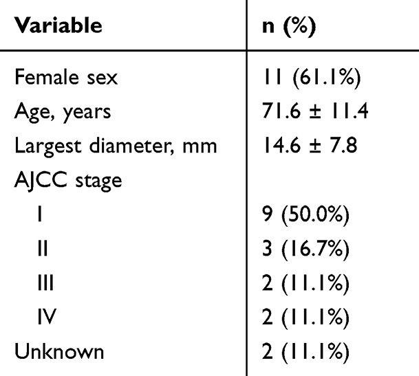 |
Table 2 Demographics and Tumor Characteristics at Diagnosis |
Upper-eyelid involvement predominated (66.7%), followed by lower eyelid (16.7%), and lateral canthus, caruncle, and lacrimal gland (5.6% each), as shown in Figure 1.
 |
Figure 1 Location of Periocular MCC. Distribution of periocular Merkel cell carcinoma cases by anatomical location. |
Treatment
Excision alone was performed in 6 patients (33.3%); excision with adjuvant radiotherapy in 5 (27.8%); excision + radiotherapy + chemotherapy in 3 (16.7%); excision + radiotherapy + immunotherapy in 2 (11.1%); and one quadruple-modality case combined surgery, radiotherapy, immunotherapy, and chemotherapy. One patient received excision, chemotherapy, and immunotherapy without radiotherapy. The distribution of initial treatment modalities is presented in Table 3.
 |
Table 3 Distribution of Treatment Modalities in Periocular MCC |
Surgical Outcomes and Complications in Periocular MCC
We further analyzed the surgical and ophthalmologic outcomes of patients with periocular MCC. Functional eye preservation was achieved in 77.8% of cases (n = 14), with 16.7% (n = 3) experiencing loss of eye function due to the need for oncological resection or ophthalmic complications. Data on eye preservation were unavailable for one case. Patients underwent an average of 1.6 resections of the primary tumor to achieve R0 status or until a decision was made to preserve function or due to the presence of advanced disease.
In our series, reconstruction with the Cutler–Beard flap was performed in 33.3% of cases (n = 6), making it the predominant primary technique. The inverse Tenzel flap followed at 16.7% (n = 3), and the Hughes flap in 11.1% (n = 2). Less commonly, procedures such as orbitotomy and exenteration each accounted for fewer than 6% of reconstructions. Extensive tissue defects necessitated combined approaches, most often the addition of free eyelid grafts, in 16.7% of patients (n = 3). Ophthalmologic complications were noted in seven cases: lagophthalmos (incomplete eyelid closure) and severe corneal injury requiring transplantation each occurred in 11.1% (n = 2), while trichiasis, ectropion, and fistula formation were all observed in under 6% of patients. Overall, the Cutler–Beard flap emerged as the preferred method, and the vast majority of eyes retained functional integrity. One patient underwent a Cutler–Beard flap followed by exenteration; this case was categorized under the primary technique (Cutler–Beard) for Table 4. Comprehensive surgical and ophthalmic outcomes are summarized in Table 4.
 |
Table 4 Clinical and Surgical Outcomes of Periocular MCC |
Survival Analysis
Median follow-up was 16.8 months (interquartile range, 2.9–48 months) and a total of four deaths occurred during follow-up. Median overall survival (OS) was 8.5 years (95% CI, 4.6–12.3), and the 2-year OS estimate was 88.9% (Figure 2).
 |
Figure 2 Overall Survival of Periocular MCC. Kaplan–Meier survival curve showing overall survival (OS) for patients with periocular MCC. |
Median progression-free survival (PFS) was 6.4 years (95% CI, 0.1–12.8) with a 2-year PFS of 54.7%. Among the six PFS events, two were locoregional (one regional lymph-node recurrence and one primary-site recurrence), three were distant metastases (first sites: liver, skin, and pancreas/duodenum), and one was death as the first event. In two cases, progression types were synchronous (local recurrence with liver metastasis; nodal recurrence with pancreatic/duodenal metastasis) and, per the prespecified hierarchy, were classified as metastasis. Survival curves are shown in Figures 2 and 3.
 |
Figure 3 Progression-Free Survival of Periocular MCC. Kaplan–Meier survival curve illustrating progression-free survival (PFS) for patients with periocular MCC. |
These results highlight the early stage at presentation, the predominance of upper-eyelid tumors, the feasibility of eye-sparing surgery, and encouraging long-term survival in periocular MCC.
Discussion
Our data provide insight into the clinical characteristics, presentation, management, and survival of Merkel cell carcinoma (MCC) confined to the periocular region.
Periocular MCC remained uncommon, with zero to three cases recorded per calendar year. Although small numbers preclude firm conclusions about secular trends, the pattern broadly echoes the steady rise in overall MCC cases described in population-based reports.5,21,22 Increasing incidence is probably multifactorial, driven by an aging population, higher cumulative ultraviolet (UV) exposure, and improved diagnostic vigilance.5,21–24
Women constituted 61.1% of our cohort, confirming the female preponderance previously noted for eyelid MCC.9 This contrasts with the male predominance reported for cutaneous MCC in general,8,25 suggesting that anatomic site, or associated behavioral and environmental factors, may influence sex distribution. The mean age at diagnosis was 71.6 ± 11.4 years, slightly younger than the seventh-to-eighth-decade peak reported for head-and-neck MCC overall,8 which could reflect earlier health-care seeking for lesions near the eye or distinct site-specific etiologies.
Two-thirds of tumors occurred in the upper eyelid, consistent with prior reports.7,9 Half of lesions presented at AJCC stage I and the mean largest diameter was 14.6 mm, indicating relatively early clinical detection despite the disease’s recognized aggressiveness.26
Management remained complex. Wide local excision was the backbone of therapy, yet nearly 40% of patients required adjuvant radiotherapy and more than one-fifth received systemic treatment, underscoring the need for individualized, multidisciplinary care. Functional globe preservation was achieved in 77.8% of cases, but severe complications such as lagophthalmos and sight-threatening keratopathy still occurred in 11.1% each, highlighting the delicate balance between oncologic clearance and ocular function. Stage I presentation, in 50.0% of patients, likely facilitated eye-sparing surgery and likely contributed to favorable outcomes, given the well-documented survival advantage of early excision with clear margins.16,27,28
Survival results were encouraging: the Kaplan–Meier 2-year overall survival (OS) and progression-free survival (PFS) rates reached 88.9% and 54.7%, respectively, and median OS was 8.5 years. These figures compare favorably with other MCC cohorts where 2-year OS often falls below 70%.29 While wide confidence intervals necessitate further multicenter cohorts with a larger sample size, data suggest that early-stage periocular MCC treated with eye-sparing surgery can achieve durable disease control.
The rarity of periocular MCC continues to hamper prospective data collection. Growing incidence nevertheless mandates greater clinical awareness, multicenter registries to validate survival benchmarks, and prospective trials that integrate advances such as immunotherapy, refined reconstructive techniques, and patient-reported outcome measures into personalized, multidisciplinary care models.
Limitations
This study has several limitations that should be kept in mind when interpreting the findings. First, it is a retrospective review that relies on registry and electronic‐health-record data; some comorbidities, immunosuppression status, and operative details may therefore be incomplete or misclassified. Second, the cohort is small (18 patients), resulting in wide confidence intervals for survival estimates and limited power to explore prognostic factors. Third, median follow-up was only 17 months, so late recurrences or treatment-related ocular sequelae may be under-reported. Fourth, the data originate from a single tertiary center; referral patterns, reconstructive expertise, and thresholds for adjuvant therapy may differ elsewhere, which could temper generalizability. Finally, the study period spans more than a decade, during which immunotherapy, sentinel-node biopsy, and radiotherapy techniques evolved, introducing temporal heterogeneity.
Despite these constraints, our series is among the largest single-institution cohorts focused exclusively on periocular MCC, provides granular surgical and ophthalmic outcome data rarely reported in the literature, and contributes the one of the first Kaplan–Meier survival benchmarks specific to this anatomic site.
Conclusion
This retrospective, single-center cohort delivers site-specific benchmarks for periocular MCC that align with our aim to characterize presentation, management, ophthalmic morbidity, and survival. Most tumors were detected early (AJCC I) with small size, and upper-eyelid predominance was common; together with multidisciplinary care, this enabled eye-sparing surgery in most patients with acceptable complication rates. Survival estimates at two years were encouraging, providing practical figures for counseling and reconstruction planning. These results support our objective of combining functional outcomes with survival metrics to inform care and to anchor future multicenter validation. Given the small sample and limited follow-up, the estimates should be interpreted as preliminary; harmonized, multicenter datasets are needed to refine these benchmarks and test generalizability.
Data Sharing Statement
All data generated or analyzed during this study are included in this article and in Supplementary Table 1. Further enquiries can be directed to the corresponding author.
Statement of Ethics
This study protocol was reviewed and approved by the Ethics Committee of Charité – University Medicine Berlin (Ref. EA1/085/22) and was conducted in accordance with the Declaration of Helsinki.
Consent to Participate Statement
Individual written informed consent was not obtained because this retrospective chart review used de-identified data and involved no direct patient contact, in accordance with the approved protocol (Ref. EA1/085/22).
Funding
This study was not supported by any sponsor or funder.
Disclosure
The authors have no conflicts of interest to declare in this work.
References
1. Becker JC. Merkel cell carcinoma. Ann Oncol. 2010;21(Suppl 7):vii81–9. doi:10.1093/annonc/mdq366
2. Toker C. Trabecular carcinoma of the skin. Arch Dermatol. 1972;105(1):107–110. doi:10.1001/archderm.1972.01620040075020
3. Tang C-K, Toker C. Trabecular carcinoma of the skin. An ultrastructural study. Cancer. 1978;42(5):2311–2321. doi:10.1002/1097-0142(197811)42:5<2311::AID-CNCR2820420531>3.0.CO;2-L
4. Pedersen EA, Verhaegen ME, Joseph MK, Harms KL, Harms PW. Merkel cell carcinoma: updates in tumor biology, emerging therapies, and preclinical models. Front Oncol. 2024;14:1413793. doi:10.3389/fonc.2024.1413793
5. Paulson KG, Park SY, Vandeven NA, et al. Merkel cell carcinoma: current US incidence and projected increases based on changing demographics. J Am Acad Dermatol. 2018;78(3):457–463.e2. doi:10.1016/j.jaad.2017.10.028
6. Becker JC, Stang A, DeCaprio JA. Merkel cell carcinoma. Nat Rev Dis Primers. 2017;3(1):17077. doi:10.1038/nrdp.2017.77
7. Agelli M, Clegg LX. Epidemiology of primary Merkel cell carcinoma in the United States. J Am Acad Dermatol. 2003;49(5):832–841. doi:10.1016/S0190-9622(03)02108-X
8. Silling S, Kreuter A, Gambichler T, Meyer T, Stockfleth E, Wieland U. Epidemiology of Merkel cell polyomavirus infection and Merkel cell carcinoma. Cancers. 2022;14(24):6176. doi:10.3390/cancers14246176
9. North VS, Habib LA, Yoon MK. Merkel cell carcinoma of the eyelid: a review. Surv Ophthalmol. 2019;64(5):659–667. doi:10.1016/j.survophthal.2019.03.002
10. Kase S, Ishijima K, Ishida S, Rao NA. Merkel cell carcinoma of the conjunctiva. Ophthalmology. 2010;117(3):637.e1–637.e2. doi:10.1016/j.ophtha.2009.10.011
11. Badiu IM, Korecka K, Orzan AO. A review of non-invasive skin imaging in Merkel cell carcinoma: diagnostic utility and clinical implications. Cancers. 2024;16(21):3586. doi:10.3390/cancers16213586
12. Chan JK, Suster S, Wenig BM, Tsang WY, Chan JB, Lau AL. Cytokeratin 20 immunoreactivity distinguishes Merkel cell (primary cutaneous neuroendocrine) carcinomas and salivary gland small cell carcinomas from small cell carcinomas of various sites. Am J Surg Pathol. 1997;21(2):226–234. doi:10.1097/00000478-199702000-00014
13. Miettinen M. Keratin 20: immunohistochemical marker for gastrointestinal, urothelial, and Merkel cell carcinomas. Mod Pathol. 1995;8(4):384–388.
14. Heath M, Jaimes N, Lemos B. Clinical characteristics of Merkel cell carcinoma at diagnosis in 195 patients: the AEIOU features. J Am Acad Dermatol. 2008;58(3):375–381. doi:10.1016/j.jaad.2007.11.020
15. Mojica P, Smith D, Ellenhorn JDI. Adjuvant radiation therapy is associated with improved survival in Merkel cell carcinoma of the skin. J Clin Oncol. 2007;25(9):1043–1047. doi:10.1200/JCO.2006.07.9319
16. Schmults CD, Blitzblau R, Aasi SZ. NCCN guidelines® insights: Merkel cell carcinoma, version 1.2024. J Natl Compr Canc Netw. 2024;22(1D):e240002. doi:10.6004/jnccn.2024.0002
17. Gupta SG, Wang LC, Peñas PF, Gellenthin M, Lee SJ, Nghiem P. Sentinel lymph node biopsy for evaluation and treatment of patients with Merkel cell carcinoma: the Dana-Farber experience and meta-analysis of the literature. Arch Dermatol. 2006;142(6):685–690. doi:10.1001/archderm.142.6.685
18. Borgognoni L, Susini P, Gerlini G, Brandani P, Giannotti V, Sestini S. Sentinel lymph node biopsy: is there a role in non-melanoma skin cancer? A systematic review. Cancers. 2024;16(24):4279. doi:10.3390/cancers16244279
19. Pham JP, Staeger R, Joshua AM. An updated review of immune checkpoint inhibitors in cutaneous oncology: beyond melanoma. Eur J Cancer. 2025;214:115121. doi:10.1016/j.ejca.2024.115121
20. Glutsch V, Schummer P, Goebeler M, Gesierich A, Schilling B. Re-exposition to ipilimumab plus nivolumab in metastatic Merkel cell carcinoma. Front Immunol. 2024;15(15):1495004. doi:10.3389/fimmu.2024.1495004
21. Zaar O, Gillstedt M, Lindelöf B, Wennberg-Larkö A-M, Paoli J. Merkel cell carcinoma incidence is increasing in Sweden. J Eur Acad Dermatol Venereol. 2016;30(10):1708–1713. doi:10.1111/jdv.13698
22. Lyhne D, Lock-Andersen J, Dahlstrøm K. Rising incidence of Merkel cell carcinoma. J Plastic Surg Hand Surg. 2011;45(6):274–280. doi:10.3109/2000656X.2011.613233
23. Yu Z-W, Zheng M, Fan H-Y, Liang X-H, Tang Y-L. Ultraviolet (UV) radiation: a double-edged sword in cancer development and therapy. Mol Biomed. 2024;5(1):49. doi:10.1186/s43556-024-00209-8
24. Popp S, Waltering S, Herbst C, Moll I, Boukamp P. UV‐B‐type mutations and chromosomal imbalances indicate common pathways for the development of Merkel and skin squamous cell carcinomas. Intl J Cancer. 2002;99(3):352–360. doi:10.1002/ijc.10321
25. Olsen CM, Pandeya N, Whiteman DC. International increases in Merkel cell carcinoma incidence rates between 1997 and 2016. J Invest Dermatol. 2021;141(11):2596–2601.e1. doi:10.1016/j.jid.2021.04.007
26. Moshiri AS, Doumani R, Yelistratova L. Polyomavirus-negative Merkel cell carcinoma: a more aggressive subtype based on analysis of 282 cases using multimodal tumor virus detection. J Invest Dermatol. 2017;137(4):819–827. doi:10.1016/j.jid.2016.10.028
27. Allen PJ, Bowne WB, Jaques DP, Brennan MF, Busam K, Coit DG. Merkel cell carcinoma: prognosis and treatment of patients from a single institution. J Clin Oncol. 2005;23(10):2300–2309. doi:10.1200/JCO.2005.02.329
28. Harms KL, Healy MA, Nghiem P. Analysis of prognostic factors from 9387 merkel cell carcinoma cases forms the basis for the new 8th edition AJCC staging system. Ann Surg Oncol. 2016;23(11):3564–3571. doi:10.1245/s10434-016-5266-4
29. Bowe CM, Bean T, Loke R. Merkel cell carcinoma of the head and neck in the south-east of England. Br J Oral Maxillofac Surg. 2021;59(10):1280–1286. doi:10.1016/j.bjoms.2021.06.009
 © 2026 The Author(s). This work is published and licensed by Dove Medical Press Limited. The
full terms of this license are available at https://www.dovepress.com/terms
and incorporate the Creative Commons Attribution
- Non Commercial (unported, 4.0) License.
By accessing the work you hereby accept the Terms. Non-commercial uses of the work are permitted
without any further permission from Dove Medical Press Limited, provided the work is properly
attributed. For permission for commercial use of this work, please see paragraphs 4.2 and 5 of our Terms.
© 2026 The Author(s). This work is published and licensed by Dove Medical Press Limited. The
full terms of this license are available at https://www.dovepress.com/terms
and incorporate the Creative Commons Attribution
- Non Commercial (unported, 4.0) License.
By accessing the work you hereby accept the Terms. Non-commercial uses of the work are permitted
without any further permission from Dove Medical Press Limited, provided the work is properly
attributed. For permission for commercial use of this work, please see paragraphs 4.2 and 5 of our Terms.