Back to Journals » Clinical, Cosmetic and Investigational Dermatology » Volume 16
Classic Eosinophilic Pustular Folliculitis in an Immunocompetent Patient with Syphilis: Are They Related?
Authors Li Y , Nie R , Cao X, Wan C
Received 22 October 2022
Accepted for publication 7 December 2022
Published 10 January 2023 Volume 2023:16 Pages 67—70
DOI https://doi.org/10.2147/CCID.S393841
Checked for plagiarism Yes
Review by Single anonymous peer review
Peer reviewer comments 2
Editor who approved publication: Dr Jeffrey Weinberg
Yuchen Li,* Ruxiao Nie,* Xianwei Cao, Chuan Wan
Department of Dermatology, The First Affiliated Hospital of Nanchang University, Nanchang, People’s Republic of China
*These authors contributed equally to this work
Correspondence: Chuan Wan, Department of Dermatology, The First Affiliated Hospital of Nanchang University, Yong Wai Zheng Street 17#, Nanchang, 330006, People’s Republic of China, Tel +86-18070052970, Email [email protected]
Abstract: Eosinophilic pustular folliculitis (EPF) is a rare, chronic, non-infectious inflammatory skin disease. Although the pathogenesis of EPF is unknown, eosinophilic pustular folliculitis may be associated with human immunodeficiency virus (HIV) infection, malignancies or syphilis. Here, we report the first case of EPF associated with syphilis, indicating that syphilis and EPF are correlated with T-helper type 2 immune responses. A 48-year-old man gradually developed erythema and pustules on the face, neck. Physical examination revealed multiple infiltrative red patches and plaques on the face, neck with tiny pustules. Skin biopsy results revealed that the dermal follicular sebaceous gland unit was infiltrated by a large number of neutrophils and eosinophils, forming eosinophilic microabscesses. Therefore, the patient was diagnosed with EPF associated with syphilis and received drug treatment. After the treatment, the pustules markedly decreased, leaving behind pigmentation. Furthermore, the patient is still being followed up.
Keywords: eosinophilic pustular folliculitis, secondary syphilis, pathogenesis, T-lymphocyte immune response
Case Report
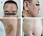
A 48-year-old Chinese man presented to the dermatology department with erythema and pustules on the face, neck and trunk (Figure 1A and B). The patient had developed red papules on the face and neck more than 3 months ago, which gradually increased and fused into patches and plaques with small pustules at the edges. A similar rash developed and spread to the abdomen and back, accompanied by pronounced itching (Figure 1C and D). He had been treated with anti-allergy therapy without any improvement. His medical history included hypertension. Additionally, he had high-risk sex a year ago. Physical examination revealed multiple infiltrative red patches and plaques on the face, neck and trunk with outwardly expanding edges in a circular pattern and tiny pustules distributed in a collar pattern.
Laboratory examination determined peripheral blood eosinophilia (2310/μL), relative eosinophilia (14.8%) and leukocytosis (15,600/μL). Treponema pallidum particle agglutination test (reactive) and rapid plasma reagin test (titre of 1:4) provided a serologic confirmation of the diagnosis. However, patient’s wife screened negative for syphilis. Furthermore, CD3 (+)-T-lymphocyte count was found to be 889/µL (1185–1901/µL) while CD4/CD8 ratio was at 0.95 (1.36–2.61). Evaluations of fungal microscopies and human immunodeficiency virus tests reported negative. Skin biopsy results revealed that the epidermis was normal and the dermal follicular sebaceous gland unit was infiltrated by a large number of neutrophils and eosinophils, forming eosinophilic microabscesses (Figure 2A and B). Amylase pretreatment PAS staining, Gram staining and immunohistochemical staining for syphilis spirochetes on skin tissue were reported negative.
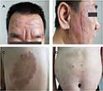
Therefore, a diagnosis of EPF associated with syphilis was made based on the typical clinical, laboratory and histopathological features. Treatment included oral indomethacin tablets (50 mg daily) and topical 0.03% tacrolimus cream. Furthermore, he was treated with an intramuscular dose of benzathine penicillin G (2.4 mIU) once a week for 3 times. After 2 weeks of treatment, the lesions subsided, leaving behind pigmentation (Figure 3A–D). The drug was gradually tapered and discontinued after six weeks once the disease was under control. The blood cell analysis was repeated, revealing that the percentage of leukocytes, eosinophils and eosinophils were normalized. Furthermore, the patient is still being followed up.
 |
Figure 3 Resolution of lesions. The lesions on the face (A and B), abdomen (C) and back (D) subsided, leaving behind pigmentation. |
Discussion
Eosinophilic pustular folliculitis (EPF) presents as an aseptic pustular skin disease with eosinophilia. Variants of EPF include classic EPF (Ofuji disease), immunosuppression-associated EPF and infancy-associated EPF.1 And EPFs presenting with other diseases have been reported, such as EPF associated with cutaneous angiosarcoma, EPF associated with pregnancy and EPF associated with herpes zoster.2–4 While the pathogenesis of EPF remains uncertain, various studies indicate that Th2-mediated immunologic mechanisms and prostaglandin D2 (PGD2) are involved in the pathogenesis of EPF. Th2 cytokines, such as IL-4, IL-5 and IL-13, promote the production of eosinophil chemokines via the component cells of the skin (such as lymphocytes).5 Additionally, PGD2 and its metabolite 15d-PGJ2 induce eotaxin-3 upregulation in sebaceous gland cells, leading to the infiltration and activation of eosinophils in the pilosebaceous units.6 Furthermore, indomethacin not only inhibited eosinophil migration to PGD2 and eotaxin, but also the initiating effect of 15d-PGJ2. Therefore, it can be concluded that the drug was effective in treating this case of EPF.
Th1 cytokines (IL-2 and IFN-r) are mainly expressed in stage I syphilis, while stage II syphilis mainly expresses Th2 cytokines (IL-4 and IL-5). The T lymphocytes subpopulation is dysregulated in patients with stage II syphilis, with the cellular immunity shifting from a Th1 to Th2 population.7 Moreover, the peripheral blood CD4 (+)-T-lymphocyte and CD4/CD8 ratio of patients with stage II syphilis was reported to be significantly lower than those of normal subjects, indicating cellular immune imbalance and immune dysregulation in patients.8 This phenomenon could reduce the body’s ability to resist and clear Treponema pallidum, leaving patients in a state of persistent infection. Thus, the Th2 cell-dominated immune response in stage II syphilis is hypothesised to be the probable cause of EPF in this patient.
Consent Statement
Informed consent for publication of the case details and associated images was obtained from the patient, and all procedures were performed in accordance with the Helsinki Declaration. Institutional approval was not required to publish the case details.
Funding
This work was supported by grants from the National Natural Science Foundation of China (No. 81860484), the Natural Science Foundation of Jiangxi Province of China (No. 20202BABL206107), Science and Technology plan of Health and Family Planning Commission of Jiangxi Province of China (No.20191054).
Disclosure
Yuchen Li and Ruxiao Nie are co-first authors for this study. The authors report no conflicts of interest in this work.
References
1. Nomura T, Katoh M, Yamamoto Y, Miyachi Y, Kabashima K. Eosinophilic pustular folliculitis: a proposal of diagnostic and therapeutic algorithms. J Dermatol. 2016;43(11):1301–1306. doi:10.1111/1346-8138.13359
2. Matsudate Y, Miyaoka Y, Urano Y. Two cases of eosinophilic pustular folliculitis associated with pregnancy. J Dermatol. 2016;43(2):218–219. doi:10.1111/1346-8138.13179
3. Jiang YY, Zeng YP, Jin HZ. Eosinophilic pustular folliculitis associated with cutaneous angiosarcoma. Chin Med J. 2018;131(1):115–116.
4. Lee JH, Lee SK, Kim JH, Kim HY, Kim MS, Lee UH. A case of Eosinophilic pustular folliculitis associated with herpes zoster. Am J Dermatopathol. 2021;43(4):298–299. doi:10.1097/DAD.0000000000001818
5. Kagami S, Saeki H, Komine M, et al. Interleukin-4 and interleukin-13 enhance CCL26 production in a human keratinocyte cell line, HaCaT cells. Clin Exp Immunol. 2005;141(3):459–466. doi:10.1111/j.1365-2249.2005.02875.x
6. Nakahigashi K, Doi H, Otsuka A, et al. PGD2 induces eotaxin-3 via PPARγ from sebocytes: a possible pathogenesis of eosinophilic pustular folliculitis. J Allergy Clin Immunol. 2012;129(2):536–543. doi:10.1016/j.jaci.2011.11.034
7. Yan Y, Wang J, Qu B, et al. CXCL13 and TH1/Th2 cytokines in the serum and cerebrospinal fluid of neurosyphilis patients. Medicine. 2017;96(47):e8850. doi:10.1097/MD.0000000000008850
8. Yang RD, Cheng JP, Tian GN, et al. 梅毒持续性阳性的梅毒患者外周血淋巴细胞亚群的检测 [Detection of peripheral blood lymphocyte subsets in treated syphilis patients with persistent positivity for rapid plasma reagin]. Di Yi Jun Yi Da Xue Xue Bao. 2003;23(12):1307–1309. Chinese
 © 2023 The Author(s). This work is published and licensed by Dove Medical Press Limited. The full terms of this license are available at https://www.dovepress.com/terms.php and incorporate the Creative Commons Attribution - Non Commercial (unported, v3.0) License.
By accessing the work you hereby accept the Terms. Non-commercial uses of the work are permitted without any further permission from Dove Medical Press Limited, provided the work is properly attributed. For permission for commercial use of this work, please see paragraphs 4.2 and 5 of our Terms.


