
Back to Journals » International Journal of Women's Health » Volume 18
Chylous Lymphocyst Following Retroperitoneal Lymphadenectomy: A Case Report and Review of Treatment
Authors Sukjariangporn H  , Thongaram M, Kittikhun R, Samrarn J , Wetcho T, Kantathavorn N
Received 16 December 2025
Accepted for publication 12 February 2026
Published 20 February 2026 Volume 2026:18 588265
DOI https://doi.org/10.2147/IJWH.S588265
Checked for plagiarism Yes
Review by Single anonymous peer review
Peer reviewer comments 2
Editor who approved publication: Dr Matteo Frigerio
Hathaipat Sukjariangporn,1 Man Thongaram,1 Ruai Kittikhun,1 Jidapa Samrarn,1 Thanita Wetcho,1 Nuttavut Kantathavorn2
1Department of Obstetrics and Gynecology, Princess Srisavangavadhana College of Medicine, Chulabhorn Royal Academy, Bangkok, Thailand; 2Princess Srisavangavadhana Faculty of Medicine, Chulabhorn Royal Academy, Bangkok, Thailand
Correspondence: Hathaipat Sukjariangporn, Department of Obstetrics and Gynecology, Princess Srisavangavadhana College of Medicine, Chulabhorn Royal Academy, 906, Kamphaeng Phet 6 Road, Talat Bang Khen, Laksi, Bangkok, 10210, Thailand, Tel +66 2 7655700, Email [email protected]
Abstract: Chylous lymphocyst or lymphocele following pelvic and para-aortic lymphadenectomy is rare. Clinically, patients usually present with abdominal distension or abdominal pain. The milky appearance of the cystic fluid reflects its high triglyceride content. Although specific guidelines for managing chylous lymphocyst are limited, the approaches used for chylous ascites can generally be applied. More than 70% of patients respond to conservative treatment, which includes a modified diet consisting of high protein, fat restriction, and medium-chain triglyceride supplementation. We present the case of a 45-year-old woman who developed a symptomatic 15-cm pelvic lymphocyst one month after surgical staging for a mucinous borderline ovarian tumor. Following the placement of a percutaneous catheter drainage (PCD), the initial output was 300– 400 mL/day of clear serous fluid. Notably, a delayed chylous conversion occurred on the 20th day of drainage, the fluid turned milky and biochemical analysis confirmed a triglyceride level of 493 mg/dL. Initiation of a modified high-protein, fat-restricted diet resulted in a rapid decrease in drainage volume to 30 mL/day within 24 hours, alongside a return to clear yellowish appearance. This study aims to report the clinical manifestations and to summarize current therapeutic options for patients with chylous leakage.
Keywords: chylous lymphocyst, chyloma, lymphocele, ovarian cancer, retroperitoneal lymphadenectomy
Introduction
Chylous lymphocyst is the accumulation of chyle within a lymphatic fluid–filled cyst. The diagnosis is typically made based on the milky appearance of the fluid, resulting from high levels of triglycerides, particularly chylomicrons. A triglyceride level of more than 200 mg/dL usually supports the diagnosis, although the cutoff may vary. Patients typically present with abdominal distention. Conservative treatment, often involving a modified diet, usually provides satisfactory results.
In this study, we present a case of chylous lymphocyst following surgical staging with retroperitoneal lymphadenectomy for ovarian cancer.
Methods
This case report was reviewed and approved by the Institutional Review Board (IRB) of Chulabhorn Royal Academy (EC 012/2567). Written informed consent was obtained from the patient for publication. The protocols of human experiments were followed based on international guidelines for human research protection, including the Declaration of Helsinki, The Belmont Report, CIOMS Guidelines, and the International Conference on Harmonisation of Technical Requirements for Registration of Pharmaceuticals for Human Use – Good Clinical Practice (ICH-GCP).
Case Presentation
A 45-year-old woman presented to the hospital with a complaint of abdominal distension for the past month. Physical and pelvic examinations revealed a smooth-surfaced, firm, midline pelvic mass, approximately the size of a 20-week pregnancy. The mass was slightly movable during rectovaginal examination. Transvaginal and transabdominal ultrasonography showed an anteverted uterus measuring 5.51 × 3.36 × 4.21 cm, along with a large multiloculated cystic adnexal mass measuring 15.76 × 11.10 × 14.95 cm (Figure 1A). The mass consisted of solid parts, constituting approximately 50% of its structure, and was located anteriorly to the uterus.
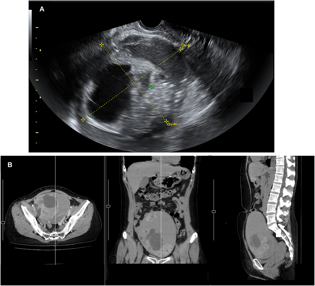 |
Figure 1 Preoperative imaging of the ovarian mass. (A) Transvaginal ultrasonography revealed a cystic-solid mass measuring 15.76 × 11.10 × 14.95 cm. The green cross is placed at the ovarian tumor, and the yellow lines represent the electronic calipers used to measure the size and dimensions of the mass. (B) The thoracic-abdomino-pelvic CT scan revealed a large well-defined cystic lesion with a multilocular and enhancing solid component or calcification at left adnexal region. |
Tumor markers for epithelial ovarian cancer were obtained, revealing normal levels for cancer antigen 125 at 19.9 U/mL (normal < 35.0 U/mL), human epididymis protein 4 at 37.3 pmol/L (normal < 140 pmol/L), and carcinoembryonic antigen at 0.9 ng/mL (normal range < 4.7 ng/mL). However, the carbohydrate antigen 19–9 level was slightly elevated at 134 U/mL (normal < 37 U/mL).
A thoracic-abdomino-pelvic CT scan (Figure 1B) confirmed a large, well-defined cystic lesion with a multilocular and enhancing solid component/calcification in the left adnexa region, raising suspicion of ovarian mucinous cystadenoma or carcinoma. No metastatic diseases were detected on imaging. Cervical cytology also showed negativity for intraepithelial lesion or malignancy.
The patient underwent an exploratory laparotomy for an ovarian tumor. Based on intraoperative findings of the left ovarian specimen (Figure 2), which revealed a multilocular cystic mass with a 50% solid component, malignancy could not be excluded. Complete surgical staging was performed, encompassing bilateral pelvic and para-aortic lymphadenectomy along with an infracolic omentectomy. While lymph node dissection techniques vary among surgeons at our institution, in this specific case, electrosurgical coagulation was systematically employed at the proximal and distal surgical margins following the nodal dissection to ensure lymphatic vessel sealing. The surgery was uneventful, with no residual tumor identified, and the patient was discharged from the hospital after 72 hours.
 |
Figure 2 Intraoperative specimens included the uterus removed intact with the right ovary and fallopian tube, while the left ovarian tumor and fallopian tube were removed separately without rupture. Gross examination of the left ovarian tumor revealed a multilocular mass with a smooth external surface and gelatinous content. |
Two weeks after the surgery, the patient returned for a follow-up. Pathological examination of the surgical specimen revealed a mucinous borderline ovarian tumor with microinvasion of the left ovary. Lymph nodes in the pelvic and para-aortic regions were negative for malignancy (n = 0/9 in the right pelvic region, n = 0/19 in the left pelvic region, and n = 0/9 in the para-aortic region). Additionally, the uterus, right ovary, both fallopian tubes, omentum, and peritoneal washing showed negative for malignancy. The patient reported vaginal discharge after the surgery. Pelvic examination revealed small amounts of clear yellowish content with an intact vaginal stump. Transvaginal ultrasonography showed a 2.0 × 2.0 cm collection. She received a one-week prescription of oral antibiotics and was scheduled for a follow-up at two weeks.
At the one-month follow-up, the vaginal discharge had resolved, but the patient complained of left pelvic pain and a palpable pelvic mass for one week. Physical examination revealed a 15-cm pelvic mass with cystic consistency in the left lower quadrant. Bedside transabdominal ultrasound confirmed a 10.16 × 7.13 cm unilocular anechoic cyst on the left side, highly suspected to be a lymphocyst. Consultation with an interventionist led to the decision for percutaneous catheter drainage (PCD). A complementary abdomino-pelvic CT scan revealed a newly identified 6.2 × 7.8 × 17.0 cm thin-walled cystic lesion in the left lower abdomen and pelvis, located anterior to the left psoas muscle and along the left common and external iliac vessels. A smaller cystic lesion was also noted in the right pelvis, measuring 2.20 × 1.75 cm (Figure 3). A percutaneous catheter was successfully inserted to relieve the left-sided lymphocyst (Figure 4).
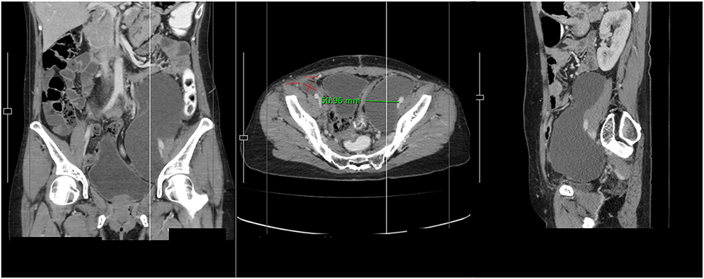 |
Figure 3 A 6.2 × 7.8 × 17.0 cm thin-walled cystic lesion in the left abdomen (green-colored line), suggestive of a lymphocyst, was identified on CT scan. A smaller thin-walled cystic lesion was also noted on the right side (the intersecting red-colored lines forming a cross to measure width × length), measuring 2.20 × 1.75 cm. |
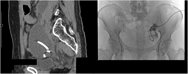 |
Figure 4 Placement of the percutaneous catheter drainage (PCD) was performed in the inferior aspect of the rim-enhancing fluid collection located in the left-sided pelvis. |
She was hospitalized for 10 days, during which the drainage of clear yellowish fluid averaged around 300–400 mL daily. A sample of the fluid was analyzed, showing negative results for granuloma or malignancy in cytology, and negative results for Gram stain and culture. Following a discussion with the patient, she was discharged from the hospital with a retained percutaneous drainage tube and bag. The patient was advised to visit the hospital every 2 days for wound irrigation and dressing, during which the fluid continued to drain at a rate of approximately 300–400 mL daily. However, on the 20th day after the percutaneous drainage insertion, a milky appearance was observed in the drainage fluid (Figure 5A). Due to the suspicion of chyle leakage secondary to the previous trauma to lymphatic channels, a sample of the fluid was sent for triglyceride level analysis, revealing a level of 493 mg/dL.
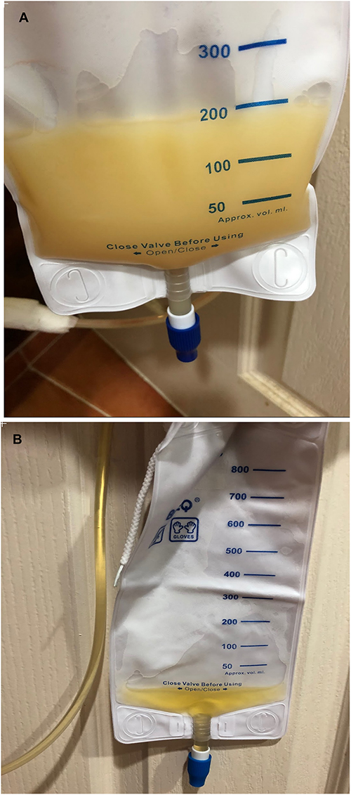 |
Figure 5 (A) On the diagnosis date of chylous lymphocyst, the fluid exhibited a milky appearance. (B) Following treatment with a modified diet, the fluid transitioned to a transparent, clear, yellowish state. |
The patient was diagnosed with a chylous lymphocyst secondary to lymphatic duct injury from a previous lymphadenectomy. After initiation of a high-protein, fat-restricted diet, the drainage output decreased markedly from 300 mL to 30 mL on the first day, and the fluid became clear and yellowish in appearance (Figure 5B). As symptoms improved and the abdomino-pelvic CT revealed a reduced size of the lymphocyst to 2.71 × 8.79 × 1.82 cm (Figure 6), the percutaneous drainage was removed. A repeat triglyceride fluid level also decreased to 78 mg/dL. The patient continued the modified diet for two months, during which no lymphocyst was detected, and no abnormal symptoms were reported during follow-up. She was then advised to resume her usual diet. At her last follow-up in December 2025, approximately two and a half years after surgery, she remained disease-free with no evidence of lymphocyst recurrence.
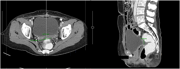 |
Figure 6 Follow-up abdomino-pelvic CT scan demonstrated improvement of the left pelvic lymphocyst after 10 days of modified dietary management, with the lesion reduced in size to 2.71 × 8.79 × 1.82 cm (green-colored lines). |
Discussion
Postoperative lymphatic complications are uncommon, with an incidence varying from 3% to 9%. These complications encompass lymphatic ascites, lymphocele, lymphorrhea, lymphatic fistula, and special forms characterized by chylous leakage, such as chylous ascites, chylorrhea, chyloperitoneum, and chylothorax.1
While chylous lymphocysts or lymphoceles are less common than chylous ascites, and there are limited treatment options specific to this condition, the existing knowledge and management principles for chylous ascites can be applied to these cases. Chylous ascites is characterized by a milky appearance of the fluid, resulting from elevated levels of triglycerides, especially chylomicrons. Patients typically present with abdominal distention, and some may experience abdominal pain, diarrhea, malnutrition, edema, and early satiety. In severe cases, dyspnea may also develop.2 The etiology of chylous ascites is typically categorized into trauma and non-trauma causes, resulting from a disruption of the lymphatic system due to injury or obstruction. Traumatic causes, most commonly observed after surgery, especially retroperitoneal lymphadenectomy, are prevalent. Non-traumatic causes, including neoplasia, hepatic cirrhosis, heart failure, and congenital factors, are the most frequent etiologies.2
The diagnosis of chylous ascites is typically established by the milky appearance of fluid, accompanied by a triglyceride value exceeding 200 mg/dl.1 Imaging plays a crucial role in assessing the presence and location of the leakage. Lymphangiography is considered the gold standard diagnostic tool in cases of lymphatic obstruction, while lymphoscintigraphy can provide valuable mapping of the lymphatics.3
Although many studies have aimed to identify risk factors and preventive techniques, the results remain inconclusive. Thiel et al4 indicated that patients with chylous ascites had a larger mean number of lymph nodes removed. In a retrospective case-control study by Chen et al,5 two cycles of neoadjuvant chemotherapy, a decrease in hemoglobin level of 20 g/L or more after surgery, and a postoperative albumin level less than 35 g/L were significantly associated with postoperative lymphatic leakage. A literature review by Achouri et al6 suggests some surgical techniques that may decrease the incidence of lymphocyst formation after lymphadenectomy for gynecological malignancies, such as leaving the peritoneum open over the lymphadenectomy sites and avoiding the placement of drains. Omentoplasty might offer potential benefits, but further studies are needed, and postoperative octreotide therapy may provide promising results.6
The treatments for chylous ascites involve a multidisciplinary approach and depend on the underlying etiology. Conservative treatment typically achieves a success rate of 67–75%. This approach includes a modified diet with high-protein and fat-restricted intake (less than 10 g of fats), supplemented with medium-chain triglycerides.4,7,8 Prolonged dietary management may also include fat-soluble vitamins and essential fatty acid supplements. Total parenteral nutrition (TPN) has shown success, and somatostatin and octreotide demonstrate promising results in reducing lymph excretion.9 Other drugs such as diuretics, orlistat, etilefrine, and ethylephrine have also demonstrated benefits when addressing the underlying cause.
In cases where conservative treatment fails or in severe instances, surgical interventions are indicated. Interventional radiology options have been introduced in recent years, with satisfactory results such as a continuous low-pressure drainage system,10 lymphangiography with embolization has a success rate of 37–70% without additional procedures.11–13 Peritoneovenous (Denver) shunting, transjugular intrahepatic portosystemic shunting (TIPSS)3 and radiation therapy in lymphoproliferative malignancies also provide therapeutic benefits. Surgical interventions, such as ligation of the disrupted lymphatic duct14 or lymphatic-venous anastomosis,15 show promising results. The examples of current therapeutic options for chylous ascites are listed in Table 1.
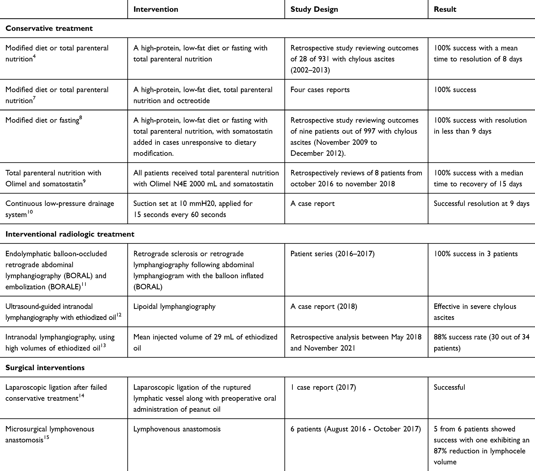 |
Table 1 Review of Therapeutic Options for Chylous Ascites |
Conclusions
This report details a rare instance of chylous lymphocyst following retroperitoneal lymphadenectomy. A significant clinical observation is the potential for delayed chylous conversion in postoperative lymphoceles; therefore, any alteration in drainage appearance warrants immediate reassessment through visual inspection and triglyceride analysis. A high-protein, fat-restricted diet remains the preferred first-line therapy to swiftly decrease chyle production. More invasive strategies, including surgical ligation or lymphangiography with embolization, should be strictly reserved for patients with high-output leakage or those who do not respond to initial dietary interventions.
Acknowledgment
This research project is supported by Chulabhorn Royal Academy.
Disclosure
The authors report no conflicts of interest in this work.
References
1. Lv S, Wang Q, Zhao W, et al. A review of the postoperative lymphatic leakage. Oncotarget. 2017;8(40):69062–8. doi:10.18632/oncotarget.17297
2. Han D, Wu X, Li J, Ke G. Postoperative chylous ascites in patients with gynecologic malignancies. Int J Gynecol Cancer. 2012;22(2):186–190. doi:10.1097/IGC.0b013e318233f24b
3. Lizaola B, Bonder A, Trivedi HD, Tapper EB, Cardenas A. Review article: the diagnostic approach and current management of chylous ascites. Aliment Pharmacol Ther. 2017;46(9):816–824. doi:10.1111/apt.14284
4. Thiel FC, Parvanta P, Hein A, et al. Chylous ascites after lymphadenectomy for gynecological malignancies. J Surg Oncol. 2016;114(5):613–618. doi:10.1002/jso.24354
5. Chen L, Lin L, Li L, et al. Lymphatic leakage after pelvic lymphadenectomy for cervical cancer: a retrospective case-control study. BMC Cancer. 2021;21(1):1242. doi:10.1186/s12885-021-08984-1
6. Achouri A, Huchon C, Bats AS, Bensaid C, Nos C, Lecuru F. Postoperative lymphocysts after lymphadenectomy for gynaecological malignancies: preventive techniques and prospects. Eur J Obstet Gynecol Reprod Biol. 2012;161(2):125–129. doi:10.1016/j.ejogrb.2011.12.021
7. Var T, Gungor T, Tonguc E, Ozdener T, Mollamahmutoglu L. The conservative treatment of postoperative chylous ascites in gynecologic cancers: four case reports. Arch Gynecol Obstet. 2012;285(3):849–851. doi:10.1007/s00404-011-2050-3
8. Zhao Y, Hu W, Hou X, Zhou Q. Chylous ascites after laparoscopic lymph node dissection in gynecologic malignancies. J Minim Invasive Gynecol. 2014;21(1):90–96. doi:10.1016/j.jmig.2013.07.005
9. Scaletta G, Quagliozzi L, Cianci S, et al. Management of postoperative chylous ascites after surgery for ovarian cancer: a single-institution experience. Updates Surg. 2019;71(4):729–734. doi:10.1007/s13304-019-00656-x
10. Shibuya Y, Asano K, Hayasaka A, et al. A novel therapeutic strategy for chylous ascites after gynecological cancer surgery: a continuous low-pressure drainage system. Arch Gynecol Obstet. 2013;287(5):1005–1008. doi:10.1007/s00404-012-2666-y
11. Srinivasa RN, Gemmete JJ, Osher ML, Hage AN, Chick JFB. Endolymphatic Balloon-Occluded Retrograde Abdominal Lymphangiography (BORAL) and Embolization (BORALE) for the diagnosis and treatment of chylous ascites: approach, technical success, and clinical outcomes. Ann Vasc Surg. 2018;49:49–56. doi:10.1016/j.avsg.2017.10.020
12. Verhaeghe L, Holsbeeck AV, Bonne L, et al. Therapeutic lymphangiography with ethiodized oil for the management of lymphoceles and chylous ascites. Diagn Interv Imaging. 2023;104(10):500–505. doi:10.1016/j.diii.2023.05.003
13. Nakamura K, Nakayama K, Minamoto T, et al. Ultrasound-guided intranodal lymphangiography with lipiodol for treatment of chylous ascites following surgery for ovarian cancer: a case report. Mol Clin Oncol. 2018;9(2):178–180. doi:10.3892/mco.2018.1647
14. Liu J, Zhang H, Du Q, Yao S. Successful laparoscopic treatment of chylous ascites after pelvic lymphadenectomy: a case report and peanut oil application. J Minim Invasive Gynecol. 2018;25(5):923–926. doi:10.1016/j.jmig.2017.12.028
15. Yamashita S, Kitamura Y, Fujisawa K, et al. Microsurgical lymphovenous anastomosis for pelvic lymphoceles after gynecological cancer surgery. J Plast Reconstr Aesthet Surg. 2022;75(3):1142–1149. doi:10.1016/j.bjps.2021.09.056
 © 2026 The Author(s). This work is published and licensed by Dove Medical Press Limited. The
full terms of this license are available at https://www.dovepress.com/terms
and incorporate the Creative Commons Attribution
- Non Commercial (unported, 4.0) License.
By accessing the work you hereby accept the Terms. Non-commercial uses of the work are permitted
without any further permission from Dove Medical Press Limited, provided the work is properly
attributed. For permission for commercial use of this work, please see paragraphs 4.2 and 5 of our Terms.
© 2026 The Author(s). This work is published and licensed by Dove Medical Press Limited. The
full terms of this license are available at https://www.dovepress.com/terms
and incorporate the Creative Commons Attribution
- Non Commercial (unported, 4.0) License.
By accessing the work you hereby accept the Terms. Non-commercial uses of the work are permitted
without any further permission from Dove Medical Press Limited, provided the work is properly
attributed. For permission for commercial use of this work, please see paragraphs 4.2 and 5 of our Terms.