Back to Journals » International Journal of Women's Health » Volume 7
Cervical cancer screening and treatment of cervical intraepithelial neoplasia in female sex workers using “screen and treat” approach
Authors Joshi S, Kulkarni V ![]() , Darak T, Mahajan U, Srivastava Y, Gupta S, Krishnan S, Mandolkar M, Bharti AC
, Darak T, Mahajan U, Srivastava Y, Gupta S, Krishnan S, Mandolkar M, Bharti AC
Received 9 January 2015
Accepted for publication 5 March 2015
Published 4 May 2015 Volume 2015:7 Pages 477—483
DOI https://doi.org/10.2147/IJWH.S80624
Checked for plagiarism Yes
Review by Single anonymous peer review
Peer reviewer comments 4
Editor who approved publication: Professor Elie Al-Chaer
Video abstract presented by Smita Joshi.
Views: 2180
Smita Joshi,1 Vinay Kulkarni,2 Trupti Darak,2 Uma Mahajan,1 Yogesh Srivastava,3 Sanjay Gupta,3 Sumitra Krishnan,1 Mahesh Mandolkar,2 Alok Chandra Bharti3
1Hirabai Cowasji Jehangir Medical Research Institute (HCJMRI), Jehangir Hospital Premises, Pune, Maharashtra, India; 2Prayas Health Group, Amrita Clinic, Pune, India; 3Institute for Cytology and Preventive Oncology, Indian Council of Medical Research, New Delhi, India
Objective: Female sex workers (FSWs) are at an increased risk of human immunodeficiency virus (HIV) as well as human papillomavirus (HPV) infections and thus have an increased risk of cervical intraepithelial neoplasia (CIN) and cervical cancer. We evaluated the feasibility of “screen and treat approach” for cervical cancer prevention and the performance of different screening tests among FSWs.
Methods: Women were screened using cytology, VIA (visual inspection with acetic acid), and VILI (visual inspection with Lugol’s iodine) and underwent colposcopy, biopsy, and immediate treatment using cold coagulation, if indicated, at the same visit.
Results: We screened 300 FSWs of whom 200 (66.67%) were HIV uninfected and 100 (33.34%) were HIV infected. The overall prevalence of CIN 2–3 lesions was 4.7%. But all women with CIN 2–3 lesions were HIV infected, and thus the prevalence of CIN 2–3 lesions in HIV-infected FSWs was 14/100 (14%, 95% confidence interval: 7.2–20.8). All of them screened positive by all three screening tests. Cold coagulation was well tolerated, with no appreciable side effects.
Conclusion: Cervical cancer prevention by “screen and treat” approach using VIA, followed by ablative treatment, in this high-risk group of women is feasible and can be implemented through various targeted intervention programs.
Keywords: cytology, VIA, VILI, CIN, cold coagulation, cervical cancer, HPV, FSWs
Introduction
Female sex workers (FSWs) are at an increased risk of many sexually transmitted infections, including human papillomavirus (HPV) and human immunodeficiency virus (HIV) infections. These infections interact with each other in many ways given the overlap of risk factors; thus HPV as well as HIV prevalence is very high in FSWs. A meta-analysis of 14 studies involving more than 4,000 FSWs in nine Asian countries has reported that HPV prevalence among FSWs in this region ranged from 12.8% to 84.8% and that FSWs had a nearly tenfold risk of HPV infection than women in the general population.1
HPV-associated cancers occur at increased rates in persons infected with HIV.2 The prevalence of HPV is more in HIV-infected women. They are infected with a broad range of HPV genotypes, and multiple concurrent infections are also common.3 HPV infections are more likely to persist in HIV-positive women than in HIV-negative women.4–6 This persistent HPV infection contributes to a higher prevalence of HPV infection among HIV-infected women, leading to higher risk of cervical intraepithelial neoplasia (CIN).7–9
Prevalence of HIV in FSWs varies in different regions of the world, and it is considerably higher than in women in the general population. The overall prevalence of HIV in FSWs in India during 2008–2009 was 4.77% (95% confidence interval [CI]: 4.60–4.94) as compared to 0.6% (95% CI: 0.52–0.67) among antenatal women who represent women at low risk of HIV.10 HIV prevalence in FSWs in certain states such as Andhra Pradesh, Maharashtra, Karnataka, Manipur, and Nagaland was >10%.10
HIV infection increases the risk of new HPV infections and decreases the rate of HPV clearance.11 There is increasing evidence suggesting that HPV-infected individuals are at higher risk of acquiring HIV infection.12–14
It is now well established that persistence of HPV infection is the strongest epidemiologic factor associated with cervical intraepithelial neoplasia and cancer of the cervix.15–20 Thus, FSWs face a dual risk and dual burden of both: HPV and HIV infections, and cervical cancer screening and treatment of CIN must be offered to all FSWs.
Cytology-based cervical cancer screening programs have not really worked well in developing countries in reducing cervical cancer incidence and mortality. Visual inspection of the cervix after application of acetic acid (VIA) is an alternative screening test which has been shown to be more sensitive than cytology in many studies in the developing regions. VIA offers an opportunity to immediately treat screen-positive women in a single visit “screen and treat” approach and has been recommended by the World Health Organization (WHO).21
We evaluated the feasibility of offering cervical cancer prevention programme for to this very high-risk and vulnerable population using “screen and treat” approach. We also concurrently evaluated the performance of conventional cytology, VIA, and visual inspection with Lugol’s iodine (VILI) in cervical cancer screening and the feasibility and acceptability of immediate treatment for CIN using an ablative method.
Materials and methods
The project was approved by the ethics committee of the Hirabai Cowasji Jehangir Medical Research Institute (HCJMRI) and Jehangir Clinical Development Centre, Pune, India. We conducted a cross-sectional study between October 2012 and February 2013. We contacted non-governmental organizations (NGOs) working in three high HIV-prevalent districts in Maharashtra, namely, Pune, Sangli, and Ahmednagar, and data were collected from a convenience sample of 300 FSWs. The peer educators and counselors working with these NGOs were provided with study information, and they referred willing FSWs to our designated study clinic for cervical cancer screening. FSWs between the age group of 18 and 60, willing to sign or provide a thumb impression on the informed consent form, willing to undergo study procedures, who were nonpregnant, with no history of prolapse, no history of treatment for cervical neoplasia, and having an intact uterus were enrolled in the study.
The study was conducted at a designated cervical cancer screening clinic set up at Prayas (a non-governmental, nonprofit organization) by a collaborative effort between HCJMRI and Prayas. After a written informed consent and HIV pre-test counseling, demographic information, history regarding sexual practices, years in sex work, number of sex partners, condom use with regular partner/client, etc were collected using a structured questionnaire. Approximately 10 mL of venous blood sample was collected for HIV testing (if a documented HIV test report was not available), HBsAg testing, syphilis serology, and CD4 and HIV viral load testing (for HIV-infected FSWs only). HIV testing was done using HIV TriDot (J. Mitra & Co. Pvt. Ltd., New Delhi, India) and Instachk (Transasia Bio-Medicals Limited, Mumbai, India) for the detection of anti-HIV antibodies, and HBsAg testing was done using the One Step Cassette Style HBsAg Test (IND Diagnostic Inc., British Columbia, Canada). Syphilis testing was done using rapid plasma regain (RPR) test (Tulip, Goa, India), and if RPR was positive, a second test was performed using Instachk TP test (Transasia Bio-Medicals Limited) for confirmation. Appropriate post-test counseling was done to participants detected with HIV infection. Treatment for syphilis and other sexually transmitted infections was provided following National AIDS Control Organization’s guidelines.22
After completing the case report forms and blood collection, a speculum examination was done with women lying down in the modified lithotomy position. A trained nurse exposed the cervix with a bivalved speculum and collected cervical specimen for cytology using a Cervex-Brush and prepared a cervical smear on a glass slide, which was fixed immediately in 95% ethanol.
Cervical cancer screening using VIA was performed by two nurses who were trained in visual screening methods, and colposcopy was done by a trained doctor using the manual developed by the International Agency for Research on Cancer (IARC), WHO.23 The nurse applied 5% acetic acid on the cervix with a cotton swab for 1 minute. Findings were recorded as VIA negative, VIA positive (if there was an acetowhite lesion touching the squamocolumnar junction), or VIA positive (suspicious of invasive cancer).23
Colposcopy was done on all women by a trained doctor, and the nurse independently recorded the findings of VILI when the doctor applied Lugol’s iodine as a part of colposcopic evaluation. Colposcopic findings were reported as normal, inflammation, probable low- or high-grade precancerous lesions, or suspected invasive cancer following the IARC manual.23 If there was a lesion suggestive of any colposcopic abnormality (low- as well as high-grade), colposcopic findings were explained to the woman and multiple punch biopsies were collected using a Tischler punch biopsy forceps. Women having lesions not involving more than three-fourths of the transformation zone and not entering into the endocervical canal, and those with fully visible squamocolumnar junction with no suspicion of an invasive cancer were treated immediately using cold coagulation. Cold coagulation (WISAP® Medical Technology GmbH, Brunnthal/Hofolding, Germany) is actually a misnomer and utilizes heat to ablate the epithelium at 105°C when applied for 45 seconds. Multiple overlapping applications of 45 seconds each were used during the same sitting without any local anesthesia to cover the entire transformation zone.
Cervical smears for cytology were stained by standard Papanicolaou staining method and the results reported by a trained cytologist using the Bethesda 2001 system of reporting cervicovaginal smears.24 Cervical biopsy specimens were analyzed and the results were reported by two pathologists using CIN terminology independently and then a consensus diagnosis was reached.
Reference standard for final diagnosis
All women underwent colposcopy irrespective of the visual screening test results. If colposcopy was suggestive of any abnormality, multiple punch biopsies were obtained from the abnormal area for histopathology. Thus, the reference standard was a normal colposcopy or histopathology. We did not perform cervical biopsies for histological diagnosis on all participants to avoid unnecessary intervention; however, colposcopy was performed on all participants to minimize the bias, and colposcopy or histopathology was taken as a reference test to derive the test characteristics of various screening modalities.
Data entry was done using MS Access 2007 software, and the data were analyzed using the STATA 12.0 software. Standard formulae were used to calculate the sensitivity, specificity, and positive (PPV) and negative predictive values (NPV) (and their exact 95% CIs) of the screening tests to detect CIN 2 and 3 lesions.
Results
We enrolled 300 FSWs in this cross-sectional study. The mean age of the participants was 33.6 (range, 21–58 years; SD, 8.7; median, 30.5). Their demographic characteristics are presented in Table 1. Of the 300 FSWs enrolled, 196 (65.3%) were natives of Maharashtra, while 104 (34.7%) were migrants from other states (Karnataka, Andhra Pradesh, West Bengal, Uttar Pradesh, Bihar, and Orissa) who relocated to Maharashtra for sex work. Majority (273/300, 91%) were brothel based and the remaining 9% were street based. Seventy-five percent of the sex workers had no formal education and 25% had some education. Smoking was reported by 14% of the participants, but majority (62.3%) reported tobacco chewing. Age at first sex before 18 years of age was reported by a substantial number of FSWs (241/286, 84.3%), and about 68% reported being in the occupation of sex work for less than 10 years. Three-quarters of the FSWs reported condom use with each and every partner, including regular partners, and about 20% reported condom use with all partners except with their regular partners. Majority reported at least one pregnancy (77%), and induced abortions were reported by 10% of the FSWs, with the number of maximum abortions exceeding more than eight. Of the 300 FSWs enrolled, 100 (33.3%) FSWs were HIV infected, 10% (30/300) were positive for syphilis serology, and 2% (6/300) were positive for HBsAg. We did not observe any participant characteristics that were significantly associated with CIN 2–3 lesions.
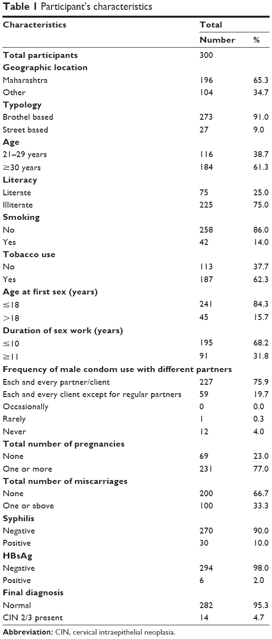 | Table 1 Participant’s characteristics |
Of the 300 enrolled, 296 completed all three cervical cancer screening tests and 5 were excluded from final analysis due to unsatisfactory cytology smears. VIA was positive in 45/296 (15.2%), VILI was positive in 36/296 (12.2%), and cytology at ASC-US (atypical squamous cells of undetermined significance) threshold was positive in 66/291 (22.7%) women. Distribution of final disease status by results from different screening tests is presented in Table 2. Histopathology of cervical punch biopsies revealed CIN 1 in eleven women, CIN 2 in five women, and CIN 3 in nine women. The overall prevalence of CIN 2–3 lesions was 4.7%. But all women with CIN 2–3 lesions were HIV infected, and thus the prevalence of CIN 2–3 lesions in HIV-infected FSWs was 14/100 (14%, 95% CI: 7.2–20.8). None of them were detected with invasive carcinoma.
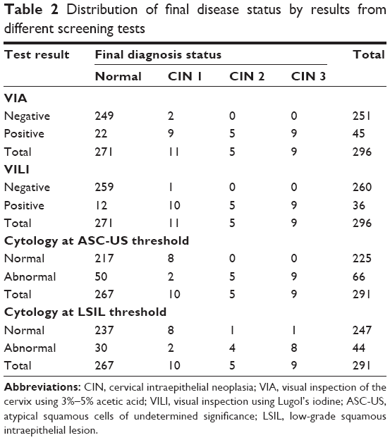 | Table 2 Distribution of final disease status by results from different screening tests |
Sensitivity, specificity, and PPV and NPV of VIA, VILI, and cytology (at ASC-US threshold) to detect CIN2+ lesions are presented in Table 3. Sensitivity of VIA, VILI, as well as cytology (at ASC-US threshold) was 100%. Specificity of VIA was 89%, that of VILI was 92.2%, and that of cytology was 81.2%. PPV of VIA, VILI, and cytology was 31.1%, 38.9%, and 21.2%, and NPV was 100% for all the three tests.
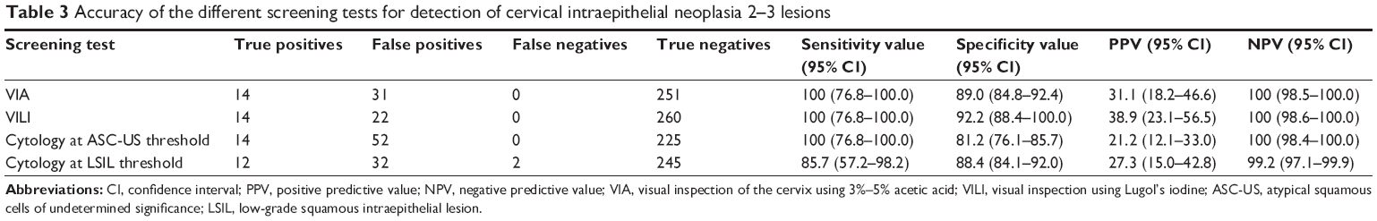 | Table 3 Accuracy of the different screening tests for detection of cervical intraepithelial neoplasia 2–3 lesions |
All FSWs with colposcopy suggestive of CIN (low-grade as well as high-grade) underwent cervical punch biopsies and were eligible for ablative treatment using cold coagulation. Of the 296 FSWs included in final analysis, 27 (9.12%) FSWs underwent cervical punch biopsies and treatment using cold coagulation. All of them accepted biopsies and treatment during the same screening visit except for one FSW, who reported to our study clinic after about 2 months following repeated counseling by the study staff and social workers of the NGO working in their area. Treatment with cold coagulation was well tolerated by all of them, and none had any appreciable side effects after treatment.
Discussion
In our study, 14% of the HIV-infected FSWs were detected with CIN 2–3 lesions; this emphasizes the need for screening and appropriate treatment of CIN in HIV-infected FSWs. There is a complex interplay between HIV and HPV infections, and FSWs constitute a very high-risk group for HIV as well as HPV infections. We concurrently evaluated the test characteristics of VIA, VILI, and cytology in detecting CIN 2–3 lesions. The sensitivity of all the three tests was 100%; however, this study was underpowered for evaluating sensitivity and NPV, and our results should be considered with caution.
It is important to note that visual screening tests, which are low-cost screening tests, did not miss any single high-grade lesion. In a pooled analysis involving about 58,000 women, cytology had the lowest sensitivity as compared to visual screening tests.25 However, considering our small sample size, it is possible that this may not be the true reflection of the screening tests. In our study, the specificity of cytology was lower than those reported in the reports published earlier.25 However, it is possible that a large percentage of FSWs were infected with high-risk HPV, resulting in minor cytological abnormalities that could not be detected on colposcopy. Histopathology report of the cervical biopsy is the gold standard, and although colposcopy was done on all participants to minimize verification bias, we did not perform cervical biopsies on all the participants without colposcopic abnormalities due to ethical reasons. In a study conducted in Hong Kong, the prevalence of any CIN among FSWs using cytology screening was 9.7%, and poorer compliance for the follow-up visit was seen among those with an abnormal result.26 This underscores the importance of the “screen and treat” approach. In spite of these limitations, our study provides important information about the feasibility of “screen and treat” strategy for preventing cervical cancer, which has been recommended by the WHO for low-resource countries.21
Visual screening tests provide an opportunity to treat screen-positive lesions at the same time; this is particularly important for this highly mobile population. Single-visit “screen and treat” approach was well accepted by FSWs, and thus it is feasible for various organizations providing care and support to FSWs to include cervical cancer screening in their program. Ablative treatment using cold coagulation was well tolerated and accepted by the FSWs. This method has several advantages over cryotherapy and has comparable cure rates for the treatment of all grades of CIN.27
Another incidental finding of our study is about the very high rate of induced abortions in this population in spite of the high rate of reported condom use, and therefore, it is essential that FSWs are counseled and provided effective temporary contraceptive choices in addition to condom counseling for HIV prevention.
Early age of initiation of sexual activity is one of the strongest risk factors for cervical cancer,18 and majority of the sex workers in our study reported age at initiation of sex work below 18 years. The first few reports on increased risk of cervical cancers among sex workers date back to 1950s and 1960s, but to our knowledge, prevention of cervical cancer among FSWs has remained neglected. It is possible to reach this highly vulnerable population and provide comprehensive reproductive health care that includes contraception and cervical cancer screening and treatment of CIN through the targeted intervention programs. We did not find any CIN in HIV-uninfected FSWs; however, high risk of CIN among them cannot be precluded due to the small sample size of our study, and all FSWs should be offered cervical cancer screening irrespective of their HIV status.
Primary prevention of infection with high-risk HPV (HPV-16 and -18) is now possible with advancement in the development of HPV vaccines, and these vaccines have been approved in several countries. The WHO recommends HPV vaccine for 9- to 13-year-old girls for preventing HPV-16 and -18 infections,28 which are responsible for 70% of the cancers. These vaccines also provide some cross-protection29,30 against additional high-risk HPV types and have the potential to substantially reduce the incidence of CIN caused by these high-risk HPV types. Because of the increased risk of HIV infection in HPV-coinfected individuals, HPV vaccines may be of help in reducing HIV acquisition and may reduce HIV incidence in high HPV-prevalent populations, in addition to preventing cervical cancer, if validated in future studies.14
To conclude, cervical cancer screening and treatment of CIN using “screen and treat” strategy needs to be integrated into targeted interventions that are ongoing for prevention of HIV infection and care and support of HIV-infected FSWs. An integrated approach of HPV vaccination and screening will help in reduction of cervical cancers in this highly vulnerable population.
Acknowledgments
This study was supported by Indian Council of Medical Research (ICMR), New Delhi, India (IRIS ID number 2010-0973A), and we thank the council for the same. We express our gratitude toward the sex workers who volunteered to be a part of this study and representatives of Sangram (Sangli), Snehalaya (Ahmednagar), and Kayakalpa (Pune) for logistics with referral of FSWs.
Author contributions
SJ, VK, AB designed and implemented the study, interpreted the data, and prepared the manuscript; TD implemented the study and prepared the manuscript; UM analyzed the data and prepared the manuscript; YS, SG, SK and MM were involved with cytology and histology reporting, interpretation of data and preparation of the manuscript.
Disclosure
The authors report no conflicts of interest in this work.
References
Peng R-R, Li H-M, Chang H, et al. Prevalence and genotype distribution of cervical human papillomavirus infection among female sex workers in Asia: a systematic literature review and meta-analysis. Sex Health. 2012;9(2):113–119. Available from: http://www.ncbi.nlm.nih.gov/pubmed/22498154. Accessed October 29, 2012. | ||
Frisch M, Biggar RJ, Goedert JJ. Human papillomavirus-associated cancers in patients with human immunodeficiency virus infection and acquired immunodeficiency syndrome. J Natl Cancer Inst. 2000;92(18):1500–1510. Available from: http://www.ncbi.nlm.nih.gov/pubmed/10995805. Accessed March 31, 2014. | ||
Strickler HD, Burk RD, Fazzari M, et al. Natural history and possible reactivation of human papillomavirus in human immunodeficiency virus-positive women. J Natl Cancer Inst. 2005;97(8):577–586. | ||
Ahdieh L, Klein RS, Burk R, et al. Prevalence, incidence, and type-specific persistence of human papillomavirus in human immunodeficiency virus (HIV)-positive and HIV-negative women. J Infect Dis. 2001;184(6):682–690. | ||
Minkoff H, Feldman J, DeHovitz J, Landesman S, Burk R. A longitudinal study of human papillomavirus carriage in human immunodeficiency virus-infected and human immunodeficiency virus-uninfected women. Am J Obstet Gynecol. 1998;178(5):982–986. | ||
Moscicki AB, Ellenberg JH, Farhat S, Xu J. Persistence of human papillomavirus infection in HIV-infected and -uninfected adolescent girls: risk factors and differences, by phylogenetic type. J Infect Dis. 2004;190(1):37–45. | ||
Ellerbrock TV, Chiasson MA, Bush TJ, et al. Incidence of cervical squamous intraepithelial lesions in HIV-infected women. JAMA. 2000;283(8):1031–1037. | ||
Hawes SE, Critchlow CW, Faye Niang MA, et al. Increased risk of high-grade cervical squamous intraepithelial lesions and invasive cervical cancer among African women with human immunodeficiency virus type 1 and 2 infections. J Infect Dis. 2003;188(4):555–563. | ||
La Ruche G, You B, Mensah-Ado I, et al. Human papillomavirus and human immunodeficiency virus infections: relation with cervical dysplasia-neoplasia in African women. Int J Cancer. 1998;76(4):480–486. | ||
National AIDS Control Organisation (NACO), Ministry of Health and Family Welfare. Annual HIV Sentinel Surveillance. Country Report 2008–2009. New Delhi, India: National AIDS Control Organisation (NACO), Ministry of Health and Family Welfare. Available from: http://naco.gov.in/upload/Surveillance/Reports&Publication/HIVSentinelSurveillanceIndiaCountryReport,2008-09.pdf. Accessed March 31, 2014. | ||
Mbulawa ZZ, Marais DJ, Johnson LF, Coetzee D, Williamson AL. Impact of human immunodeficiency virus on the natural history of human papillomavirus genital infection in South African men and women. J Infect Dis. 2012;206(1):15–27. | ||
Auvert B, Marais D, Lissouba P, et al. High-risk human papillomavirus is associated with HIV acquisition among South African female sex workers. Infect Dis Obstet Gynecol. 2011;2011:692012. Available from: http://www.pubmedcentral.nih.gov/articlerender.fcgi?artid=3143430&tool=pmcentrez&rendertype=abstract. Accessed September 18, 2013. | ||
Tobian AA, Grabowski MK, Kigozi G, et al. High-risk human papillomavirus prevalence is associated with HIV infection among heterosexual men in Rakai, Uganda. Sex Transm Infect. 2013;89(2):122–127. Available from: http://www.ncbi.nlm.nih.gov/pubmed/22628661. Accessed October 13, 2013. | ||
Houlihan CF, Larke NL, Watson-Jones D, et al. Human papillomavirus infection and increased risk of HIV acquisition. A systematic review and meta-analysis. AIDS. 2012;26(17):2211–2222. Available from: http://www.ncbi.nlm.nih.gov/pubmed/22874522. Accessed June 2, 2013. | ||
Zur Hausen H. Papillomaviruses and cancer: from basic studies to clinical application. Nat Rev Cancer. 2002;2(5):342–350. | ||
Ferlay J, Shin HR, Bray F, et al. Estimates of worldwide burden of cancer in 2008: GLOBOCAN 2008. Int J Cancer. 2010;127(12):2893–2917. | ||
Bosch FX, Lorincz A, Munoz N, Meijer CJ, Shah KV. The causal relation between human papillomavirus and cervical cancer. J Clin Pathol. 2002;55(4):244–265. | ||
Munoz N, Bosch FX, de Sanjose S, et al. The causal link between human papillomavirus and invasive cervical cancer: a population-based case-control study in Colombia and Spain. Int J Cancer. 1992;52(5):743–749. | ||
Schiffman MH, Bauer HM, Hoover RN, et al. Epidemiologic evidence showing that human papillomavirus infection causes most cervical intraepithelial neoplasia. J Natl Cancer Inst. 1993;85(12):958–964. | ||
Walboomers JM, Jacobs MV, Manos MM, et al. Human papillomavirus is a necessary cause of invasive cervical cancer worldwide. J Pathol. 1999;189(1):12–19. | ||
World Health Organization. Comprehensive Cervical Cancer Prevention and Control – A Healthier Future for Girls and Women. Switzerland, Geneva: World Health Organization; 2013. Available from: http://www.who.int/reproductivehealth/publications/cancers/9789241505147/en/. Accessed March 27, 2014. | ||
National AIDS Control Organisation, Ministry of Health and Family Welfare, Government of India. National Guidelines on Prevention, Management and Control of Reproductive Tract Infections including Sexually Transmitted Infections. New Delhi, India: National AIDS Control Organisation, Ministry of Health and Family Welfare, Government of India. Available from: http://www.naco.gov.in/upload/STI RTI services/National_Guidelines_on_PMC_of_RTI_Including_STI 1.pdf. Accessed April 7, 2014. | ||
Sellors JW, Sankaranarayanan R. Colposcopy and Treatment of Cervical Ntraepithelial Neoplasia: A Beginners’ Manual. Lyon: IARC Press; 2003. | ||
Apgar BS, Zoschnick L, Wright TC Jr, Wright TC. The 2001 Bethesda System terminology. Am Fam Physician. 2003;68(10):1992–1998. Available from: http://www.ncbi.nlm.nih.gov/pubmed/14655809. Accessed March 10, 2013. | ||
Arbyn M, Sankaranarayanan R, Muwonge R, et al. Pooled analysis of the accuracy of five cervical cancer screening tests assessed in eleven studies in Africa and India. Int J Cancer. 2008;123(1):153–160. Available from: http://www.ncbi.nlm.nih.gov/pubmed/18404671. Accessed March 20, 2014. | ||
Wong WC, Wun YT, Chan KW, Liu Y. Silent killer of the night: a feasibility study of an outreach well-women clinic for cervical cancer screening in female sex workers in Hong Kong. Int J Gynecol Cancer. 18(1):110–115. Available from: http://www.ncbi.nlm.nih.gov/pubmed/17466035. Accessed February 17, 2015. | ||
Dolman L, Sauvaget C, Muwonge R, Sankaranarayanan R. Meta-analysis of the efficacy of cold coagulation as a treatment method for cervical intraepithelial neoplasia: a systematic review. BJOG. 2014;121(8):929–942. Available from: http://www.ncbi.nlm.nih.gov/pubmed/24597779. Accessed September 8, 2014. | ||
World Health Organization (WHO). Global Advisory Committee on Vaccine Safety, 12–13 June 2013. Switzerland, Geneva: World Health Organization; 2013. Available from: http://www.who.int/vaccine_safety/committee/reports/wer8829.pdf?ua=1. Accessed April 14, 2014. | ||
Paavonen J, Naud P, Salmerón J, et al. Efficacy of human papillomavirus (HPV)-16/18 AS04-adjuvanted vaccine against cervical infection and precancer caused by oncogenic HPV types (PATRICIA): final analysis of a double-blind, randomised study in young women. Lancet. 2009;374(9686):301–314. Available from: http://www.ncbi.nlm.nih.gov/pubmed/19586656. Accessed May 27, 2013. | ||
Brown DR, Kjaer SK, Sigurdsson K, et al. The impact of quadrivalent human papillomavirus (HPV; types 6, 11, 16, and 18) L1 virus-like particle vaccine on infection and disease due to oncogenic nonvaccine HPV types in generally HPV-naive women aged 16–26 years. J Infect Dis. 2009;199(7):926–935. Available from: http://www.ncbi.nlm.nih.gov/pubmed/19236279. Accessed May 27, 2013. |
 © 2015 The Author(s). This work is published and licensed by Dove Medical Press Limited. The
full terms of this license are available at https://www.dovepress.com/terms
and incorporate the Creative Commons Attribution
- Non Commercial (unported, 3.0) License.
By accessing the work you hereby accept the Terms. Non-commercial uses of the work are permitted
without any further permission from Dove Medical Press Limited, provided the work is properly
attributed. For permission for commercial use of this work, please see paragraphs 4.2 and 5 of our Terms.
© 2015 The Author(s). This work is published and licensed by Dove Medical Press Limited. The
full terms of this license are available at https://www.dovepress.com/terms
and incorporate the Creative Commons Attribution
- Non Commercial (unported, 3.0) License.
By accessing the work you hereby accept the Terms. Non-commercial uses of the work are permitted
without any further permission from Dove Medical Press Limited, provided the work is properly
attributed. For permission for commercial use of this work, please see paragraphs 4.2 and 5 of our Terms.